physiology of kidney
TRANSCRIPT
Lect. 8Lect. 8Physiology of Physiology of
kidney and kidney and excretionexcretion
bybyDr: Nassar Dr: Nassar
AyoubAyoub

Urinary systemUrinary system • Organ system that produces, stores, and carries urine• Includes two kidneys, two ureters, the urinary bladder, two
sphincter muscles, and the urethra. • Humans produce about 1.5 liters of urine over 24 hours,
although this amount may vary according to the circumstances.
• Increased fluid intake generally increases urine production.• Increased perspiration and respiration may decrease the
amount of fluid excreted through the kidneys. • Some medications interfere directly or indirectly with urine
production, such as diuretics.


Function of urinary systemFunction of urinary system• Excretion• Keeping homeostasis• Keeping acid-base balance• Secretion (rennin, erytropoetin)
Excreted products:• Product of the metabolism• Water• Hormones• Vitamins• Toxic substances

KidneysKidneys • Morphology
– It is paired organ (weight about 300 g)– Compound from two parts cortex
(isotonic urine) and medulla (hypertonic urine)
– Cortex: Glomerular apparatus– Medulla: Divided: Outer and Inner
• Consists of about 1 million filtering units termed nephrons (basic structural and functional unit)
• The kidney plays a crucial role in regulating electrolytes in the human blood (e.g. Na+, K+, Ca2+).
• It clears urea, a nitrogenous waste product from the metabolism of amino acids.
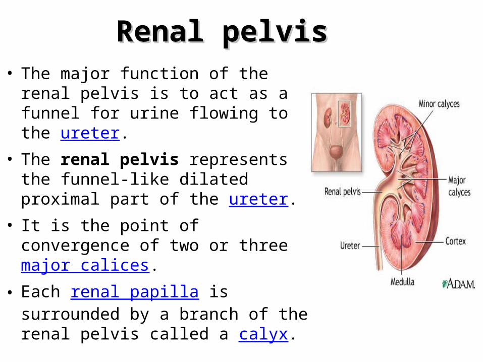
Renal pelvisRenal pelvis • The major function of the renal
pelvis is to act as a funnel for urine flowing to the ureter.
• The renal pelvis represents the funnel-like dilated proximal part of the ureter.
• It is the point of convergence of two or three major calices.
• Each renal papilla is surrounded by a branch of the renal pelvis called a calyx.

UretersUreters
• Urine is collected in the renal pelvis (or pyelum), which connects to the ureters, which carry urine to the bladder.
• The ureters are about 200 to 250 mm long.
• Smooth muscular tissue in the walls of the ureters peristaltically force the urine downward.

Urinary bladderUrinary bladder• The urinary bladder is a hollow
muscular organ shaped like a balloon.
• It is located in the pelvic fossa and held in place by ligaments attached to the pelvic bones.
• The bladder stores urine - up to 500 ml of urine comfortably for 2 to 5 hours.
• Sphincters (circular muscles) regulate the flow of urine from the bladder. – Internal urethral sphincter = in
the beginning of urethra smooth muscle – not under our voluntary control
– External urethral sphincter = skeletal muscle – we can control it

Urinary bladderUrinary bladder• The detrusor muscle is a layer of the urinary
bladder wall, made up of smooth muscle fibers arranged in inner and outer longitudinal layers and a middle circular layer.
• Contraction of the detrusor muscle causes the bladder to expel urine through the urethra.
• Problems with this muscle can lead to incontinence.

UrethraUrethra• The urethra has an excretory function in both sexes,
to pass urine to the outside, and also a reproductive function in the male, as a passage for sperm.
• The external urethral sphincter is a striated smooth muscle that allows voluntary control over urination.
• Urethral sphincters:– Internal– External
• In males the internal and external urethral sphincters are more powerful, able to retain urine for twice as long as females

UrethraUrethra
• Women: shorter• Mans: Longer
(together with genital efferent system)

Urination (micturition)Urination (micturition)• The process of disposing
urine from the urinary bladder through the urethra to the outside of the body.
• The process of urination is usually under voluntary control.
• Urinary incontinence is inability to control urination, and is more common in women than men.
• Urinary retention refers to the inability to urinate.
• Nocturnal Enuresis = incontinence during the night

MMicturition reflexicturition reflex • Activated when the urinary bladder wall is stretched; it results
in urination. • This reflex occurs in the spinal cord, specifically in the sacral
region that is modified by the higher centers in the brain: the pons and cerebrum.
• The presence of urine in the bladder stimulates the stretch receptors, which produces action potential.
• The action potentials are carried by sensory neurons to the sacral segments of the spinal cord through the pelvic nerves, the parasympathetic fibers carry the action potentials to the urinary bladder in the pelvic nerves.
• The pressure in the urinary bladder increases rapidly once its volume exceeds approximately 400-500 ml.

NephronNephron• A nephron (1-1.2 millions) is the
basic structural and functional unit of the kidney.
• Its chief function is to regulate water and soluble substances by filtering the blood, reabsorbing what is needed and excreting the rest as urine.
• Each nephron is composed of an initial filtering component (the renal corpuscle) and a tubule specialized for reabsorption and excretion (the renal tubule).
• The renal corpuscle filters out large solutes from the blood, delivering water and small solutes to the renal tubule for modification.
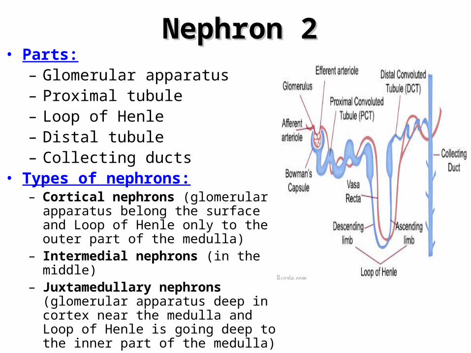
Nephron 2Nephron 2• Parts:
– Glomerular apparatus– Proximal tubule– Loop of Henle– Distal tubule– Collecting ducts
• Types of nephrons:– Cortical nephrons (glomerular
apparatus belong the surface and Loop of Henle only to the outer part of the medulla)
– Intermedial nephrons (in the middle)– Juxtamedullary nephrons (glomerular
apparatus deep in cortex near the medulla and Loop of Henle is going deep to the inner part of the medulla)
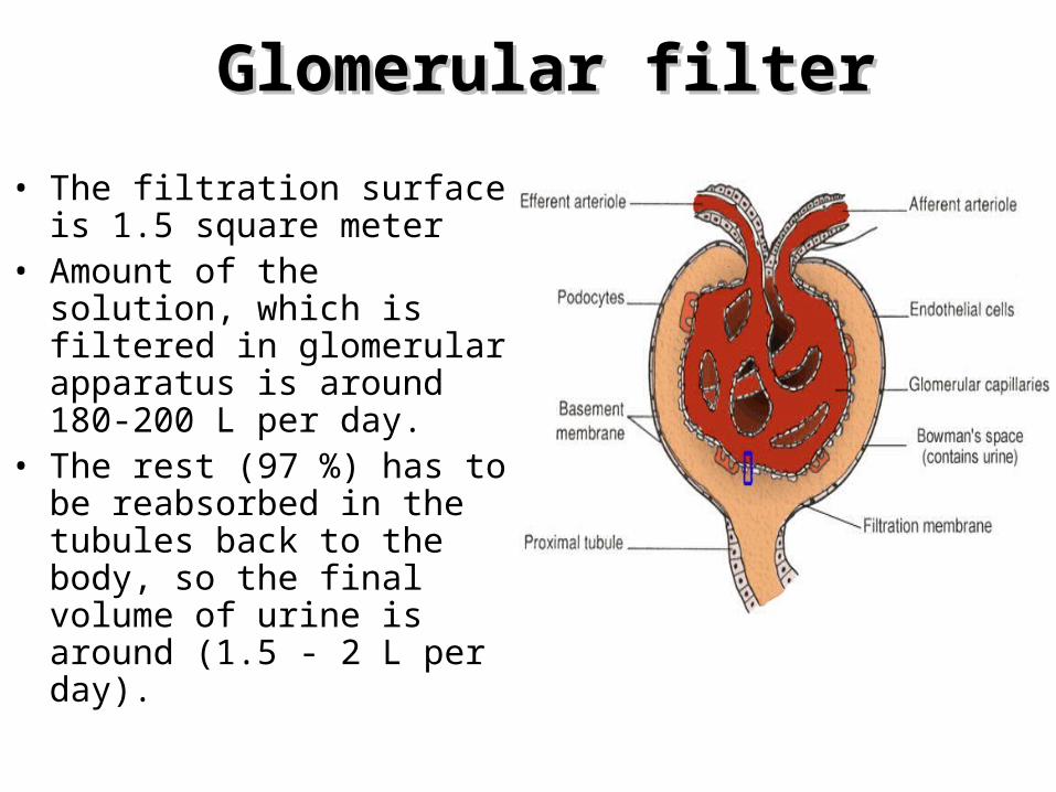
Glomerular filterGlomerular filter
• The filtration surface is 1.5 square meter
• Amount of the solution, which is filtered in glomerular apparatus is around 180-200 L per day.
• The rest (97 %) has to be reabsorbed in the tubules back to the body, so the final volume of urine is around (1.5 - 2 L per day).

GLOMERULAR GLOMERULAR FILTRATIONFILTRATION • Depends on:
– Pressure gradient across the filtration slit (endothelium, basal membrane, epithelium = podocytes)
– Blood circulation throughout the kidneys– Permeability of the filtration barrier– Filtration surface
• The solution after filtration is very similar like plasma, but should be WITHOUT PROTEINS

CLEARANCECLEARANCE• The ability of kidneys to clear plasma from different products. GLOMERULAR FILTRATION RATE (GFR)• describes the flow rate of filtered fluid through the kidney.• can be measured by measuring the excretion and plasma
level of a substance that freely filtered through the glomeruli and neither secreted nor reabsorbed by the tubules, such as INULIN (polymer of fructose).
GFR = U x V/PU = concentration of inulin in urineV = volume of the urineP = concentration of inulin in plasma• Normal GFR is around 125 ml/min (7.5 l/h)

Juxtaglomerular apparatusJuxtaglomerular apparatus• The
juxtaglomerular cells are cells that synthesize, store, and secrete the enzyme renin.
• Specialized smooth muscle cells in the wall of the afferent arteriole that are in contact with distal tubule.
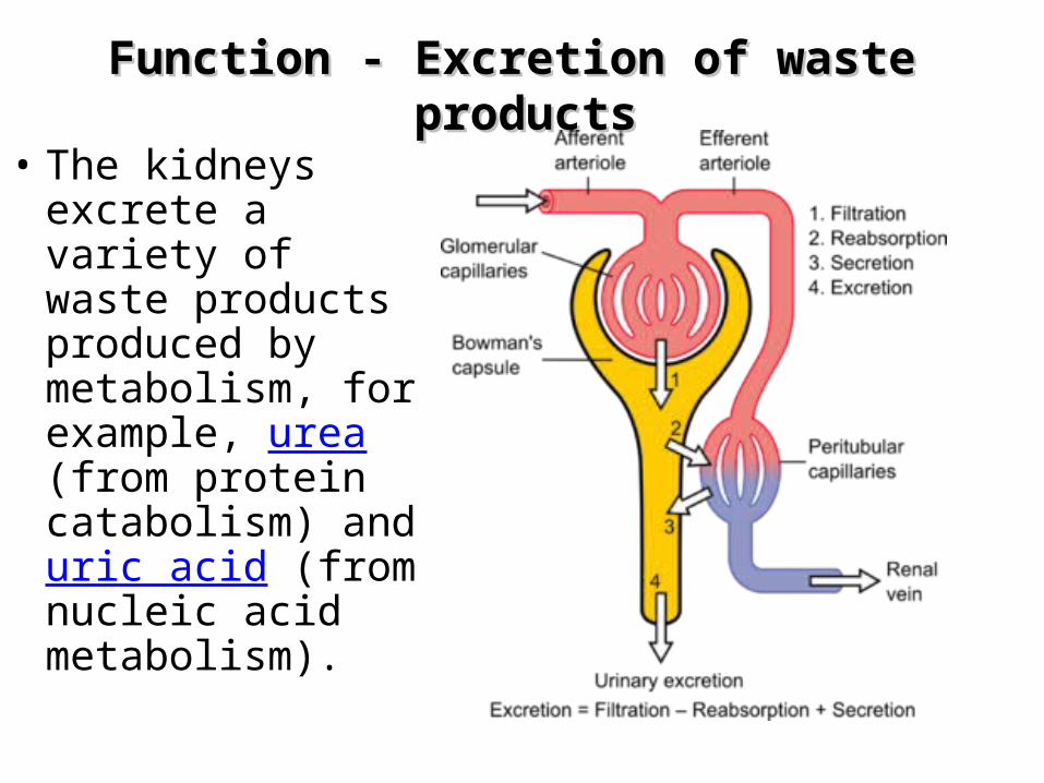
Function - Excretion of waste productsFunction - Excretion of waste products
• The kidneys excrete a variety of waste products produced by metabolism, for example, urea (from protein catabolism) and uric acid (from nucleic acid metabolism).

Function - HomeostasisFunction - Homeostasis• Acid-Base Balance
– The kidneys regulate the pH, mineral ion concentration, and water composition of the blood.
– Urine, on the other hand, becomes either acidic at pH 5 or alkaline at pH 8.
• Water Balance – Aldosterone
• Plasma Volume – ADH

AldosteroneAldosterone• A steroid hormone (mineralocorticoid) synthesized
from cholesterol by the enzyme aldosterone synthase. • It is formed in the outer-section (zona glomerulosa) of
the adrenal cortex of the adrenal gland.• It helps regulate the body's electrolyte balance by
acting on the mineralocorticoid receptor (MR). • It diminishes the excretion of Na+ ions and therefore
water, and stimulates the excretion of K+ ions by the kidneys.
• Aldosterone is synthesized in reaction to increases of angiotensin II or plasma potassium, which are present in proportion to sodium deficiencies.

Control of aldosterone Control of aldosterone releaserelease
• The role of baroreceptors – Baroreceptors in the human body detect the
pressure of blood flowing though them, and can send messages to the central nervous system to increase or decrease total peripheral resistance and cardiac output.
• The role of the juxtaglomerular apparatus • The role of sympathetic nerves • The role of the renin-angiotensin system

ADH (ADH (VASOPRESSINVASOPRESSIN))• A human hormone that is mainly released when the body is low on
water. • It causes the kidneys to conserve water by concentrating the urine. • If there is not enough water in the body
– The osmotic activity of the EC solution is increased → stimulation of the OSMOTIC RECEPTORS in the hypothalamus → stimulation of posterior lobe of the pituitary gland → activation of VASOPRESSIN → increase of the permeability of collecting ductus for the water → reabsorption → HYPERTONIC URINE
• If there is too much water in the body– The increase volume stimulates VOLUME RECEPTORS in the
heart and big veins and arteries → decrease of the activation of VASOPRESSIN → decrease of the permeability of collecting ductus for the water → water is not reabsorbed → ISO- or HYPOOSMOTIC URINE

Renin-angiotensin system 1Renin-angiotensin system 1 • A hormone system that helps regulate long-term
blood pressure and blood volume in the body. • The system can be activated when there is a loss of blood
volume or a drop in blood pressure (such as in a hemorrhage).
• If the perfusion of the juxtaglomerular apparatus in the kidneys decreases, then the juxtaglomerular cells release the enzymatic hormone renin.
• Activation: – from VOLUME RECEPTORS in afferent arteriole → decrease in
perfusion → decrease in tonus of afferent arteriole– from CHEMORECEPTORS in macula densa → decrease of NaCl in
macula densa cells

Renin-angiotensin system 3Renin-angiotensin system 3• Renin activates the renin-angiotensin system
by cleaving angiotensinogen, produced in the liver, to yield angiotensin I, which is further converted into angiotensin II by specialized cells of the lung capillaries.
• Angiotensin II then constricts blood vessels, increases the secretion of ADH and aldosterone, and stimulates the hypothalamus to activate the thirst reflex, all these actions leading to increased blood pressure.

ReninRenin • Also known as angiotensinogenase, is a circulating
enzyme released mainly by juxtaglomerular cells of the kidneys in response to low blood volume or low body NaCl content.
• Actions of renin:– Vasoconstriction in efferent arteriole (increase of glomerular
filtration)– Peripheral vasoconstriction (increase in blood pressure)– Secretion of aldosterone (reabsorption of Na+ and water)
ProstaglandinsProstaglandins• A prostaglandin is any member of a group of lipid
compounds that are derived from fatty acids and have important functions in the animal body.
• Every prostaglandin contains 20 carbon atoms, including a 5-carbon ring.
• Hormone-like substances• Function:
– Vasodilatation– Increase of perfusion– Decrease of water reabsorption– Decrease of active Na+ transport in tubules

Parathyroid hormoneParathyroid hormone• PTH is secreted by the parathyroid glands
• Function:– regulation of calcium and phosphates excretion by
urine– increase of Ca2+ reabsorption in distal tubule and
collecting ductus– inhibition of phosphates reabsorption in proximal
and distal tubules (increase of their excretion)– decrease in natrium and bicarbonates
reabsorption = decrease in water reabsorption