physical training in patients with stable chronic …jacc vol. 25. no. 6 1239 may 1995:1239-49...
TRANSCRIPT

JACC Vol. 25. No. 6 1239 May 1995:1239-49
Physical Training in Patients With Stable Chronic Heart Failure: Effects on Cardiorespiratory Fitness and Ultrastructural Abnormalities of Leg Muscles
R A I N E R H A M B R E C H T , M D , J O S E F N I E B A U E R , M D , * E D U A R D F I E H N , BS,*
B A R B A R A K A _ L B E R E R , RN,* B E R T R A M O F F N E R , M D , K L A U S H A U E R , PHD,*
U R S R I E D E , M D , t G I ~ I N T E R S C H L I E R F , M D , * W O L F G A N G KI~IBLER, M D , F A C C , *
G E R H A R D S C H U L E R , M D
Karlsruhe, Heidelberg and Freiburg, Germany
Objectives. The present study was designed to evaluate the effect of an ambulatory training program on ultrastructural morphol- ogy and the oxidative capacity of skeletal muscle and its relation to central and peripheral hemodynamic variables in patients with chronic heart failure.
Background. Clinical evidence supports the hypothesis that exercise intolerance in patients with chronic heart failure is not only a consequence of low cardiac output, but is also a result of alterations in oxidative metabolism of skeletal muscle.
Methods. Twenty-two patients were prospectively randomized either to a training group (mean [-+SD] ejection fraction 26 -'!- 9%,
n = 12) participating in an ambulatory training program or to a physically inactive control group (ejection fraction 27 -+ 10%, n = 10). At baseline and after 6 months, patients underwent symptom- limited bicycle exercise testing, and central and peripheral hemo- dynamic variables were measured. Percutaneous needle biopsy samples of the vastus lateralis muscle were obtained at baseline and after 6 months. The ultrastructure of skeletal muscle was analyzed by ultrastructural morphometry.
Results. After 6 months, patients in the training group achieved an increase in oxygen uptake at the ventilatory threshold of 23% (from 0.86 + 0.2 to 1.07 _+ 0.2 liters/rain, p < 0.01 vs. control group) and at peak exercise of 31% (from 1.49 -+ 0.4 to 1.95 + 0.4
liters/rain, p < 0.01 vs. control group). There was no significant change in oxygen uptake at the ventilatory threshold and at peak exercise in the control group. The total volume density of mito- chondria and volume density of cytochrome c oxidase-positive mitochondria increased significantly by 19% (from 4.7 -+ 1.5 to 5.6 -+ 1.5 vol%, p < 0.05 vs. control group) and by 41% (from 2.2 +
1.0 to 3.1 -+ 1.0 vol%, p < 0.05 vs. control group) after 6 months of regular physical exercise. Cardiac output at rest and at submaximal exercise remained unchanged hut increased during maximal symptom-limited exercise from 11.9 -+ 4.0 to 14.1 -+ 3.3 liters/rain in the training group (p < 0.05 vs. baseline; p = NS vs. control group). Peak leg oxygen consumption increased signifi- cantly by 45% (from 510 + 172 to 740 -+ 254 mi/min, p < 0.01 vs. control group). Changes in cytochrome c oxidase-positive mito- chondria were significantly related to changes in oxygen uptake at the ventilatory threshold (r = 0.82, p < 0.0001) and at peak exercise (r = 0.87, p < 0.0001).
Conclusions. Regular physical training increases maximal ex- ercise tolerance and delays anaerobic metabolism during sub- maximal exercise in patients with stable chronic heart failure. Improved functional capacity is closely linked to an exercise- induced increase in the oxidative capacity of skeletal muscle.
(J Am Coll Cardiol 1995;25:1239-49)
Exercise intolerance and fatigue are hallmarks in patients with chronic heart failure. Paradoxically, exercise intolerance is only poorly correlated with central hemodynamic abnormalities in this syndrome (1-4). Several studies (5-11) have demonstrated that alterations in peripheral hemodynamic variables and
From the St. Vincentius-Krankenh~user Karlsruhe, Abteilung l l l - Kardiologie, Karlsruhe; *Medizinische Universitfitsklinik, Abteilung IIl- Kardiologie, Heidelberg; and +Pathologisches Institut, Univcrsitfit Freiburg, Freiburg, Germany. This study was supported by Grant Schu 399/4-1 from the Deutsche Forschungsgemeinschaft (DFG), Bonn, Germany and Zeneca Phar- maceutical Company, Plankstadt, Germany.
Manuscript received September 1, I994; revised manuscript received De- cember 19, 1994, accepted December 20, 1994.
Address for correspondence: Dr. Rainer Hambrecht, St. Vincentius- Krankenh/iuser, Innere Medizin, Abteilung llI--Kardiologie, Edgar-von- Gierke-Strasse 2, 76135 Karlsruhe, Germany.
intrinsic abnormalities in skeletal muscle structure and metab- olism are responsible for the early onset of anaerobic metab- olism during exercise and contribute substantially to the re- duced exercise capacity of patients with chronic heart failure. The mechanism for the intrinsic muscle alterations remains unclear, although detailed evidence favors deconditioning as a possible explanation. Moreover, evidence is available that exercise intolerance in patients with chronic heart failure may be corrected at least partially by improving peripheral metab- olism by means of regular physical exercise (12,13).
The purpose of the present study was to determine the effects of regular physical exercise in patients with chronic heart failure on oxidative capacity and ultrastructural morphol- ogy in the working skeletal muscle and its relation to central and peripheral hemodynamic variables during exercise.
©1995 by the American College of Cardiology 0735-1097/95/$9.50 I)735-1097(94)00568-B

1240 HAMBRECHT ET AL. JACC Vol. 25, No. 6 PHYSICAL TRAINING IN CHRONIC HEART FAILURE May 1995:1239-49
M e t h o d s
Patient selection. Male patients with clinical, radiologic and echocardiographie signs of chronic heart failure (New York Heart Association functional classes II and III) as a result of dilated cardiomyopathy or ischemic heart disease were asked to participate in this study. Inclusion criteria were reduced left ventricular ejection fraction (<40%) as assessed by radionuclide scintigraphy and a reduced fractional shorten- ing <30% assessed by echocardiography; willingness to partic- ipate in the study for at least 6 months; and permanent residence within 25 km of the training facilities. Physical work capacity at baseline was >25 W without signs of myocardial ischemia (i.e., angina pectoris or ST segment depression). Furthermore, patients had to be in clinically stable condition for at least 3 months before entry into the study. Exclusion criteria were exercise-induced myocardial ischemia or ventric- ular tachyarrhythmias (higher than Lown class IVa), valvular heart disease, uncontrolled hypertension, peripheral vascular disease, chronic obstructive pulmonary disease and orthopedic or other conditions precluding regular participation in exercise sessions.
Study protocol. All studies were performed according to a research protocol approved by the Ethics Committee of the University of Heidelberg. Before entrance into the study, patients were required to be in clinically stable condition. Cardiac medications were titrated for 3 months to achieve optimal afterload reduction; the regimens applied included angiotensin-converting enzyme inhibitors in all patients. At baseline two percutaneous needle biopsy samples of the vastus lateralis muscle were obtained. Two days later, before exercise testing, patients ate a light breakfast and received their cardiac medications. A 5F thermodilution Swan-Ganz catheter was positioned in the right femoral vein as described by Sullivan et al. (14), and a 7F Swan-Ganz catheter was introduced into the right pulmonary artery through thc right antecubital vein; thereafter patients were transferred to the exercise facility. After a rest period of 30 rain, hemodynamic and gas-exchange measurements, as well as blood samples for determination of blood lactate and plasma catecholamine levels, were simulta- neously obtained at rest and at each work load during bicycle exercise. Exercise testing was performed on a calibrated, electronically braked bicycle in an upright position. Work load was increased progressively every 3 rain in steps of 25 W beginning at 50 W. Exercise was terminated when patients were physically exhausted or developed severe dyspnea or dizziness.
Training group. Patients assigned to the training program remained in an intermediate care ward for the initial 3 weeks. Training sessions were conducted individually under strict supervision for the first 3 weeks. Patients exercised six times daily for 10 min on a bicycle ergometer. Work loads were adjusted so that 70% of the symptom-limited maximal oxygen uptake was reached. Before discharge from hospital, maximal symptom-limited ergospirometry was performed to calculate training target heart rate for home training, which was defined
as the heart rate reached at 70% of the maximal oxygen uptake during symptom limited exercise. On discharge from hospital, bicycle ergometers were loaned to the patients, and they were asked to exercise close to their target heart rate twice daily for a minimum of 40 min altogether. In addition, they were expected to participate in at least two group training sessions of 60 rain each per week. Exercise sessions consisted of walking, calisthenics and ball games. To minimize the risk of exercise-induced arrhythmia, electrocardiographic (ECG)- based pulse rate monitoring was used during exercise outside the hospital. Patients were asked to terminate exercise when- ever their heart rate increased above their target heart rate.
Control group. Patients assigned to the control group spent 3 days in an intermediate care ward for baseline evaluation. After discharge, medical therapy was continued, and patients were supervised by their private physicians.
Follow-up studies. Exercise testing was repeated at 3 and 6 months. Muscle biopsy samples were obtained 6 months after entry into the study.
Respiratory gas exchange variables. Respiratory gas ex- change data were determined continuously throughout the exercise test with a commercially available system (Jaeger EOS-Sprint). Ventilatory threshold was defined as oxygen uptake before the systematic increase in the ventilatory equiv- alent for oxygen without a concomitant increase in the venti- latory equivalent for carbon dioxide (15). The ventilatory threshold was evaluated by two independent observers unin- formed of tbe patient's identity or the sequence of exercise tests performed.
Hemodynamic measurements. Heart rate was measured by continuous ECG monitoring. Cardiac output was obtained by a thermodilution catheter (Swan-Ganz 93A-131-7F, Edwards Laboratories) that was interfaced to a cardiac output computer (COM-2, Edwards Laboratories). Blood samples were taken at rest and during the last minute of each work load and were kept in an ice bath immediately after collection. Oxygen content and saturation of femoral and mixed venous blood samples were measured on a calibrated OSM2-Oximeter (Ra- diometer, Copenhagen, Denmark). Arterial oxygen saturation was obtained by pulse-oximetry (ASAT pulse-oximeter, Ed- wards Laboratories). Femoral and mixed venous blood lactate concentrations were determined enzymatically (16). Free and conjugated plasma catecholamines were analyzed by high pressure liquid chromatography with amperometric detection as described by Weicker (17).
Leg blood flow. Femoral venous blood flow was measured with a thermodilution catheter (model 93A-105-5F, Edwards Laboratories) that was interfaced to a cardiac output com- puter. Bolus injections of 5 ml of cooled saline solution were used to obtain two or three blood flow measurements at rest and at the end of each work load, and these were then averaged. To determine the variability of leg blood flow and cardiac output measurements, five patients with chronic heart failure performed two maximal bicycle exercise tests separated by a 2-h rest period. The variability of duplicate measurements at rest and at submaximal and maximal work loads was 16 +
JACC Vol. 25, No. 6 HAMBRECHT ET AL. 1241 May 1995:1239 49 PHYSICAI~ TRAINING IN CHRONIC HEART FAILURE
8%, 9 + 3% and 4 -+ 1%, respectively, for leg blood flow. The corresponding data for cardiac output were 12 + 5%, 8 +_ 3%, and 8 + 2%, respectively. Regression of paired leg blood flow determinations at maximal exercise showed a highly significant correlation (r = 0.98, p < 0.001).
Echocardiography. At baseline and after 6 months (4 weeks in patients in the high risk training group [see Results, High risk training group]) two-dimensional echocardiography was performed to determine left ventricular end-systolic and end-diastolic dimensions. At least five consecutive cardiac cycles were analyzed and averaged for each patient by an observer working without knowledge of patient status.
Assessment of leisure time physical activities. Energy ex- penditure in leisure time physical activity was estimated using a modified Minnesota Leisure Time Physical Activity Ques- tionnaire (18). Patients were interviewed at least twice during the treatment period by the same technician. Activities re- corded were those performed during the previous weekend and previous 2 days. From each interview, energy expended per week (kcal/week) in leisure time physical activity was calculated.
Muscle biopsy. Two days before exercise testing, percuta- neous needle biopsy samples from the middle part of the vastus lateralis muscle were obtained at baseline and after 6 months under local anesthesia as described by Bergstr6m (19). The specimens were examined using an EM 200 Philips electron microscope. Each biopsy sample was cut into four blocks, and 15 microphotographs were randomly taken from each block. The samples were photographed at a primary magnification of × 15,500 and analyzed at a final magnification of x60,000 with the aid of a 1,089-point and 121-test line multipurpose test grid superimposed over each microphotograph. According to stan- dard stereologic principles, the volume density of mitochon- dria was analyzed (as counted points/total points of grid) as reported previously (i.e., the mitochondrial volume fraction per unit volume tissue, expressed as volume percent) (20,21).
The diaminobenzidine cytochrome oxidase reaction was performed in mitochondria as described elsewhere (5) using a modification of the technique reported by Perotti et al. (22) to provide a qualitative assessment of the mitochondrial cyto- chrome oxidase activity in skeletal muscle. Sixty microphoto- graphs from each patient were analyzed to determine thc volume density of mitochondria, as described earlier. The mitochondria were classified as cyctochrome oxidase positive when diaminobenzidine staining was visible within the mito- chondria. Diaminobenzidine-negative mitochondria may re- flect low cytochrome oxidase activity. All specimens for ultra- structural morphometry were coded and analyzed by an independent technician who was unaware of clinical data or group assignment.
For consecutive duplicate biopsy samples, the variation regarding fiber-type distribution has been shown to be on the order of 5% to 6% (23). The photographs of the first 10 biopsies were analyzed independently by two investigators unaware of each other's results. The interobserver variability
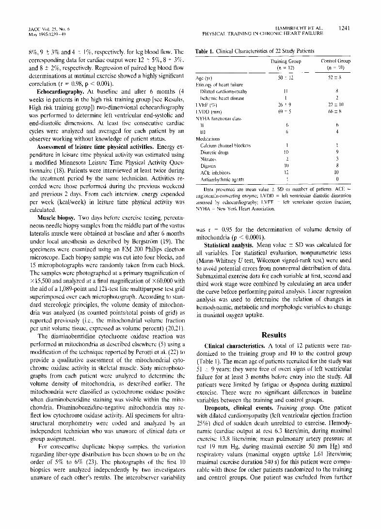
Table 1. Clinical Characteristics of 22 Study Patients
Training Group Control Group (n = 12) (n = 10)
Age (yr) 50 : 12 52 _+ 8 Etiology of heart failure
Dilated cardiomyopathy 11 8 lschemic heart disease 1 2
LVEF (%) 26 _+ 9 27 +_ 10 LVDD (mm) 69 +- 5 66 -+ 8 NYHA functional class
II 6 6 Ill 6 4
Medications Calcium channel blockers 1 1 Diuretic drugs 10 9 Nitrates 2 3 Digoxin 10 8 ACE inhibitors 12 10 Antiarrhythmic agents 1 0
Data presented are mean value _+ SD or number of patients. ACE =
angiotensin-converting enzyme; LVDD - left ventricular diastolic dimension assessed by echocardiography; LVEF - left ventricular ejection fraction;
NYHA New York Heart Association.
was r - 0.95 for the determination of volume density of mitochondria (p < 0.0001).
Statistical analysis. Mean value _+ SD was calculated for all variables. For statistical evaluation, nonparametric tests (Mann-Whitney U test, Wileoxon signed-rank test) were used to avoid potential errors from nonnormal distribution of data. Submaximal exercise data for each variable at first, second and third work stage were combined by calculating an area under the curve before performing paired analysis. Linear regression analysis was used to determine the relation of changes in hemodynamic, metabolic and morphologic variables to change in maximal oxygen uptake.
R e s u l t s
Clinical characteristics. A total of 12 patients were ran- domized to the training group and 10 to the control group (Table 1). The mean age of patients recruited for the study was 51 + 9 years; they were free of overt signs of left ventricular failure for at least 3 months before entry into the study. All patients were limited by fatigue or dyspnea during maximal exercise. There were no significant differences in baseline variables between the training and control groups.
Dropouts, clinical events. Training group. One patient with dilated cardiomyopathy (left ventricular ejection fraction 25%) died of sudden death unrelated to exercise. Hemody- namic (cardiac output at rest 6.3 liters/rain, during maximal exercise 13.8 liters/rain; mean pulmonary artery pressure at rest 19 mm Hg, during maximal exercise 50 mm Hg) and respiratory, values (maximal oxygen uptake 1.61 liters/rain; maximal exercise duration 540 s) for this patient were compa- rable with those for other patients randomized to the training and control groups. One patient was excluded from further

1242 HAMBRECHT ET AL. JACC Vol. 25, No. 6 PHYSICAL TRAINING IN CHRONIC HEART FAILURE May 1995:1239-49
Table 2. Hemodynamic Variables in the Training and Control Groups
Baseline 6 mo Change (%)
Training Group
HR at rest (liters/rain) 88 _+ 18 82 _+ 18"? 6 _+ 12( -7%)?
Maximal HR (liters/rain) 163 = 27 174 _+ 22* 11 _+ 13 (+3%)~
SBP at rest (mm Hg) 118 _+ 10 119 + 15 1 + 14 (~-1%)
Maximal SBP (mm Hg) 172 + 17 182 + 31 10 _+ 24 (+6%)
Maximal exercise time (s) 536 + 180 700 + 199' 164 + 169 (+26%)§
Maximal mean PAP (ram Hg) 46 _+ 17 41 _+ 16 - 5 _+ 18(-10%) Maximal CO (liters/rain) 11.9 + 4.0 14.1 + 3.3* 2.2 + 2.5 (+19%)~:
Control Group
HR at rest (Iiters/min) 90 _+ 15 92 + I3 2 +_ 8 (~-2%)
Maximal HR (liters/rain) 159 + 13 159 _+ 22 0 + 12 (+0%)
SBP at rest (ram Hg) 119 + 12 125 + 25 6 + 14 (+5%)
Maximal SBP (ram Hg) 167 + 35 169 -+ 28 2 _+ 20 (+ 1%)
Maximal exercise time (s) 581 ± 266 563 _+ 283 -17 _+ 25 ( -2%) Maximal mean PAP (ram Hg) 43 + 2I 42 _+ 2I -1 _+ 17 ( -2%)
Maximal CO (liters/rain) 13.0 _+ 5.6 13.3 + 4.7 0.3 +_ 2.5 (+2%)
*p < 0.05, significantly different from baseline. ?p < 0.05, §p < 0.01, significantly different from control group. Sp = 0.I, tendentially different from control group. CO = cardiac output; HR = heart rate; PAP = pulmonary artery pressure; SBP = systolic blood pressure.
participation because of atrioventricular node reentrant tachy- cardia. One patient in clinically stable condition (dilated cardiomyopathy, left ventricular ejection fraction 33%) refused follow-up examinations. Data for the remaining nine patients were used for sequential analysis.
Control group. One patient withdrew his consent after baseline examinations. One patient had right heart failure during the study period and was readmitted to the hospital for an additional 2 weeks. He was then discharged from hospital, and 6-month measurements could be obtained. At 6 months, complete data were available in nine patients.
Functional class. Training improved functional class from 2.5 + 0.7 to 1.7 _+ 0.7 in the training group (p < 0.005 vs. control group). Functional class did not change in the control group (2.3 + 0.5 vs. 2.3 + 0.5).
Medical treatment. Initially, all patients were taking angiotensin-converting en~me inhibitors and 83% and 100% were taking diuretic drugs and 83% and 80% digoxin in the training and control groups, respectively (p = NS) (Table 1). Drug treatment did not change during the last 4 weeks before or during the study in any patient completing the trial.
Energy expenditure in leisure time physical activity. In the training group, mean attendance for the training sessions was 62 _+ 24%, and compliance for home training was calculated to be 70%, amounting to a total of 4.5 h/week leisure time physical activity. The mean energy expenditure in leisure time physical activity/week increased in the training group from 921 z 730 to 2,510 _-2- 1,177 kcal/week (p < 0.0005 vs. control group). In the control group there was no significant change observed (615 + 486 vs. 482 _+ 514 kcal/week, p = NS).
Central hemodynamic and respiratory variables. Training group. After 22 _+ 6 days of supervised in-hospital bicycle training, mean submaximal heart rate reached at a constant
submaximal work load decreased significantly from 117 _+ 13 to 105 _+ 13 beats/min (p < 0.001 vs. begin). After 6 months of training, rest heart rate decreased by 7% (from 88 + 18 to 82 + 18 beats/rain, p < 0.05 vs. control group) (Table 2). Oxygen uptake at ventilatory threshold increased by 23% (from 0.86 _+ 0.2 to 1.07 _+ 0.2 liters/rain, p < 0.01 vs. control group) and at peak exercise by 31% (from 1.49 _+ 0.4 to 1.95 _+ 0.4 liters/rain, p < 0.01 vs. control group), respectively (Table 3). Maximal exercise time increased by 26% (from 536 _+ 180 to 700 _+ 199 s, p < 0.01 vs. control group). Cardiac output was nearly unchanged at rest and at submaximal exercise but demonstrated an increase at maximal exercise (from 11.9 _+ 4.0 to 14.1 + 3.3 liters/rain, p < 0.05 vs. baseline, p = 0.1 vs. control group) (Fig. 1). Rest and exercise mean arterial and mean right atrial pressure were unchanged at the end of the study compared with that at baseline. During the initial stress test, mean pulmonary artery pressure increased from 19 _+ 12 mm Hg at rest to 32 _+ 18 mm Hg during submaximal exercise (75 W) and to 46 _-2- 17 mm Hg at peak exercise. The corresponding values after the training period were 15 + 5 mm Hg (p = NS vs. baseline), 32 _+ 15 mm Hg (pNS vs. baseline) and 41 + 16 mm Hg (p = NS vs. baseline). Stroke volume at rest did not change but tended to increase during submaximal (from 63.1 _+ 28 to 72.5 _+ 23 ml/min, p = 0.17) and peak exercise (from 74.4 + 28 to 81.6 _+ 20 ml/min, p -- 0.15). Left ventricular end-diastolic dimension, assessed by echocardiography, decreased from 68.6 + 4.9 to 64.2 _+ 3.1 mm (p < 0.05 vs. baseline, p - NS vs. control group) after training. Left ventricular end-systolic dimension was not significantly altered by training (55.1 _+ 6.6 vs. 53.8 ___ 6.4 mm).
Control group. There was no significant change in rest and exercise cardiac output, stroke volume, mean arterial pressure, mean pulmonary artery pressure or oxygen uptake at ventila-

JACC Vol. 25, No. 6 HAMBRECHT ET AL. 1243 May 1995:1239-49 PHYSICAI~ TRAINING IN CHRONIC HEART FAILURE
Table 3. Respiratory Variables
Baseline 6 mo Change (%)
Training Group
Exercise time Vt (s) 320 + 115 406 -+ 192't 86 + 149 (+27%)'1"
Vo2-Vt (liters/rain) 0.86 + 0.23 1.07 - 0.24*:}: 0.20 -+ 0.18 (+23%)-
Vo~max (liters/rain) 1.49 _+ 0.42 1.95 - 0.39~§ 0.46 _+ 0.28 (+31%)-
Vo~max (ml/kg per rain) 17.5 + 5.1 23.3 - 4.2¢§ 5.8 -+ 3.6 (+33%)~
V~:max (liters/min) 62.4 _+ 18 75.3 - 12'1 12.9 + 13 (+20%):]:
Max RER 1.10 -+/).14 1.07 _- 0.16 0.03 _+ 0.24 ( -3%)
Control Group
Exercise time Vt (s) 307 + 132 26g = 90 38 +_ 77 ( 1 2 % )
Vo2-VI (liters/min) 0.91 + 0.23 (I.81 + (I.20 0.10 + 0.12 ( l ( / % )
Vozmax (liters/rain) 1.53 + 0.58 1.54 _+ (I.52 0.0 _+ 0.25 (=0%)
Vozmax (ml/kg per min) 17.9 + 5.6 17.9 -+ 5.6 0.0 + 2.7 (-+0%)
Vzmax (liters/rain) 65.9 + 21 6(!.4 + g 5.5 + 16 ( 8 % )
Max RER l.J)l _+. 0.11 1.00 + 0.14 -0.01 + 0 .04( -1%)
*p < 0.05, §p < 0.01, significantly different from baseline. -p < 0.05, Sp < 0.01, significantly different from control group. Exercise time Vt time at which ventilatory threshold occurs; Max RER - respiratory exchange ratio (carbon
dioxide uptake/oxygen uptake) at peak exercise; VEmax - maximal minute ventilation; Vo2max = o~,gen uptake at peak exercise: Vo,-Vt ~ oxygen uptake at ventilatory threshold.
tory threshold or at peak exercise. After 6 months, rest heart rate, oxygen uptake at ventilatory threshold and peak exercise, exercise time at which ventilatory threshold occurred and peak exercise time in the training group differed significantly from the corresponding variables in the control group.
Figure 1. Change in cardiac output in patients in the training (top) and control (bottom) groups at baseline (triangles) and at 6-month follow-up (circles). Max = maximal exercise. Sp < 0.05 versus baseline.
17,5
15
12,5
10
7,5
5
2,5
0
[lfmin]
50~W ~ ~ Rest 75 W 100 W Max
17,5
15
12,5
10
7,5
5
2,5-
0
[l/rain]
I I ; i i
Rest 50 W 75 W 100 W Max
Peripheral hemodynamic variables. Training group. Leg blood flow tended to increase at submaximal exercise (75 W) by 15% (from 3.02 to 3.47 liters/min, p = 0.1). Peak leg blood flow and peak leg oxygen consumption increased significantly by 28% (from 3.6 + 1.6 to 4.6 _+ 1.4 liters/rain, p < 0.01 vs. control group) and by 45% (from 510 _+ 172 to 740 _+ 254 ml/min, p < 0.01 vs. control group), respectively, com- pared with that in the control group (Fig. 2). There was a significant decrease in leg vascular resistance at rest (from 326 : 109 to 218 _+ 73 mm Hg.min/liter, p < 0.01 vs. control group) and at submaximal (from 120 _+ 62 to 87 _+ 36 mm Hg.min/liter, p < 0.05 vs. control group) and peak exercise (38 _+ 15 to 28 _+ 8 mm Hg.min/liter, p < 0.01 vs. control group) in the training group. Mixed venous and femoral venous oxygen saturation were unchanged at rest and at submaximal exercise. At peak exercise, femoral venous oxygen saturation decreased by 32% (from 22 _+ 9% to 15 _+ 6%) in the training group (p < 0.05 vs. control group).
Control group. There were no significant changes observed with regard to leg blood flow, leg vascular resistance, leg oxygen consumption, leg arteriovenous oxygen difference, fem- oral venous and mixed venous saturation at rest and during exercise.
Predictor of response to exercise. A significant correlation between training-induced reduction of mean submaximal heart rate during initial in-hospital bicycle exercise training and changes in maximal oxygen uptake after 6 months (r = 0.78, p < 0.05) was observed. There was no significant correlation between changes in maximal oxygen uptake after training and patient age, left ventricular ejection fraction, left ventricular end-diastolic dimension, functional status or hemodynamic variables measured during baseline exercise evaluation.
Metabolic response to exercise training. After 6 months of exercise training, femoral venous lactate concentrations were

1244 HAMBRECHT ET AL. JACC Vol. 25, No. 6 PHYSICAL TRAINING IN CHRONIC HEART FAILURE May 1995:1239-49
[ml/min]
# s
2 # $
l I I I I
Rest 50 W 75 W 100 W M a x
1000-
800
600
400
200
0
mllmin]
J • I I I I
Rest 5 0 W 7 5 W 1 0 0 W Max
significantly reduced during submaximal exercise (p < 0.05 vs. control group) (Fig. 3). No change was observed at rest or at peak exercise. There were no changes in femoral venous lactate concentrations at rest or at submaximal or peak exer- cise in the control group.
Oxidative capacity of skeletal muscle. Baseline volume density of cytochrome c oxidase-positive mitochondria (r = 0.71, p < 0.0001) as well as total volume density of mitochon- dria (r = 0.64, p < 0.01) were significantly correlated with oxygen uptake at peak exercise (Table 4, Fig. 4).
Training group. Cytochrome c oxidase-positive volume den- sity of mitochondria increased significantly by 41% (from 2.2 + 1.0 to 3.1 _+ 1.0 vol%, p < 0.05 vs. control group), whereas cytochrome c oxidase-negative volume density of mitochondria remained unchanged. Total volume density of mitochondria increased after 6 months of training by 19% (from 4.7 ± 1.5 to 5.6 -+ 1.5 vol%, p < 0.05 vs. control group) (Fig. 5).
Control group. There was no significant change in volume density of mitochondria. Changes in total volume density of mitochondria and changes in cytochrome c oxidase-positive volume density of mitochondria were significantly correlated with both changes in oxygen uptake at the ventilato~ threshold and peak oxygen uptake (Fig. 4). No correlation was found between the cytochrome c oxidase-positive volume density of mitochondria at study beginning or the change in cytochrome oxidase-positive volume density of mitochondria and patient age, duration of heart failure or functional status.
To assess whether patients with severe or only mild to
Figure 2. Change in leg oxygen consumption in patients in the training (top) and control (bottom) groups at baseline (diamonds) and at 6-month follow-up (triangles). Max = maximal exercise. $p < 0.05 versus baseline. #p < 0.05 versus control group.
Figure 3. Change in plasma lactate concentration in patients in the training (top) and control (bottom) groups at baseline (diamonds) and at 6-month follow-up (triangles). Max = maximal exercise. Sp < 0.05 versus baseline. #p < 0.05 versus control group.
10
8
6
4
2
0
"nrnol/I]
I I I | 1
Rest 50 W 75 W 100 W Max
10
B
6
4
2
0
rnmol/ll
i 501W 751W i i Rest 100 W Max

JACC Vo]. 25, No. 6 HAMBRECHT ET AL. 1245 May 1995:1239 40 PHYSICAL TRAINING IN CHRONIC HEART FAILURE
Table 4. Cytochrome c Oxidase Activity in Mitochondria of Skeletal Muscle
Baseline 6 mo Change (%)
Training Group
Vvm-cox+ (vol%) 2.2 + 1.0 3.1 + 1.0*t 0.9 _+ 0.5 (+41%)~
Vvm-cox (w~l~>; -) 2.5 + 0.8 2.5 + 0.7 0.0 _+ 0.2 (0%)
Vvm-total (vol%) 4.7 _+ 1.5 5.6 + 1.5t$ 0.9 + 0.5 (+19%)'~
Control group
Vvm-cox+ (w~lC~) 1.9 + 1.0 1.7 + 1.0 0.2 + 0 .6 ( -11%)
Vvm-cox- (vol%) 2.3 +_ (1.6 2.4 + 0.5 0.1 + 0.5 (+5%)
Vvm-tntal (vol%) 4.2 + 1.3 4.1 + 1.3 0.l + 0 . 7 ( - 2 % )
*p < 0.01, Sp < 0.05, significantly different from baseline. ~p < 0.05, significantly different from control group.
Vvm-cox+ (Vvm-cox-) = volume densiW of cytochromc c oxidase-positive (negative) mitochondria; Vvm-total = total density, of mitochondria.
moderate left ventricular dysfunction would benefit more from exercise, we analyzed the following subgroups: subgroup 1 = left ventricular ejection fraction -<20% (n = 3); sub- group 2 = left ventricular ejection fraction between 21% and 30% (n = 3); subgroup 3 = left ventricular ejection fraction between 31% and 39% (n = 3). The greatest increase in volume density of cytochrome c oxidase-positive mitochondria and maximal oxygen uptake could be observed in subgroup 1 (increase in volume density of cytochrome oxidase-positive mitochondria 1.03 _+ 0.64 vol%, change +64%) (increase in maximal ventilation 7.2 - 5.7 ml/min per kg; change +43%), whereas these changes were less impressive in subgroup 2 (0.9 _+ 0.24 vol%, change +41%; and 5.6 _+ 1.7 ml/min per kg, change +30%, respectively) and subgroup 3 (0.68 _+ 0.5 vol%, change +22%; 4.6 _+ 3.1 ml/min per kg, change +26%, respectively).
To determine whether patients with severe or only moder- ate exercise intolerance would benefit more from exercise training, we performed another subgroup analysis: subgroup I - beginning maximal oxygen uptake -<17 ml/kg per min (n = 6); subgroup 2 = >17 ml/kg per min (n = 3). The greatest change in volume density of cytochrome oxidase-positive mi- tochondria (subgroup 1, 1.1 _+ 0.3 vol%, change +62%; subgroup 2, 0.42 _+ 0.56 vol%, change + 15%) and change in peak ovgen uptake (subgroup 1, 6.8 _+ 3.6 ml/kg per min, change +47%; subgroup 2, 3.9 + 3.2 ml/kg per min, change +17%) was observed in subgroup 1.
Effects of training on plasma catecholamine levels. After 6 months of training, plasma norepinephrine levels at rest and at submaximal exercise decreased significantly by 52% (from 2.9 +_ 2.8 to 1.4 + 0.5 nmol/liter, p < 0.05) and by 46% (from 13.4 _+ 10.1 to 7.2 + 2.8 nmol/liter, p < 0.05), respectively, compared with those in the control group. Plasma epinephrine at rest also decreased significantly by 50% (from 0.4 _+ 0.3 to 0.2 + 0.1 nmol/liters, p < 0.05 vs. control group) in the training group. There was no change in plasma catecholamine levels at rest or during exercise in the control group. Changes in norepinephrine and epinephrine at rest and during submaxi- mal exercise were not significantly correlated with changes in leg blood flow or leg vascular resistance.
High risk training group. The high risk training group included 6 patients who were scheduled for heart transplanta-
tion, and whose individual risk associated with home-based, nonsupervised physical exercise was considered to be unac- ceptably high because of repetitive ventricular arrhythmias (Lown class IVb) and severely depressed left ventricular performance (left ventricular ejection fraction <20%). These patients were examined to determine the lower cutoff point beyond which untoward side effects and risks associated with regular physical exercise outweighed the expected benefit. Each of these patients exercised daily -1 h in 10- to 15-min intervals over a period of 4 weeks at an individually prescribed work intensity under strict supervision. They underwent iden- tical baseline and follow-up tests as described for the random- ized study groups. However, training sessions were conducted under strict and individual supervision. Additionally, the fre- quency of repetitive ventricular tachyarrhythmias (more than three consecutive ventricular ectopic beats) was assessed at baseline and after 2 and 4 weeks by 24-h Holter monitoring.
The reduction in left ventricular ejection fraction in the high risk training group (16 + 5%) was significantly more pronounced compared with that in patients in the randomized groups (p < 0.05). All patients in the high risk training group had repetitive ventricular tachyarrhythmias documented by 24-h Holter monitoring. Two patients in the high risk training group required cardiopulmonary resuscitation as a result of cardiac arrest before entry into the study. Training improved functional class from 3.0 _+ 0.0 to 2.3 _+ 0.5 (p < 0.01 vs. baseline). After training, the following variables differed sig- nificantly from baseline values: exercise time at which ventila- tory threshold occurred (215 +_ 96 vs. 240 _+ 107 s, p < 0.05), maximal exercise time (483 _= 175 vs. 590 _+ 195 s, p < 0.05), oxygen uptake at ventilatory threshold (0.69 _+ 0.18 vs. 0.80 + 0.12 liters/min, p < 0.05) and peak exercise (1.07 _+ 0.30 vs. 1.27 _+ 0.21 liters/min, p < 0.05). There was no change in the number of ventricular ectopic beats between baseline (72 _+ 23 beats/h) and 2 weeks (70 _+ 25 beats/h) and 4 weeks of in-hospital training (66 _+ 25 beats/h). At baseline, cyto- chrome c oxidase-positive volume density of mitochondria was significantly reduced compared with that in the randomized groups (p < 0.05). There was a tendency toward an increase in cytochrome c oxidase-positive volume density of mitochondria by 30% from 1.0 + 0.4 to 1.3 _+ 0.5 vol% (p = 0.1 vs. beginning) after the training period.

1 2 4 6 HAMBRECHT ET AL. JACC Vol. 25, No. 6 PHYSICAL TRAINING IN CHRONIC HEART FAILURE May 1995:1239-49
dVvm-cox* [vot~]
-1
I 5 110
dVO2-Vl" [mi/mln kg I
y = 0 1 6 * 0 2 4 ~ f = 0 8 2 0 p • 0001
dVvm-cox+ [vol%] :/j aVO~m~ [mllm~ kg]
r = 0 8 7 4 p <.0001
Figure 4. Changes in volume density of cytochrome c oxidase-positive mitochondria (dVvm-cox+) correlated with change in oxygen uptake at ventilato~ threshold (dVO2-Vt) (top) and change in peak oxygen uptake (dVO2max) (bottom).
Discussion Two important findings emerge from this study. 1) Regular
physical training increases maximal exercise tolerance and delays anaerobic metabolism during submaximal exercise in patients with chronic heart failure. 2) Improved aerobic exer- cise capacity is closely linked to an exercise-induced increase in oxidative capacity of the working skeletal muscle. The results of this study confirm that alterations in skeletal muscle from deconditioning play an independent role in the pathophysiol-
Figure 5. Electron micrographs of cytochrome c oxidase in a patient in the training group before (top) and after (bottom) 6 months of exercise training. Enzyme activity within the mitochondria (black) is increased aftcr training.
ogy of exercise intolerance in chronic heart failure and indicate that these alterations are at least in part reversible by an exercise training program tailored to individual requirements.
Training effects on skeletal muscle. Previous studies (5,8,11,24-26) have demonstrated alterations in the oxidative metabolism of skeletal muscle in patients with chronic heart failure by ultrastructural, cytochemical and biochemical anal- ysis as well as by magnetic resonance imaging (MRI) spectros- copy. Furthermore, it has been shown (5) that changes in oxidative capacity of skeletal muscles are closely correlated with changes in exercise capacity. In the present study, cyto- chemical analysis of cytochrome oxidase activity was per- formed as a measure of oxidative capacity of the working skeletal muscle. At baseline, a positive cytochrome oxidase reaction was present in 30 + 11%, 28 _+ 13% and 18 _+ 5% of mitochondria in the training, control and high risk groups. In contrast, this reaction has been reported (5) to be present in -60% of mitochondria in normal subjects. Furthermore, total volume of mitochondria was also clearly reduced by -47% in our patients compared with that in normal subjects (5). One important result of the present study is the observation that changes in oxidative capacity of skeletal muscles are reversible

JACC Vol. 25, No. 6 HAMt3RECHT ET AL. 1247 May 1995:1239-49 PHYSICAL TRAINING IN CHRONIC H E A R T FAILURE
by an effective home-based "training therapy." This beneficial training effect in patients with chronic heart failure is in agreement with results of various investigations in animals and normal subjects that have demonstrated major adaptations in skeletal muscles after training, such as increases in mitochon- drial content, capillary density and oxidative capacity (27-32). However, immobilization has canceled these beneficial train- ing effects on skeletal muscles in normal subjects and is related to decreased activity of mitochondrial oxidative enzymes (33- 35). To our knowledge, the present study is the first random- ized trial documenting the beneficial effect of exercise training on skeletal muscle morphology and oxidative capacity in patients in stable condition with moderate to severe chronic heart failure. Baseline volume density of cytochrome c oxidase- positive mitochondria was related to oxygen uptake at peak exercise. The significant increase in oxidative capacity of skeletal muscle, assessed by change in volume density of cytochrome oxidase-positive mitochondria, after endurance training was significantly correlated with changes in functional capacity and peak oxygen uptake (Fig. 4). In the present study, exercise-induced changes in oxidative capacity of skeletal muscle and in peak oxygen uptake were most impressive in patients in the training group with severe exercise intolerance or severe left ventricular dysfunction, or both, compared with patients who had only moderate exercise intolerance or mod- erate ventricular dysfunction, or both.
Recently published studies have focused on the effect of exercise training on skeletal muscle metabolism in patients with chronic heart failure. Minotti et al. (12) used localized conditioning that improved both the metabolic and functional characteristics of forearm muscles in patients with chronic heart failure who participated in an uncontrolled trial. Al- though there was no change in forearm muscle size or exercise blood flow, the endurance of the wrist flexors after 28 days of unilateral forearm training was significantly increased. Correc- tion of skeletal muscle metabolic abnormalities by physical exercise could be demonstrated using phosphorus-31 MRI (13). However, training-induced MRI spectroscopic changes in the phosphocreatine/(phosphocreatine + inorganic phos- phate) ratio observed in these studies could theoretically have resulted from performing the same work with more muscle as a result of endurance training. The semiquantitative determi- nation of skeletal muscle oxidative capacity by cytochemistry used in the present study represents a method well correlated with biochemical determination of oxidative enzymes (35,36) that is largely independent from muscle mass.
Although there is evidence that endurance training can improve muscle metabolism and oxidative capacity, the effect of training appears to be nonspecific (36) and does not imply that deconditioning is the sole cause of the initial muscle abnormalities because muscle dysfunction occurs also in mus- cles, (i.e., small hand muscles) not expected to be subject to deconditioning (37,38).
Several additional factors are responsible for intrinsic alter- ations of skeletal muscle:
1) Quantitative ultrastructural analysis of upper arm skele-
tal muscle suggests that the myopathy observed in patients with dilated cardiomyopathy may not be limited to the myocardium but may also involve skeletal muscles (39).
2) Previously demonstrated increase in sarcoplasmic triads (5) and reduction in sodium-potassium pump activity (40) in skeletal muscles of some patients with severe chronic heart failure due to dilated cardiomyopathy seem to contribute to alterations in muscle metabolism by changes in intracellular homeostasis.
3) Mutations of mitochondrial deoxyribonucleic acid (DNA) due to excessive accumulation of free radicals induced by recurrent episodes of hypoxemia and reperfusion may account for intrinsic alterations in skeletal muscle of patients with chronic heart failure (41). Free radicals are known to mu- tagenize mitochondrial DNA as well as enzymes involved in oxidative phosphorylation.
Respiratory and hemodynamic variables. The 31% im- provement in maximal oxygen uptake shown here in a con- trolled study is similar to that reported by Sullivan et al. (7,14) in a retrospective, uncontrolled report of training and that reported by Coats et al. (42) in a controlled, crossover trial of rehabilitation in a highly selected study group of severely affected patients. The percent increase in exercise time or peak oxygen consumption is also comparable to that seen in training programs in normal subjects and in patients with coronary artery disease without heart failure (43,44).
In both training groups cardiac output at rest and during submaximal exercise remained unchanged during the course of the study. However, the effect of training-induced bradycardia on cardiac output during submaximal exercise was offset by a trend toward increased stroke volume. Several previous studies (45,46) have shown that patients with coronary artery disease can improve exercise stroke volume or left ventricular contrac- tility after 1 year of intensive training. Sullivan et al. (14) could also not demonstrate an improvement in left ventricular systolic function at rest, although exercise stroke volume tended to increase, after 4 to 6 months of training in patients with left ventricular dysfunction. In the present study, maximal cardiac output increased significantly after long-term exercise in both training groups. The change in maximal cardiac output was significantly correlated with the change in peak oxygen uptake, suggesting that central hemodynamic adaptations con- tribute to improved exercise performance after regular physi- cal exercise in patients with chronic heart failure.
Ventilato~ threshold. The results of the present study dem- onstrate that physical exercise at a specified intensity caused a delay in the onset of ventilatory threshold by 86 _+ 149 s (+27%) in the training group and 25 __* 35 s (+12%) in the high risk training group. Consequently, patients reached a small but significant increase in oxygen uptake at ventilatory threshold of 23%, and 16%, respectively. The amount of work accomplished between ventilatory threshold and peak exercise was nearly identical, suggesting a similar contribution of anaerobic work at baseline and after the training period. In our study, change in oxygen uptake at ventilatory threshold was significantly correlated with change in volume density of

1248 HAMBRECHT ET AL. JACC Vol. 25, No. 6 PHYSICAL TRAINING IN CHRONIC HEART FAILURE May 1995:1239-49
cytochrome c oxidase-positive mitochondria in skeletal muscle, indicating that an increased oxidative capacity of the mitochon- dria may account for a delayed onset of ventilatory threshold after endurance training.
Metabolic and peripheral hemodynamic variables. Leg blood flow at rest and during exercise were reduced in our study patients compared with that in normal subjects (47). Several adaptive mechanisms were activated by regular physi- cal training: 1) increase in aerobic capacity resulting in a decrease in submaximal blood lactate levels; 2) increase in peak leg blood flow; and 3) improved utilization of blood oxygen-carrying capacity as indicated by an increase in peak arteriovenous oxygen difference.
Submaximal blood lactate levels were decreased after reg- ular exercise in both training groups despite an unchanged leg blood flow at submaximal work stages, indicating a delay in onset of leg anaerobic lactate production. There was only a weak correlation between changes in peak leg blood flow and changes in volume density of mitochondria, suggesting that the correction of abnormal muscle function in patients with chronic heart failure is not solely by improved leg blood flow and oxygen delivery but involves other mechanisms as well.
Autonomic function. In the present study, physical training led to a significant decrease in plasma catecholamine levels at rest and at submaximal exercise with a concomitant decrease in heart rate, indicating reduced sympathetic drive in patients with chronic heart failure after training. However, no signifi- cant correlation between training-induced reduction of plasma catecholamine levels and decrease in leg vascular resistance at rest and during exercise could be detected. Consequently, a decrease in vascular resistance rather than an increase in driving force was the mechanism responsible for improved peak leg blood flow. The missing correlation between changes in plasma catecholamine levels and leg vascular resistance is not an unexpected finding because peripheral vasoconstriction and reduced vasodilatatory capacity in patients with chronic heart failure are caused by several additional mechanisms, such as water and sodium storage in the vessel wall, activation of the renin-angiotensin-aldosterone system, increase of plasma endothelin levels and endothelial dysfunction (48-50).
High risk training group. Patients in the high risk group were scheduled for heart transplantation because of end-stage left ventricular dysfunction associated with ventricular tachy- arrhythmias. In this group, left ventricular failure had pro- gressed significantly further with respect to left ventricular ejection fraction, functional class and initial physical activity in leisure time compared with that in both randomized groups. The frequency of ectopic beats did not increase during the study period, and daily exercise was well tolerated. The effects of training on central and peripheral hemodynamic variables, plasma catecholamine levels and oxidative capacity of skeletal muscle after 4 weeks were similar to but less pronounced than those seen in the randomized training group after 6 months. The results achieved in these high risk patients with severely compromised left ventricular performance indicate that there is no clear-cut limit beyond which no benefit from regular
physical exercise can be expected. If carefully supervised and adapted to individual requirements, physical training may result in significant improvements in exercise performance without deterioration in cardiac function.
Clinical implications. The results of the present study show that patients with stable chronic heart failure benefit from an ambulatory cardiac rehabilitation program similar to those prescribed for patients with coronary heart disease. Patients willing to devote their leisure time partly to physical exercise are regularly rewarded with an upward shift of their anaerobic threshold during submaximal exercise and an im- provement of maximal oxygen uptake predominantly caused by an increased oxidative capacity of skeletal muscle. Even pa- tients with severe depression of left ventricular performance, who are otherwise confined to physical inactivity, may benefit from a training-induced increase in aerobic capacity of periph- eral muscle. Therefore, it appears prudent to offer a carefully and individually tailored program of physical activity to pa- tients with compromised cardiac performance to maintain peripheral muscle function and prevent the deleterious effects of deconditioning.
We thank Brigitte Plessow-Freudenberg and Susanne B~ihrle, MD, for technical assistance.
References
1. Franciosa JA, Park M, Levine TB. Lack of correlation between exercise capacity and indexes of resting left ventricular performance in heart failure. Am J Cardiol 1981;47:33-9.
2. Myers J, Froehlicher VF. Hemodynamic determinants of exercise capacity in chronic heart failure. Ann Intern Med 1991;115:377-86.
3. Szlachcic J, Massie BM, Kramer BL, Topic N, Yubau J. Correlates and prognostic implication of exercise capacity in chronic heart failure. Am J Cardiol 1985;55:1037-42.
4. Higginbotham MB, Morris KG, Conn EH, Coleman RE, Cobb FR. Deter- minants of variable exercise performance among patients with severe left ventricular dysfunction. Am J Cardiol 1983;51:52-60.
5. Drexler H, Riede U, M/2nzel T, K6nig H, Funke E, Just H. Alterations of skeletal muscle in chronic heart failure. Circulation 1992;85:1751-9.
6. Wilson JR, Mancini DM, Dunkman WB. Exertional fatigue due to skeletal muscle dysfunction in patients with heart failure. Circulation 1993;87:470-5.
7. Sullivan M J, Higginbotham MB, Cobb FR. Exercise training in patients with chronic heart failure delays ventilatory anaerobic threshold and improves submaximal exercise performance. Circulation 1989;79:324-9.
8. Sullivan M J, Green HJ, Cobb FR. Skeletal muscle biochemistry and histology in ambulatory patients with long-term heart failure. Circulation 1990;81:518-27.
9. Zelis R, Flaim SF. Alterations in vasomotor tone in chronic heart failure. Prog Cardiovasc Dis 1982;24:437-59.
10. Mancini DM, Walter G, Reichek N, et al. Contribution of skeletal muscle atrophy to exercise intolerance and altered muscle metabolism in heart failure. Circulation 1992;85:1364-73.
11. Massic B, Conway M, Yonge R, et al. Skeletal muscle metabolism in patients with chronic heart failure: relation to clinical severity and blood flow. Circulation 1987;76:1009-19.
12. Minotti JR, Johnson EC, Hudson TL, et al. Skeletal muscle response to exercise training in chronic heart failure. J Clin Invest 1990;86:751-8.
13. Adamopoulos S, Coats MS, Brunotte F, et al. Physical training improves skeletal muscle metabolism in patients with chronic heart failure. J Am Coll Cardiol 1993;21:1101-6.
14. Sullivan MJ, Higginbotham MB, Cobb FR. Exercise training in patients with

JACC Vol. 25, No. 6 HAMBRECHT ET AL. 1249 May 1995:1239-49 PItYSICAL TRAINING IN CHRONIC HEART FAILURE
severe left ventricular dysfunction: hcmodynamic and metabolic effccts. Circulation 1988;78:506-15.
15. Wassermann K, Whipp B J, Koyal SH, Beaver WL. Anaerobic threshold and respiratory, gas exchange during exercise. J Appl Physiol 1973;35:236-43.
16. Hambrecht R, Schuler G, Muth T, et al. Greater diagnostic sensitivity of treadmill versus cycle exercise testing of asymptomatic men with coronary. arte~ disease. Am J Cardiol 1992;711:141-6.
17. Weickcr H. Bestimung der freien und konjugierten Katecholamine mit HPLC unter amperometrischer Dctektion. Lab Med 1986;33:122 32.
18. Taylor HL, Jacobs DR, Schucker B, Knudsen J, Leon AS, DeBacker G. A questionnaire for the assessment of leisure time physical activities. J Chron Dis 1978;31:741-55.
19. BergstrOm J. Muscle electrolytes in man. Stand J Clin Lab invest 1962;14 Suppl 68:7-110.
20. Hoppelcr H, Uithi P, Claassen H, Weibel ER, Howald H. The ultrastructure of the normal human skeletal muscle: a morphomctric analysis on untrained men, women, and well-trained orientcers. Pflugers Arch 1973;344:217-32.
21. Riedc U, Rcith A. Morphomet~' in Pathology. Stuttgart: Gustav Fischer Verlag, 1980:1 90.
22. Perotti ME, Anderson WA. Swift H. Quantitative cytochemistry of the diaminobenzidine cytochrome oxidase reaction product in mitochondria of cardiac muscle and pancreas. J Histochem Cytochem 1983;31:351-65.
23. Blomstrand E, Ekblom B. The needle biopsy technique for type determina- tion in human skeletal muscle: a methodological study. Acta Physiol Scand 1982; 116:437-42.
24. Massie B, Conway M, Rajagopalan B, et al. Skcletal muscle metabolism during exercise under ischemic conditions in chronic heart failure: evidence for abnormalities unrelated to blood flow. Circulation 1988;78:320-6.
25. Mancini DM, Coyle E, Coggan A, et al. Contribution of intrinsic skeletal muscle changes to 31p-NMR skeletal muscle metabolism abnormalities in paticnts with chronic heart failure. Circulation 1989;81):1338-46.
26. Wiener DH, Fink Ll, Marls J, Jones RA, Chance B, Wilson JR. Abnormal skeletal muscle bioenergetics during exercise in patients with heart failure: role of reduccd muscle blood flow. Circulation 1986:73:1127-36.
27. Howald H, Training-induced morphological and functional changes in skelctal muscle. Int J Sports Med 1982:3:1-12.
28. Blomquist CG, Saltin B. Cardiovascular adaptations to physical training. Annu Rev Physiol 1983;45:169-89.
29. Holloszy JO, Coylc EF. Adaptations of skeletal muscle to endurance exercise and their metabolic consequcnccs. J AppI Physiol 1984;56:831-8.
30. Gollnik P, King WD. Effect of exercise and training on mitochondria of rat skeletal muscle. Am J Physiol 1969;216:15112-9.
31. Alway SE, MacDougall JD, Sale DG. Sutton JR, McComas AJ. Functiomd and structural adaptations in skeletal muscle of trained athletes. J Appl Physiol 1988;64:1114-20.
32. Hoppeler H, Howald H, Conlcy K, e! al. Endurance training in humans: aerobic capaci~ and structure of skeletal muscle. J Appl Physiol 1985;59: 320 -7.
33. Coyle EF, Martin WH, Bloomfield SA. Ix)wry OH, Holloszy JO. Effects of
detraining on responses to submaximal exercise. J Appl Physiol 1985;59: 853-9.
34. Sullivan M J, Binklcy PF, Unverferth DV, et al. Prevention of bedrest- induced physical deconditioning by daily dohutamine infusions: implications for drug-induced physical conditioning. J Clin Invest 1985;76:1632-42.
35. Moore RL, Thacker EM, Kelley GA, et al. Effect of training/detraining on suhmaximal exercise responses in humans. J Appl Physiol 1987;63:1719-24.
36. Minotti JR, Dudley G. Pathophysiology of exercise intolerance and the role of exercise training in chronic heart failure. Curr Opin Cardiol 1993;8:397- 403.
37. Buller NP, Jones D, Poole-Wilson PA. Direct measurement of skeletal muscle fatigue in patients with chronic heart failure. Br Heart J 1991;65: 21)- 4.
38. Minotti JR, Pillay P, Chang L, Wells L, Massic BM. Neurophysiological assessment of skeletal muscle in patients with chronic heart failure. Circu- lation 1992;86:903-8.
39. Benditt DG, Dunnigan A, Milstein S, Limas S. Coexistence of skeletal muscle abnormalities in cardiomyopathy. J Am Coil Cardiol 1989;6:1474-5.
40. Norgaard A, Bjerregaard P, Baandrup U, Kjeldsen K, Reske-Nielsen E, Bloch Thomsen PE. The concentration of the Na,K-pump in skeletal and heart muscle in chronic heart failure. Int J Cardiol 1990;26:185-90.
41. Corral-Debrinski M, Stepien G, Shoffner JM, Lott MT, Kanter K, Wallace DC. Hypoxemia is associated with mitochondrial DNA damage and gene induction. Implications for cardiac disease. JAMA 1991;266:1812-6.
42. Coats AJS, Adamopoulos S, Meyer TE, Conway J, Sleight P. Effects of physical training in chronic heart failure. Lancet 1990;335:63-6.
43. Clausen JP. Circulaton' adjustments to dynamic exercise and effect of physical training in normal subjects and in patients with coronary artery disease. Ping Cardiovasc Dis 1976;18:459-95.
44. Hambrecht R, Niebauer J, Marburger CH, et al. Various intensities of leisure time physical activity in patients with coronaw artery disease: effects on cardiorespiratory fitness and progression of coronary' atherosclerotic lesions. J Am Coil Cardiol 1993;22:468-77.
45. Ehsani AA, Biello DR, Schultz J, Sobel BE, Holloszy JO. Improvement of left ventricular contractile function by exercise training in patients with coronary artery, disease. Circulation 1986;74:351)-8.
46. Hagberg JM, Ehsani AA, Holloszy JO. Effect of 12 months of intensive exercise training on stroke volume in patients with coronary artery disease. Circulation 1983;67:1194-9.
47. Sullivan MJ. Binkley PF, Unverferth DV, Leier CV. Hemodynamic and metabolic responses of the exercising lower limb of humans. J Lab Clin Med 1987;1111:145-52.
48. Forfar JC. Ncuroendocrine activation in chronic heart failure. Am J Cardiol 1991;67:3C 15C.
49. Kubo SH, Rector TC, Williams RE, Hcifritz SM, Bank AJ. Endothelium- dependent vasodilation is attenuated in patients with heart failure. Circula- tion 1991;84:1589 96.
511. Wei CM, Lcrman A, Rodeheffer R J, ct al. Endothelin in human chronic heart failure. Circulation 1994;89:1580-6.