photolabile precursors photochemical - proceedings of the ... · photolabile precursorsofglutamate:...
TRANSCRIPT

Proc. Nadl. Acad. Sci. USAVol. 91, pp. 8752-8756, September 1994Chemistry
Photolabile precursors of glutamate: Synthesis, photochemicalproperties, and activation of glutamate receptors on amicrosecond time scale
(caged compounds/caged glutamate)
RAYMOND WIEBOLDT*, KYLE R. GEEt, Li NIu*, DORAISWAMY RAMESH*1, BARRY K. CARPENTER§,AND GEORGE P. HESS*¶*Section of Biochemistry, Molecular and Cell Biology, Division of Biological Sciences, 216 Biotechnology Building, and §Department of Chemistry, CornellUniversity, Ithaca, NY 14853-2703; and tMolecular Probes, Inc., P.O. Box 22010, Eugene, OR 97402
Contributed by George P. Hess, June 1, 1994
ABSTRACT Newly synthesized photolabile derivatives ofglutamate, caged glutamate, that release free glutamate on amicrosecond time scale after a pulse of UV laser light aredescribed. 2-Nitrobenzyl derivatives were attached to theamino or carboxyl groups of glutamate. Substitution with a-COj group at the benzylic carbon accelerates the photolysisreaction when compared to -H and -CH3 substituents. y-O-(a-Carboxy-2.nitrobenzyl)glutamate is stable at neutral pH. In100 mM phosphate buffer at pH 7.0, the compound is photo-lyzed at 308 nm with a quantum product yield of 0.14. Thehalf-life of the major component of the photolytic reaction, asjudged by the transient absorbance change at 430 nm, is 21 ps(90%); the half-life of a minor component (=10%) is 0.2 ms.The amine-linked derivatives have half-lives in the milisecondregion and a 4-fold lower quantum yield. The potential of thenewly synthesized compound for use in rapid chemical kineticinvestigations of glutamate receptors is demonstrated. (i) Thecaged glutamate at 1 mM concentration does not desensitizeglutamate receptors in rat hippocampal neurons. (a") Cagedglutamate (1 mM) does not inhibit activation of the receptorsby 50 ,uM glutamate. (u) Photolysis of the compound inducesrapid onset of transmembrane currents in rat hippocampalneurons.
The main excitatory receptors in the central nervous systemare members ofa family ofmembrane-bound proteins and areactivated by glutamate (1, 2). The receptors are believed tobe involved in learning and memory and pathological phe-nomena such as ischemia-related neuronal death (reviewed inref. 2). Activation of signal transmission in the mammaliannervous system can occur on a submillisecond time scale.Rapid desensitization (transient inactivation) of glutamatereceptors in the millisecond time region has been observed(3). For this reason, chemical kinetic investigations of suchprocesses must employ techniques with an equivalent (orbetter) time resolution (4).One strategy to overcome limitations in time resolution
imposed by slow diffusion and mixing of reactants is pho-tolytic release of an active substance from a precursor of thedesired compound (a "caged" compound) (for reviews, seerefs. 5-7). The first practical example of a photolabile pre-cursor ofa neurotransmitter was a 2-nitrobenzyl derivative ofcarbamoylcholine, a specific ligand for the acetylcholinereceptor (8). Introduction of the a-carboxyl-2-nitrobenzyl(aCNB) group to protect the amino group of carbamoylcho-line (9) led to a compound that could be photolyzed by ananosecond pulse of UV light to carbamoylcholine with aquantum yield of 0.8 and a tlq2 value of 45 As. The compound
has been used in kinetic investigations of the acetylcholinereceptor in BC3H1 muscle cells (10, 11) and in mapping thedistribution ofreceptor sites on the surface ofthese cells (12).
Photolytic release of glutamate from the a-(4,5-dimethoxy-2-nitrobenzyl) ester of L-glutamate (13) has been used to mapfunctional connections between neurons in tissue slice prepa-rations from mammalian cortex (14). Another caged glutamatederivative, N-1-(2-nitrophenyl)ethoxycarbonyl-L-glutamate,was used to provide evidence for the role of glutamate as aneurotransmitter in the squid giant axon (15).We report here the synthesis and photochemical charac-
teristics of two additional series of caged glutamate deriva-tives. The first series links the photolabile protecting group tothe a-amino position of glutamate, while the second series isprotected at either the a- or ycarboxyl group. The carboxyl-linked compound has a 4-fold higher product quantum yield.The experiments-described indicate that the aCNB group hasthe most desirable properties of those so far discovered as aphotolabile protecting group for the carboxyl group of neu-rotransmitters and amino acids.
MATERIALS AND METHODSSynthesis. 1-(2-Nitrophenyl)ethyl bromide (1). A yellow
solution of 1-(2-nitrophenyl)ethanol (1.00 g; 5.98 mmol) (16)in 33% HBr/HOAc (20 ml) was heated at 75TC for 1 hr andthen diluted with water. The resulting mixture was extractedwith EtOAc (2 x 30 ml). The extract was washed withsaturated sodium bicarbonate (1 x 20 ml), dried (sodiumsulfate), and concentrated to give a yellow liquid. This liquidwas purified by flash chromatography (10-20% EtOAc/hexanes) to give 1 as 1.12 g of a yellow liquid (81%): Rf 0,78(5% EtOAc/CHCl3); 1H NMR (C2HC13) 7.89 (d, J = 8.1 Hz,1H, C3 H), 7.83 (d, J = 8.1 Hz, 1H, C6 H), 7.64 (t, J = 8.2Hz, 1H, C5 H), 7.43 (t, J = 8.2 Hz, 1H, C4 H), 5.81 (q, J =6.9 Hz, 1H, CHBr), 2.08 (d, J = 7.1 Hz, 3H, CH3).
t-Butyl-(2-bromo-2-nitrophenyl) acetate (2). t-Butyl-(2-nitrophenyl) acetate was prepared by reaction of 2-nitrophe-nylacetic acid with oxalyl chloride to produce the acidchloride and then esterified with t-butyl alcohol. To a solutionof t-butyl-(2-nitrophenyl) acetate (1.81 g; 7.63 mmol) andbenzoyl peroxide (40 mg) in CC14 (75 ml) was added N-bro-mosuccinimide (NBS) (1.42 g; 8.00 mmol). The resultingmixture was refluxed for 90 hr and then cooled and filtered.The filtrate was charged with more benzoyl peroxide (40 mg)and NBS (1.44 g; 8.1 mmol) and refluxing was resumed for afurther 48 hr. The filtrate was again recharged with benzoyl
Abbreviations: aCNB, a-carboxy-2-nitrobenzyl; RT, room temper-ature; TFA, trifluoroacetic acid.tPresent address: CalBiochem-Nova Biochem, 10394 Pacific CenterCourt, San Diego, CA 92121-4340.$To whom reprint requests should be addressed.
8752
The publication costs of this article were defrayed in part by page chargepayment. This article must therefore be hereby marked "advertisement"in accordance with 18 U.S.C. §1734 solely to indicate this fact.

Proc. Natl. Acad. Sci. USA 91 (1994) 8753
peroxide (30 mg) and NBS (1.3 g; 7.3 mmol) and refluxed for48 hr. After cooling and filtration, the filtrate was concen-trated in vacuo to give a red-yellow oil that contained 2 andunreacted starting ester. Compound 2 was obtained as 1.72 g(71%) of a pale yellow solid via flash chromatography (100 gof silica gel; dichloromethane/hexanes, 1:1); m.p. 29-310C;Rf 0.36 (dichloromethane/hexanes, 1:1; two elutions); IHNMR (C2HC13) 8.0 (dd, J = 8.1, 1.3 Hz, 2H, C3H and C5 H),7.68 (dt, J = 8.0, 1.2 Hz, 1H, C4 H), 7.51 (dt, J = 7.7, 1.3 Hz,1H, C6H), 5.96 (s, 1H, CHBr), 1.47 [s, 9H, C(CH3)3].Analysis calculated for C12H14NO4Br: C, 45.59; H, 4.46; N4.43. Found: C, 45.06; H, 4.44; N, 4.31.
y-(a-Carboxy-2-nitrobenzyl)-L-glutamic acid ester (3). Asolution of N-t-Boc-L-glutamic acid, a-t-butyl ester (240 mg;0.79 mmol; Sigma), t-butyl-(2-bromo-2-nitrophenyl) acetate(2; 250 mg; 0.79 mmol), and 1,8-diazabicyclo[5.4.0]undec-7-ene (DBU) (125 mg; 0.82 mmol) in benzene (10 ml) wasrefluxed for 5 hr. After cooling, the reaction mixture waspartitioned between ethyl acetate (20 ml) and water (20 ml).The organic layer was dried (sodium sulfate) and concen-trated to give a pale brown oil, which was purified by flashchromatography (0-5% EtOAc/CHCl3) to give the alkylatedand still protected product as 0.34 g (80%) ofa clear colorlessoil: Rf 0.35 (5% EtOAc/CHCl3). Deprotection was accom-plished at room temperature (RT) under argon by addition of30% trifluoroacetic acid (TFA)/CHC13 (10 ml) to the productof the previous reaction and letting the mixture stand for 3 hr.The volatile compounds were removed in vacuo, and toluene(2 x 15 ml) was evaporated from the residual pale brown oil.This oil was purified by chromatography on Sephadex LH-20, using water as eluant, giving 3 as 135 mg (50%o) of a fluffywhite powder after lyophilization; m.p. 122-125°C (dec.); 1HNMR (2H20) 8.13 (d, J = 7.8 Hz, 1H, C3 H), 7.80 (m, 1H, C4H), 7.7 (m, 2H, C4 H and C6 H), 6.68 (s, 1H, ArCH), 4.08 (t,J = 6.4 Hz, 1H, CO2CHN), 2.7 (m, 2H, CO2CH2CIi2), 2.25(m, 2H, CO2CH2CH2). Analysis calculated forC15H15N201,F3: C, 40.92; H, 3.43; N, 6.36. Found: C, 42.27;H, 3.81; N, 6.74.
a-(a-Carboxy-2-nitrobenzyl)-L-glutamic acid ester (4). Asolution of N-t-Boc-L-glutamic acid, y-t-butyl ester (221 mg;0.73 mmol), bromide 2 (0.21 g, 0.66 mmol), andDBU (116 mg;0.76 mmol) in benzene (10 ml) was refluxed overnight. Aftercooling, water (30 ml) was added and the layers separated.The aqueous layer was extracted with ethyl acetate (2 x 15ml). The combined organic portions were washed with brineonce, dried over sodium sulfate, and concentrated in vacuoto give 0.37 g of a brown oil, which was purified by flashchromatography (0-5% EtOAc/CHCl3) to give the protectedproduct as 0.30 g (84%) ofa clear colorless oil as a 1:1 mixtureof diastereomers. A solution of the alkylated product (0.29 g;0.54 mmol) in 40%o TFA/CHCl3 (10 ml) was allowed to standat RT for 24 hr and then concentrated in vacuo. Benzene (2x 10 ml) was evaporated from the residue, leaving a palebrown oil. This oil was dissolved in water (20 ml) andlyophilized, giving the title compound as 22 mg (94%) of apale brown crystalline solid that was a mixture of diaste-reomers: 1HNMR (2H2O) 8.17 (d, J = 8.1 Hz, 1H, C3 H), 7.84(t, J = 7.6 Hz, 1H, C5 H), 7.70 (m, 2H, C4 H and C6 H), 6.8(two s, 1H, ArCHCO2), 4.4 (two t, J = 6.6 Hz, 1H,CO2CHN), 2.76 (dt, J = 8.0, 1.9 Hz, 1H, C02CH2CH2),2.6-2.2 (m, 3H, CO2CH2CHI2). An analytical sample wasprepared by chromatography on Sephadex LH-20, giving afluffy white powder; m.p. 130-134°C (dec.). Analysis calcu-lated for Cj5H15N20joF3: C, 40.92; H, 3.43; N, 6.36. Found:C, 41.45; H, 3.82; N, 6.62.
y-(a-Methyl-2-nitrobenzyl)glutamic acid ester (5). A mix-ture of 2-nitroacetophenone hydrazone (180 mg; 1.0 mmol),MnO2 (700 mg; 8.05 mmol), and CHC13 (8 ml) was stirred for10 min in the dark at RT. The solution was filtered througha pad of Celite, washed with more CHCl3, and cooled to 0C.
N-t-Boc-a-t-butyl-L-glutamic acid (150 mg; 0.5 mmol) wasadded to the diazo solution and was stirred overnight. Thesolution was filtered, dried over sodium sulfate, and thenslurried with silica gel. The product was eluted twice with Sml of hexane/diethyl ether (1:1). The solvent was removed invacuo to yield 130 mg (60%) of yellow solid. Deprotectionwas accomplished by mixing the entire product with 2 ml ofethyl acetate saturated with dry HCO cooled with a dryice/ethanol bath. The mixture was allowed to warm to RTovernight. The solid product was recovered by filtration andwashing with ethyl acetate. 1H NMR (C2HC13): 7.95 (d, J =8.15, 1H), 7.63 (m, 2H), 7.42 (m, 1H), 6.32 (q, J = 7.35, 1H,benzylic CH), 4.19 (m, 1H, a-CH), 2.4 (m, 2H, C02CH2CH2),1.8 (m, 2H, CO2CH2CHI2). Analysis calculated for C13H706-N2CI: C, 46.93, H, 5.15, N, 8.42; Found: C, 46.72, H, 5.15,N, 8.22.
N-(a-Carboxy-2-nitrobenzyl)glutamic acid (6). To a solu-tion of L-glutamic di(t-butyl) ester hydrochloride (300 mg;1.01 mmol) and bromide 2 (304 mg; 0.96 mmol) in acetonitrile(7 ml) was added potassium carbonate (304 mg; 2.2 mmol).The resulting mixture was stirred at 50°( for 48 hr; moreglutamate diester (44 mg; 0.15 mmol) was added, and heatingwas continued for another 24 hr. Water was added to dissolvethe solids, and the resulting mixture was extracted with ethylacetate (2 x 20 ml). The extract was dried over sodium sulfateand concentrated in vacuo to give 0.51 g of a pale yellow oil.The diastereomeric products were separated by flash chro-matography, using CHCl3/hexanes (3:1-1:0) as eluant. Thediastereomers were treated separately with 40% TFA/dichloromethane (10 ml). After 20 hr at RT, the volatilecompounds were removed in vacuo to give pale brownimmobile oils. The crude product from the faster elutingdiastereomer was purified by chromatography on SephadexLH-20, using water as eluant, giving 44 mg of a fluffy whitepowder (36%); m.p. 112-116'C (dec.); 1H NMR (2H20) 8.30(d,J= 8.1 Hz, 1H, C3 H), 7.90 (t, J = 7.4 Hz, 1H, C5 H), 7.82(t, J = 7.7 Hz, 1H, C4 H), 7.70 (d, J = 7.4 Hz, 1H, C6 H), 5.60(s, 1H, ArCHCO2), 3.98 (t, J = 6.0 Hz, 1H CO2CHN), 2.6 (m,2H, CO2CH2CH2), 2.25 (in, 2H, CO2CH2CH2). Analysiscalculated for C1sH15N201,F3: C, 40.92; H, 3.43; N, 6.36.Found: C, 41.61; H, 3.45; N, 6.39. The crude product fromthe slower-eluting diastereomer was purified similarly to give90 mg of a fluffy, pale yellow powder (60%6); m.p. 126-1290C(dec.). Preparation of 6 has been reported in a review (6) butsynthesis and characterization of the compound have notbeen published.
N-(a-Methyl-2-nitrophenyl)-L-glutamic acid (7). To a paleyellow solution of y-t-butyl glutamic acid ester (900 mg; 3.04mmol) and 1-(2-nitrophenyl)ethyl bromide (1; 0.64 g; 2.8mmol) in anhydrous acetonitrile (20 ml) at RT were addedpotassium carbonate (0.90 g; 6.5 mmol) and catalytic sodiumiodide (50 mg). The resulting mixture was stirred at 609C for5 days and then filtered. The filtrate was concentrated invacuo to give a pale brown residue, which was purified byflash chromatography using EtOAc/CHCl3 (0-5%) as eluant.The product diastereomers were obtained together as 0.60 gof a clear, pale brown oil (53%): Rf 0.54, 0.48 (5% EtOAc/CHCl3). A solution of the protected products (0.17 g; 0.42mmol) in 40% TFA/CHCl3 (5 ml) was allowed to stand at RTovernight. The volatile compounds were removed in vacuo,and benzene (1 x 10 ml) was evaporated from the residue,leaving a brown oil. This oil was purified by chromatographyon Sephadex LH-20 using water as eluant, giving the titleproduct as a mixture of diastereomers as 0.16 g of a palebrown hygroscopic foam (94%): 1H NMR (2H20) 8.06 (d, J =8.1 Hz, 2H, C3 H), 7.80 (m, 4H, C4H and C5 H), 7.69 (m, 2H,C6 H), 5.19 (q, J = 6.7 Hz, 1H, ArCH), 5.14 (q, J = 6.8 Hz,1H, ArCH), 4.06 (dd, J = 8.4, 5.0 Hz, 1H, C02CHN), 3.81(t, J = 6.6 Hz, 1H, C02CHN), 2.5 (m, 4H, C02CI2CH2),2.3-2.1 (m, 4H, C02CH2CHj2), 1.75 (t, J = 6.5 Hz, 6H,
Chemistry: Wieboldt et al.
Proc. Natl. Acad. Sci. USA 91 (1994)
ArCHCkI3). Analysis calculated for C15Hi7N208F3 H20: C,42.06; H, 4.47; N, 6.54. Found: C, 41.95; H, 4.00; N, 6.95.
Laser-Pulse Photolysis and Transient Spectral Measure-ments. The equipment used for transient absorption spec-troscopy has been described (17). Briefly, pulses of 308-nmlight from an excimer laser initiated photolysis, and transientabsorbance signals were digitized at rates up to 2 MHz. Anonlinear least-squares analysis program, GENPLOT (Com-puter Graphics, Ithaca, NY), was used to fit one- or two-component exponential functions to the transient decaysignals measured between 380 and 500 nm.Quantum Product Yield. A 15-jd aliquot of caged glutamate
solution [1-5 mM in 100 mM phosphate buffer (pH 7.4)] wasphotolyzed in a 0.2-mm quartz cuvette with 308-nm pulsedUV light from the excimer laser; 15-25 mJ was absorbed bythe solution in each trial as measured by an energy meter(Gentec, Palo Alto, CA). Analytical HPLC was performedwith a Waters Novapak C18 reversed-phase column (300 x
4.5 mm) to determine the amount of free glutamate releasedin the photolysis. o-Phthaldialdehye labeling (OPA reagent;Sigma) was used to derivatize the free amino acid, whichelutes at k' = 3.4 using fluorescence detection and isocraticelution at 1 ml/min with 27% methanol/70% 50 mM phos-phate buffer, pH 6.4.
Cell Culture and Whole-Cell Current Recording. Neuronsfrom hippocampi of 1-day-old rats were enzymatically andmechanically separated and cultured on plates treated withcollagen (18). Isolated neurons were chosen for whole-cellcurrent recording (19) and lifted intact with attached neuritesfrom the substrate or were caused to form a vesicle by pullinga large membrane patch from the soma of neurons stronglyadherent to the substrate (20, 21). The cell or vesicle sus-pended by the recording electrode was positioned in front ofa U-tube flow device (22) used for rapid equilibration ofligands with receptors on the cell or vesicle surface. Anoptical fiber was used to deliver UV light at 343 nm (300-600pJ per pulse) from a dye laser to the cell (11). The extracel-lular recording buffer consisted of 145 mM NaCl, 1 mMCaCl2, 10 mM glucose, and 10 mM Hepes (pH 7.4); no Mg2+or glycine was added. The intracellular electrode buffer was140mM CsCl, 1 mM CaCl2, 10mM EGTA, and 10mM Hepes(pH 7.2).
RESULTSRate of Photoproduct Release. The characteristics of the
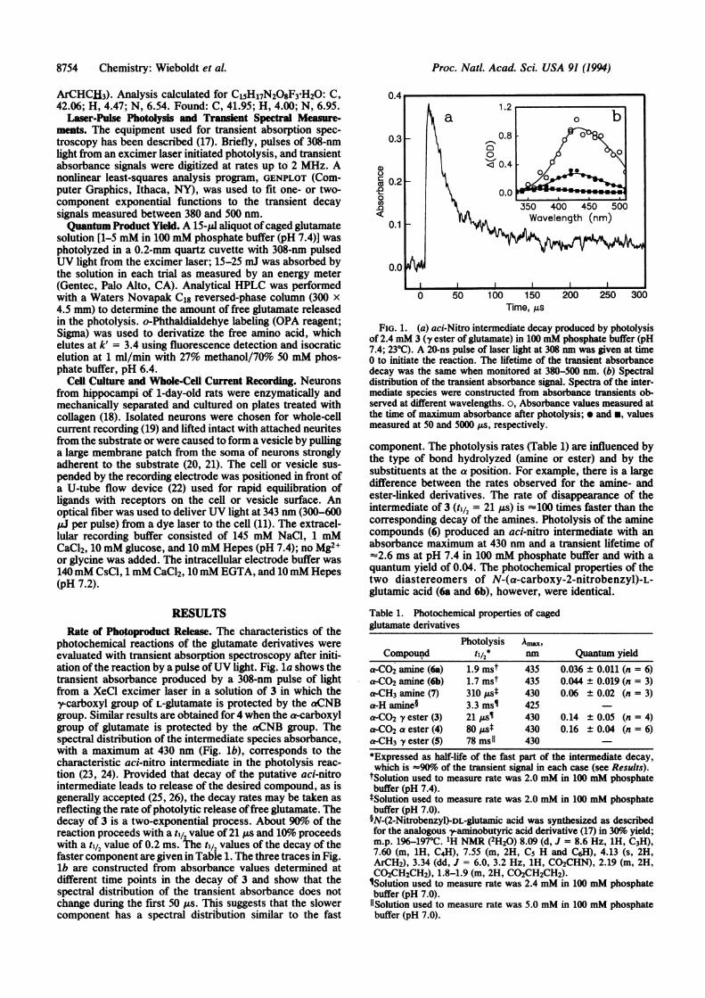
photochemical reactions of the glutamate derivatives wereevaluated with transient absorption spectroscopy after initi-ation of the reaction by a pulse ofUV light. Fig. la shows thetransient absorbance produced by a 308-nm pulse of lightfrom a XeCl excimer laser in a solution of 3 in which they--carboxyl group of L-glutamate is protected by the aCNB
group. Similar results are obtained for 4 when the a-carboxylgroup of glutamate is protected by the aCNB group. Thespectral distribution of the intermediate species absorbance,with a maximum at 430 nm (Fig. lb), corresponds to thecharacteristic aci-nitro intermediate in the photolysis reac-tion (23, 24). Provided that decay of the putative aci-nitrointermediate leads to release of the desired compound, as isgenerally accepted (25, 26), the decay rates may be taken asreflecting the rate ofphotolytic release offree glutamate. Thedecay of 3 is a two-exponential process. About 90%6 of thereaction proceeds with a til2 value of 21 ps and 10%o proceedswith a til2 value of 0.2 ms. The tl12 values of the decay of thefaster component are given in Table 1. The three traces in Fig.lb are constructed from absorbance values determined atdifflerent time points in the decay of 3 and show that the
spectral distribution of the transient absorbance does notchange during the first 50 Us. This suggests that the slowercomponent has a spectral distribution similar to the fast
6 0.20.0 0
~~~~~~~350400 450 500
0.1 ~~~~~Wavelength (nm)
0.0
0 50 100 150 200 250 300Time, As
FIG. 1. (a) aci-Nitro intermediate decay produced by photolysisof 2.4 mM 3 (y ester of glutamate) in 100 mM phosphate buffer (pH7.4; 230C). A 20-ns pulse of laser light at 308 nm was given at time0 to initiate the reaction. The lifetime of the transient absorbancedecay was the same when monitored at 380-500 nm. (b) Spectraldistribution of the transient absorbance signal. Spectra of the inter-mediate species were constructed from absorbance transients ob-served at different wavelengths. o, Absorbance values measured atthe time of maximum absorbance after photolysis; * and *, valuesmeasured at 50 and 5000 ,us, respectively.
component. The photolysis rates (Table 1) are influenced bythe type of bond hydrolyzed (amine or ester) and by thesubstituents at the a position. For example, there is a largedifference between the rates observed for the amine- andester-linked derivatives. The rate of disappearance of theintermediate of 3 (til2 = 21 us) is =100 times faster than thecorresponding decay of the amines. Photolysis of the aminecompounds (6) produced an aci-nitro intermediate with anabsorbance maximum at 430 nm and a transient lifetime of-%2.6 ms at pH 7.4 in 100 mM phosphate buffer and with aquantum yield of 0.04. The photochemical properties of thetwo diastereomers of N-(a-carboxy-2-nitrobenzyl)-L-glutamic acid (6a and 6b), however, were identical.
Table 1. Photochemical properties of cagedglutamate derivatives
Photolysis Ama,Compound tl/2* Quantum yield
a-CO2 amine (6a) 1.9 inst 435 0.036 ± 0.011 (n = 6)a-CO2 amine (6b) 1.7 mst 435 0.044 ± 0.019 (n = 3)a-CH3 amine (7) 310 pst 430 0.06 ± 0.02 (n = 3)a-H amine§ 3.3 msi 425a-CO2 fy ester (3) 21 psi 430 0.14 ± 0.05 (n = 4)a-CO2 a ester (4) 80 ,ust 430 0.16 ± 0.04 (n = 6)a-CH3 y ester (5) 78 ms1 430
*Expressed as half-life of the fast part of the intermediate decay,which is =90%6 of the transient signal in each case (see Results).
tSolution used to measure rate was 2.0 mM in 100 mM phosphatebuffer (pH 7.4).tSolution used to measure rate was 2.0 mM in 100 mM phosphatebuffer (pH 7.0).§N-(2-Nitrobenzyl)-DL-glutamic acid was synthesized as describedfor the analogous y-aminobutyric acid derivative (17) in 30% yield;m.p. 196-197rC. 1H NMR (2H20) 8.09 (d, J = 8.6 Hz, 1H, C3H),7.60 (m, 1H, C4H), 7.55 (m, 2H, Cs H and C6H), 4.13 (s, 2H,ArCH2), 3.34 (dd, J = 6.0, 3.2 Hz, 1H, CO2CHN), 2.19 (m, 2H,C02CH2CH2), 1.8-1.9 (m, 2H, CO2CH2CH2).ISolution used to measure rate was 2.4 mM in 100 mM phosphatebuffer (pH 7.0)."Solution used to measure rate was 5.0 mM in 100 mM phosphatebuffer (pH 7.0).
8754 Chemistry: Wieboldt et al.

Proc. Natl. Acad. Sci. USA 91 (1994) 8755
The a substituent affects both ester and amine derivatives.The rate of intermediate decay is slower when -CO2 isreplaced by -CH3 in the ester-linked series but is slightlyfaster in the amines. Similar effects have been observed foranalogous 2-nitrobenzyl derivatives ofamides (27). Also, they ester of glutamate (3) photolyzes more rapidly than the aester (4). Therefore, photolysis is sensitive to changes inconfiguration close to the bond that hydrolyzes and tosubstitution at the a position of the protecting group.Quantum Yield and Hydrolysis. The product quantum
yields measured for the derivatives are given in Table 1.Deprotection of the amine bond leads to an overall productyield for released glutamate that is -4-fold lower than theyield ofcorresponding esters. The a substituents do not exerta large influence on the yield for either the amine or esterlinkage.
Buffered physiological saline solutions of4 used for whole-cell studies were found to have a background level of freeglutamate that increased with time. Solutions (1 mM) of3 and4 were prepared in 10 mM Hepes buffer (pH 7.4). Thesolutions were held at 27TC; aliquots were sampled at 1, 2, 4,6, 24, and 48 hr, derivatized with o-phthaldialdehyde, andanalyzed for free glutamate by HPLC with fluorescencedetection. Under these conditions, the a-glutamate ester (4)hydrolyzes to produce free glutamate with a til2 of '45 hr,whereas free glutamate in a solution of3 increased <2% after2 days.
Activation ofNeuronal Glutamate Receptors by Photolysis ofCaged Glutamate. Photolysis of the caged glutamate deriva-tives induced a transmembrane ion current in rat hippocam-pal neurons and membrane vesicles as monitored by whole-cell recording. Fig. 2 presents examples of currents initiatedin neurons by photolysis of caged glutamate (Fig. 2a) or byfree glutamate used with a cell-flow technique (29) (Fig. 2b).The tlq2 value of the rising phase of the current (1.1 ms in Fig.2a) represents the lifetime of an open glutamate channel(s).Values reported for the lifetime of open glutamate channelsmeasured by single-channel current recordings are in the1-ms time scale (e.g., ref. 20). The current decay, indicative
3.0-01a < ffh b
2.5 "~E O.l
.2.0 00.05a) I0 500 1000
.' 1.5 _ \ Time (ms)B _0.20F
C IIC
(b~ ~ ~ ?0.10L5
0.0 _ Time (ins)0 10 20 30
Time, ms
FIG. 2. Comparison of photolysis (10, 11, 28) and flow (22, 29)methods used to activate whole-cell current response of glutamatereceptors in rat hippocampal neurons. Measurements were made inphysiological saline at RT and the transmembrane voltage wasclamped at -60 mV. (a) Current produced when 500 ,uM 3 wasphotolyzed over a neuron detached from the culture dish. Nobackground response from the caged glutamate itself was observed.(b) Maximum signal produced when 300 ,M free glutamate flowedrapidly over the same cell. (c) Response to rapid flow of 600 AM freeglutamate over a membrane vesicle obtained from a hippocampalneuron.
of receptor desensitization, occurs in two time zones; onlythe rapid phase is shown. The maximum observed current, 3nA, is 20 times larger than the current amplitude observedwith the same cell when 300 ,LM glutamate was delivered tothe cell by a flow device (22) (Fig. 2b). In addition, in thecell-flow experiment only a relatively slow falling phase ofthe current is observed. The explanation is that in the flowexperiment, receptor desensitization occurs on a time scalecomparable to equilibration of receptors on the cell surfacewith glutamate. Consequently, the receptor form associatedwith the rapid inactivation phase is not seen. In the experi-ment illustrated in Fig. 2c, a 600 AuM solution of free gluta-mate in physiological saline was applied rapidly to an -7-,Amround vesicle obtained from the membrane of the neuron.The total rise time for the response was 14 ms; the rapiddesensitization (220 so1) that takes place during this equili-bration period prevents a direct measurement of the truecurrent amplitude. The slow phase associated with desensi-tization (shown in Fig. 2b) can no longer be detected becauseof the smaller signal when vesicles are used. The area of thecell membrane forming the vesicle is 10 times smaller thanthat of the intact neuron and the number of receptor sites isconsequently expected to be smaller. In separate experi-ments, 1.0 mM 3 applied in the absence and presence of (50jLM) free glutamate neither potentiated nor inhibited thecurrent response to free glutamate.
DISCUSSIONThe goal of the present investigation was to further developphotolabile derivatives of neurotransmitters (9, 13, 17, 27)suitable for rapid chemical kinetic investigations ofreceptorson the surface of single cells (10, 11) and for mapping thedistribution of receptors on cell surfaces (12) and functionalconnections between neurons (14).The results in Fig. 2 demonstrate the usefulness of the
caged compound in rapid chemical kinetic investigations ofglutamate receptors. The effect of glutamate concentrationon the current rise time, the maximum current amplitude, andthe falling phase of the current (Fig. 2a) can be determined ina single experiment that can give information about the rateand equilibrium constants for channel opening and receptordesensitization (10). Fig. 2 b and c shows that when tech-niques with inappropriate time resolution are used importantinformation about the reaction is lost. The experiment in Fig.2b gives information about only the receptorforms associatedwith the slow desensitization. By using a vesicle the timeneeded for the receptors to equilibrate with glutamate isconsiderably reduced (because the surface area of the mem-brane in contact with flowing solutions of glutamate isdecreased), but the signal is also reduced and informationabout receptor forms that desensitize slowly (the minorcomponent; Fig. 2b) is not obtained.The aCNB group was introduced to protect the amino
group of carbamoylcholine (9). The resulting caged com-pound was found to be suitable for rapid chemical kineticinvestigations of the nicotinic acetylcholine receptor in thesubmillisecond time region (10, 11). The results describedsuggest that the aCNB group, successfully used in caging theamino group of carbamoylcholine, may be equally useful incaging the carboxyl group of neurotransmitters and otherinteresting biological compounds with carboxyl groups, in-cluding N-methyl-D-aspartic acid and a-amino-3-hydroxy-5-methyl-4-isoxazolepropionic acid, which are activating li-gands for glutamate receptor subtypes.
We thank Dr. Ying Chen Lynn for assistance with cell culture andwhole-cell recording, Dr. Christof Grewer for the data in Fig. 1, andProf. Jack Kaplan (University of Pennsylvania) and Prof. LawrenceKatz (Duke University Medical Center) for helpful comments. This
Chemistry: Wieboldt et al.
Proc. Natl. Acad. Sci. USA 91 (1994)
work was supported by a grant (GM04842) from the NationalInstitutes of Health.
1. Kandel, E. R., Schwartz, J. H. & Jessel, T. M. (1991) Princi-ples ofNeural Science (Elsevier, New York), 3rd Ed.
2. Cunningham, M. D., Ferkany, J. W. & Enna, S. J. (1994) LifeSci. 54, 135-148.
3. Trussell, L. O., Thio, L. L., Zorumski, C. F. & Fischbach,G. D. (1988) Proc. Natd. Acad. Sci. USA 85, 4562-4566.
4. Hammes, G. G. (1982) Enzyme Catalysis and Regulation (Ac-ademic, New York).
5. Kaplan, J. H. (1990) Annu. Rev. Physiol. 52, 897-914.6. Corrie, J. E. T. & Trentham, D. R. (1993) in Bioorganic Pho-
tochemistry, ed. Morrison, H. (Wiley, New York), Vol. 2, pp.243-305.
7. Adams, S. R. & Tsien, R. Y. (1993) Annu. Rev. Physiol. 55,755-783.
8. Walker, J. W., McCray, J. A. & Hess, G. P. (1986) Biochem-istry 25, 1799-1805.
9. Milburn, T., Matsubara, N., Billington, A. P., Udgaonkar,J. B., Walker, J. W., Carpenter, B. K., Webb, W. W.,Marque, J., Denk, W., McCray, J. A. & Hess, G. P. (1989)Biochemistry 28, 49-55.
10. Matsubara, N., Billington, A. P. & Hess, G. P. (1992) Bio-chemistry 31, 5477-5487.
11. Niu, L. & Hess, G. P. (1993) Biochemistry 32, 3831-3835.12. Denk, W. (1994) Proc. Natl. Acad. Sci. USA 91, 6629-6633.13. Wilcox, M., Viola, R. W., Johnson, K. W., Billington, A. P.,
Carpenter, B. K., McCray, J. A., Guzikowski, A. P. & Hess,G. P. (1990) J. Org. Chem. 55, 1585-1589.
14. Callaway, E. M. & Katz, L. C. (1993) Proc. Natl. Acad. Sci.USA 90, 7661-7665.
15. Corrie, J. E. T., DeSantis, A., Katayama, Y., Khodakhah, K.,Messenger, J. B., Ogden, D. C. & Trentham, D. R. (1993) J.Physiol. (London) 465, 1-8.
16. Kaplan, J. H., Forbush, B. & Hoffman, J. F. (1978) Biochem-istry 17, 1929-1935.
17. Wieboldt, R., Ramesh, D., Carpenter, B. K. & Hess, G. P.(1994) Biochemistry 33, 1526-1533.
18. Romijn, H. J., van Huizen, F. & Wolters, P. S. (1984) Neuro-sci. Behav. Rev. 8, 703-742.
19. Hamill, 0. P., Marty, E., Neher, E., Sakmann, B. & Sigworth,F. J. (1981) Pflugers Arch. 391, 85-100.
20. Sather, W., Dieudonne, S., Macdonald, J. F. & Ascher, P.(1992) J. Physiol. (London) 450, 643-672.
21. Walstrom, K. M. & Hess, G. P. (1994) Biochemistry 33, 7718-7730.
22. Krishtal, 0. A. & Pidoplichko, V. I. (1980) Neuroscience 5,2325-2327.
23. McCray, J. A., Herbette, L., Kihara, T. & Trentham, D. R.(1980) Proc. Natl. Acad. Sci. USA 77, 7237-7241.
24. Schupp, H., Wong, W. K. & Schnabel, W. (1987) J. Photo-chem. 36, 85-97.
25. Walker, J. W., Reid, G. P., McCray, J. A. & Trentham, D. R.(1988) J. Am. Chem. Soc. 110, 7170-7177.
26. Wootton, J. F. & Trentham, D. R. (1989) NATO Adv. StudyInst. Ser. C 272, 277-2%.
27. Ramesh, D., Wieboldt, R., Billington, A. P., Carpenter, B. K.& Hess, G. P. (1993) J. Org. Chem. 58, 4599-4605.
28. Billington, A. P., Matsubara, N., Webb, W. W. & Hess, G. P.(1992) Tech. Protein Chem. 11, 417-427.
29. Udgaonkar, J. B. & Hess, G. P. (1987) Proc. Natl. Acad. Sci.USA 84, 8758-8762.
8756 Chemistry: Wieboldt et al.