phospholipase a2 in skin biology: new insights from gene
TRANSCRIPT
REVIEW Open Access
Phospholipase A2 in skin biology: newinsights from gene-manipulated mice andlipidomicsMakoto Murakami1,2, Kei Yamamoto3,4 and Yoshitaka Taketomi1*
Abstract
The skin represents one of the tissues that are most profoundly influenced by alterations in the quality of lipids(lipoquality). Lipids not only constitute cellular membranes, but also serve as bioactive lipid mediators and essentialcomponents of the skin barrier. Phospholipase A2 (PLA2) enzymes supply fatty acids and lysophospholipids frommembrane phospholipids, thereby variably affecting cutaneous homeostasis. Accordingly, perturbation ofparticular PLA2-driven lipid pathways can be linked to various forms of skin disease. In this review article, wehighlight the roles of several PLA2 subtypes in cutaneous pathophysiology, as revealed by transgenic/knockout studiesin combination with comprehensive lipidomics. We focus mainly on secreted PLA2 group IIF (sPLA2-IIF), whichis associated with epidermal hyperplasia through mobilization of a unique lipid metabolite. We also addressthe distinct roles of sPLA2-IIE in hair follicles and sPLA2-IID in lymphoid immune cells that secondarily affectcutaneous inflammation, and provide some insights into species differences in sPLA2s. Additionally, we brieflyoverview the patatin-like phospholipase PNPLA1, which belongs to the Ca2+-independent PLA2 (iPLA2) family,as a key regulator of skin barrier function through catalysis of a unique non-PLA2 reaction. These knowledgeson lipid metabolism driven by various PLA2 subtypes will open novel opportunities for translated studies towarddiagnosis and therapy of human skin diseases.
Keywords: Knockout mouse, Lipid mediator, Lipidomics, Phospholipase A2, Skin
BackgroundThe skin consists of the outer epidermis, beneath whichare the dermis and subcutaneous tissue. Epidermal kera-tinocytes undergo a tightly regulated program of prolif-eration and differentiation leading to formation of thestratified epidermis, which consists of four layers includ-ing the stratum basale (SB), the stratum spinosum (SS),the stratum granulosum (SG), and the stratum corneum(SC) from the inside to the outside. For survival in a dryterrestrial environment, the epidermis constitutes a life-sustaining skin barrier, which not only prevents waterloss (inside-out barrier), but also protects against invasionof environmental substances or microorganisms (outsi-de-in barrier) [1]. In the uppermost SC, corneocytes are
embedded in a lipid-rich extracellular matrix that formslamellar membranes composed of ceramides, cholesterol,and fatty acids in a mildly acidic environment [2]. The epi-dermis also has immunologic functions, protecting theskin from ultraviolet damage via pigmentation of melano-cytes and from external harmful stimuli by releasing vari-ous bioactive factors such as cytokines, chemokines,DAMPs (danger-associated molecular patterns), and lipidmediators, which relay signals to specialized immune cellsresiding in the epidermis and dermis [3].Another important component of the skin is the hair
follicle, whose morphogenesis is regulated by interac-tions between epidermal keratinocytes committed to hairfollicle differentiation and dermal fibroblasts committedto formation of the dermal papilla of developing hair fol-licles [4]. These epithelial-mesenchymal interactions cul-minate in the formation of the hair shaft, which issurrounded by the multilayered inner root sheath andouter root sheath, the latter comprising an outermost
* Correspondence: [email protected] of Microenvironmental and Metabolic Health Science, Center forDisease Biology and Integrative Medicine, Graduate School of Medicine, TheUniversity of Tokyo, 7-3-1 Hongo, Bunkyo-ku, Tokyo 113-8655, JapanFull list of author information is available at the end of the article
Inflammation and Regeneration
© The Author(s). 2018 Open Access This article is distributed under the terms of the Creative Commons Attribution 4.0International License (http://creativecommons.org/licenses/by/4.0/), which permits unrestricted use, distribution, andreproduction in any medium, provided you give appropriate credit to the original author(s) and the source, provide a link tothe Creative Commons license, and indicate if changes were made. The Creative Commons Public Domain Dedication waiver(http://creativecommons.org/publicdomain/zero/1.0/) applies to the data made available in this article, unless otherwise stated.
Murakami et al. Inflammation and Regeneration (2018) 38:31 https://doi.org/10.1186/s41232-018-0089-2

concentric layer of keratinocytes. Hair follicles undergorepeated cycles of growth (anagen), regression (catagen),and rest (telogen) during their life span, representingone of the most regenerative organs in the body. Withinthe apex of the follicle are sebaceous glands, which pro-duce sebum. The adipocyte layer within the hypodermisalso constitutes a significant compartment of the skin,contributing to hair follicle activation [5], skin regener-ation [6], and cold-induced adaptive thermogenesis [7].Lipids play fundamental roles in skin physiology and
pathology. Dysregulated production of polyunsaturatedfatty acid (PUFA)- or lysophospholipid-derived lipid medi-ators can be linked to skin disorders including alopecia,inflammation, and cancer. For instance, arachidonic acid(AA; ω6 C20:4)-derived lipid mediators such as prosta-glandins (PGs) and leukotrienes (LTs) have diverse roles inimmune responses and keratinocyte activation [8, 9], ei-cosapentaenoic acid (EPA; ω3 C20:5)- or docosahexaenoicacid (DHA; ω3 C22:6)-derived resolvins attenuate skin im-mune responses [10, 11], and lysophosphatidic acid (LPA)controls hair homeostasis [12, 13]. Apart from these sig-naling lipids, linoleic acid (LA; ω6 18:2), by far the mostabundant PUFA in the epidermis, is esterified to theω-hydroxyl group of ultra-long chain fatty acids in cera-mides, thus forming ω-O-acylceramide, a structural lipidthat is essential for skin barrier function [14]. Fatty acidshave also been proposed to be important for SC acidifica-tion [15].Release of fatty acids and lysophospholipids from gly-
cerophospholipids (phospholipids hereafter) is catalyzedby phospholipase A2 (PLA2) enzymes, which are classi-fied into several families as shown in Table 1 [16]. Untilrecently, however, it has remained obscure as to whichPLA2 subtype(s) is important in the skin, which lipidspecies serve as the substrates and products for thePLA2(s), and how the PLA2-driven lipid metabolitesaffect skin pathophysiology. In this review, we highlightthe distinct roles of several secreted PLA2s (sPLA2s) andthe patatin-like phospholipase PNPLA1, whose functionshave been revealed by recent studies using gene-ma-nipulated (transgenic and knockout) mice in combinationwith mass spectrometry-based analytical techniques re-ferred to collectively as lipidomics. Importantly, these en-zymes are linked to unique lipid pathways distinct fromcanonical AA metabolism. The localizations and functionsof particular PLA2s in the skin, as described in this review,are summarized in Fig. 1.
sPLA2-IIF, an epidermal sPLA2
The sPLA2 family consists of 11 isoforms with distinctsubstrate specificities and tissue distributions [17, 18].Historically, several sPLA2s have been detected in mouseand human skin, but by using semi-quantitative RT-PCRand immunoblotting which have uncertain specificity
[19–23]. sPLA2s have also been suggested to supply fattyacids for formation of the SC acid mantle, a hypothesisthat stems primarily from the observation that SC acid-ity is perturbed by non-specific sPLA2 inhibitors [15,23–25]. However, the molecular identity of any particu-lar sPLA2(s) that participates in skin homeostasis anddiseases has remained unclear until recently. Now, it hasbecome obvious that sPLA2-IIF is a bona fide “epidermalsPLA2” that controls keratinocyte differentiation, hyper-proliferation, and function [26].Among the group II subfamily sPLA2s (which include
sPLA2-IIA, sPLA2-IIC, sPLA2-IID, sPLA2-IIE, sPLA2-IIF,and sPLA2-V), sPLA2-IIF has several unique features[27, 28]. sPLA2-IIF has a uniquely long C-terminal ex-tension that is proline-rich and contains a single cyst-eine, which raises the possibility that it might form acovalent homodimer, although this hypothesis has notbeen confirmed. In contrast to other group II subfamilysPLA2s that are basic proteins and catalytically active atneutral to mildly basic pH, sPLA2-IIF is an acidic protein(pI ~ 5.8) and retains its full enzymatic activity even atmildly acidic pH. This property may be related to thedistribution of this enzyme in the upper epidermis (seebelow), which has a mildly acidic environment [15]. Fur-thermore, sPLA2-IIF is more hydrophobic than othersPLA2s, and probably because of this, it has a uniqueability to penetrate and disrupt lipid monolayers and bi-layers in vitro; when added exogenously, it rapidly entersthe cells in an endocytosis-independent manner to formunusual aggregates [29]. Moreover, when overexpressed,sPLA2-IIF also tends to aggregate within the cells andcan undergo N-glycosylation at three positions, possiblyincreasing its water solubility and thereby decreasing theunusual accumulation of sPLA2-IIF aggregates. However,it remains unknown whether or not endogenous sPLA2-IIF (or any other sPLA2s) is N-glycosylated in vivo. In aPLA2 enzyme assay using a phospholipid mixture ex-tracted from mouse skin as a substrate (natural membraneassay [30]), a physiologically relevant concentration ofsPLA2-IIF preferentially hydrolyzes phosphatidylethanol-amine (PE; particularly plasmalogen-type PE) containingPUFAs (particularly DHA) to yield plasmalogen-typelysoPE (P-LPE) and DHA in preference to AA [26].Therefore, although sPLA2-IIF is capable of releasingAA when overexpressed in mammalian cells at super-physiological levels [31], it may mobilize lipid metabo-lites separately from canonical AA metabolism underphysiological conditions (see below).It is now obvious that sPLA2-IIF is a major sPLA2
expressed in mouse epidermis, where it is distributed inthe suprabasal SS, SG, and SC layers [26]. Developmen-tal expression of Pla2g2f in mouse skin is far greaterthan that of other sPLA2s (except for Pla2g2e, see below),gradually increasing before birth to reach a maximum
Murakami et al. Inflammation and Regeneration (2018) 38:31 Page 2 of 10

level by P5 (Fig. 2a). sPLA2-IIF expression is markedly in-duced during Ca2+-induced differentiation and also ro-bustly upregulated in primary keratinocytes followingstimulation with the Th17 cytokines IL-22 and IL-17A.Moreover, sPLA2-IIF is induced in mouse skin treatedwith imiquiod, an inducer of experimental psoriasis, andalso highly expressed in the hyperplasic epidermis of pa-tients with psoriasis. Strikingly, global or skin-specifictransgenic mice overexpressing mouse sPLA2-IIF (Pla2g2f-TG) spontaneously develop psoriasis-like epidermal hyper-plasia and alopecia, with increased expression of variouspsoriasis markers such as S100A9 and IL-36α [26], sug-gesting that increased expression of this sPLA2 alonecould trigger psoriasis.In a basal state, Pla2g2f−/− mice have mild abnormalities
in the skin (particularly the abdominal skin, probably be-cause it is continuously exposed to friction against the
ground surface), as revealed by a fragile stratum corneumwith modest perturbation of skin barrier function andacidity [26]. After tape stripping of the SC, Pla2g2f−/−
mice display delayed recovery from the epidermal bar-rier perturbation [23]. In primary culture, keratinocytesfrom Pla2g2f−/− mice fail to differentiate and becomeproperly activated (Fig. 2b), and similar defects are evi-dent when WT keratinocytes are treated with a pan-sPLA2 inhibitor or an sPLA2-IIF-directed siRNA. Mostimportantly, in pathological settings, Pla2g2f−/− miceare protected from epidermal hyperplasia and associ-ated inflammation in models of Th17-dependent psor-iasis and Th1-dependent contact hypersensitivity (CHS)[26]. Consistent with this, Pla2g2f deficiency in kerati-nocytes markedly impairs the induction of several psor-iasis markers in response to IL-17A or IL-22. Moreover,Pla2g2f−/− mice are also protected from skin carcinogenesis,
Table 1 The classification of PLA2 family. sPLA2, cPLA2, and iPLA2/PNPLA are the original big three among the PLA2 family.The sPLA2 family contains 10 catalytically active isoforms (IB, IIA, IIC, IID, IIE, IIF, III, V, X, XIIA) and 1 inactive isoform (XIIB) inmammals. The cPLA2 family comprises 6 isoforms (α-ζ). The human genome encodes 9 iPLA2 enzymes. These enzymes arenow more generally referred to as PNPLA (1–9). In this review, the biological roles of particular PLA2s in the context of skinhomeostasis and diseases were described
Family Isoforms Gene name Common name Alias Enzyme property
sPLA2 11 PLA2G1B sPLA2-IB Pancreatic sPLA2 PLA2
PLA2G2A sPLA2-IIA Non-pancreatic sPLA2 PLA2
PLA2G2C sPLA2-IIC PLA2
PLA2G2D sPLA2-IID PLA2
PLA2G2E sPLA2-IIE PLA2
PLA2G2F sPLA2-IIF PLA2
PLA2G3 sPLA2-III PLA2
PLA2G5 sPLA2-V PLA2
PLA2G10 sPLA2-X PLA2
PLA2G12A sPLA2-XIIA PLA2
PLA2G12B sPLA2-XIIB Inactive
cPLA2 6 PLA2G4A cPLA2α PLA2
PLA2G4B cPLA2β PLA1/A2
PLA2G4C cPLA2γ PLA2, transacylase, lysopholipase
PLA2G4D cPLA2δ PLA1/A2
PLA2G4E cPLA2ε PLA1/A2, N-acyltransferase
PLA2G4F cPLA2ζ PLA1/A2
iPLA2/PNPLA 9 PNPLA1 Transacylase
PNPLA2 iPLA2ζ ATGL TG lipase
PNPLA3 iPLA2ε ADPN TG lipase, acyltransferase, restiny-esteryl lipase
PNPLA4 iPLA2η GS2 Retinyl-esteryl lipase?
PNPLA5 GS2L TG lipase
PNPLA6 iPLA2δ NTE Lysophospholipase
PNPLA7 NRE Lysophospholipase
PNPLA8 iPLA2γ PLA1/A2
PNPLA6 iPLA2β PNPLA9 PLA2
Murakami et al. Inflammation and Regeneration (2018) 38:31 Page 3 of 10

whereas Pla2g2f-TG mice conversely develop largerskin tumors than WT mice [26]. Mechanistically, sPLA2-IIF preferentially hydrolyzes plasmalogen-type PE secretedfrom keratinocytes to yield P-LPE, a unique lysophospho-lipid that facilitates the differentiation and activation ofkeratinocytes, leading to exacerbation of epidermal hyper-plasia and inflammation. Indeed, the skin levels of P-LPEare correlated well with those of sPLA2-IIF expression inseveral skin disease models, and topical application ofP-LPE to Pla2g2f−/− skin in vivo or supplementation ofPla2g2f−/− keratinocytes with P-LPE ex vivo restoresthe psoriasis-related phenotypes.Taken together, these results indicate that sPLA2-IIF
promotes epidermal hyperplasic diseases including psor-iasis and skin cancer and that P-LPE, a primary sPLA2-IIF product, represents a biomarker and bioactive lipidthat reflects the expression and function of sPLA2-IIF.Given that sPLA2-IIF is expressed in the epidermis ra-ther specifically and that Pla2g2f−/− mice display moreprofound skin phenotypes in diseases than in homeosta-sis, inhibition of this particular sPLA2 may be useful fortreatment of psoriasis, skin cancer, or other conditionsinvolving epidermal hyperplasia. It remains to be deter-mined, however, whether sPLA2-IIF-driven P-LPE would
act on keratinocytes through a specific receptor orthrough other mechanism(s). It is also possible that DHA,another sPLA2-IIF-driven product, would be metabolizedto certain metabolites that could affect skin homeostasis,since DHA or its pro-resolving metabolites can facilitateskin wound healing, suppress psoriasis, and prevent neo-plastic transformation of keratinocytes [32–34].
sPLA2-IIE, a hair follicular sPLA2
Although sPLA2-IIE is not substantially expressed in theepidermis, it is a major “hair follicular sPLA2” in mice,being expressed in hair follicles in synchrony with haircycling [35]. Thus, during the anagen phase, sPLA2-IIEis distributed in companion cells of the outer root sheathand cuticular cells of the inner root sheath in growinghair follicles. At P10–15, when hair follicles are max-imally developed in the initial hair cycle, the expressionof sPLA2-IIE becomes maximal, even exceeding that ofsPLA2-IIF in the whole mouse skin (Fig. 2a). In contrast,during the catagen to telogen phase, when hair folliclesregress, sPLA2-IIE expression promptly decreases to anegligible level, and then rises again in correlation withentry into the next anagen.
Fig. 1 Expressions and functions of various PLA2s in mouse skin. sPLA2-IIF is localized in the suprabasal epidermis and produces P-LPE, whichpromotes epidermal hyperplasic diseases such as psoriasis and skin cancer. Epidermal sPLA2-IIF expression and thereby P-LPE production areaugmented by IL-17A and IL-22 supplied by γδ T and Th17 cells in psoriasis. sPLA2-IIE is localized in hair follicles in synchrony with the growingphase (anagen) of hair cycling and may regulate hair homeostasis. sPLA2-IID is constitutively expressed in DCs and M2 macrophages in regionalLNs and produces ω3 PUFA-derived anti-inflammatory lipid mediators, which put a brake on Th1 or Th17 immunity, thereby sequestering CHSand psoriasis and promoting skin cancer. PNPLA1 is expressed in the border of SG and SC, where it produces ω-O-acylceramide that is essentialfor skin barrier function. For details, please see the text
Murakami et al. Inflammation and Regeneration (2018) 38:31 Page 4 of 10
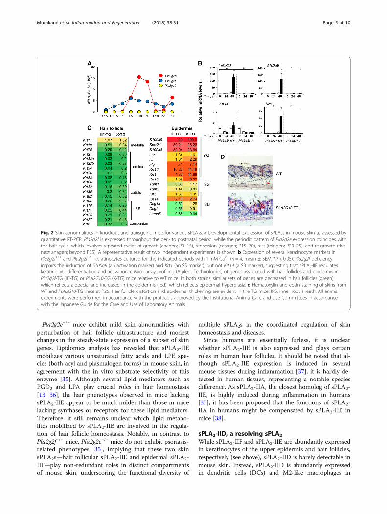
Pla2g2e−/− mice exhibit mild skin abnormalities withperturbation of hair follicle ultrastructure and modestchanges in the steady-state expression of a subset of skingenes. Lipidomics analysis has revealed that sPLA2-IIEmobilizes various unsaturated fatty acids and LPE spe-cies (both acyl and plasmalogen forms) in mouse skin, inagreement with the in vitro substrate selectivity of thisenzyme [35]. Although several lipid mediators such asPGD2 and LPA play crucial roles in hair homeostasis[13, 36], the hair phenotypes observed in mice lackingsPLA2-IIE appear to be much milder than those in micelacking synthases or receptors for these lipid mediators.Therefore, it still remains unclear which lipid metabo-lites mobilized by sPLA2-IIE are involved in the regula-tion of hair follicle homeostasis. Notably, in contrast toPla2g2f−/− mice, Pla2g2e−/− mice do not exhibit psoriasis-related phenotypes [35], implying that these two skinsPLA2s—hair follicular sPLA2-IIE and epidermal sPLA2-IIF—play non-redundant roles in distinct compartmentsof mouse skin, underscoring the functional diversity of
multiple sPLA2s in the coordinated regulation of skinhomeostasis and diseases.Since humans are essentially furless, it is unclear
whether sPLA2-IIE is also expressed and plays certainroles in human hair follicles. It should be noted that al-though sPLA2-IIE expression is induced in severalmouse tissues during inflammation [37], it is hardly de-tected in human tissues, representing a notable speciesdifference. As sPLA2-IIA, the closest homolog of sPLA2-IIE, is highly induced during inflammation in humans[37], it has been proposed that the functions of sPLA2-IIA in humans might be compensated by sPLA2-IIE inmice [38].
sPLA2-IID, a resolving sPLA2
While sPLA2-IIF and sPLA2-IIE are abundantly expressedin keratinocytes of the upper epidermis and hair follicles,respectively (see above), sPLA2-IID is barely detectable inmouse skin. Instead, sPLA2-IID is abundantly expressedin dendritic cells (DCs) and M2-like macrophages in
Fig. 2 Skin abnormalities in knockout and transgenic mice for various sPLA2s. a Developmental expression of sPLA2s in mouse skin as assessed byquantitative RT-PCR. Pla2g2f is expressed throughout the peri- to postnatal period, while the periodic pattern of Pla2g2e expression coincides withthe hair cycle, which involves repeated cycles of growth (anagen; P0–15), regression (catagen; P15–20), rest (telogen; P20–25), and re-growth (thenext anagen; beyond P25). A representative result of two independent experiments is shown. b Expression of several keratinocyte markers inPla2g2f+/+ and Pla2g2f−/− keratinocytes cultured for the indicated periods with 1 mM Ca2+ (n = 4, mean ± SEM, *P < 0.05). Pla2g2f deficiencyimpairs the induction of S100a9 (an activation marker) and Krt1 (an SS marker), but not Krt14 (a SB marker), suggesting that sPLA2-IIF regulateskeratinocyte differentiation and activation. c Microarray profiling (Agilent Technologies) of genes associated with hair follicles and epidermis inPla2g2f-TG (IIF-TG) or PLA2G10-TG (X-TG) mice relative to WT mice. In both strains, similar sets of genes are decreased in hair follicles (green),which reflects alopecia, and increased in the epidermis (red), which reflects epidermal hyperplasia. d Hematoxylin and eosin staining of skins fromWT and PLA2G10-TG mice at P25. Hair follicle distortion and epidermal thickening are evident in the TG mice. IRS, inner root sheath. All animalexperiments were performed in accordance with the protocols approved by the Institutional Animal Care and Use Committees in accordancewith the Japanese Guide for the Care and Use of Laboratory Animals
Murakami et al. Inflammation and Regeneration (2018) 38:31 Page 5 of 10

secondary lymphoid organs such as the spleen and lymphnodes (LNs) of mice and humans [39, 40]. The expressionof sPLA2-IID is downregulated, rather than upregulated,following inflammatory stimuli [39, 41]. This property isunique among the sPLA2 isoforms and probably reflectsits role as a “resolving sPLA2” that counteracts inflamma-tion [18, 39]. Despite the low expression of sPLA2-IID inskin, Pla2g2d deficiency leads to exacerbation of CHS andpsoriasis. This is most likely because sPLA2-IID attenuatesadaptive immunity in the LNs, thereby sequestering Th1and Th17 immune responses [39, 40].In a model of CHS, the resolution of inflammation in
the skin and regional LNs is delayed in Pla2g2d−/− mice[39]. In this state, expression of the Th1 cytokines IFN-γand IL-12 is robustly elevated in the LNs. Likewise, in amodel of psoriasis, Pla2g2d−/− mice display more severeepidermal hyperplasia than do Pla2g2d+/+ mice, with in-creased IL-17A+ or IL-22+ T cells in the affected skinand LNs [40]. Furthermore, DCs isolated from Pla2g2d−/−
mice are hyper-activated even in the absence of stimu-lation. Mechanistically, sPLA2-IID in the LNs constitu-tively hydrolyzes PUFA-containing PE species (likely inmicroparticle membranes) to mobilize ω3 PUFA-derivedanti-inflammatory lipid mediators, which can put abrake on DC-committed adaptive immunity. Indeed,the steady-state levels of ω3 PUFAs and their metabo-lites, such as DHA-derived resolvin D1 (RvD1), aremarkedly reduced in the LNs of Pla2g2d−/− mice rela-tive to Pla2g2d+/+ mice. Conversely, Pla2g2d-TG micedisplay milder inflammation in the CHS and psoriasismodels, with increased levels of ω3 PUFA metabolites[40]. ω3 PUFA-derived resolvins and maresins suppressacquired immunity by attenuating migration and activa-tion of DCs, antigen presentation to T cells, and IgEclass switching in B cells [10, 39, 42, 43]. Moreover,these ω3 PUFA-derived lipid mediators have the abilityto facilitate the polarization of anti-inflammatory M2macrophages [44, 45], consistent with the fact that fewerM2 macrophages are present in the LNs of Pla2g2d−/−
mice [40].On the other hand, the beneficial role of sPLA2-IID in
counteracting harmful Th1/Th17 immune responses canbe conversely disadvantageous in some situations suchas host defense against infection and cancer [40, 46]. In-deed, sPLA2-IID promotes, rather than prevents, the de-velopment of skin tumors, likely because it attenuatesanti-tumor Th1 immunity. Accordingly, Pla2g2d−/− miceare protected against skin carcinogenesis, with increasednumbers of tumor-suppressing cytotoxic T cells and M1macrophages [40]. Thus, the immunosuppressive func-tion of sPLA2-IID provides “good” or “bad” outcomes indistinct disease settings, protecting against skin inflam-mation and exacerbating skin cancer. In the latter con-text, specific inhibition of sPLA2-IID in patients with
certain types of cancer would be a potentially attractivetherapeutic intervention for restoration of immuno-logical functions, a concept reminiscent of “immunecheckpoint” therapy.
Recalling sPLA2-IIA and sPLA2-X: a matter ofspecies differenceAs in the case of transgenic mice overexpressing sPLA2-IIF [26], those overexpressing human sPLA2-IIA orsPLA2-X (PLA2G2A-TG and PLA2G10-TG, respectively)also develop alopecia and epidermal hyperplasia, accom-panied by cyst formation, sebaceous gland hyperplasia,and a disturbed hair stem cell fate (Fig. 2c, d) [47–49].However, since neither sPLA2-IIA nor sPLA2-X is en-dogenously detected in mouse skin at a substantial level[26, 50], the intrinsic roles of these two sPLA2s in theskin have remained elusive. The discovery of sPLA2-IIFas a bona fide “epidermal sPLA2” in mice [26] has led tospeculation that the skin phenotypes observed in PLA2-G2A-TG or PLA2G10-TG mice may reflect the fact thatsPLA2-IIA or sPLA2-X mimics the intrinsic actions ofsPLA2-IIF when artificially overexpressed in the skin, orthat endogenous sPLA2-IIF is upregulated in the hyper-plasic epidermis of these transgenic mice. In support ofthe latter idea, the skin of PLA2G10-TG mice has ele-vated expression of sPLA2-IIF, with increased hydrolysisof DHA-containing PE species [26, 49], and microarraygene profiling of the skin reveals similar changes in geneexpression between PLA2G2F-TG and PLA2G10-TGmice (Fig. 2c).However, considering the species difference between
mice and humans, as already pointed out for the rela-tionship between sPLA2-IIA and sPLA2-IIE (see above),it seems important to reconcile the expression of sPLA2-IIA or sPLA2-X in human keratinocytes. Indeed, beyondthe uncertainty regarding the specificity of the detectionmethods employed, previous studies have demonstratedthe expression of various sPLA2s in human keratinocytes[21]. Moreover, under the assumption that sPLA2-X isexpressed in keratinocytes, exogenously added sPLA2-Xcan stimulate dendricity and pigmentation of humanmelanocytes through a mechanism dependent uponlysophosphatidylcholine [51]. We therefore reevaluatedthe expression of sPLA2s in human keratinocytes byquantitative RT-PCR. As in mouse primary epidermalkeratinocytes (MPEKs) (Fig. 3a), PLA2G2F was in-duced following Ca2+-induced differentiation, whereasother sPLA2s including PLA2G1B, PLA2G2A, PLA2G2D,PLA2G2E, PLA2G5, and PLA2G10 were barely detectable,in human primary epidermal keratinocytes (HPEKs)(Fig. 3b). In contrast, in the transformed human keratino-cyte cell line HaCaT, there was robust Ca2+-induced up-regulation of PLA2G2A and PLA2G10, which was evengreater than that of PLA2G2F as well as PLA2G5 (Fig. 3c).
Murakami et al. Inflammation and Regeneration (2018) 38:31 Page 6 of 10

These results suggest that not only sPLA2-IIF, butalso sPLA2-IIA, sPLA2-X, and possibly sPLA2-V can beexpressed in transformed, rather than normal, human ker-atinocytes. Thus, although it is possible that sPLA2-IIAand sPLA2-X might participate in certain forms of skinpathology such as cancer, it is nonetheless likely thatsPLA2-IIF is the primary sPLA2 acting in the epidermis ofboth mice and humans under physiological conditions.This is reminiscent of the fact that sPLA2-V is upregulatedin the transformed mouse macrophage cell line P388D1
[52], whereas it is not induced, but rather downregulated,in primary mouse macrophages [38], after stimulationwith LPS or zymosan. Therefore, caution should be exer-cised when interpreting the data obtained from studiesusing transformed cell lines.
PNPLA1, an ω-O-acylceramide synthase(transacylase)The epidermis contains a unique class of ceramides withω-hydroxy ultra-long chain fatty acids (C30–C36) esterified
specifically with LA. This particular ceramide class is calledω-O-acylceramide, a key lipid component essential for skinbarrier function [53]. The unique structure and highhydrophobicity of ω-O-acylceramide are important forthe organization and function of lipid lamellae in theSC, where this unique lipid serves as a “molecular rivet”that connects adjacent lamellar membrane structures.ω-O-acylceramide also acts as a precursor of protein-bound ceramides for formation of the cornified lipidenvelope, where a lipid monolayer is covalently boundto the cornified envelope. A series of recent studies onpatients with congenital ichthyosis have revealed thatmany of the causal genes are related to the biosynthesisand metabolism of ω-O-acylceramide [54]. The entirepicture of ω-O-acylceramide metabolism has been com-prehensively summarized in other recent reviews [14, 55].A recent breakthrough in this research area has been
the identification of PNPLA1, a member of the iPLA2
family, as a long-sought ω-O-acylceramide synthase, whosegenetic mutations in humans and dogs cause congenital
Fig. 3 Expression of sPLA2s in mouse and human keratinocytes. Quantitative RT-PCR of various sPLA2s in MPEKs (a), HPEKs (b), and HaCaT cells (c)that were cultured for the indicated periods with 1 mM Ca2+ (n = 4, mean ± SEM, *P < 0.05). PLA2G2F is the dominant sPLA2 expressed in MPEKsand HPEKs, whereas PLA2G2A > PLA2G10 > PLA2G2F > PLA2G5 are expressed in HaCaT cells
Murakami et al. Inflammation and Regeneration (2018) 38:31 Page 7 of 10
ichthyosis [56] and deletion in mice leads to neonataldeath due to excessive transepidermal dehydration result-ing from severe skin barrier defect [57–59]. PNPLA1 cata-lyzes the unique transacylase reaction, whereby the LAmoiety cleaved from triacylglycerol through the lipase-likereaction of this enzyme is directly transferred to theω-hydroxy moiety of ultra-long chain fatty acid in cer-amide (ω-O-hydroxyceramide), with the ω-hydroxy group,instead of water, serving as an acyl (linoleoyl) acceptor[60]. Thus, on the basis of PLA2 biology, PNPLA1 is par-ticularly unique in that it (i) is involved in the metabolismof sphingolipids rather than glycerophospholipids, (ii) cat-alyzes transacylation rather than hydrolysis of target sub-strates, and (iii) recognizes the specific lipoquality of LAand ultra-long chain fatty acids.Of additional note, PLA2G15 (also known as lyso-
somal PLA2 or LPLA2) has the capacity to catalyze thebiosynthesis of 1-O-acylceramide through transacylationof fatty acid from the sn-2 position of phospholipid tothe 1-hydroxy group of ceramide [61]. 1-O-acylceramideis a natural component of human and mouse epidermis[62]. However, the biological importance of this uniquelipid and the contribution of PLA2G15 to its biosyn-thesis in vivo are unclear.
ConclusionsHealthy skin depends on a unique lipid profile to form abarrier that confers protection and prevents excessivewater loss, aids cell-cell communication, and regulatescutaneous homoeostasis and inflammation. Alterationsin the cutaneous lipid profile often have severe conse-quences for skin health and have been implicated invarious skin diseases. Recent developments in lipidomicstechnologies now allow in-depth qualitative and quanti-tative investigation of a wide variety of cutaneous lipids,providing insight into their roles and mechanistic actions[63]. Cross-communication between various types ofbioactive lipids suggests that their cutaneous activitiesshould be considered as part of a wider metabolic net-work that can be targeted to maintain skin health, con-trol inflammation, and improve skin pathologies [64].Given that PLA2s are crucial enzymes for the control
of lipoquality, it is of particular importance to under-stand the expression and function of each PLA2 in a spe-cific skin niche. In addition to sPLA2s and PNPLA1,which we have focused on here, several biochemical andpharmacological studies have suggested potential contri-butions of other PLA2s such as cytosolic PLA2s (cPLA2αand cPLA2δ) to skin inflammation [65–68], althoughthese findings should be confirmed by genetic studiesusing knockout mice for these enzymes. Our preliminarystudy has revealed that several other PLA2s are alsoexpressed in different cell populations and may play dis-tinct roles in skin homeostasis and inflammation. Thus,
unveiling the entire view of lipid metabolism driven byvarious forms of PLA2s will support translational studiesexploring the involvement of lipids in skin health anddisease.
AbbreviationsAA: Arachidonic acid; CHS: Contact hypersensitivity; DC: Dendritic cells;DHA: Docosahexaenoic acid; EPA: Eicosapentaenoic acid; HPEKs: Humanprimary epidermal keratinocytes; iPLA2: Ca
2+-independent phospholipaseA2; LA: Linoleic acid; LN: Lymph nodes; LPA: Lysophosphatidic acid;LT: Leukotriene; MPEKs: Mouse primary epidermal keratinocytes;PE: Phosphatidylethanolamine; PG: Prostaglandin; P-LPE: Plasmalogen-typelysoPE; PNPLA: Patatin-like phospholipase; PUFA: Polyunsaturated fattyacid; Rv: Resolvin; SB: Stratum basale; SC: Stratum corneum; SG: Stratumgranulosum; sPLA2: Secreted phospholipase A2; SS: Stratum spinosum
AcknowledgementsWe thank all the lab members and collaborators who contributed to this work.
FundingThis work was supported by JSPS KAKENHI Grant Numbers JP18K06624(to Y.T.), JP15H05905, JP16H02613, and JP18H05025 (to M.M.) from theMinistry of Education, Culture, Sports, Science and Technology of Japan,and AMED-CREST gm0710006h9906 (to M.M.) and PRIME gm5910012 (toK.Y.) from the Japan Agency for Medical Research and Development.
Availability of data and materialsAll data generated or analyzed during this study are included in this publishedarticle.
Authors’ contributionsMM and YT wrote the manuscript. KY and YT performed the experiments inFigs. 2 and 3, respectively. All authors read and approved the final manuscript.
Ethics approval and consent to participateNot applicable.
Consent for publicationNot applicable.
Competing interestsThe authors declare that they have no competing interests.
Publisher’s NoteSpringer Nature remains neutral with regard to jurisdictional claims in publishedmaps and institutional affiliations.
Author details1Laboratory of Microenvironmental and Metabolic Health Science, Center forDisease Biology and Integrative Medicine, Graduate School of Medicine, TheUniversity of Tokyo, 7-3-1 Hongo, Bunkyo-ku, Tokyo 113-8655, Japan.2AMED-CREST, Japan Agency for Medical Research and Development, 1-7-1Otemachi, Chiyoda-ku, Tokyo 100-0004, Japan. 3PRIME, Japan Agency forMedical Research and Development, 1-7-1 Otemachi, Chiyoda-ku, Tokyo100-0004, Japan. 4Division of Bioscience and Bioindustry, Graduate School ofTechnology, Industrial and Social Sciences, Tokushima University, Tokushima770-8513, Japan.
Received: 1 October 2018 Accepted: 21 November 2018
References1. Elias PM. The skin barrier as an innate immune element. Semin Immunopathol.
2007;29:3–14.2. Nestle FO, Di Meglio P, Qin JZ, Nickoloff BJ. Skin immune sentinels in
health and disease. Nat Rev Immunol. 2009;9:679–91. https://doi.org/10.1038/nri2622.
3. Elias PM, Williams ML, Holleran WM, Jiang YJ, Schmuth M. Pathogenesis ofpermeability barrier abnormalities in the ichthyoses: inherited disorders of
Murakami et al. Inflammation and Regeneration (2018) 38:31 Page 8 of 10

lipid metabolism. J Lipid Res. 2008;49:697–714. https://doi.org/10.1194/jlr.R800002-JLR200.
4. Fuchs E. Scratching the surface of skin development. Nature. 2007;445:834–42. https://doi.org/10.1038/nature05659.
5. Festa E, Fretz J, Berry R, Schmidt B, Rodeheffer M, Horowitz M, et al. Adipocytelineage cells contribute to the skin stem cell niche to drive hair cycling. Cell.2011;146:761–71. https://doi.org/10.1016/j.cell.2011.07.019.
6. Plikus MV, Guerrero-Juarez CF, Ito M, Li YR, Dedhia PH, Zheng Y, et al.Regeneration of fat cells from myofibroblasts during wound healing.Science. 2017;355:748–52. https://doi.org/10.1126/science.aai8792.
7. Harms M, Seale P. Brown and beige fat: development, function andtherapeutic potential. Nat Med. 2013;19:1252–63. https://doi.org/10.1038/nm.3361.
8. Kabashima K, Sakata D, Nagamachi M, Miyachi Y, Inaba K, Narumiya S.Prostaglandin E2-EP4 signaling initiates skin immune responses by promotingmigration and maturation of Langerhans cells. Nat Med. 2003;9:744–9. https://doi.org/10.1038/nm872.
9. Liu M, Saeki K, Matsunobu T, Okuno T, Koga T, Sugimoto Y, et al. 12-Hydroxyheptadecatrienoic acid promotes epidermal wound healing byaccelerating keratinocyte migration via the BLT2 receptor. J Exp Med. 2014;211:1063–78. https://doi.org/10.1084/jem.20132063.
10. Sawada Y, Honda T, Hanakawa S, Nakamizo S, Murata T, Ueharaguchi-Tanada Y, et al. Resolvin E1 inhibits dendritic cell migration in the skin andattenuates contact hypersensitivity responses. J Exp Med. 2015;212:1921–30.https://doi.org/10.1084/jem.20150381.
11. Kim SN, Akindehin S, Kwon HJ, Son YH, Saha A, Jung YS, et al. Anti-inflammatory role of 15-lipoxygenase contributes to the maintenanceof skin integrity in mice. Sci Rep. 2018;8:8856. https://doi.org/10.1038/s41598-018-27221-7.
12. Kazantseva A, Goltsov A, Zinchenko R, Grigorenko AP, Abrukova AV, MoliakaYK, et al. Human hair growth deficiency is linked to a genetic defect in thephospholipase gene LIPH. Science. 2006;314:982–5. https://doi.org/10.1126/science.1133276.
13. Inoue A, Arima N, Ishiguro J, Prestwich GD, Arai H, Aoki J. LPA-producingenzyme PA-PLA1α regulates hair follicle development by modulating EGFRsignalling. EMBO J. 2011;30:4248–60. https://doi.org/10.1038/emboj.2011.296.
14. Elias PM, Gruber R, Crumrine D, Menon G, Williams ML, Wakefield JS, et al.Formation and functions of the corneocyte lipid envelope (CLE). BiochimBiophys Acta. 2014;1841:314–8. https://doi.org/10.1016/j.bbalip.2013.09.011.
15. Fluhr JW, Behne MJ, Brown BE, Moskowitz DG, Selden C, Mao-Qiang M, et al.Stratum corneum acidification in neonatal skin: secretory phospholipase A2and the sodium/hydrogen antiporter-1 acidify neonatal rat stratumcorneum. J Invest Dermatol. 2004;122:320–9. https://doi.org/10.1046/j.0022-202X.2003.00204.x.
16. Murakami M, Taketomi Y, Miki Y, Sato H, Hirabayashi T, Yamamoto K. Recentprogress in phospholipase A2 research: from cells to animals to humans.Prog Lipid Res. 2011;50:152–92. https://doi.org/10.1016/j.plipres.2010.12.001.
17. Murakami M, Sato H, Miki Y, Yamamoto K, Taketomi Y. A new era ofsecreted phospholipase A2. J Lipid Res. 2015;56:1248–61. https://doi.org/10.1194/jlr.R058123.
18. Murakami M, Yamamoto K, Miki Y, Murase R, Sato H, Taketomi Y. The rolesof the secreted phospholipase A2 gene family in immunology. Adv Immunol.2016;132:91–134. https://doi.org/10.1016/bs.ai.2016.05.001.
19. Schadow A, Scholz-Pedretti K, Lambeau G, Gelb MH, Furstenberger G,Pfeilschifter J, et al. Characterization of group X phospholipase A2 as themajor enzyme secreted by human keratinocytes and its regulation by thephorbol ester TPA. J Invest Dermatol. 2001;116:31–9. https://doi.org/10.1046/j.1523-1747.2001.00179.x.
20. Gurrieri S, Furstenberger G, Schadow A, Haas U, Singer AG, Ghomashchi F,et al. Differentiation-dependent regulation of secreted phospholipases A2 inmurine epidermis. J Invest Dermatol. 2003;121:156–64. https://doi.org/10.1046/j.1523-1747.2003.12315.x.
21. Haas U, Podda M, Behne M, Gurrieri S, Alonso A, Furstenberger G, et al.Characterization and differentiation-dependent regulation of secretedphospholipases A in human keratinocytes and in healthy and psoriatichuman skin. J Invest Dermatol. 2005;124:204–11. https://doi.org/10.1111/j.0022-202X.2004.23513.x.
22. Sato H, Taketomi Y, Isogai Y, Masuda S, Kobayashi T, Yamamoto K,et al. Group III secreted phospholipase A2 transgenic micespontaneously develop inflammation. Biochem J. 2009;421:17–27.https://doi.org/10.1042/BJ20082429.
23. Ilic D, Bollinger JM, Gelb M, Mauro TM. sPLA2 and the epidermal barrier.Biochim Biophys Acta. 2014;1841:416–21. https://doi.org/10.1016/j.bbalip.2013.11.002.
24. Mao-Qiang M, Jain M, Feingold KR, Elias PM. Secretory phospholipase A2activity is required for permeability barrier homeostasis. J Invest Dermatol.1996;106:57–63.
25. Fluhr JW, Kao J, Jain M, Ahn SK, Feingold KR, Elias PM. Generation of freefatty acids from phospholipids regulates stratum corneum acidification andintegrity. J Invest Dermatol. 2001;117:44–51. https://doi.org/10.1046/j.0022-202x.2001.01399.x.
26. Yamamoto K, Miki Y, Sato M, Taketomi Y, Nishito Y, Taya C, et al. Therole of group IIF-secreted phospholipase A2 in epidermal homeostasisand hyperplasia. J Exp Med. 2015;212:1901–19. https://doi.org/10.1084/jem.20141904.
27. Valentin E, Ghomashchi F, Gelb MH, Lazdunski M, Lambeau G. On thediversity of secreted phospholipases A2. Cloning, tissue distribution, andfunctional expression of two novel mouse group II enzymes. J Biol Chem.1999;274:31195–202.
28. Valentin E, Singer AG, Ghomashchi F, Lazdunski M, Gelb MH, Lambeau G.Cloning and recombinant expression of human group IIF-secretedphospholipase A2. Biochem Biophys Res Commun. 2000;279:223–8.https://doi.org/10.1006/bbrc.2000.3908.
29. Wijewickrama GT, Albanese A, Kim YJ, Oh YS, Murray PS, Takayanagi R, et al.Unique membrane interaction mode of group IIF phospholipase A2. J BiolChem. 2006;281:32741–54. https://doi.org/10.1074/jbc.M606311200.
30. Yamamoto K, Miki Y, Sato H, Murase R, Taketomi Y, Murakami M. Secretedphospholipase A2 specificity on natural membrane phospholipids. MethodsEnzymol. 2017;583:101–17. https://doi.org/10.1016/bs.mie.2016.09.007.
31. Murakami M, Yoshihara K, Shimbara S, Lambeau G, Gelb MH, Singer AG,et al. Cellular arachidonate-releasing function and inflammation-associatedexpression of group IIF secretory phospholipase A2. J Biol Chem. 2002;277:19145–55. https://doi.org/10.1074/jbc.M112385200.
32. Nikolakopoulou Z, Nteliopoulos G, Michael-Titus AT, Parkinson EK. Omega-3polyunsaturated fatty acids selectively inhibit growth in neoplastic oralkeratinocytes by differentially activating ERK1/2. Carcinogenesis. 2013;34:2716–25. https://doi.org/10.1093/carcin/bgt257.
33. Arantes EL, Dragano N, Ramalho A, Vitorino D, de-Souza GF, Lima MH,et al. Topical docosahexaenoic acid (DHA) accelerates skin woundhealing in rats and activates GPR120. Biol Res Nurs. 2016;18:411–9.https://doi.org/10.1177/1099800415621617.
34. Xu J, Duan X, Hu F, Poorun D, Liu X, Wang X, et al. Resolvin D1 attenuatesimiquimod-induced mice psoriasiform dermatitis through MAPKs andNF-κB pathways. J Dermatol Sci. 2018;89:127–35. https://doi.org/10.1016/j.jdermsci.2017.10.016.
35. Yamamoto K, Miki Y, Sato H, Nishito Y, Gelb MH, Taketomi Y, et al. Expressionand function of group IIE phospholipase A2 in mouse skin. J Biol Chem. 2016;291:15602–13. https://doi.org/10.1074/jbc.M116.734657.
36. Garza LA, Liu Y, Yang Z, Alagesan B, Lawson JA, Norberg SM, et al.Prostaglandin D2 inhibits hair growth and is elevated in bald scalp of menwith androgenetic alopecia. Sci Transl Med. 2012. https://doi.org/10.1126/scitranslmed.3003122.
37. Suzuki N, Ishizaki J, Yokota Y, Higashino K, Ono T, Ikeda M, et al. Structures,enzymatic properties, and expression of novel human and mouse secretoryphospholipase A2s. J Biol Chem. 2000;275:5785–93.
38. Sato H, Taketomi Y, Ushida A, Isogai Y, Kojima T, Hirabayashi T, et al. Theadipocyte-inducible secreted phospholipases PLA2G5 and PLA2G2E playdistinct roles in obesity. Cell Metab. 2014;20:119–32. https://doi.org/10.1016/j.cmet.2014.05.002.
39. Miki Y, Yamamoto K, Taketomi Y, Sato H, Shimo K, Kobayashi T, et al.Lymphoid tissue phospholipase A2 group IID resolves contacthypersensitivity by driving antiinflammatory lipid mediators. J Exp Med.2013;210:1217–34. https://doi.org/10.1084/jem.20121887.
40. Miki Y, Kidoguchi Y, Sato M, Taketomi Y, Taya C, Muramatsu K, et al. Dualroles of group IID phospholipase A2 in inflammation and cancer. J BiolChem. 2016;291:15588–601. https://doi.org/10.1074/jbc.M116.734624.
41. von Allmen CE, Schmitz N, Bauer M, Hinton HJ, Kurrer MO, Buser RB, et al.Secretory phospholipase A2-IID is an effector molecule of CD4+CD25+
regulatory T cells. Proc Natl Acad Sci U S A. 2009;106:11673–8. https://doi.org/10.1073/pnas.0812569106.
42. Chiurchiu V, Leuti A, Dalli J, Jacobsson A, Battistini L, Maccarrone M, et al.Proresolving lipid mediators resolvin D1, resolvin D2, and maresin 1 are
Murakami et al. Inflammation and Regeneration (2018) 38:31 Page 9 of 10

critical in modulating T cell responses. Sci Transl Med. 2016;8:353ra111.https://doi.org/10.1126/scitranslmed.aaf7483.
43. Kim N, Ramon S, Thatcher TH, Woeller CF, Sime PJ, Phipps RP. Specializedproresolving mediators (SPMs) inhibit human B-cell IgE production. Eur JImmunol. 2016;46:81–91. https://doi.org/10.1002/eji.201545673.
44. Titos E, Rius B, Gonzalez-Periz A, Lopez-Vicario C, Moran-Salvador E,Martinez-Clemente M, et al. Resolvin D1 and its precursordocosahexaenoic acid promote resolution of adipose tissueinflammation by eliciting macrophage polarization toward an M2-likephenotype. J Immunol. 2011;187:5408–18. https://doi.org/10.4049/jimmunol.1100225.
45. Dalli J, Zhu M, Vlasenko NA, Deng B, Haeggstrom JZ, Petasis NA, et al. Thenovel 13S,14S-epoxy-maresin is converted by human macrophages tomaresin 1 (MaR1), inhibits leukotriene A4 hydrolase (LTA4H), and shiftsmacrophage phenotype. FASEB J. 2013;27:2573–83. https://doi.org/10.1096/fj.13-227728.
46. Vijay R, Hua X, Meyerholz DK, Miki Y, Yamamoto K, Gelb M, et al. Critical roleof phospholipase A2 group IID in age-related susceptibility to severe acuterespiratory syndrome-CoV infection. J Exp Med. 2015;212:1851–68. https://doi.org/10.1084/jem.20150632.
47. Grass DS, Felkner RH, Chiang MY, Wallace RE, Nevalainen TJ, Bennett CF, etal. Expression of human group II PLA2 in transgenic mice results inepidermal hyperplasia in the absence of inflammatory infiltrate. J ClinInvest. 1996;97:2233–41. https://doi.org/10.1172/JCI118664.
48. Mulherkar R, Kirtane BM, Ramchandani A, Mansukhani NP, Kannan S, NareshKN. Expression of enhancing factor/phospholipase A2 in skin results inabnormal epidermis and increased sensitivity to chemical carcinogenesis.Oncogene. 2003;22:1936–44. https://doi.org/10.1038/sj.onc.1206229.
49. Yamamoto K, Taketomi Y, Isogai Y, Miki Y, Sato H, Masuda S, et al. Hairfollicular expression and function of group X secreted phospholipase A2
in mouse skin. J Biol Chem. 2011;286:11616–31. https://doi.org/10.1074/jbc.M110.206714.
50. MacPhee M, Chepenik KP, Liddell RA, Nelson KK, Siracusa LD, Buchberg AM.The secretory phospholipase A2 gene is a candidate for the Mom1 locus, amajor modifier of ApcMin-induced intestinal neoplasia. Cell. 1995;81:957–66.
51. Scott GA, Jacobs SE, Pentland AP. sPLA2-X stimulates cutaneous melanocytedendricity and pigmentation through a lysophosphatidylcholine-dependent mechanism. J Invest Dermatol. 2006;126:855–61. https://doi.org/10.1038/sj.jid.5700180.
52. Balboa MA, Balsinde J, Winstead MV, Tischfield JA, Dennis EA. Novel group Vphospholipase A2 involved in arachidonic acid mobilization in murineP388D1 macrophages. J Biol Chem. 1996;271:32381–4.
53. Uchida Y, Holleran WM. Omega-O-acylceramide, a lipid essential formammalian survival. J Dermatol Sci. 2008;51:77–87. https://doi.org/10.1016/j.jdermsci.2008.01.002.
54. Akiyama M. Corneocyte lipid envelope (CLE), the key structure for skinbarrier function and ichthyosis pathogenesis. J Dermatol Sci. 2017;88:3–9.https://doi.org/10.1016/j.jdermsci.2017.06.002.
55. Kihara A. Synthesis and degradation pathways, functions, and pathology ofceramides and epidermal acylceramides. Prog Lipid Res. 2016;63:50–69.https://doi.org/10.1016/j.plipres.2016.04.001.
56. Grall A, Guaguere E, Planchais S, Grond S, Bourrat E, Hausser I, et al. PNPLA1mutations cause autosomal recessive congenital ichthyosis in goldenretriever dogs and humans. Nat Genet. 2012;44:140–7. https://doi.org/10.1038/ng.1056.
57. Grond S, Eichmann TO, Dubrac S, Kolb D, Schmuth M, Fischer J, et al.PNPLA1 deficiency in mice and humans leads to a defect in the synthesis ofomega-O-acylceramides. J Invest Dermatol. 2017;137:394–402. https://doi.org/10.1016/j.jid.2016.08.036.
58. Hirabayashi T, Anjo T, Kaneko A, Senoo Y, Shibata A, Takama H, et al.PNPLA1 has a crucial role in skin barrier function by directingacylceramide biosynthesis. Nat Commun. 2017;8:14609. https://doi.org/10.1038/ncomms14609.
59. Pichery M, Huchenq A, Sandhoff R, Severino-Freire M, Zaafouri S, Opalka L,et al. PNPLA1 defects in patients with autosomal recessive congenitalichthyosis and KO mice sustain PNPLA1 irreplaceable function in epidermalomega-O-acylceramide synthesis and skin permeability barrier. Hum MolGenet. 2017;26:1787–800. https://doi.org/10.1093/hmg/ddx079.
60. Ohno Y, Kamiyama N, Nakamichi S, Kihara A. PNPLA1 is a transacylaseessential for the generation of the skin barrier lipid ω-O-acylceramide. NatCommun. 2017;8:14610. https://doi.org/10.1038/ncomms14610.
61. Hiraoka M, Abe A, Shayman JA. Cloning and characterization of a lysosomalphospholipase A2, 1-O-acylceramide synthase. J Biol Chem. 2002;277:10090–9. https://doi.org/10.1074/jbc.M111977200.
62. Rabionet M, Bayerle A, Marsching C, Jennemann R, Grone HJ, Yildiz Y, et al.1-O-acylceramides are natural components of human and mouse epidermis.J Lipid Res. 2013;54:3312–21. https://doi.org/10.1194/jlr.M040097.
63. Kendall AC, Koszyczarek MM, Jones EA, Hart PJ, Towers M, Griffiths CEM,et al. Lipidomics for translational skin research: a primer for the uninitiated.Exp Dermatol. 2018;27:721–8. https://doi.org/10.1111/exd.13558.
64. Kiezel-Tsugunova M, Kendall AC, Nicolaou A. Fatty acids and related lipidmediators in the regulation of cutaneous inflammation. Biochem Soc Trans.2018;46:119–29. https://doi.org/10.1042/BST20160469.
65. Yamamoto M, Haruna T, Imura K, Hikita I, Furue Y, Higashino K, et al.Inhibitory effect of a potent and selective cytosolic phospholipase A2αinhibitor RSC-3388 on skin inflammation in mice. Pharmacology. 2008;81:301–11. https://doi.org/10.1159/000117816.
66. Roebrock K, Wolf M, Bovens S, Lehr M, Sunderkotter C. Inhibition ofbenzalkonium chloride-induced skin inflammation in mice by an indol-1-ylpropan-2-one inhibitor of cytosolic phospholipase A2α. Br J Dermatol.2012;166:306–16. https://doi.org/10.1111/j.1365-2133.2011.10637.x.
67. Cheung KL, Jarrett R, Subramaniam S, Salimi M, Gutowska-Owsiak D, ChenYL, et al. Psoriatic T cells recognize neolipid antigens generated by mast cellphospholipase delivered by exosomes and presented by CD1a. J Exp Med.2016;213:2399–412. https://doi.org/10.1084/jem.20160258.
68. Omland SH, Habicht A, Damsbo P, Wilms J, Johansen B, Gniadecki R. Arandomized, double-blind, placebo-controlled, dose-escalation first-in-man study (phase 0) to assess the safety and efficacy of topicalcytosolic phospholipase A2 inhibitor, AVX001, in patients with mild tomoderate plaque psoriasis. J Eur Acad Dermatol Venereol. 2017;31:1161–7. https://doi.org/10.1111/jdv.14128.
Murakami et al. Inflammation and Regeneration (2018) 38:31 Page 10 of 10