phosphatase and tensin homolog is a growth repressor of ... · phosphatase and tensin homolog is a...
TRANSCRIPT
Phosphatase and Tensin Homolog Is a GrowthRepressor of Both Rhizoid and GametophoreDevelopment in the Moss Physcomitrella patens1[OPEN]
Laura Saavedra2*, Rita Catarino, Tobias Heinz, Ingo Heilmann, Magdalena Bezanilla, and Rui Malhó*
Universidade de Lisboa, Faculdade de Ciências, Biosystems and Integrative Sciences Institute, Campo Grande,1749–016 Lisboa, Portugal (L.S., R.C., R.M.); Institute of Biochemistry and Biotechnology/CellularBiochemistry, Martin-Luther-University Halle-Wittenberg, 06120 Halle, Germany (T.H., I.H.); and University ofMassachusetts, Amherst, Massachusetts 01003 (M.B.)
ORCID IDs: 0000-0002-2638-0334 (L.S.); 0000-0002-8052-0932 (T.H.); 0000-0001-6124-9916 (M.B.); 0000-0001-5287-869X (R.M.).
Phosphatase and tensin homolog deleted on chromosome 10 (PTEN) is a lipid phosphatase implicated in cellular proliferation andsurvival. In animal cells, loss of PTEN leads to increased levels of phosphatidylinositol (3,4,5)-trisphosphate, stimulation of glucoseand lipid metabolism, cellular growth, and morphological changes (related to adaptation and survival). Intriguingly, in plants,phosphatidylinositol (3,4,5)-trisphosphate has not been detected, and the enzymes that synthesize it were never reported. In this study weperformed a genetic, biochemical, and functional characterization of the moss Physcomitrella patens PTEN gene family. P. patens has fourPTENs, which are ubiquitously expressed during the entire moss life cycle. Using a knock-in approach, we show that all four genes areexpressed in growing tissues, namely caulonemal and rhizoid cells. At the subcellular level, PpPTEN-green fluorescent protein fusionslocalized to the cytosol and the nucleus. Analysis of single and double knockouts revealed no significant phenotypes at differentdevelopmental stages, indicative of functional redundancy. However, compared with wild-type triple and quadruple pten knockouts,caulonemal cells grew faster, switched from the juvenile protonemal stage to adult gametophores earlier, and produced more rhizoids.Furthermore, analysis of lipid content and quantitative real-time polymerase chain reaction data performed in quadruple mutantsrevealed altered phosphoinositide levels [increase in phosphatidylinositol (3,5)-bisphosphate and decrease in phosphatidylinositol3-phosphate] and up-regulation of marker genes from the synthesis phase of the cell cycle (e.g. P. patens proliferating cell nuclearantigen, ribonucleotide reductase, and minichromosome maintenance) and of the retinoblastoma-related protein gene P. patens retinoblastoma-related protein1. Together, these results suggest that PpPTEN is a suppressor of cell growth and morphogenic development in plants.
Human Phosphatase and Tensin Homolog (PTEN;located on chromosome 10q23, EC 3.1.3.67) was firstidentified as a protein frequently disrupted in multiple
sporadic tumor types (Li et al., 1997; Steck et al., 1997).Its role as a tumor suppressor gene belonging to themost frequently mutated genes in human cancer isnow firmly established (Chalhoub and Baker, 2009).PTEN is a dual phosphatase that can act both on poly-peptide and phosphoinositide (PPI) substrates, witha high preference toward PPIs, specifically the signal-ingmolecule phosphatidylinositol (3,4,5)-trisphosphate[PtdIns(3,4,5)P3], which it dephosphorylates to phos-phatidylinositol (4,5)-bisphosphate [PtdIns(4,5)P2](Maehama and Dixon, 1998; Myers et al., 1998). In an-imal cells, PTEN acts in a haploinsufficient mannerand is a negative regulator of the phosphatidylinositol3-kinase (PI3K)-serine/threonine-protein kinase (AKT)signaling pathway. Mutation of PTEN in mice leads tothe accumulation of PtdIns(3,4,5)P3 (Goebbels et al.,2010) and activation of a subset of proteins that containpleckstrin homology domains, including AKT familymembers and phosphatidylinositol-dependent kinase1(PDK1), which play crucial roles in cell survival, cellproliferation, angiogenesis, and anabolic metabolism(Song et al., 2012). In addition, PTEN has been impli-cated in a wide array of other functions. Nuclear PTENregulates cellular senescence by enhancing the activityof the anaphase-promoting complex/cyclosome (Songet al., 2011), and by up-regulating the transcription ofthe eukaryote homolog of bacterial RecA (Rad51), a key
1 This work was supported by the Fundação Ciência e Tecnologia(Ministry of Science, Technology, and Higher Education/Programa deInvestimentos e Despesas de Desenvolvimento da AdministraçãoCentral, Portugal; Postdoctoral Fellowship/63619/2009 to L.S., PhDgrant no. PD/BD/52493/2014 to R.C., and research funds PEst–OE/BIA/UI4046/2014 and UID/MULTI/04046/2013 to R.M.), the Funda-ção Ciência e Tecnologia/Deutscher Akademischer Austauschdienstprogram (grant no. daad130757760421333), and the National ScienceFoundation (MCB–1330171 to M.B.).
2 Present address: Cátedra de Fisiología Vegetal, Facultad deCiencias Exactas, Físicas y Naturales, Universidad Nacional deCórdoba, Av. Vélez Sársfield 299, CP 5000, Córdoba, Argentina.
* Address correspondence to [email protected] [email protected].
The author responsible for distribution of materials integral to thefindings presented in this article in accordance with the policy de-scribed in the Instructions for Authors (www.plantphysiol.org) is:Laura Saavedra ([email protected]).
L.S. conceived and carried out the experiments, designed and carriedout the data analysis, and cowrote the article; R.C. carried out the exper-iments and the data analysis; T.H. performed the lipid analysis; I.H. andM.B. cowrote and revised the article; R.M. conceived the experiments,carried out the data analysis, and cowrote and revised the article.
[OPEN] Articles can be viewed without a subscription.www.plantphysiol.org/cgi/doi/10.1104/pp.15.01197
2572 Plant Physiology�, December 2015, Vol. 169, pp. 2572–2586, www.plantphysiol.org � 2015 American Society of Plant Biologists. All Rights Reserved. www.plantphysiol.orgon March 31, 2019 - Published by Downloaded from
Copyright © 2015 American Society of Plant Biologists. All rights reserved.
protein involved in double-strand break repair, PTENhelps control chromosomal integrity (Shen et al., 2007).In the cytoplasm of mammalian cells, PTEN is involvedin the PI3K/AKT/target of rapamycin (mTOR) path-way, which integrates nutrient status and stress re-sponses with cell growth (Slomovitz and Coleman,2012). PTEN also regulates actin remodelling and isinvolved in controlling the size of DNA-damaged cells(Kim et al., 2011).Structurally, animal PTENs consist of four functional
domains: a short N-terminal PtdIns(4,5)P2-bindingdomain, a phosphatase domain, a C2 domain, and aC-terminal tail containing a PEST sequence (Pro, Glu, Ser,and Thr), which was shown to have regulatory features(Chalhoub and Baker, 2009). The majority of the mis-sense mutations found in human tumors occur in thephosphatase domain affecting PTEN catalytic activity,highlighting the importance of its phosphatase activityin tumor suppression (Eng, 2003). Due to PTEN’s pivotalrole, the regulation of its gene (which in humans is a singlecopy gene) includes epigenetic silencing, transcriptionalrepression, microRNA regulation, and posttranslationalmodifications such as phosphorylation, acetylation, oxi-dation, and ubiquitination (Song et al., 2012). While earlystudies suggested an exclusively cytosolic localization forPTEN protein (Furnari et al., 1997; Li et al., 1997), subse-quent work has shown PTEN to associate with severalorganelles, such as the nucleus, mitochondria, and en-doplasmic reticulum (Trotman et al., 2007; Bononi et al.,2013; Liang et al., 2014).PTEN function has been studied in diverse model
organisms. In Mus musculus and in Drosophila mela-nogaster, PTEN homozygous knockouts are embryoniclethal, but in Caenorhabditis elegans, they are not. How-ever, in all three organisms, reduced levels of PTEN arelinked to the stimulation of the PI3K/AKT pathwaydriving cell survival, cell proliferation, and angiogenesis(Huang et al., 1999; Rouault et al., 1999; Dupont et al.,2002). In Dictyostelium discoideum, PTEN is involved inthe regulation of chemotaxis and motility to cAMP gra-dients, a process for which proper local gradients ofPtdIns(3,4,5)P3 in the plasma membrane are essential(Funamoto et al., 2002; Iijima and Devreotes, 2002).In contrast to animal cells, PtdIns(3,4,5)P3 has never
been detected in plant cells. The only reports of PtdIns(3,4,5)P3 production in plants are in vitro activity assaysusing heterologous-expressed Arabidopsis phosphati-dylinositol phosphate5-kinase1 (AtPIP5K1) andPpPIPK1kinases (Elge et al., 2001; Saavedra et al., 2009). However,the phosphatidylinositol (3,4)-bisphosphate [PtdIns(3,4)P2]substrate for this reaction is likely not present inplants, and sequences coding for phosphatidylinositol3-phosphate (PtdIns3P) kinase Class I, the enzyme re-sponsible for the synthesis of PtdIns(3,4,5)P3, have notbeen identified in sequencedplant genomes (Boss and Im,2012). The current data thus suggest that, in plants, PTENmay act on substrates other than PtdIns(3,4,5)P3 and/orexerts its function through another signaling cascade. Inthe plant lineage, two distinct phylogenetic groups ofPTEN genes have been described (Grunt et al., 2008;
Pribat et al., 2012). These two groups are based on dif-ferent amino acid composition in the catalytic phos-phatase domain. The Arabidopsis (Arabidopsis thaliana)genome contains three PTEN genes, AtPTEN1, in thetype I group, andAtPTEN2a andAtPTEN2b in the type IIgroup. AtPTEN1 is specifically expressed in pollen andwas shown to dephosphorylate PtdIns(3,4,5)P3 in vitro,but no other PPI specieswere tested as substrates (Guptaet al., 2002). Silencing AtPTEN1 levels by RNA interfer-ence caused pollen cell death after mitosis, showing itsessential role for pollen viability (Gupta et al., 2002).Using a transient overexpression approach in tobacco(Nicotiana tabacum) pollen tubes, AtPTEN1 was sug-gested to regulate autophagy by disrupting the dy-namics of PtdIns3P (Zhang et al., 2011). The otherArabidopsis isoforms, AtPTEN2a and AtPTEN2b, havebeen biochemically characterized. AtPTEN2a wasfound to be the most active with strong preferences for3-phosphorylated PPI substrates, specifically PtdIns3P,PtdIns(3,4)P2, and PtdIns(3,5)P2 but not PtdIns(3,4,5)P3.AtPTEN2b was reported to be almost inactive towardPPIs substrates (Pribat et al., 2012). Thus, the function ofthe PTEN-type class II group remains elusive.
To address this question, we have studied PTENfunction in the moss Physcomitrella patens, a multicel-lular plant with a much simpler developmental patternthan most flowering plants. The gametophytic genera-tion consists of protonemata (filamentous tissue thatgerminates from the spore) and gametophores (shootsthat develop off protonemata). Protonemata are com-posed of two cells types: chloronemata and caulonemata,which differ in chloroplast number and morphology, celllength, tip growth rate, and orientation of the cell platesto the long axis of the filament (Vidali and Bezanilla,2012). The gametophore is composed of a photosyntheticnonvascularized stem, which carries the phyllids, thereproductive organs, and the filamentous rhizoids,and on top of the gametophore, the sporophytic gen-eration develops (Schaefer and Zrÿd, 2001). P. patenshas four PTEN genes, all belonging to the class II group.Phylogenetically and based on sequence similarity,these genes can be divided into two subgroups:PpPTENA and PpPTENB in one group and PpPTENCand PpPTEND in the other. Previously, it was shownthat PpPTENA but not PpPTEND could functionallyreplace the N-terminal domain of a class II formin (For2A),rescuing the compromised growth phenotype obtainedafter silencing all For2As (van Gisbergen et al., 2012).This study showed that PpPTENA but not PpPTENDbinds PtdIns(3,5)P2 in vitro and that the N-terminalPTEN domain functions to localize formin to sites onthe membrane rich in PtdIns(3,5)P2 (van Gisbergenet al., 2012).
Here, we performed a functional characterization ofthe PpPTEN family using single, double, triple, andquadruple pten knockout mutants as well as over-expression lines. Our data shows that while PpPTENgenes are expressed throughout themoss life cycle, theirexpression is enriched in caulonemata and rhizoids. Ourresults further suggest that PpPTENs function to regulate
Plant Physiol. Vol. 169, 2015 2573
PTEN Regulates Cell Cycle Progression in Moss
www.plantphysiol.orgon March 31, 2019 - Published by Downloaded from Copyright © 2015 American Society of Plant Biologists. All rights reserved.
the transition from the two-dimensional filamentous pro-tonemal stage to the three-dimensional adult gametophore,possibly by regulating transition to the synthesis (S) phaseof the cell cycle.
RESULTS
The P. patens Genome Contains Four PTEN-Like Class IIType Genes
Using Arabidopsis PTEN protein sequences as queries,we identified four PTEN orthologs in the P. patens ge-nome (v1.6, http://phytozome.jgi.doe.gov/pz/portal.html). An extensive phylogenetic analysis previouslyperformed on proteins bearing PTEN domains fromdifferent organisms (Grunt et al., 2008) identified twodifferent clades for plant PTENs: AtPTEN1-type andAtPTEN2/AtPTEN3-type, later renamed as type I andtype II PTENs (Pribat et al., 2012). The PTEN type I cladeincorporates sequences from angiosperms, gymnosperms,lycophytes, and from Chlamydomonas reinhardtii, whereasthe PTEN type II clade comprises sequences from angio-sperms, lycophytes, and bryophytes. The four PpPTENsequences belong to the type II group,which clustered intotwo subgroups, with PpPTENA and PpPTENB in oneand PpPTENC and PpPTEND in the other (SupplementalFig. S1; Grunt et al., 2008); the proximity of these sub-groups is in agreement with a recent duplication of theP. patens genome (Rensing et al., 2008).
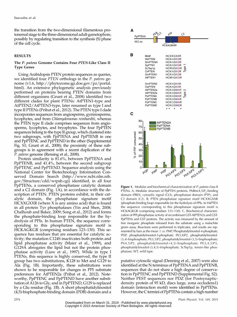
Protein similarity is 81.6%, between PpPTENA andPpPTENB, and 41.4%, between the second subgroupPpPTENC and PpPTEND. Sequence analysis using theNational Center for Biotechnology Information Con-served Domain Search (http://www.ncbi.nlm.nih.gov/Structure/cdd/wrpsb.cgi) identified, in all fourPpPTENs, a conserved phosphatase catalytic domainand a C2 domain (Fig. 1A), in accordance with the de-scription of PTEN. PTEN proteins exhibit, in their cat-alytic domain, the phosphatase signature motifHCXXGXXR (where X is any amino acid) that is foundin all protein Tyr phosphatases (Yin and Shen, 2008;Chalhoub and Baker, 2009; Song et al., 2012) and formsthe phosphate-binding loop responsible for the hy-drolysis of PPIs. In human PTEN, the sequence corre-sponding to this phosphatase signature motif isHCKAGKGR (comprising residues 123–130). This se-quence has residues that are essential for catalytic ac-tivity; the mutation C124S inactivates both protein andlipid phosphatase activity (Maier et al., 1999), andG129A abrogates the lipid but not the protein phos-phatase activity (Liaw et al., 1997). While in type IPTENs, this sequence is highly conserved, the type IIgroup has two substitutions, K128 to Met and G129 toAla (Fig. 1B). Importantly, these substitutions wereshown to be responsible for changes in PPI substratepreferences for AtPTEN2a (Pribat et al., 2012). Note-worthy, PpPTENC and PpPTEND have another substi-tution of A126 toGly, and in PpPTEND,G129 is replacedby a Glu residue (Fig. 1B). A short phosphatidylinositol(4,5)-bisphosphate-binding domain (PDB) domain and a
putative cytosolic signal (Denning et al., 2007) were alsoidentified at theN terminus of PpPTENAand PpPTENB,sequences that do not share a high degree of conserva-tion in PpPTENC and PpPTEND (Supplemental Fig. S2).Neither PEST sequences nor PDZ (for Postsynaptic-density protein of 95 kD, discs large, zona occludens1)domain (interaction motif) were identified in PpPTENs.However, theC termini of PpPTENs contain ahighnumber
Figure 1. Modular and biochemical characterization of P. patens class IIPTENs. A, Modular structure of PpPTEN proteins. PtdIns(4,5)P2-bindingdomain (PBD), cytosolic signal (CLS), phosphatase domain (PTP), andC2 domain (C2). B, PTEN phosphatase signature motif HCXXGXXR(phosphate-binding loop) responsible for the hydrolysis of PPIs. In HsPTENthe sequence corresponding to this phosphatase signature motif isHCKAGKGR (comprising residues 123–130). C, Biochemical characteri-zation of PPI phosphatase activity of recombinant GST-AtPTEN2a andGST-PpPTENs and GST proteins. The activity was measured by the amount offree inorganic phosphate released from the substrate using a malachitegreen assay. Reactions were performed in triplicates, and results are rep-resented by bars as themean6 SD. PI4P, Phosphatidylinositol-4-phosphate;PI5P, phosphatidylinositol-5-phosphate; PI(3,4)P2, phosphatidylinositol-(3,4)-bisphosphate; PI(3,5)P2, phosphatidylinositol-(3,5)-bisphosphate;PI(4,5)P2, phosphatidylinositol-(4,5)-bisphosphate; PI(3,4,5)P3,phosphatidylinositol-(3,4,5)-trisphosphate; ScTep1p, tensin-like phos-phatase; WT, wild type.
2574 Plant Physiol. Vol. 169, 2015
Saavedra et al.
www.plantphysiol.orgon March 31, 2019 - Published by Downloaded from Copyright © 2015 American Society of Plant Biologists. All rights reserved.
of Thr and Ser residues, which could reflect a mode ofregulation of these proteins that is similar to the PEST se-quences of their animal counterparts.
PpPTENA and PpPTENB Have Lipid Phosphatase Activity
It is well known that PTENs are dual phosphatasesthat can act on both polypeptide and PPI substrates,with a high preference toward the three-position of theinositol ring of PPIs. To test the specificity of PpPTENsagainst PPI substrates, their open reading frames wereexpressed in Escherichia coli as fusion proteins with aglutathione S-transferase (GST)-tag, and their phos-phatase activity was tested in vitro toward the sevenPPI isomers (Fig. 1C). GST alone was used as a negativecontrol, and the previously characterized AtPTEN2a(Pribat et al., 2012) was included in the assay as a pos-itive control due to its close phylogenetic relationshipwith the PpPTENs. Recombinant GST-PpPTENs weresuccessfully expressed, with the exception of GST-PpPTENC, despite multiple attempts. Therefore, nofunctional statements can be made for PpPTENC. Theother isoforms, expressed as fusions to N-terminal GST-tags, were purified, and size and identity of the re-combinant proteins were confirmed by western blotusing anti-GST antibodies (Supplemental Fig. S3).We found that PpPTENA efficiently (and to similarextent) dephosphorylated PtdIns3P, PtdIns(3,4)P2,PtdIns(3,5)P2, and PtdIns(3,4,5)P3, whereas PpPTENBpreferentially dephosphorylated PtdIns3P with decreas-ing activity toward PtdIns(3,4)P2 and PtdIns(3,5)P2(Fig. 1C). Interestingly, PpPTEND had no phosphataseactivity against any of the seven PPI isomers tested.This could be due to a specific substitution observedin PpPTEND (G129E), which, in humans, was shownto abrogate PTEN lipid phosphatase activity whilemaintaining protein phosphatase activity (Myers et al.,1997).Among the monophosphorylated PPI species, both
PpPTENA and PpPTENB only dephosphorylated thephosphate in the D3 position. Similarly, PpPTENA andPpPTENB preferred the bisphosphorylated PPI in theD3 position [PtdIns(3,4)P2 and PtdIns(3,5)P2] comparedwith PtdIns(4,5)P2 as substrate. These data indicatethat PpPTENs have a strong preference for dephos-phorylating the D3 position. Given that AtPTEN2a,PpPTENA, and PpPTENB share identical phosphate-binding loop sequences, it was surprising thatPpPTENA dephosphorylated PtdIns(3,4,5)P3 in vitro,suggesting that substrate selection likely extends be-yond the phosphate-binding loop.
PpPTEN Genes Are Expressed throughout the MossLife Cycle
The simple life cycle of P. patens, with a clear cellulardifferentiation, makes this moss ideal to study PTENexpression from a developmental point of view. Theexpression pattern of PpPTEN genes was analyzed in
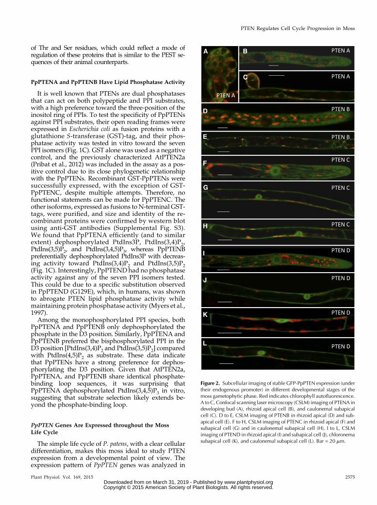
Figure 2. Subcellular imaging of stable GFP-PpPTEN expression (undertheir endogenous promoter) in different developmental stages of themoss gametophytic phase. Red indicates chlorophyll autofluorescence.A to C, Confocal scanning laser microscopy (CSLM) imaging of PTENA indeveloping bud (A), rhizoid apical cell (B), and caulonemal subapicalcell (C). D to E, CSLM imaging of PTENB in rhizoid apical (D) and sub-apical cell (E). F to H, CSLM imaging of PTENC in rhizoid apical (F) andsubapical cell (G) and in caulonemal subapical cell (H). I to L, CSLMimaging of PTEND in rhizoid apical (I) and subapical cell (J), chloronemasubapical cell (K), and caulonemal subapical cell (L). Bar = 20 mm.
Plant Physiol. Vol. 169, 2015 2575
PTEN Regulates Cell Cycle Progression in Moss
www.plantphysiol.orgon March 31, 2019 - Published by Downloaded from Copyright © 2015 American Society of Plant Biologists. All rights reserved.
the gametophytic generation, and expression of thefour genes was detected in both chloronemal andcaulonemal cells and in gametophores (SupplementalFig. S4). Our results confirmed the PpPTEN expressiondata deposited in the Genevestigator database, whichadditionally includes expression of the whole PTENfamily in the sporophyte.
To investigate PpPTEN’s cellular and subcellularlocalization, we integrated sequences encoding mono-meric enhanced GFP (mEGFP) in frame and down-stream of each PpPTEN gene. Using this strategy,expression of the PpPTEN fusion proteins is underthe control of their endogenous promoters, therebyminimizing possible overexpression artifacts. StablePpPTENpro:PpPTEN-mEGFP lines were obtained forthe four PpPTEN genes, all of which were verified byPCR analysis (Supplemental Fig. S5B). At a tissue leveland except for differences in fluorescence intensitieswithin the different lines, the same cellular localizationpattern was observed for all PpPTEN genes. In agree-ment with a role in cellular growth and proliferation,PpPTEN-GFP fusions exhibited higher fluorescencein the apical cells of chloronemata and caulonemataand new developing buds, whereas in developed ga-metophores, the signal was mainly observed in theemerging rhizoids cells (Fig. 2; Supplemental Fig. S5,D and E). In the case of PpPTENApro:PpPTENA-mEGFP, the fluorescent signal was often too weakfor adequate imaging. We therefore generated also aPpPTENpro:PpPTEN-3XmEGFP to complement our
observations. Using this construct for ectopic ex-pression, fluorescence was visible in developingbuds, as well as in actively growing caulonemal andrhizoid cells (Fig. 2, A–C).
At a subcellular level, and in agreement with animalPTEN localization, a primarily cytosolic signal wasobserved for all PpPTEN genes (Fig. 2, A–L). The iso-forms PpPTENC and PpPTEND were also clearly ob-served in the nucleus of chloronemal, caulonemal, andrhizoid cells (Fig. 2, F–L). Weak PpPTENB-mEGFP flu-orescence was found in the nucleus (Fig. 2E), in agree-ment with its putative cytosolic signal functioningalso as a noncanonical signal for nuclear trafficking(Denning et al., 2007). Similar observations were madefor PpPTENA-mEGFP in few caulonemal subapicalcells, but PpPTENA-3XmEGFP fluorescence was foundonly in the cytosol, which is likely a consequence ofthe fusion to three tandem GFP molecules, therebypreventing normal protein transit through the nuclearpores. Although a nuclear localization for PTENAand PTENB is not to be excluded, these observationssuggest that the two subgroups of PpPTENs may haveevolved different functions and localizations, withPpPTENA and PpPTENB playing their role mostly inthe cytosol and PpPTENC and PpPTEND acting bothat nuclei and cytosol. Additionally, we also observedfluorescent punctate structures in the cytoplasm of cellsexpressing both PpPTENA-mEGFP and PpPTENB-mEGFP (Fig. 2, B and E), which could be related to thelocalization of specific PPIs intracellular pools.
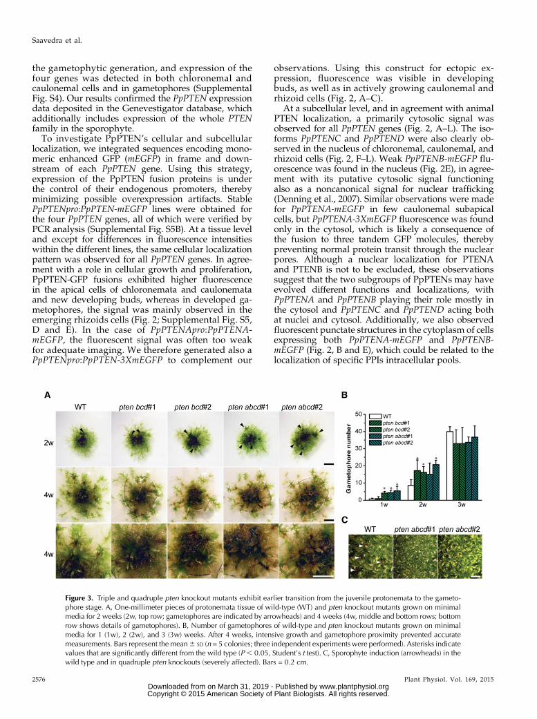
Figure 3. Triple and quadruple pten knockout mutants exhibit earlier transition from the juvenile protonemata to the gameto-phore stage. A, One-millimeter pieces of protonemata tissue of wild-type (WT) and pten knockout mutants grown on minimalmedia for 2 weeks (2w, top row; gametophores are indicated by arrowheads) and 4 weeks (4w, middle and bottom rows; bottomrow shows details of gametophores). B, Number of gametophores of wild-type and pten knockout mutants grown on minimalmedia for 1 (1w), 2 (2w), and 3 (3w) weeks. After 4 weeks, intensive growth and gametophore proximity prevented accuratemeasurements. Bars represent the mean6 SD (n = 5 colonies; three independent experiments were performed). Asterisks indicatevalues that are significantly different from the wild type (P , 0.05, Student’s t test). C, Sporophyte induction (arrowheads) in thewild type and in quadruple pten knockouts (severely affected). Bars = 0.2 cm.
2576 Plant Physiol. Vol. 169, 2015
Saavedra et al.
www.plantphysiol.orgon March 31, 2019 - Published by Downloaded from Copyright © 2015 American Society of Plant Biologists. All rights reserved.
Triple and Quadruple pten Knockout Mutants’ Transitionfrom Protonemata to Gametophores Occurs Earlier Thanthe Wild Type
To characterize the functional role of PpPTEN genes,we generated pten knockout lines by homologous re-combination using targeted gene disruption. Putativeknockout lines resistant to the corresponding antibioticcassettes were selected and further analyzed by PCRand real-time (RT)-PCR to confirm the disruption of thegene of interest (Supplemental Figs. S6 and S7;Supplemental Table S1). When more than one PpPTENwas targeted, the addition of each gene was performedprogressively.Analysis of single ptena, ptenb, ptenc, and ptend knock-
out lines, under normal growth conditions, exhibited nosignificant differences compared with the wild type(data not shown). Because the four PpPTEN genes areexpressed in all tissues, functional redundancy is thelikely explanation for these results. In agreement withthis hypothesis, double knockout lines, either generatedfrom members of same subgroup (e.g. ptencd) or dif-ferent subgroup (e.g. ptenbd), exhibited nonsignificantdifferences to the wild type when grown under normalconditions. Thus, we generated triple (ptenbcd) andquadruple (ptenabcd) knockout lines (SupplementalFigs. S6 and S7; Supplemental Table S1). Two lines for
each knockout were isolated from independent trans-formations and used for detailed analyses. We foundthat triple and quadruple pten knockout lines devel-oped gametophores earlier than the wild type (Fig. 3A).After 2 weeks of growth on minimal media, knockoutprotonemata of approximately 1-mm diameter devel-oped almost twice the number of gametophores whencomparedwith the wild type (Fig. 3B). This observationsuggests that, at earlier stages of moss development,the disruption of pten leads to a faster transition fromprotonemata to gametophores. However, later in de-velopment, wild-type colonies hadmore gametophoresand were larger (Supplemental Fig. S8). After 4 to5 weeks of growth in minimal media, quadruple ptenknockouts exhibited signs of cell death (e.g. brownishcolor), which was not observed in the wild type (Fig.3A). Furthermore, even though sporophytes could bedeveloped in quadruple knockout lines, only one to twocould be detected in approximately 1 cm2 of gameto-phores grown in jiffy pellets (Fig. 3C).
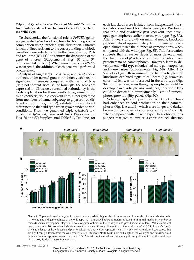
Notably, triple and quadruple pten knockout lineshad enhanced rhizoid production on their gameto-phores (Fig. 4, A and B), which were longer and darkerbrown but composed of shorter cells (Fig. 4, C and D),when compared with the wild type. These observationssuggest that pten mutant cells enter into cell division
Figure 4. Triple and quadruple pten knockout mutants exhibit higher rhizoid number and longer rhizoids with shorter cells.A, Twenty-day-old gametophytes of the wild type (WT) and pten knockout mutants growing in minimal media. B, Number ofrhizoids versus development stage (n˚ leaves) of gametophores of the wild type and pten knockout mutants. Values representmean 6 SD (n $ 10). Asterisks indicate values that are significantly different from the wild type (P , 0.05, Student’s t test).C, Rhizoid length of the wild type and pten knockout mutants. Values represent mean6 SD (n$ 10). Asterisks indicate values thatare significantly different from the wild type (P, 0.05, Student’s t test). D, Rhizoid cell length of the wild type and pten knockoutmutants. Values represent mean 6 SD (n $ 30). Asterisks indicate values that are significantly different from the wild type(P , 0.001, Student’s t test). Bar = 0.1 cm.
Plant Physiol. Vol. 169, 2015 2577
PTEN Regulates Cell Cycle Progression in Moss
www.plantphysiol.orgon March 31, 2019 - Published by Downloaded from Copyright © 2015 American Society of Plant Biologists. All rights reserved.
sooner (higher division rate) upon reaching a lowercritical size threshold compared with the wild type.
Caulonemal Cell Growth Is Accelerated in ptenKnockout Lines
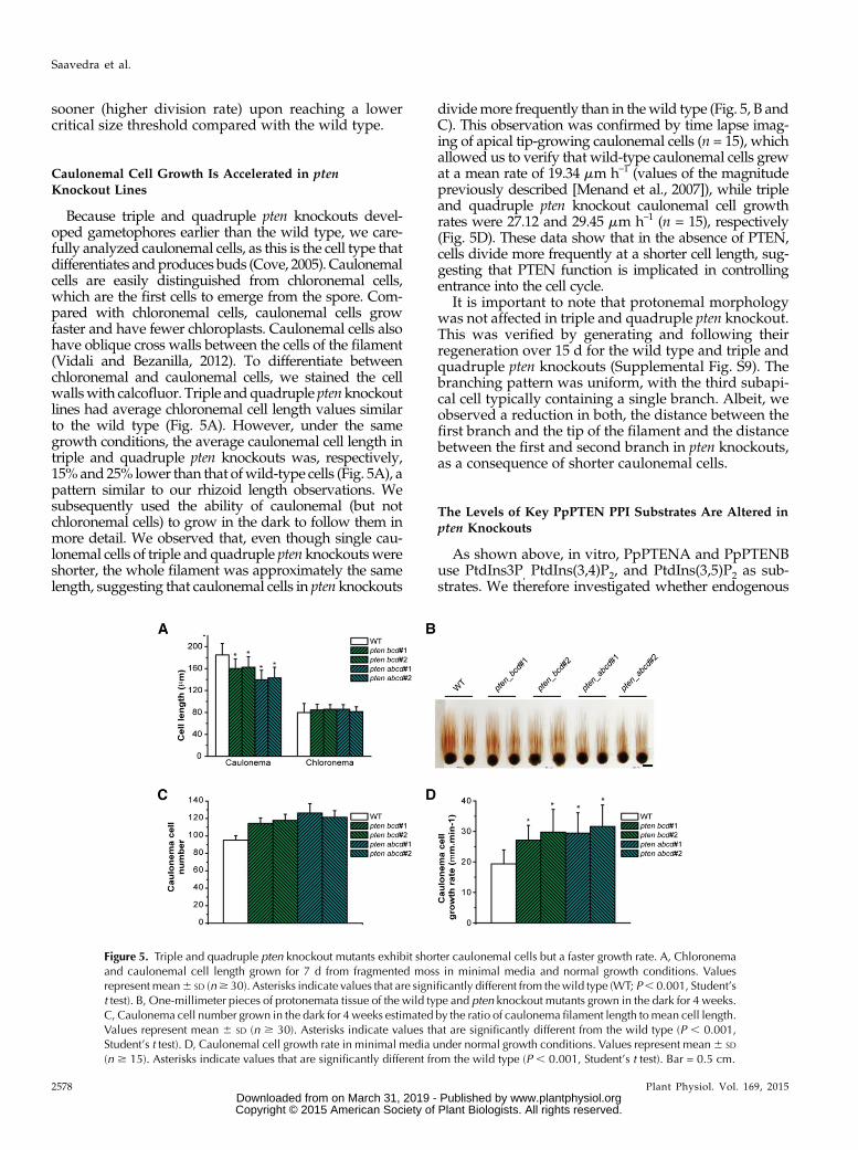
Because triple and quadruple pten knockouts devel-oped gametophores earlier than the wild type, we care-fully analyzed caulonemal cells, as this is the cell type thatdifferentiates andproduces buds (Cove, 2005). Caulonemalcells are easily distinguished from chloronemal cells,which are the first cells to emerge from the spore. Com-pared with chloronemal cells, caulonemal cells growfaster and have fewer chloroplasts. Caulonemal cells alsohave oblique cross walls between the cells of the filament(Vidali and Bezanilla, 2012). To differentiate betweenchloronemal and caulonemal cells, we stained the cellwallswith calcofluor. Triple andquadruple ptenknockoutlines had average chloronemal cell length values similarto the wild type (Fig. 5A). However, under the samegrowth conditions, the average caulonemal cell length intriple and quadruple pten knockouts was, respectively,15%and 25% lower than that ofwild-type cells (Fig. 5A), apattern similar to our rhizoid length observations. Wesubsequently used the ability of caulonemal (but notchloronemal cells) to grow in the dark to follow them inmore detail. We observed that, even though single cau-lonemal cells of triple and quadruple pten knockoutswereshorter, the whole filament was approximately the samelength, suggesting that caulonemal cells in pten knockouts
dividemore frequently than in thewild type (Fig. 5, B andC). This observation was confirmed by time lapse imag-ing of apical tip-growing caulonemal cells (n = 15), whichallowed us to verify that wild-type caulonemal cells grewat a mean rate of 19.34 mm h–1 (values of the magnitudepreviously described [Menand et al., 2007]), while tripleand quadruple pten knockout caulonemal cell growthrates were 27.12 and 29.45 mm h–1 (n = 15), respectively(Fig. 5D). These data show that in the absence of PTEN,cells divide more frequently at a shorter cell length, sug-gesting that PTEN function is implicated in controllingentrance into the cell cycle.
It is important to note that protonemal morphologywas not affected in triple and quadruple pten knockout.This was verified by generating and following theirregeneration over 15 d for the wild type and triple andquadruple pten knockouts (Supplemental Fig. S9). Thebranching pattern was uniform, with the third subapi-cal cell typically containing a single branch. Albeit, weobserved a reduction in both, the distance between thefirst branch and the tip of the filament and the distancebetween the first and second branch in pten knockouts,as a consequence of shorter caulonemal cells.
The Levels of Key PpPTEN PPI Substrates Are Altered inpten Knockouts
As shown above, in vitro, PpPTENA and PpPTENBuse PtdIns3P, PtdIns(3,4)P2, and PtdIns(3,5)P2 as sub-strates. We therefore investigated whether endogenous
Figure 5. Triple and quadruple pten knockout mutants exhibit shorter caulonemal cells but a faster growth rate. A, Chloronemaand caulonemal cell length grown for 7 d from fragmented moss in minimal media and normal growth conditions. Valuesrepresent mean6 SD (n$ 30). Asterisks indicate values that are significantly different from thewild type (WT; P, 0.001, Student’st test). B, One-millimeter pieces of protonemata tissue of the wild type and pten knockout mutants grown in the dark for 4 weeks.C, Caulonema cell number grown in the dark for 4 weeks estimated by the ratio of caulonema filament length tomean cell length.Values represent mean 6 SD (n $ 30). Asterisks indicate values that are significantly different from the wild type (P , 0.001,Student’s t test). D, Caulonemal cell growth rate in minimal media under normal growth conditions. Values represent mean6 SD
(n $ 15). Asterisks indicate values that are significantly different from the wild type (P , 0.001, Student’s t test). Bar = 0.5 cm.
2578 Plant Physiol. Vol. 169, 2015
Saavedra et al.
www.plantphysiol.orgon March 31, 2019 - Published by Downloaded from Copyright © 2015 American Society of Plant Biologists. All rights reserved.
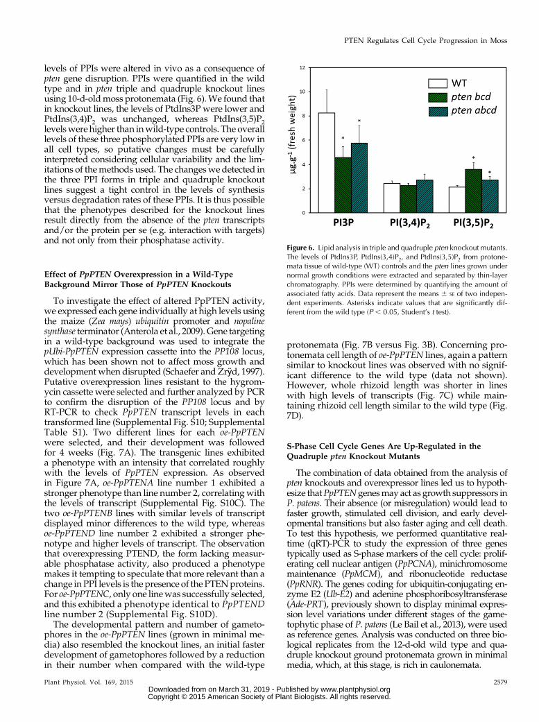
levels of PPIs were altered in vivo as a consequence ofpten gene disruption. PPIs were quantified in the wildtype and in pten triple and quadruple knockout linesusing 10-d-oldmoss protonemata (Fig. 6).We found thatin knockout lines, the levels of PtdIns3P were lower andPtdIns(3,4)P2 was unchanged, whereas PtdIns(3,5)P2levelswere higher than inwild-type controls. The overalllevels of these three phosphorylated PPIs are very low inall cell types, so putative changes must be carefullyinterpreted considering cellular variability and the lim-itations of themethods used. The changeswe detected inthe three PPI forms in triple and quadruple knockoutlines suggest a tight control in the levels of synthesisversus degradation rates of these PPIs. It is thus possiblethat the phenotypes described for the knockout linesresult directly from the absence of the pten transcriptsand/or the protein per se (e.g. interaction with targets)and not only from their phosphatase activity.
Effect of PpPTEN Overexpression in a Wild-TypeBackground Mirror Those of PpPTEN Knockouts
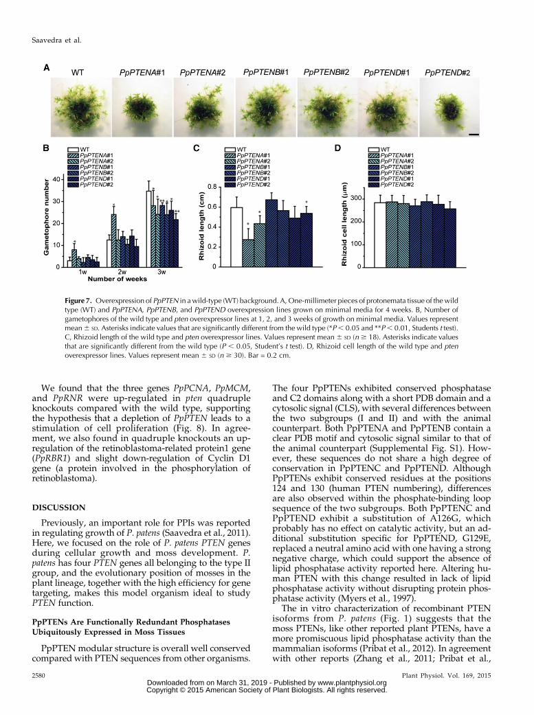
To investigate the effect of altered PpPTEN activity,we expressed each gene individually at high levels usingthe maize (Zea mays) ubiquitin promoter and nopalinesynthase terminator (Anterola et al., 2009). Gene targetingin a wild-type background was used to integrate thepUbi-PpPTEN expression cassette into the PP108 locus,which has been shown not to affect moss growth anddevelopment when disrupted (Schaefer and Zrÿd, 1997).Putative overexpression lines resistant to the hygrom-ycin cassette were selected and further analyzed by PCRto confirm the disruption of the PP108 locus and byRT-PCR to check PpPTEN transcript levels in eachtransformed line (Supplemental Fig. S10; SupplementalTable S1). Two different lines for each oe-PpPTENwere selected, and their development was followedfor 4 weeks (Fig. 7A). The transgenic lines exhibiteda phenotype with an intensity that correlated roughlywith the levels of PpPTEN expression. As observedin Figure 7A, oe-PpPTENA line number 1 exhibited astronger phenotype than line number 2, correlatingwiththe levels of transcript (Supplemental Fig. S10C). Thetwo oe-PpPTENB lines with similar levels of transcriptdisplayed minor differences to the wild type, whereasoe-PpPTEND line number 2 exhibited a stronger phe-notype and higher levels of transcript. The observationthat overexpressing PTEND, the form lacking measur-able phosphatase activity, also produced a phenotypemakes it tempting to speculate that more relevant than achange in PPI levels is the presence of the PTENproteins.For oe-PpPTENC, only one linewas successfully selected,and this exhibited a phenotype identical to PpPTENDline number 2 (Supplemental Fig. S10D).The developmental pattern and number of gameto-
phores in the oe-PpPTEN lines (grown in minimal me-dia) also resembled the knockout lines, an initial fasterdevelopment of gametophores followed by a reductionin their number when compared with the wild-type
protonemata (Fig. 7B versus Fig. 3B). Concerning pro-tonemata cell length of oe-PpPTEN lines, again a patternsimilar to knockout lines was observed with no signif-icant difference to the wild type (data not shown).However, whole rhizoid length was shorter in lineswith high levels of transcripts (Fig. 7C) while main-taining rhizoid cell length similar to the wild type (Fig.7D).
S-Phase Cell Cycle Genes Are Up-Regulated in theQuadruple pten Knockout Mutants
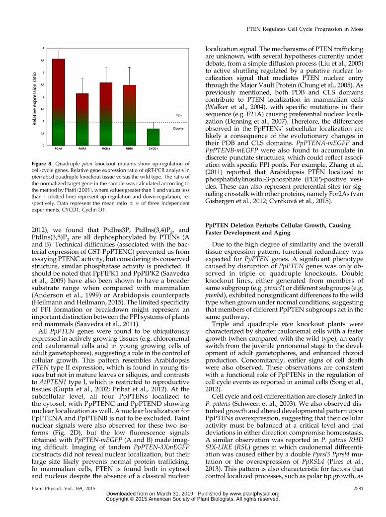
The combination of data obtained from the analysis ofpten knockouts and overexpressor lines led us to hypoth-esize thatPpPTENgenesmayact as growth suppressors inP. patens. Their absence (or misregulation) would lead tofaster growth, stimulated cell division, and early devel-opmental transitions but also faster aging and cell death.To test this hypothesis, we performed quantitative real-time (qRT)-PCR to study the expression of three genestypically used as S-phase markers of the cell cycle: prolif-erating cell nuclear antigen (PpPCNA), minichromosomemaintenance (PpMCM), and ribonucleotide reductase(PpRNR). The genes coding for ubiquitin-conjugating en-zyme E2 (Ub-E2) and adenine phosphoribosyltransferase(Ade-PRT), previously shown to display minimal expres-sion level variations under different stages of the game-tophytic phase of P. patens (Le Bail et al., 2013), were usedas reference genes. Analysis was conducted on three bio-logical replicates from the 12-d-old wild type and qua-druple knockout ground protonemata grown in minimalmedia, which, at this stage, is rich in caulonemata.
Figure 6. Lipid analysis in triple and quadruple pten knockout mutants.The levels of PtdIns3P, PtdIns(3,4)P2, and PtdIns(3,5)P2 from protone-mata tissue of wild-type (WT) controls and the pten lines grown undernormal growth conditions were extracted and separated by thin-layerchromatography. PPIs were determined by quantifying the amount ofassociated fatty acids. Data represent the means 6 SE of two indepen-dent experiments. Asterisks indicate values that are significantly dif-ferent from the wild type (P , 0.05, Student’s t test).
Plant Physiol. Vol. 169, 2015 2579
PTEN Regulates Cell Cycle Progression in Moss
www.plantphysiol.orgon March 31, 2019 - Published by Downloaded from Copyright © 2015 American Society of Plant Biologists. All rights reserved.
We found that the three genes PpPCNA, PpMCM,and PpRNR were up-regulated in pten quadrupleknockouts compared with the wild type, supportingthe hypothesis that a depletion of PpPTEN leads to astimulation of cell proliferation (Fig. 8). In agree-ment, we also found in quadruple knockouts an up-regulation of the retinoblastoma-related protein1 gene(PpRBR1) and slight down-regulation of Cyclin D1gene (a protein involved in the phosphorylation ofretinoblastoma).
DISCUSSION
Previously, an important role for PPIs was reportedin regulating growth of P. patens (Saavedra et al., 2011).Here, we focused on the role of P. patens PTEN genesduring cellular growth and moss development. P.patens has four PTEN genes all belonging to the type IIgroup, and the evolutionary position of mosses in theplant lineage, together with the high efficiency for genetargeting, makes this model organism ideal to studyPTEN function.
PpPTENs Are Functionally Redundant PhosphatasesUbiquitously Expressed in Moss Tissues
PpPTEN modular structure is overall well conservedcompared with PTEN sequences from other organisms.
The four PpPTENs exhibited conserved phosphataseand C2 domains along with a short PDB domain and acytosolic signal (CLS), with several differences betweenthe two subgroups (I and II) and with the animalcounterpart. Both PpPTENA and PpPTENB contain aclear PDB motif and cytosolic signal similar to that ofthe animal counterpart (Supplemental Fig. S1). How-ever, these sequences do not share a high degree ofconservation in PpPTENC and PpPTEND. AlthoughPpPTENs exhibit conserved residues at the positions124 and 130 (human PTEN numbering), differencesare also observed within the phosphate-binding loopsequence of the two subgroups. Both PpPTENC andPpPTEND exhibit a substitution of A126G, whichprobably has no effect on catalytic activity, but an ad-ditional substitution specific for PpPTEND, G129E,replaced a neutral amino acid with one having a strongnegative charge, which could support the absence oflipid phosphatase activity reported here. Altering hu-man PTEN with this change resulted in lack of lipidphosphatase activity without disrupting protein phos-phatase activity (Myers et al., 1997).
The in vitro characterization of recombinant PTENisoforms from P. patens (Fig. 1) suggests that themoss PTENs, like other reported plant PTENs, have amore promiscuous lipid phosphatase activity than themammalian isoforms (Pribat et al., 2012). In agreementwith other reports (Zhang et al., 2011; Pribat et al.,
Figure 7. Overexpression of PpPTEN in awild-type (WT) background. A, One-millimeter pieces of protonemata tissue of the wildtype (WT) and PpPTENA, PpPTENB, and PpPTEND overexpression lines grown on minimal media for 4 weeks. B, Number ofgametophores of the wild type and pten overexpressor lines at 1, 2, and 3 weeks of growth on minimal media. Values representmean6 SD. Asterisks indicate values that are significantly different from the wild type (*P, 0.05 and **P, 0.01, Students t test).C, Rhizoid length of the wild type and pten overexpressor lines. Values represent mean 6 SD (n $ 18). Asterisks indicate valuesthat are significantly different from the wild type (P , 0.05, Student’s t test). D, Rhizoid cell length of the wild type and ptenoverexpressor lines. Values represent mean 6 SD (n $ 30). Bar = 0.2 cm.
2580 Plant Physiol. Vol. 169, 2015
Saavedra et al.
www.plantphysiol.orgon March 31, 2019 - Published by Downloaded from Copyright © 2015 American Society of Plant Biologists. All rights reserved.
2012), we found that PtdIns3P, PtdIns(3,4)P2, andPtdIns(3,5)P2 are all dephosphorylated by PTENs (Aand B). Technical difficulties (associated with the bac-terial expression of GST-PpPTENC) prevented us fromassaying PTENC activity, but considering its conservedstructure, similar phosphatase activity is predicted. Itshould be noted that PpPIPK1 and PpPIPK2 (Saavedraet al., 2009) have also been shown to have a broadersubstrate range when compared with mammalian(Anderson et al., 1999) or Arabidopsis counterparts(Heilmann andHeilmann, 2015). The limited specificityof PPI formation or breakdown might represent animportant distinction between the PPI systems of plantsand mammals (Saavedra et al., 2011).All PpPTEN genes were found to be ubiquitously
expressed in actively growing tissues (e.g. chloronemaland caulonemal cells and in young growing cells ofadult gametophores), suggesting a role in the control ofcellular growth. This pattern resembles ArabidopsisPTEN type II expression, which is found in young tis-sues but not in mature leaves or siliques, and contraststo AtPTEN1 type I, which is restricted to reproductivetissues (Gupta et al., 2002; Pribat et al., 2012). At thesubcellular level, all four PpPTENs localized tothe cytosol, with PpPTENC and PpPTEND showingnuclear localization as well. A nuclear localization forPpPTENA and PpPTENB is not to be excluded. Faintnuclear signals were also observed for these two iso-forms (Fig. 2D), but the low fluorescence signalsobtained with PpPTEN-mEGFP (A and B) made imag-ing difficult. Imaging of tandem PpPTEN-3XmEGFPconstructs did not reveal nuclear localization, but theirlarge size likely prevents normal protein trafficking.In mammalian cells, PTEN is found both in cytosoland nucleus despite the absence of a classical nuclear
localization signal. The mechanisms of PTEN traffickingare unknown, with several hypotheses currently underdebate, from a simple diffusion process (Liu et al., 2005)to active shuttling regulated by a putative nuclear lo-calization signal that mediates PTEN nuclear entrythrough the Major Vault Protein (Chung et al., 2005). Aspreviously mentioned, both PDB and CLS domainscontribute to PTEN localization in mammalian cells(Walker et al., 2004), with specific mutations in theirsequence (e.g. F21A) causing preferential nuclear locali-zation (Denning et al., 2007). Therefore, the differencesobserved in the PpPTENs’ subcellular localization arelikely a consequence of the evolutionary changes intheir PDB and CLS domains. PpPTENA-mEGFP andPpPTENB-mEGFP were also found to accumulate indiscrete punctate structures, which could reflect associ-ation with specific PPI pools. For example, Zhang et al.(2011) reported that Arabidopsis PTEN localized tophosphatidylinositol-3-phosphate (PI3P)-positive vesi-cles. These can also represent preferential sites for sig-naling crosstalkwith other proteins, namely For2As (vanGisbergen et al., 2012; Cvr�cková et al., 2015).
PpPTEN Deletion Perturbs Cellular Growth, CausingFaster Development and Aging
Due to the high degree of similarity and the overalltissue expression pattern, functional redundancy wasexpected for PpPTEN genes. A significant phenotypecaused by disruption of PpPTEN genes was only ob-served in triple or quadruple knockouts. Doubleknockout lines, either generated from members ofsame subgroup (e.g. ptencd) or different subgroups (e.g.ptenbd), exhibited nonsignificant differences to the wildtype when grown under normal conditions, suggestingthat members of different PpPTEN subgroups act in thesame pathway.
Triple and quadruple pten knockout plants werecharacterized by shorter caulonemal cells with a fastergrowth (when compared with the wild type), an earlyswitch from the juvenile protonemal stage to the devel-opment of adult gametophores, and enhanced rhizoidproduction. Concomitantly, earlier signs of cell deathwere also observed. These observations are consistentwith a functional role of PpPTENs in the regulation ofcell cycle events as reported in animal cells (Song et al.,2012).
Cell cycle and cell differentiation are closely linked inP. patens (Schween et al., 2003). We also observed dis-turbed growth and altered developmental pattern uponPpPTENs overexpression, suggesting that their cellularactivity must be balanced at a critical level and thatdeviations in either direction compromise homeostasis.A similar observation was reported in P. patens RHDSIX-LIKE (RSL) genes in which caulonemal differenti-ation was caused either by a double Pprsl3 Pprsl4 mu-tation or the overexpression of PpRSL4 (Pires et al.,2013). This pattern is also characteristic for factors thatcontrol localized processes, such as polar tip growth, as
Figure 8. Quadruple pten knockout mutants show up-regulation ofcell-cycle genes. Relative gene expression ratio of qRT-PCR analysis inpten abcd quadruple knockout tissue versus the wild type. The ratio ofthe normalized target gene in the sample was calculated according tothe method by Pfaffl (2001), where values greater than 1 and values lessthan 1 (dotted line) represent up-regulation and down-regulation, re-spectively. Data represent the mean ratio 6 SE of three independentexperiments. CYCD1, Cyclin D1.
Plant Physiol. Vol. 169, 2015 2581
PTEN Regulates Cell Cycle Progression in Moss
www.plantphysiol.orgon March 31, 2019 - Published by Downloaded from Copyright © 2015 American Society of Plant Biologists. All rights reserved.
previously observed by us. In Arabidopsis pollen tubesand root hairs, knocking down or overexpressingphosphatidylinositol-4-monophosphate 5-kinasesresulted in abnormal morphologies and reducedgrowth (Kusano et al., 2008; Ischebeck et al., 2008; Sousaet al., 2008; Stenzel et al., 2008).
PpPTENs Exert Their Role through Changes in PPI Levelsand Regulation of S-Phase Cell Cycle Genes
It remains unclear how PPIs might exert regulatoryeffects on cell division. The observed overexpressionphenotypes, similar for the four PpPTENs, suggestthat their effect is not solely due to dephosphorylationof PPI substrates. As reported here (Fig. 1C), PpPTENsexhibited different affinities for PPI isomers, includingno lipid phosphatase activity for PpPTEND. Analogousobservationwasmade for themoss Ser/Thr protein kinasePDK1, which was reported to lack a phospholipid-bindingdomain (Dittrich andDevarenne, 2012), suggesting thatin nonvascular land plants, lipid regulation evolveddifferently.
Unlike some observations in animal cells (Goebbelset al., 2010), a clear increase in a PPI isomer was notobserved in our moss pten knockout lines. Only PtdIns(3,5)P2 showed increased levels in the pten knockouts(Fig. 6), whereas other PPIs were not significantly al-tered or showed even reduced levels. The promiscuityof plant PTENs versus different PPI substrates in vitromakes it thus difficult to link the observed growth ef-fects to changes in any particular PPI in vivo. Inter-pretation is further limited by a lack of knowledge ofhow some of the lipids shown to be substrates forPpPTENs are metabolically connected. Compensatoryeffects by other enzymes in response to misregulatedPPI levels are likely, as have previously been proposedfor Arabidopsis mutants with defects in the expressionof PPI phosphatases of the suppressor of actin family(Nováková et al., 2014). Rapid metabolite turnover andsystem compensation are characteristics of resilientorganisms like Physcomitrella spp. Nevertheless, it istempting to speculate that the reduced PtdIns3P levelsmeasured in triple and quadruple knockouts resultfrom amisregulation of PI3K. In animal cells, PTEN is amajor antagonist of PI3K activity and of the PI3K/AKT/mTOR, an intracellular signaling pathway im-portant in regulating the cell cycle (Song et al., 2012).Plants do not contain AKT kinase genes, but themTOR/ribosomal S6 kinase is well conserved, and ithas recently been shown to negatively regulate cell di-vision as part of a signaling pathway connected to theRBR1-adenovirus E2 promoter (E2F) transcriptionalmachinery (Henriques et al., 2010).
Our findings that at least three of the four PpPTENs(B, C, and D) localize also to the nucleus, together withthe biochemical data discussed above, are in agreementwith a functional role for PTENs in cell cycle extendingbeyond their lipid phosphatase activity (Lindsay et al.,2006). For example, nuclear PTEN was reported to
positively regulate DNA repair independently of itsphosphatase activity (Shen et al., 2007). Such hypothe-sis is supported by quantitative PCR data showingup-regulation of S-phase cell cycle genes. Both animalsand plants, use the same retinoblastoma (RB)/E2F/dimerization partner (DP) pathway to control theG1-to-S transition (Inzé and De Veylder, 2006). Inplants, RBR is phosphorylated in a cell cycle-specificmanner by several Cyclin/cyclin-dependent kinasecomplexes, namely cyclin D (Boniotti and Gutierrez,2001; Nakagami et al., 2002; Menges et al., 2006). Theactivated cyclin complexes phosphorylate RB and in-hibit RB binding to E2F/DP transcription factor, lead-ing to activation in transcription of genes needed forDNA replication during the S-phase of the cell cycle. InArabidopsis, RBR was also shown to be a positiveregulator of developmental switches promoting thetransition from embryonic to autotrophic plant devel-opment (Gutzat et al., 2011). The shorter caulonemalcell size observed in pten knockout plants and its highergrowth rate were thus possibly a consequence of anaccelerated entry into the S-phase of the cell cycle,manifested by an up-regulation of PpPCNA, PpRNR2,PpMCM3, and RBR1 genes and a down-regulation ofcyclin D1 gene. This is in agreement with the findingthat the chloronemal tissue is arrested in G2, whilecaulonemal cells aremainly in G1 (Schween et al., 2003).Chloronema-to-caulonema transition upon changes ingene expression have been reported (Jang and Dolan,2011). We cannot exclude the possibility of altered cellidentities in pten knockout plants. However, our ob-servation that chloronemal cell growth is not signifi-cantly affected in triple and quadruple knockouts doessuggest that PTENs’ main effect is on cell cycle (tran-sition G1/S) and not on cell differentiation.
Taken together, the data presented in this reportsupport the hypothesis that PpPTEN genes are involvedin the critical regulation of cell size and cell cycle tran-sitions (Jorgensen and Tyers, 2004), playing a funda-mental role in organism development and aging. Itfurther raises new questions about the evolution ofplant lipid regulation and how protein function isachieved beyond catalytic activity.
MATERIALS AND METHODS
Plant Material and Growth Conditions
The Gransden wild-type strain of Physcomitrella patens (Ashton and Cove,1977) was used in this study. The wild type and mutant strains were grownaxenically at 24°C under a 16-h-light/8-h-dark regime with a photon flux of55 mmol m–2 s–1. P. patens protonemal tissue was propagated routinely at 7-dintervals on cellophane disks (AA packaging) overlaying petri dishes (90-mmdiameter) that contained PP-NH4 medium (PP-NO3 [0.8 g L–1 CaNO3$4H2O,0.25 g L–1 MgSO4$7H2O, 0.0125 g L–1 FeSO4$7H2O, 0.055 mg L–1 CuSO4$5H2O,0.055 mg L–1 ZnSO4$7H2O, 0.614 mg L–1 H3BO3, 0.389 mg L–1 MnCl2$4H2O,0.055 mg L–1 CoCl2$6H2O, 0.028 mg L–1 KI, 0.025 mg L–1 Na2MoO4$2H2O, and0.25 mg L–1 KH2PO4 buffer, pH 7.0, with KOH] supplemented with 0.5 g L–1 [di]ammonium tartrate), and 7 g L–1 agar (Duchefa). Phenotypic analyses of thewild type and mutant strains were performed on PP-NO3 medium.
For gametophores and rhizoid analysis of plants, protonemal pieces of ap-proximately 1 mm were placed on PP-NO3 media and grown for 3 to 4 weeks.
2582 Plant Physiol. Vol. 169, 2015
Saavedra et al.
www.plantphysiol.orgon March 31, 2019 - Published by Downloaded from Copyright © 2015 American Society of Plant Biologists. All rights reserved.
Chloronemal and caulonemal cell length measurements were performed onthree-first subapical cells of 6-d-old protonemata stained with 10 mg mL–1
fluo-rescence brightener 28 (Sigma-Aldrich).
For cell growth rate measurements, protonemal tissue was grown on petridishes (30-mm diameter) on PP-NO3 media from 5 to 6 d. Images of apicalcaulonemal cells were acquired every 30 min during a 2.3-h period with a PCOSensicam-QE camera (Labocontrole) attached to an Olympus IX-50 microscope(Labocontrole).
For the dark growth experiments, square petri dishes with 7-d-old mossprotonematapieceswere grown for 10dunder continuous light on solidPP-NO3supplemented with 5 mM (di)ammonium tartrate and 2% (w/w) Suc and thengrown in darkness in vertical position for 4 weeks.
For sporophyte induction, fresh protonemata were grown in Jiffy7 potscovered bywater below 1 cm at 24°C under a 16-h-light/8-h-dark regime. After6weeks, cultures were transferred to 19°C under short-day conditions (8-h-lightper day) for sporophyte induction.
Data analysis was performed using IMAGEJ software (http://rsbweb.nih.gov/ij/).
Phylogenetic Analysis
The P. patens ssp. patens genome (v1.6, http://www.phytozome.net/) wasscreened for PTEN homologs using TBLASTN with query sequences fromArabidopsis (Arabidopsis thaliana). PTEN sequences were also searched for thegreen algae Chlamydomonas reinhardtii and additional 10 representative landplants (P. patens, Selaginella moellendorffii, Pinus taeda, Oryza sativa, Sorghum bi-color, Vitis vinifera, Arabidopsis, Glycine max, and Populus trichocarpa) and fromSchizosaccharomyces pombe, Saccharomyces cerevisiae, and Homo sapiens genomesdeposited at Phytozome v9.1 and at the National Center for BiotechnologyInformation. Amino acid sequences were aligned with ClustalW, and theevolutionary history was inferred using the maximum likelihood with theMEGA6 package (Tamura et al., 2013). The bootstrap consensus tree wasinferred from 500 replicates. The datamatrixwas analyzedwith heuristic searchand the default search options.
RT-PCR
For cloning coding sequences (CDs), gene expression analysis by RT-PCR, ormutant genotyping, total RNA frommoss tissue was isolated using the RNeasyPlant Mini kit (Qiagen), according to the manufacturer’s recommendations.One microgram of total RNA was treated with two units of DNaseI (RQ1,Promega) and then used as template for reverse transcription with SuperscriptIII reverse transcriptase (Invitrogen) or RevertAidHMinus Reverse Transcriptase(Thermo Fisher Scientific) and primed with an oligo(dT) primer following themanufacturer’s protocol. A one-fortieth volume of the complementary DNAwassubsequently used as template for PCR. Primers used for amplification are listedin Supplemental Table S1.
qRT-PCR
Total RNA was purified using the RNeasy Plant Mini kit (Qiagen) from12-d-old ground protonemata grown in minimal media (ppNO3). One mi-crogram of total RNAwas treated with two units of DNase I (RQ1, Promega)and then used as template for reverse transcription with RevertAid HMinusReverse Transcriptase (Thermo Fisher Scientific) and primed with an oligo(dT) primer. qRT-PCR was performed using a PikoReal 96-Well System(PikiReal Real-Time PCR System, Thermo Scientific) with Maxima SYBRGreen/ROX qPCR Master Mix (Thermo Fisher Scientific). The sequences ofprimers for qRT-PCR are described in Supplemental Table S1. The genesencoding Ub-E2 and Ade-PRT were used as controls for normalization ofmRNA content between samples, according to previously published data(Le Bail et al., 2013). For each gene, nine reactions were performed includingthree biological replicates and three technical replicates. Melting curveswere obtained to ensure that only a single product was amplified. The rel-ative quantification of the target gene was obtained using Pfaffl method(Pfaffl, 2001) in which the relative expression ratio (R) is determined usingthe Efficiency (E ) and the threshold cycle (Ct) deviation of an unknownsample versus a control compared with a reference gene according to thefollowing equation: R = [(Etarget) DCt (control–test)]/[(Eref)DCt (control–sample)]. For statistical analysis, a Student’s t test was applied (values fromthe wild type were considered as expected with P , 0.05 and 1 degree offreedom).
Gene Isolation and Protein Expression
Full-length CDs ofAtPTEN2a, PpPTENB, and PpPTENDwere amplifiedwithspecific primers (Supplemental Table S1) and cloned into pENTR/D-TOPOvector (Invitrogen) following the manufacturer’s protocol. PpPTENA andPpPTENC CDs cloned in pENTR/D-TOPO vector were obtained fromMagdalenaBenzanilla (van Gisbergen et al., 2012). PTEN CDs were then transferred to thedestination expression vector pDEST15 (Invitrogen) using LR clonase (Invitrogen).GST-PTEN open reading frames were verified by sequencing.
Escherichia coli Turner cells, harboring a plasmid for the expression of a re-combinant fusion protein of GST with PTEN were grown in Luria Broth me-dium at 37°C and 180 rpm until an optical density at 600 nm of 0.6 to 0.8 wasreached. At this point, isopropyl b-D-thiogalactoside was added to a finalconcentration of 0.2 mM. Cells were incubated overnight at 19°C and 200rev min–1 and then collected by centrifugation and frozen at –80°C. For GSTfusion protein purification, frozen cells were thawed on ice and resuspended inTris-buffered saline extraction buffer (10 mM Tris-HCl, pH 8.0, 140 mM NaClcontaining 1% [w/v] Triton X-100, 1 mM EDTA, 1 mM dithiothreitol, and 1 mM
phenylmethylsulfonyl fluoride). The crude extract was sonicated for 120 s (0.5-sintervals) and centrifuged for 15 min at 10,000g at 4°C. The supernatant wasincubated with 0.1 mL of glutathione6 Sepharose resin (Amersham PharmaciaBiotech) and pre-equilibratedwith extraction buffer for 1 h at 4°C. The resinwasthen washed with extraction buffer twice, and then proteins bound to the resinwere eluted with 10 mM glutathione (Sigma-Aldrich), 50 mM Tris-HCl, pH 8.0,and 5% (v/v) glycerol and stored at –80°C.
Lipid Phosphatase Activity Using Malachite Green Assays
PTEN hydrolyzing activity toward the different PPIs was determined invitro by the malachite green-based assay that measures the released inorganicphosphate (Odriozola et al., 2007). Briefly, 1 mg of recombinant proteinwas incubated with 100 mM of one of the seven di-C8 PPI lipids [PI3P,phosphatidylinositol-4-phosphate, phosphatidylinositol-5-phosphate, phos-phatidylinositol (3,4)-bisphosphate, phosphatidylinositol (3,5)-bisphosphate,phosphatidylinositol (4,5)-bisphosphate, or phosphatidylinositol (3,4,5)-tri-sphosphate (Echelon Biosciences)] for 60 min at 37°C in a final volume of 25 mL.The reaction buffer was 25 mM Tris-HCl, pH 7.4, 140 mM NaCl, 2.7 mM KCl, and10 mM dithiothreitol. The reaction was stopped with 100 mL of malachite greenreagent (Echelon Biosciences). The amount of phosphate releasedwas determinedby reading the A630 in an ELx800 Absorbance Microplate Reader (BioTek) andconverted to a molar amount of product using a standard curve. The amount ofprotein used and the duration of the assay were within the linear range of thereaction. Free inorganic phosphate was measured in controls (buffer, substratesolutions, and recombinant proteins alone) and subtracted for calculating PTENactivity. GST was used as negative control. For the reaction with PI3P, 38% (v/v)of the substrate was consumed at the end of the reaction. The activity assay wasperformed in triplicates and are presented as the mean 6 SE of two independentexperiments.
Lipid Analysis
Material was ground in liquid nitrogen. Phospholipids were extracted from250 mg of the ground material and analyzed exactly as described previously(König et al., 2008). For statistical analysis, a Student’s t test was applied (valuesfrom the wild type were considered as expected with P , 0.05).
Generation of Targeting Constructs
Sequences for targeting constructs were all amplified from genomic DNAisolated from moss protonemata. The PpPTEN gene knock-in constructs wereperformed using Multisite Gateway four-fragment recombination (Invitrogen).In first position, a fragment of 1,000 bp just before the stop codonwas amplifiedusing primers containing attB1 and attB5r sites (Supplemental Table S1) andcloned into pDONR 221 P1-P5r using BP clonase. At second position, EGFPcloned into pDONR 221 P5-P4 was used. At third position, pDONR 221 P4r-P3rcontaining a hygromycin resistance cassette was used, and at fourth position, afragment of 1,000 bp belonging to the 39 untranslated region of PpPTENs wasamplified using primers containing attB3 and attB2 sites (Supplemental TableS1) and cloned into pDONR 221 P3-P2 using BP clonase. Sequences of specificprimers used for cloning the 59 fragment and 39 fragment for the knock-inconstructs are listed in Supplemental Table S1. EGFP and hygromycin se-quences cloned in pEntry vectors were obtained from Magdalena Benzanilla.
Plant Physiol. Vol. 169, 2015 2583
PTEN Regulates Cell Cycle Progression in Moss
www.plantphysiol.orgon March 31, 2019 - Published by Downloaded from Copyright © 2015 American Society of Plant Biologists. All rights reserved.
As target sequences for homologous recombination for PpPTEN gene dis-ruption, two genomic DNA fragments of 1,000 bp from the 59 and 39 regions ofPpPTEN genes were cloned with specific primers (Supplemental Table S1). ThePpPTENA knockout construct was performed the using Multisite Gatewaythree-fragment recombination (Invitrogen). For this purpose, 59 PpPTENA and39 PpPTENA genomic DNA fragments were cloned into pDONR 221 P1-P4and pDONR 221 P3-P2 using BP clonase (Invitrogen). These entry vectorsand Nos_loxGentlox-R3R4 vector were cloned into the pGEM. PpPTENB,PpPTENC, and PpPTEND knockout constructs were obtained by cloning withrestriction enzymes using fragments amplified with primers listed in SupplementalTable S1 cloned into p35SZeo, pBHRF, and pBNRF, respectively. All generatedclones were verified by sequencing.
For the overexpression analysis PpPTENA to PpPTEND, CDs cloned inpENTR/D-TOPO were transferred to the destination vector uj3-pTUbiGate(Anterola et al., 2009) using LR clonase II (Invitrogen).
Sequences of all targeting vectors used for transformation were verified bysequencing. In all cases, putative transformant lines growing in the corre-sponding antibiotic selective media from knock-in, knockout, and over-expression experiments were verified by PCR and RT-PCR with primerssequences listed in Supplemental Table S1.
Transformation of P. patens
Isolation of protoplasts, polyethylene glycol-mediated transformation, re-generation, and antibiotic selection were performed as described previously(Schaefer and Zrÿd, 1997). For polyethylene glycol-mediated protoplast trans-formation, 6-d-old protonemata were treatedwith 0.5%Driselase in 8.5% (w/v)mannitol for 30 min, passed through a 100-mm sieve, incubated for 15 minat room temperature, and passed through a 50-mm sieve. The protoplasts ofthe final flowthrough were washed twice in 8.5% mannitol before further use.Protoplasts were transformed at a concentration of 1.6 3 106 protoplasts mL–1.Each transformation consisted of 0.3 mL of protoplast suspension and 15 to20 mg of linear DNA. To eliminate any episomal resistant colonies, two roundsof selection were undertaken using the appropriate antibiotic. To select forantibiotic-resistant cells, G418, Zeocin, Hygromycin, and Gentamycin all pur-chased from Duchefa were added at 50, 25, 25, and 200 mg L–1 to the medium,respectively.
Fluorescence Imaging
Moss protonemata was inoculated over cellophane of a PP-NO3 growthmedium plate and grown for 7 to 20 d. At these two time points, small proto-nemata pieces or gametophores were transferred to a slide, and microscopicfluorescence images were acquired with a Leica SP-E confocal laser scanningmicroscope. Thin time course optical sections (approximately 3 mm thick) wereacquired using a 320 Plan Apo dry objective (numerical aperture = 0.75) andless than 20% laser intensity and operating in the mode 1,024 3 1,024and 400 Hz (approximately one-third s per frame). GFP was imaged using the488-nm excitation line, and the chlorophyll signal was imaged with the 532-nmexcitation line. Images were then processed using the LAS AFL 2.6.0 (LeicaMicrosystems) and Image-Pro Plus 5.0 software (Media Cybernetics, Leiden,The Netherlands).
GFP fluorescence images from gametophores were acquired using a ZeissSteREO Lumar.V12 and an Imaging Source DFK23u274 camera.
Sequence data from this article can be found at the Phytozome database:PpPTENA (Phpat.019G062100), PpPTENB (Phpat.022G039200), PpPTENC(Phpat.021G028600), and PpPTEND (Phpat.022G052200).
Supplemental Data
The following supplemental materials are available.
Supplemental Figure S1. Maximum likelihood tree showing that theP. patens PTEN proteins fall into the type II class.
Supplemental Figure S2. PDB and CLS motifs of PpPTENs.
Supplemental Figure S3. Immunoblot of recombinant GST-PTENs.
Supplemental Figure S4. Expression analysis of PpPTENs.
Supplemental Figure S5. Genotyping and western-blot analysis ofPpPTENs transformants.
Supplemental Figure S6. Constructs used for homologous recombination.
Supplemental Figure S7. PCR genotyping analysis of wild-type, triple,and quadruple pten knockouts.
Supplemental Figure S8. Total colony area values in square centimetersfrom wild-type, triple, and quadruple pten knockouts growing in mini-mal media for 5 weeks.
Supplemental Figure S9. Effects of pten deletion on moss structural mor-phology.
Supplemental Figure S10. Overexpressing PpPTEN mutants by disruptionof the Pp108 locus.
Supplemental Table S1. Primers used in this study.
ACKNOWLEDGMENTS
We thank Drs. Andreia Figueiredo, Filipa Monteiro, and Ana MargaridaFortes (University of Lisbon) for the technical assistance in qRT-PCR, Dr. MattiasThelander (Swedish University of Agricultural Sciences) for the pUBW302 vector,Dr.MitsuyasuHasebe (National Institute Basic Biology, Japan) for the p35S-Zeovector, Dr. Pierre-François Perroud (Washington University, St. Louis) for theuj3-pTUbiGate vector, Dr. Fabien Nogue (Institut National de la RechercheAgronomique, Institut Jean-Pierre Bourgin) for the pBNRF and pBHRF vectors,and Dr. Mareike Heilmann (Martin-Luther-University Halle-Wittenberg) forhelpful discussion on cell cycle regulation.
Received July 31, 2015; accepted October 8, 2015; published October 13, 2015.
LITERATURE CITED
Anderson RA, Boronenkov IV, Doughman SD, Kunz J, Loijens JC (1999)Phosphatidylinositol phosphate kinases, a multifaceted family of sig-naling enzymes. J Biol Chem 274: 9907–9910
Anterola A, Shanle E, Perroud PF, Quatrano R (2009) Production of taxa-4(5),11(12)-diene by transgenic Physcomitrella patens. Transgenic Res 18:655–660
Ashton NW, Cove DJ (1977) Isolation and preliminary characterization ofauxotrophic and analog resistant mutants of moss Physcomitrella patens.Mol Gen Genet 154: 87–95
Boniotti MB, Gutierrez C (2001) A cell-cycle-regulated kinase activityphosphorylates plant retinoblastoma protein and contains, in Arabi-dopsis, a CDKA/cyclin D complex. Plant J 28: 341–350
Bononi A, Bonora M, Marchi S, Missiroli S, Poletti F, Giorgi C, PandolfiPP, Pinton P (2013) Identification of PTEN at the ER and MAMs and itsregulation of Ca2+ signaling and apoptosis in a protein phosphatase-dependent manner. Cell Death Differ 20: 1631–1643
Boss WF, Im YJ (2012) Phosphoinositide signaling. Annu Rev Plant Biol 63:409–429
Chalhoub N, Baker SJ (2009) PTEN and the PI3-kinase pathway in cancer.Annu Rev Pathol 4: 127–150
Chung JH, Ginn-Pease ME, Eng C (2005) Phosphatase and tensin homo-logue deleted on chromosome 10 (PTEN) has nuclear localization signal-like sequences for nuclear import mediated by major vault protein.Cancer Res 65: 4108–4116
Cove D (2005) The moss Physcomitrella patens. Annu Rev Genet 39: 339–358Cvr�cková F, Oulehlová D, �Zárský V (2015) Formins: linking cytoskeleton
and endomembranes in plant cells. Int J Mol Sci 16: 1–18Denning G, Jean-Joseph B, Prince C, Durden DL, Vogt PK (2007) A short
N-terminal sequence of PTEN controls cytoplasmic localization and isrequired for suppression of cell growth. Oncogene 26: 3930–3940
Dittrich AC, Devarenne TP (2012) Characterization of a PDK1 homologuefrom the moss Physcomitrella patens. Plant Physiol 158: 1018–1033
Dupont J, Renou JP, Shani M, Hennighausen L, LeRoith D (2002) PTENoverexpression suppresses proliferation and differentiation and en-hances apoptosis of the mouse mammary epithelium. J Clin Invest 110:815–825
Elge S, Brearley C, Xia HJ, Kehr J, Xue HW, Mueller-Roeber B (2001) AnArabidopsis inositol phospholipid kinase strongly expressed in pro-cambial cells: synthesis of PtdIns(4,5)P2 and PtdIns(3,4,5)P3 in insectcells by 5-phosphorylation of precursors. Plant J 26: 561–571
2584 Plant Physiol. Vol. 169, 2015
Saavedra et al.
www.plantphysiol.orgon March 31, 2019 - Published by Downloaded from Copyright © 2015 American Society of Plant Biologists. All rights reserved.
Eng C (2003) PTEN: one gene, many syndromes. Hum Mutat 22: 183–198Funamoto S, Meili R, Lee S, Parry L, Firtel RA (2002) Spatial and temporal
regulation of 3-phosphoinositides by PI 3-kinase and PTEN mediateschemotaxis. Cell 109: 611–623
Furnari FB, Lin H, Huang HS, Cavenee WK (1997) Growth suppression ofglioma cells by PTEN requires a functional phosphatase catalytic do-main. Proc Natl Acad Sci USA 94: 12479–12484
Goebbels S, Oltrogge JH, Kemper R, Heilmann I, Bormuth I, Wolfer S,Wichert SP, Möbius W, Liu X, Lappe-Siefke C, et al (2010) Elevatedphosphatidylinositol 3,4,5-trisphosphate in glia triggers cell-autonomousmembrane wrapping and myelination. J Neurosci 30: 8953–8964
Grunt M, Zárský V, Cvrcková F (2008) Roots of angiosperm formins: theevolutionary history of plant FH2 domain-containing proteins. BMCEvol Biol 8: 115
Gupta R, Ting JT, Sokolov LN, Johnson SA, Luan S (2002) A tumorsuppressor homolog, AtPTEN1, is essential for pollen development inArabidopsis. Plant Cell 14: 2495–2507
Gutzat R, Borghi L, Fütterer J, Bischof S, Laizet Y, Hennig L, Feil R, LunnJ, Gruissem W (2011) RETINOBLASTOMA-RELATED PROTEIN con-trols the transition to autotrophic plant development. Development 138:2977–2986
Heilmann M, Heilmann I (2015) Plant phosphoinositides-complex net-works controlling growth and adaptation. Biochim Biophys Acta 1851:759–769
Henriques R, Magyar Z, Monardes A, Khan S, Zalejski C, Orellana J,Szabados L, de la Torre C, Koncz C, Bögre L (2010) Arabidopsis S6kinase mutants display chromosome instability and altered RBR1-E2Fpathway activity. EMBO J 29: 2979–2993
Huang H, Potter CJ, Tao W, Li DM, Brogiolo W, Hafen E, Sun H, Xu T(1999) PTEN affects cell size, cell proliferation and apoptosis duringDrosophila eye development. Development 126: 5365–5372
Iijima M, Devreotes P (2002) Tumor suppressor PTEN mediates sensing ofchemoattractant gradients. Cell 109: 599–610
Inzé D, De Veylder L (2006) Cell cycle regulation in plant development.Annu Rev Genet 40: 77–105
Ischebeck T, Stenzel I, Heilmann I (2008) Type B phosphatidylinositol-4-phosphate 5-kinases mediate Arabidopsis and Nicotiana tabacum pollentube growth by regulating apical pectin secretion. Plant Cell 20: 3312–3330
Jang G, Dolan L (2011) Auxin promotes the transition from chloronema tocaulonema in moss protonema by positively regulating PpRSL1andPpRSL2 in Physcomitrella patens. New Phytol 192: 319–327
Jorgensen P, Tyers M (2004) How cells coordinate growth and division.Curr Biol 14: R1014–R1027
Kim JS, Xu X, Li H, Solomon D, Lane WS, Jin T, Waldman T (2011)Mechanistic analysis of a DNA damage-induced, PTEN-dependent sizecheckpoint in human cells. Mol Cell Biol 31: 2756–2771
König S, Hoffmann M, Mosblech A, Heilmann I (2008) Determination ofcontent and fatty acid composition of unlabeled phosphoinositide spe-cies by thin-layer chromatography and gas chromatography. Anal Bio-chem 378: 197–201
Kusano H, Testerink C, Vermeer JEM, Tsuge T, Shimada H, Oka A,Munnik T, Aoyama T (2008) The Arabidopsis PhosphatidylinositolPhosphate 5-Kinase PIP5K3 is a key regulator of root hair tip growth.Plant Cell 20: 367–380
Le Bail A, Scholz S, Kost B (2013) Evaluation of reference genes forRT-qPCR analyses of structure-specific and hormone regulatedgene expression in Physcomitrella patens gametophytes. PLoS One 8:e70998
Li J, Yen C, Liaw D, Podsypanina K, Bose S, Wang SI, Puc J, Miliaresis C,Rodgers L, McCombie R, et al (1997) PTEN, a putative protein tyrosinephosphatase gene mutated in human brain, breast, and prostate cancer.Science 275: 1943–1947
Liang H, He S, Yang J, Jia X, Wang P, Chen X, Zhang Z, Zou X, McNuttMA, Shen WH, et al (2014) PTENa, a PTEN isoform translated throughalternative initiation, regulates mitochondrial function and energy me-tabolism. Cell Metab 19: 836–848
Liaw D, Marsh DJ, Li J, Dahia PL, Wang SI, Zheng Z, Bose S, Call KM,Tsou HC, Peacocke M, et al (1997) Germline mutations of the PTENgene in Cowden disease, an inherited breast and thyroid cancer syn-drome. Nat Genet 16: 64–67
Lindsay Y, McCoull D, Davidson L, Leslie NR, Fairservice A, Gray A,Lucocq J, Downes CP (2006) Localization of agonist-sensitive PtdIns
(3,4,5)P3 reveals a nuclear pool that is insensitive to PTEN expression. JCell Sci 119: 5160–5168
Liu F, Wagner S, Campbell RB, Nickerson JA, Schiffer CA, Ross AH(2005) PTEN enters the nucleus by diffusion. J Cell Biochem 96: 221–234
Maehama T, Dixon JE (1998) The tumor suppressor, PTEN/MMAC1, de-phosphorylates the lipid second messenger, phosphatidylinositol 3,4,5-trisphosphate. J Biol Chem 273: 13375–13378
Maier D, Jones G, Li X, Schönthal AH, Gratzl O, Van Meir EG, Merlo A(1999) The PTEN lipid phosphatase domain is not required to inhibitinvasion of glioma cells. Cancer Res 59: 5479–5482
Menand B, Calder G, Dolan L (2007) Both chloronemal and caulonemalcells expand by tip growth in the moss Physcomitrella patens. J Exp Bot58: 1843–1849
Menges M, Samland AK, Planchais S, Murray JA (2006) The D-type cyclinCYCD3;1 is limiting for the G1-to-S-phase transition in Arabidopsis. PlantCell 18: 893–906
Myers MP, Pass I, Batty IH, Van der Kaay J, Stolarov JP, Hemmings BA,Wigler MH, Downes CP, Tonks NK (1998) The lipid phosphatase ac-tivity of PTEN is critical for its tumor supressor function. Proc Natl AcadSci USA 95: 13513–13518
Myers MP, Stolarov JP, Eng C, Li J, Wang SI, Wigler MH, Parsons R,Tonks NK (1997) P-TEN, the tumor suppressor from human chromo-some 10q23, is a dual-specificity phosphatase. Proc Natl Acad Sci USA94: 9052–9057
Nakagami H, Kawamura K, Sugisaka K, Sekine M, Shinmyo A (2002)Phosphorylation of retinoblastoma-related protein by the cyclin D/cyclin-dependent kinase complex is activated at the G1/S-phase tran-sition in tobacco. Plant Cell 14: 1847–1857
Nováková P, Hirsch S, Feraru E, Tejos R, van Wijk R, Viaene T, HeilmannM, Lerche J, De Rycke R, Feraru MI, et al (2014) SAC phosphoinositidephosphatases at the tonoplast mediate vacuolar function in Arabidopsis.Proc Natl Acad Sci USA 111: 2818–2823
Odriozola L, Singh G, Hoang T, Chan AM (2007) Regulation of PTENactivity by its carboxyl-terminal autoinhibitory domain. J Biol Chem282: 23306–23315
Pfaffl MW (2001) A new mathematical model for relative quantification inreal-time RT-PCR. Nucleic Acids Res 29: e45
Pires ND, Yi K, Breuninger H, Catarino B, Menand B, Dolan L (2013)Recruitment and remodeling of an ancient gene regulatory networkduring land plant evolution. Proc Natl Acad Sci USA 110: 9571–9576
Pribat A, Sormani R, Rousseau-Gueutin M, Julkowska MM, Testerink C,Joubès J, Castroviejo M, Laguerre M, Meyer C, Germain V, et al (2012)A novel class of PTEN protein in Arabidopsis displays unusual phos-phoinositide phosphatase activity and efficiently binds phosphatidicacid. Biochem J 441: 161–171
Rensing SA, Lang D, Zimmer AD, Terry A, Salamov A, Shapiro H,Nishiyama T, Perroud PF, Lindquist EA, Kamisugi Y, et al (2008) ThePhyscomitrella genome reveals evolutionary insights into the conquest ofland by plants. Science 319: 64–69
Rouault JP, Kuwabara PE, Sinilnikova OM, Duret L, Thierry-Mieg D,Billaud M (1999) Regulation of dauer larva development in Caenorhabditiselegans by daf-18, a homologue of the tumour suppressor PTEN. Curr Biol 9:329–332
Saavedra L, Balbi V, Dove SK, Hiwatashi Y, Mikami K, Sommarin M(2009) Characterization of phosphatidylinositol phosphate kinases fromthe moss Physcomitrella patens: PpPIPK1 and PpPIPK2. Plant Cell Physiol50: 595–609
Saavedra L, Balbi V, Lerche J, Mikami K, Heilmann I, Sommarin M(2011) PIPKs are essential for rhizoid elongation and caulonemal celldevelopment in the moss Physcomitrella patens. Plant J 67: 635–647
Schaefer DG, Zrÿd JP (1997) Efficient gene targeting in the moss Phys-comitrella patens. Plant J 11: 1195–1206
Schaefer DG, Zrÿd JP (2001) The moss Physcomitrella patens, now and then.Plant Physiol 127: 1430–1438
Schween G, Gorr G, Hohe A, Reski R (2003) Unique tissue-specific cellcycle in Physcomitrella. Plant Biol 5: 50–58
Shen WH, Balajee AS, Wang J, Wu H, Eng C, Pandolfi PP, Yin Y (2007)Essential role for nuclear PTEN in maintaining chromosomal integrity.Cell 128: 157–170
Slomovitz BM, Coleman RL (2012) The PI3K/AKT/mTOR pathway as atherapeutic target in endometrial cancer. Clin Cancer Res 18: 5856–5864
Song MS, Carracedo A, Salmena L, Song SJ, Egia A, MalumbresM, Pandolfi PP (2011) Nuclear PTEN regulates the APC-CDH1
Plant Physiol. Vol. 169, 2015 2585
PTEN Regulates Cell Cycle Progression in Moss
www.plantphysiol.orgon March 31, 2019 - Published by Downloaded from Copyright © 2015 American Society of Plant Biologists. All rights reserved.

tumor-suppressive complex in a phosphatase-independent manner.Cell 144: 187–199
Song MS, Salmena L, Pandolfi PP (2012) The functions and regulation ofthe PTEN tumour suppressor. Nat Rev Mol Cell Biol 13: 283–296
Sousa E, Kost B, Malhó R (2008) Arabidopsis phosphatidylinositol-4-monophosphate 5-kinase 4 regulates pollen tube growth and polarityby modulating membrane recycling. Plant Cell 20: 3050–3064
Steck PA, Pershouse MA, Jasser SA, Yung WK, Lin H, Ligon AH, LangfordLA, Baumgard ML, Hattier T, Davis T, et al (1997) Identification of acandidate tumour suppressor gene, MMAC1, at chromosome 10q23.3 thatis mutated in multiple advanced cancers. Nat Genet 15: 356–362
Stenzel I, Ischebeck T, König S, Hołubowska A, Sporysz M, Hause B,Heilmann I (2008) The type B phosphatidylinositol-4-phosphate 5-kinase 3is essential for root hair formation in Arabidopsis thaliana. Plant Cell 20:124–141
Tamura K, Stecher G, Peterson D, Filipski A, Kumar S (2013) MEGA6:Molecular Evolutionary Genetics Analysis version 6.0. Mol Biol Evol 30:2725–2729
Trotman LC, Wang X, Alimonti A, Chen Z, Teruya-Feldstein J, Yang H,Pavletich NP, Carver BS, Cordon-Cardo C, Erdjument-Bromage H,et al (2007) Ubiquitination regulates PTEN nuclear import and tumorsuppression. Cell 128: 141–156
van Gisbergen PA, Li M, Wu SZ, Bezanilla M (2012) Class II formin tar-geting to the cell cortex by binding PI(3,5)P(2) is essential for polarizedgrowth. J Cell Biol 198: 235–250
Vidali L, Bezanilla M (2012) Physcomitrella patens: a model for tip cellgrowth and differentiation. Curr Opin Plant Biol 15: 625–631
Walker SM, Leslie NR, Perera NM, Batty IH, Downes CP (2004) Thetumour-suppressor function of PTEN requires an N-terminal lipid-binding motif. Biochem J 379: 301–307
Yin Y, Shen WH (2008) PTEN: a new guardian of the genome. Oncogene27: 5443–5453
Zhang Y, Li S, Zhou LZ, Fox E, Pao J, Sun W, Zhou C, McCormick S (2011)Overexpression of Arabidopsis thaliana PTEN caused accumulation ofautophagic bodies in pollen tubes by disrupting phosphatidylinositol3-phosphate dynamics. Plant J 68: 1081–1092
2586 Plant Physiol. Vol. 169, 2015
Saavedra et al.
www.plantphysiol.orgon March 31, 2019 - Published by Downloaded from Copyright © 2015 American Society of Plant Biologists. All rights reserved.