phosphat - nucleic acids research
TRANSCRIPT

PhosPhAt: the Arabidopsis thaliana phosphorylationsite database. An updatePawel Durek1, Robert Schmidt1, Joshua L. Heazlewood2, Alexandra Jones3,
Daniel MacLean3, Axel Nagel1, Birgit Kersten1 and Waltraud X. Schulze1,*
1Max Planck Institut fur molekulare Pflanzenphysiologie, Am Muhlenberg 1, 14476 Golm, Germany2Joint BioEnergy Institute, Lawrence Berkley National Laboratory, Berkeley, CA 94720, USA and3The Sainsbury Laboratory, John Innes Centre, Norwich Research Park, Colney Lane, Norwich, NR4 7UH, UK
Received August 21, 2009; Revised September 10, 2009; Accepted September 14, 2009
ABSTRACT
The PhosPhAt database of Arabidopsisphosphorylation sites was initially launched inAugust 2007. Since then, along with 10-foldincrease in database entries, functionality ofPhosPhAt (phosphat.mpimp-golm.mpg.de) hasbeen considerably upgraded and re-designed.PhosPhAt is now more of a web application withthe inclusion of advanced search functionsallowing combinatorial searches by Boolean terms.The results output now includes interactive visual-ization of annotated fragmentation spectra andthe ability to export spectra and peptide sequencesas text files for use in other applications. Wehave also implemented dynamic links to otherweb resources thus augmenting PhosPhAt-specificinformation with external protein-related data.For experimental phosphorylation sites with infor-mation about dynamic behavior in response toexternal stimuli, we display simple time-resolveddiagrams. We have included predictions for pTand pY sites and updated pS predictions. Accessto prediction algorithm now allows ‘on-the-fly’ pre-diction of phosphorylation of any user-uploadedprotein sequence. Protein Pfam domain structuresare now mapped onto the protein sequencedisplay next to experimental and predicted phos-phorylation sites. Finally, we have implementedfunctional annotation of proteins using MAPMANontology. These new developments make thePhosPhAt resource a useful and powerful tool forthe scientific community as a whole beyond theplant sciences.
INTRODUCTION
The medium- to large-scale study of proteinphosphorylation by mass spectrometry in various organsfrom plants and under different conditions has in thepast 2 years resulted in many publications and newdatasets (1–6). Despite the multitude of new sites thesestudies that have identified, our fundamental understand-ing of the in vivo role of these phosphorylation sites islimited to only a few examples (7). More detailed studieshave examined the roles of specific phosphorylation sitesfor the transport activities of aquaporins (8,9), the activityof the plasma membrane ATPase (10) and the regula-tion of specific metabolic enzymes by phosphorylation(11–13). Recently, an essential role of threonine phosp-horylation was discovered for the function of plantammonium transporters (14), and a novel role oftyrosine phosphorylation in BRI1 signaling has been dis-covered (15).
Unfortunately, the precise molecular function of thevast majority of identified phosphorylation sites stillremain to be elucidated. To this end, it is extremelyvaluable to search and identify experimentally determinedphosphorylation sites with regards to specific experimentalconditions or tissue types. Ultimately, such resourceswould also provide quantitative information concerningchanges in phosphorylation status and enable users tounravel the role of specific protein phosphorylationevents.
Given the growing resource of mass spectrometricevidence of protein phosphorylation, the challenge forgood data accessibility and storage of the informationis of utmost importance. This is especially importantwhen considering issues of false positive and falsenegative identification rates and data quality. Such issuescan have major impacts on the design of biologicalexperiments carried out to asses the function of targeted
*To whom correspondence should be addressed. Fax: þ49 331 5678134; Email: [email protected] address:Pawel Durek, Institute of Pathology, Universitatsmedizin Charite, Chariteplatz 1, D-10117 Berlin, Germany.
D828–D834 Nucleic Acids Research, 2010, Vol. 38, Database issue Published online 30 October 2009doi:10.1093/nar/gkp810
� The Author(s) 2009. Published by Oxford University Press.This is an Open Access article distributed under the terms of the Creative Commons Attribution Non-Commercial License (http://creativecommons.org/licenses/by-nc/2.5/uk/) which permits unrestricted non-commercial use, distribution, and reproduction in any medium, provided the original work is properly cited.
Dow
nloaded from https://academ
ic.oup.com/nar/article-abstract/38/suppl_1/D
828/3112205 by guest on 05 April 2019
phosphorylation sites. In an attempt to address these typesof issues, we developed an online database for plantprotein phosphorylation. The PhosPhAt database wasinitially launched in 2007 and brought together publishedinformation on experimentally determined phosphoryla-tion sites in Arabidopsis. While the initial focus of thisresource was to catalog phosphorylation sites, it alsoprovided a searchable interface that could be used asa source of information for bioinformatics analyses.Furthermore, these data were also used to develop aserine phosphorylation prediction algorithm, which wasalso made available through PhosPhAt (16).
In the past 2 years, we have re-designed the user inter-face while the content of the database has been increasedby approximately 10-fold. New features concerningvisualization of phosphorylation sites in the contextof the whole protein, an update of the plant-specificphosphorylation site predictor, display of quantitativephosphorylation information, motif-based searchfunctions and better export functions have been addedto make the PhosPhAt resource even more useful for thecommunity of scientists interested in regulation andsignaling.
NEW AND IMPROVED FEATURES OF THEDATABASE
Database interface and availability
The user interface framework of the PhosPhAt database isnow based on ExtJS, while the database itself remains anSQL database hosted by a linux-based operating system.PhosPhAt 3.0 is freely accessible through the internet viahttp://phosphat.mpimp-golm.mpg.de using any modernweb browser. Direct links to specific protein entries canbe established through http://phosphat.mpimp-golm.mpg.de/phosphat.html?code=AtXGXXXXX, where AtXGXXXXX is the Arabidopsis gene identifier of the protein.
Extension of the phosphorylation site datasets
The most important addition as the basis for all other newfeatures is the major expansion of the datasets hosted inPhosPhAt. Two recent large-scale studies of Arabidopsisphosphorylation sites each containing over 1000 individ-ual sites (4,5) and four medium-scale analysis (1–3,6) havebeen added to the database. With that the databasecontent was increased by about 10-fold compared withthe first version of PhosPhAt (16).
The updated PhosPhAt database (version 3.0) nowcontains 10 268 unique phosphopeptides, mapping to12 457 individual phosphorylation sites in 5170 proteins.Unambiguously defined sites are distributed to STYresidues as follows: 6659 serine, 1818 threonine and 676tyrosine phosphorylation sites.
New web functionalities for database querying
The major difference to the first release of PhosPhAt forthe user is in the separation of introductory page and theapplication page. The launch screen outlines necessarybrowser parameters and provides the hyperlink that
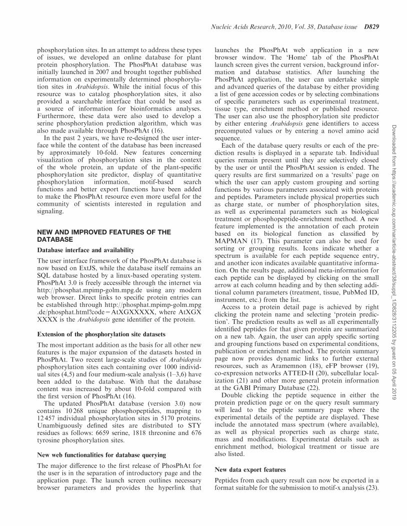
launches the PhosPhAt web application in a newbrowser window. The ‘Home’ tab of the PhosPhAtlaunch screen gives the current version, background infor-mation and database statistics. After launching thePhosPhAt application, the user can undertake simpleand advanced queries of the database by either providinga list of gene accession codes or by selecting combinationsof specific parameters such as experimental treatment,tissue type, enrichment method or published resource.The user can also use the phosphorylation site predictorby either entering Arabidopsis gene identifiers to accessprecomputed values or by entering a novel amino acidsequence.Each of the database query results or each of the pre-
diction results is displayed in a separate tab. Individualqueries remain present until they are selectively closedby the user or until the PhosPhAt session is ended. Thequery results are first summarized on a ‘results’ page onwhich the user can apply custom grouping and sortingfunctions by various parameters associated with proteinsand peptides. Parameters include physical properties suchas charge state, or number of phosphorylation sites,as well as experimental parameters such as biologicaltreatment or phosphopeptide-enrichment method. A newfeature implemented is the annotation of each proteinbased on its biological function as classified byMAPMAN (17). This parameter can also be used forsorting or grouping results. Icons indicate whether aspectrum is available for each peptide sequence entry,and another icon indicates available quantitative informa-tion. On the results page, additional meta-information foreach peptide can be displayed by clicking on the smallarrow at each column heading and by then selecting addi-tional column parameters (treatment, tissue, PubMed ID,instrument, etc.) from the list.Access to a protein detail page is achieved by right
clicking the protein name and selecting ‘protein predic-tion’. The prediction results as well as all experimentallyidentified peptides for that given protein are summarizedon a new tab. Again, the user can apply specific sortingand grouping functions based on experimental conditions,publication or enrichment method. The protein summarypage now provides dynamic links to further externalresources, such as Aramemnon (18), eFP browser (19),co-expression networks ATTED-II (20), subcellular local-ization (21) and other more general protein informationat the GABI Primary Database (22).Double clicking the peptide sequence in either the
protein prediction page or on the query result summarywill lead to the peptide summary page where theexperimental details of the peptide are displayed. Theseinclude the annotated mass spectrum (where available),as well as physical properties such as charge state,mass and modifications. Experimental details such asenrichment method, biological treatment or tissue arealso listed.
New data export features
Peptides from each query result can now be exported in aformat suitable for the submission to motif-x analysis (23).
Nucleic Acids Research, 2010, Vol. 38, Database issue D829
Dow
nloaded from https://academ
ic.oup.com/nar/article-abstract/38/suppl_1/D
828/3112205 by guest on 05 April 2019
At the level of the query result summary, the user has thechoice of hand-selecting peptides or of selecting all uniquepeptide sequences present in the query result. The outputformat is as 13-mers centered on the phosphorylatedresidue. Only clearly assigned phosphorylation sites(i.e. those annotated as pS, pT and pY) will be included.For multiply phosphorylated peptides, each phospho-rylation site will result in export of a separate 13-mersequence. It is also now possible to export PhosPhAtquery results in a spreadsheet format. The spectra of indi-vidual phosphopeptides can also be exported as an imageor in the mgf data format.
Extension of the plant-specific prediction of threonineand tyrosine
Given the growing number of experimentally determinedphosphorylation sites, the plant-specific predictor forserine phosphorylation has also been updated and newprediction algorithms for threonine and tyrosinephosphorylation have now been implemented in additionto serine phosphorylation. For prediction of threonineand tyrosine phosphorylation sites, 1818 experimentalsites for threonine and 676 experimental sites fortyrosine were used (Supplementary Data). Instead ofonly providing pre-computed phosphorylation sitepredictions for Arabidopsis proteins, it is now possible to
paste any given protein sequence into the prediction querywindow and obtain prediction results. The latter functionallows the predictors to be used independent of the plantspecies. In the original version, only Arabidopsis sites hadbeen pre-computed. Thus, this presents a novel service forprediction of plant-specific phosphorylation sites.
Visualization of site-specific phosphorylation dynamics
Where available, we have collected data from time-coursestudies of relative abundance of phosphorylation atspecific sites (10,24). This information is displayed onthe peptide summary page as simplified trend graphs. Asquantitative datasets will expand in future, the display ofthe results can help to assign biological functions tospecific phosphorylation sites through their involvementin particular biological processes or stress responses.
Extension of protein-context information
As a new feature, we have annotated each protein basedon its biological function as classified by MAPMAN (17)and provide related mapping to existing pathways.Moreover, predicted Pfam domain structures aremapped onto the protein sequence display allowing theuser to visually inspect the localization of experimentaland predicted phosphorylation sites to known domainsas displayed in the phosphorylation site prediction
Figure 1. Screenshots of new PhosPhAt web interface. (A) Entry screen with advanced search, (B) result window, (C) peptide summary and(D) protein summary.
D830 Nucleic Acids Research, 2010, Vol. 38, Database issue
Dow
nloaded from https://academ
ic.oup.com/nar/article-abstract/38/suppl_1/D
828/3112205 by guest on 05 April 2019
summary. This information can be valuable in assessingthe quality of predicted and experimentally deter-mined phosphorylation sites as certain domains areunlikely to contain phosphorylation sites. Thus, besidesprediction score and the experimentally determinedphosphorylation site assignment score [PTM score (25)and PhosCalc score (26)], the likelihood of presence of aphosphorylation site in the protein sequence can be judgedby the user.
In addition to the experimentally determined andpredicted phosphorylation sites of a given protein on theprotein summary page, information is also provided aboutany putative phosphorylating kinase based on publisheddata from the recent protein microarray-based kinaseassays (27,28). This information is being dynamicallydrawn from GabiPD (22). Such information will helpusers to establish links between specific phosphorylationsites or kinase substrates and phosphorylating kinases.Peptides containing specific kinase target motifs can beextracted from the experimental datasets using the motifsearch function. The motif search function identifiesknown kinase recognition motifs (e.g. a casein kinasemotif around the experimentally determined phosphor-ylation sites) and displays all experimentally identifiedpeptides containing the desired motif. Users can alsoenter motifs of interest by typing in the motif in singleamino acid letter code and putting alternative aminoacids in brackets. The X serves as a wild card for anyamino acid. For example, [KRH]X[KRH]XXS willretrieve the SNF1-like kinase recognition sites in frontof a phosphoserine.
Subcellular localization
While much of the data in PhosPhAt representsphosphoproteomic surveys from whole tissues or cells, itis possible to profile these data utilizing subcellular local-ization information available for Arabidopsis proteins.The SUBA database contains experimentally derivedsubcellular locations for over 8000 Arabidopsis proteinscomprising proteomic studies, fluorescent proteins,ontology data and descriptor information (16). This local-ization information was used to profile experimentallydetermined phosphorylated proteins in the PhoPhAtdatabase. A ‘winner takes all’ approach was used to
determine a single (or multiple) subcellular location foreach protein, i.e. an unweighted count of each experimen-tal location in SUBA was calculated, with the highestscore deemed the winner. Over 40% (2051) of thephosphorylated proteins in PhosPhAt had some informa-tion regarding their subcellular localization in Arabidopsis(Table 1). A comparison of localization rates of the12 subcellular locations annotated in the SUBAdatabase for phosphorylated proteins indicates that ahigher proportion seem to be localized to the plasmamembrane (�2-fold higher) and cytoskeleton (�1.5-foldhigher). While the increased number of phosphoproteinslocalized to the plasma membrane may be explained bythe number of targeted phosphoproteomic analyses of thisstructure (10,24,29,30), it also represents one of the maincontrol points within the plant cell where phosphorylationcascades are initiated and thus may be an overrepresentedlocation within the plant cell for phosphorylation. Thecytoskeleton is also known to be heavily regulated byphosphorylation especially during cell division (31).Subcellular locations with some under-representation ofprotein phosphorylation include the extracellular space,the Golgi, the mitochondrion and to a lesser extent thenucleus. It seems probable that little protein phospho-rylation would occur to secreted proteins (extracellular)while little is currently known concerning the role ofphosphorylation in plant Golgi. Interestingly, a recentsurvey of phosphorylation in Arabidopsis mitochondriaidentified very few mitochondrial phosphoproteins con-firming the findings of under-representation in thisorganelle (32). As further subcellular structures aretargeted and characterized using phosphoproteomictechniques, it will be interesting to observe if the aboveproportions are sustained, especially with regard to theplasma membrane as a major site of phosphorylationwithin the plant cell.
CONFIDENCE OF EXPERIMENTALLYDETERMINED PHOSPHORYLATION SITESAND FALSE POSITIVES
With increasing numbers of phosphorylation sites beingidentified by various experimental groups, the questionabout confidence of the phosphorylation site identification
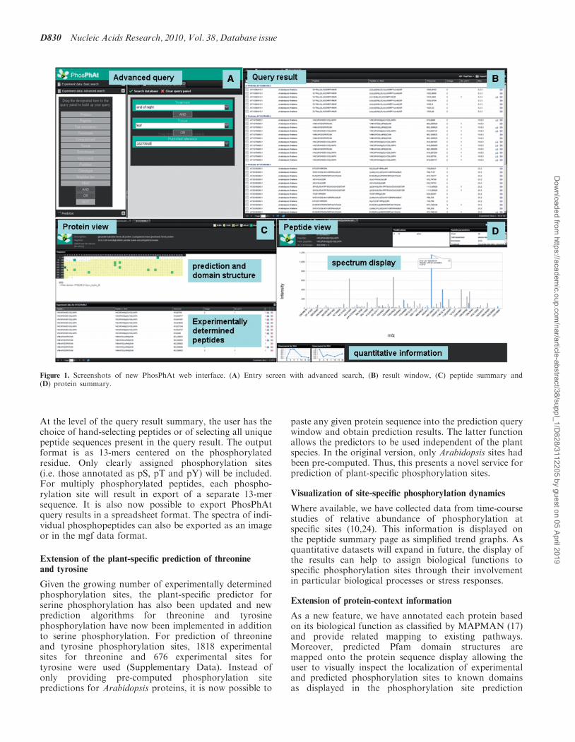
Table 1. Sub-cellular location of experimentally determined phosphoproteins in the PhosPhAt database
Subcellular location Total SUBA Total SUBA (%) PhosPhAt experiment PhosPhAt experiment (%) Fold difference
Cell plate 20 0.2 6 0.2 1.2Cytoskeleton 78 0.8 28 1.2 1.4Cytosol 515 5.4 114 4.7 0.9Endoplasmic reticulum 479 5.0 112 4.6 0.9Extracellular 565 5.9 65 2.7 0.5Golgi 183 1.9 28 1.2 0.6Mitochondrion 883 9.2 132 5.5 0.6Nucleus 2363 24.6 433 17.9 0.7Peroxisome 238 2.5 56 2.3 0.9Plasma membrane 1759 18.3 823 34.1 1.9Plastid 1894 19.7 441 18.3 0.9Vacuole 622 6.5 175 7.3 1.1
Nucleic Acids Research, 2010, Vol. 38, Database issue D831
Dow
nloaded from https://academ
ic.oup.com/nar/article-abstract/38/suppl_1/D
828/3112205 by guest on 05 April 2019
as well as the false positive rate is frequently raised.Although the extent of false positive rate for phospho-peptides and phosphorylation sites is not known andtheir experimental confirmation is not easily assessedat present, a confidence measure may be generated bystatistical analysis of the overlap of phosphorylationsites between two independent datasets. Furthermore,predictor-based strategies supporting the identification offalse positives are conceivable. A newly identifiedphosphorylation site might be further supported byassignment of the confidence value derived from anaccurate prediction approach.In this study, we evaluated the overlap of experimental
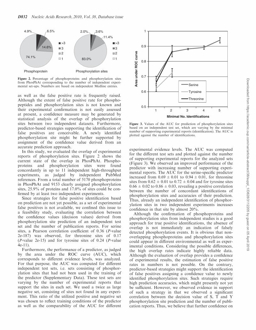
reports of phosphorylation sites. Figure 2 shows thecurrent state of the overlap in PhosPhAt. Phospho-proteins and phosphorylation sites were foundconcordantly in up to 11 independent high-throughputexperiments, as judged by independent PubMedreferences. From a total number of 5170 phosphoproteinsin PhosPhAt and 9153 clearly assigned phosphorylationsites, 25.9% of proteins and 17.0% of sites could be con-firmed by at least two independent experiments.Since strategies for false positive identification based
on prediction are not yet possible, as a set of experimentalfalse positives is not available, we confined this issue toa feasibility study, evaluating the correlation betweenthe confidence values (decision values) derived fromphosphorylation site prediction of an independent testset and the number of publication reports. For serinesites, a Pearson correlation coefficient of 0.36 (P-value2e-187) was observed, for threonine sites of 0.17(P-value 2e-15) and for tyrosine sites of 0.24 (P-value4e-11).Furthermore, the performance of a predictor, as judged
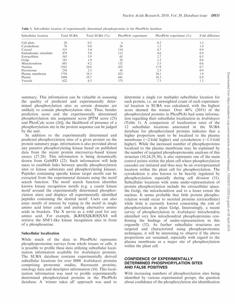
by the area under the ROC curve (AUC), whichcorresponds to different evidence levels, was analyzed.For that purpose, the AUC was computed for differentindependent test sets, i.e. sets consisting of phosphor-ylation sites that had not been used in the training ofthe predictor (Supplementary Data). These test sets arevarying by the number of experimental reports thatsupport the sites in each set. We used a twice as largenegative set, consisting of sites not found in any experi-ment. This ratio of the utilized positive and negative setwas chosen to reflect training conditions of the predictoras well as the comparability of the AUC for different
experimental evidence levels. The AUC was computedfor the different test sets and plotted against the numberof supporting experimental reports for the analyzed sets(Figure 3). We observed an improved performance of thepredictor with increasing number of supporting experi-mental reports. The AUC for the serine-specific predictorincreased from 0.69±0.01 to 0.94±0.01, for threoninesites from 0.62±0.01 to 0.72±0.04 and for tyrosine sites0.66±0.02 to 0.86±0.03, revealing a positive correlationbetween the number of concordant identifications ofphosphorylation sites and accuracies of their prediction.Thus, already an independent identification of phosphor-ylation sites in two independent experiments increasesconfidence in that site by almost 20%.
Although the confirmation of phosphoproteins andphosphorylation sites from independent studies is a goodapproach for true positive identifications, the absence ofoverlap is not immediately an indication of falselydetected phosphorylation events. It is obvious that non-overlapping phosphoproteins and phosphorylation sitescould appear in different environmental as well as exper-imental conditions. Considering the possible differences,the high overlap rates indicate highly reliable data.Although the evaluation of overlap provides a confidenceof experimental results, the estimation of false positiverates in numbers is not possible. On the contrary,predictor-based strategies might support the identificationof false positives assigning a confidence value to newlyidentified phosphorylation sites. Such strategies requirehigh prediction accuracies, which might presently not yetbe sufficient. However, we observed evidence in supportof such a strategy in that we observed a significantcorrelation between the decision value of S, T and Yphosphorylation site prediction and the number of publi-cation reports. Thus, we believe that further confidence on
Minimal No. Identifications
Are
a u
nd
er R
OC
cu
rve
1 20.5
0.6
0.7
0.8
0.9
1
3 4 5
Serine
Threonine
Tyrosine
Figure 3. Values of the AUC for prediction of phosphorylation sitesbased on an independent test set, which are varying by the minimalnumber of supporting experimental reports (identification). The AUC isplotted against the number of identifications.
Phosphoprotein Phosphorylation sites
4.9% 6.5% 2.0% 3.6%
11.4%
83.0%
>3
3
2
1
>3
3
2
1
14.6%
74.1%
Figure 2. Percentage of phosphoproteins and phosphorylation sitesfrom PhosPhAt corresponding to the number of independent experi-mental set-ups. Numbers are based on independent Medline entries.
D832 Nucleic Acids Research, 2010, Vol. 38, Database issue
Dow
nloaded from https://academ
ic.oup.com/nar/article-abstract/38/suppl_1/D
828/3112205 by guest on 05 April 2019
experimental data might indeed be provided by integra-tion with prediction results.
CONCLUSIONS
In conclusion, we believe that the PhosPhAt database haswithin the last 2 years been significantly improved, andits web service extended and is now recognized beyondthe plant research community as a basis for futuredevelopments and research around phosphorylation sitesand their biological functions.
SUPPLEMENTARY DATA
Supplementary Data are available at NAR Online.
ACKNOWLEDGEMENTS
We thank Dr Joachim Selbig and Dr Dirk Walther forhelpful discussion and input during the development ofnew functionalities. Dr Bjorn Usadel was of great helpin implementation of the MAPMAN ontology.
FUNDING
Financial support by the Bundesministerium fur Bildungund Forschung (BMBF) (GABI-FUTURE Grants0315046 and 0315049A to B.K. and A.N.). Funding foropen access charge: Max Planck Society, Germany.
Conflict of interest statement. None declared.
REFERENCES
1. de la Fuente van Bentem,S., Anrather,D., Dohnal,I., Roitinger,E.,Csaszar,E., Joore,J., Buijnink,J., Carreri,A., Forzani,C.,Lorkovic,Z.J. et al. (2008) Site-specific phosphorylation profiling ofArabidopsis proteins by mass spectrometry and peptide chipanalysis. J. Proteome Res., 7, 2458–2470.
2. Jones,A.M., Maclean,D., Studholme,D.J., Serna-Sanz,A.,Andreasson,E., Rathjen,J.P. and Peck,S.C. (2009)Phosphoproteomic analysis of nuclei-enriched fractions fromArabidopsis thaliana. J. Proteomics, 72, 439–451.
3. Li,H., Wong,W.S., Zhu,L., Guo,H.W., Ecker,J. and Li,N. (2009)Phosphoproteomic analysis of ethylene-regulated proteinphosphorylation in etiolated seedlings of Arabidopsis mutant ein2using two-dimensional separations coupled with a hybridquadrupole time-of-flight mass spectrometer. Proteomics, 9,1646–1661.
4. Reiland,S., Messerli,G., Baerenfaller,K., Gerrits,B., Endler,A.,Grossmann,J., Gruissem,W. and Baginsky,S. (2009) Large-scaleArabidopsis phosphoproteome profiling reveals novel chloroplastkinase substrates and phosphorylation networks. Plant Physiol.,150, 889–903.
5. Sugiyama,N., Nakagami,H., Mochida,K., Daudi,A., Tomita,M.,Shirasu,K. and Ishihama,Y. (2008) Large-scale phosphorylationmapping reveals the extent of tyrosine phosphorylation inArabidopsis. Mol. Syst. Biol., 4, e1–7.
6. Whiteman,S.-A., Serazetdinova,L., Jones,A.M., Sanders,D.,Rathjen,J., Peck,S.C. and Maathuis,F.J.M. (2008) Identification ofnovel proteins and phosphorylation sites in a tonoplast enrichedmembrane fraction of Arabidopsis thaliana. Proteomics, 8,3536–3547.
7. Kersten,B., Agrawal,G.K., Durek,P., Neigenfind,J., Schulze,W.,Walther,D. and Rakwal,R. (2009) Plant phosphoproteomics: anupdate. Proteomics, 9, 964–988.
8. Tornroth-Horsefield,S., Wang,Y., Hedfalk,K., Johanson,U.,Karlsson,M., Tajkhorshid,E., Neutze,R. and Kjellbom,P. (2006)Structural mechanism of plant aquaporin gating. Nature, 439,688–694.
9. Prak,S., Hem,S., Boudet,J., Viennois,G., Sommerer,N.,Rossignol,M., Maurel,C. and Santoni,V. (2008) Multiplephosphorylations in the C-terminal tail of plant plasma membraneaquaporins: role in subcellular trafficking of AtPIP2;1 in responseto salt stress. Mol. Cell. Proteomics, 7, 1019–1030.
10. Niittyla,T., Fuglsang,A.T., Palmgren,M.G., Frommer,W.B. andSchulze,W.X. (2007) Temporal analysis of sucrose-inducedphosphorylation changes in plasma membrane proteins ofArabidopsis. Mol. Cell. Proteomics, 6, 1711–1726.
11. Tripodi,K.E., Turner,W.L., Gennidakis,S. and Plaxton,W.C. (2005)In vivo regulatory phosphorylation of novel phosphoenolpyruvatecarboxylase isoforms in endosperm of developing castor oil seeds.Plant Physiol., 139, 969–978.
12. Kaiser,W.M. and Huber,S.C. (2001) Post-translational regulation ofnitrate reductase: mechanism, physiological relevance andenvironmental triggers. J. Exp. Bot., 52, 1981–1989.
13. Hardin,S.C., Larue,C.T., Oh,M.H., Jain,V. and Huber,S.C. (2009)Coupling oxidative signals to protein phosphorylationvia methionine oxidation in Arabidopsis. Biochem. J., 422,305–312.
14. Loque,D., Yuan,L., Kojima,S., Gojon,A., Wirth,J., Gazzarrini,S.,Ishiyama,K., Takahashi,H. and von Wiren,N. (2006) Additivecontribution of AMT1;1 and AMT1;3 to high-affinity ammoniumuptake across the plasma membrane of nitrogen-deficientArabidopsis roots. Plant J., 48, 522–534.
15. Oh,M.H., Wang,X., Kota,U., Goshe,M.B., Clouse,S.D. andHuber,S.C. (2009) Tyrosine phosphorylation of the BRI1 receptorkinase emerges as a component of brassinosteroid signaling inArabidopsis. Proc. Natl Acad. Sci. USA, 106, 658–663.
16. Heazlewood,J.L., Durek,P., Hummel,J., Selbig,J., Weckwerth,W.,Walther,D. and Schulze,W.X. (2008) PhosPhAt: a database ofphosphorylation sites in Arabidopsis thaliana and a plant-specificphosphorylation site predictor. Nucleic Acids Res., 36,D1015–D1021.
17. Usadel,B., Nagel,A., Thimm,O., Redestig,H., Blasing,O.E.,Palacios-Rojas,N., Selbig,J., Hannemann,J., Piques,M.C.,Steinhauser,D. et al. (2005) Extension of the visualization toolMapMan to allow statistical analysis of arrays, display ofcoresponding genes, and comparison with known responses.Plant Physiol., 138, 1195–1204.
18. Schwacke,R., Schneider,A., van der Graaff,E., Fischer,K.,Catoni,E., Desimone,M., Frommer,W.B., Flugge,U.-I. andKunze,R. (2003) ARAMEMNON, a novel databasefor Arabidopsis integral membrane proteins. Plant Physiol., 131,16–26.
19. Winter,D., Vinegar,B., Nahal,H., Ammar,R., Wilson,G.V. andProvart,N.J. (2007) An ‘‘electronic fluorescent pictograph’’ browserfor exploring and analyzing large-scale biological data sets.PLoS ONE, 2, e718.
20. Obayahi,T., Hayashi,S., Saeki,M., Ohta,H. and Kinoshita,K. (2009)ATTED-II provides coexpressed gene networks for Arabidopsis.Nucleic Acids Res., 37, D987–D991.
21. Heazlewood,J.L., Verboom,R.E., Tonti-Filippini,J., Small,I. andMillar,A.H. (2007) SUBA: the Arabidopsis Subcellular Database.Nucleic Acids Res., 35, D213–D218.
22. Riano-Pachon,D.M., Nagel,A., Neigenfind,J., Wagner,R.,Basekow,R., Weber,E., Muller-Rober,B., Diehl,S. and Kersten,B.(2009) GabiPD: the GABI primary database—a plant integrative‘omics’ database. Nucleic Acids Res., 37, D954–D959.
23. Schwartz,D. and Gygi,S.P. (2005) An iterative statistical approachto the identification of protein phosphorylation motifs from large-scale data sets. Nat. Biotechnol., 23, 1391–1398.
24. Nuhse,T.S., Bottrill,A.R., Jones,A.M. and Peck,S.C. (2007)Quantitative phosphoproteomic analysis of plasma membraneproteins reveals regulatory mechanisms of plant innate immuneresponses. Plant J., 51, 931–940.
25. Olsen,J.V., Blagoev,B., Gnad,F., Macek,B., Kumar,C.,Mortensen,P. and Mann,M. (2006) Global, in vivo, and site-specificphosphorylation dynamics in signaling networks. Cell, 127,635–648.
Nucleic Acids Research, 2010, Vol. 38, Database issue D833
Dow
nloaded from https://academ
ic.oup.com/nar/article-abstract/38/suppl_1/D
828/3112205 by guest on 05 April 2019
26. MacLean,D., Burrell,M.A., Studholme,D.J. and Jones,A.M. (2008)PhosCalc: a tool for evaluating the sites of peptide phosphorylationfrom Mass Spectrometer data. BMC Res. Notes, 1, 30.
27. Popescu,S.C., Popescu,G.V., Bachan,S., Zhang,Z., Gerstein,M.,Snyder,M. and Dinesh-Kumar,S.P. (2009) MAPK target networksin Arabidopsis thaliana revealed using functional proteinmicroarrays. Genes Dev., 23, 80–92.
28. Feilner,T., Hultschig,C., Lee,J.M., Meyer,S., Immink,R.G.H.,Koenig,A., Possling,A., Seitz,H., Beveridge,A., Scheel,D. et al.(2005) High throughput identification of potential Arabidopsismitogen-activated protein kinases substrates. Mol. Cell Proteomics,4, 1558–1568.
29. Benschop,J.J., Mohammed,S., O’Flaherty,M., Heck,A.J., Slijper,M.and Menke,F.L. (2007) Quantitative phospho-proteomics of early
elicitor signalling in Arabidopsis. Mol. Cell Proteomics, 6,1705–1713.
30. Nuhse,T.S., Stensballe,A., Jensen,O.N. and Peck,S.C. (2004)Phosphoproteomics of the Arabidopsis plasma membraneand a new phosphorylation site database. Plant Cell, 16,2394–2405.
31. Blume,Y.B., Lloyd,C.W. and Yemets,A.I. (2008) The plantcytoskeleton: a key tool for Agro-Biotechnology. Springer,The Netherlands, pp. 145–159.
32. Ito,J., Taylor,N.L., Castleden,I., Weckwerth,W., Millar,A.H. andHeazlewood,J.L. (2009) A survey of the Arabidopsis thalianamitochondrial phosphoproteome. Proteomics (in press).
D834 Nucleic Acids Research, 2010, Vol. 38, Database issue
Dow
nloaded from https://academ
ic.oup.com/nar/article-abstract/38/suppl_1/D
828/3112205 by guest on 05 April 2019