phloem loading by the pmsuc2 sucrose carrier from plantago … · the plant cell, vol. 7,...
TRANSCRIPT

The Plant Cell, Vol. 7, 1545-1554, October 1995 0 1995 American Society of Plant Physiologists
Phloem Loading by the PmSUC2 Sucrose Carrier from Plantago major Occurs into Companion Cells
Ruth Stadler,a3' Johanna Brandner,ai2 Alexander Schulz,b Manfred Gahrtz,a,' and Norbert Sauer
a Lehrstuhl für Zellbiologie und Pflanzenphysiologie, Universitat Regensburg, D-93040 Regensburg, Germany Botanisches Institut, Universitat Kiel, Olshausenstrasse 40, D-24098 Kiel, Germany
High levels of mRNA for the sucrose-H+ symporter PmSUC2 have been found in the vascular bundles of petioles from Plantago major. The possible role of PmSUC2 in phloem loading was studied with antiserum raised against the recom- binant PmSUC2 protein. This antiserum labeled a single 35-kD protein band in detergent extracts of R major vascular bundles. It showed no cross-reaction with the R majorsucrose carrier PmSUCl, which was tested with detergent extracts from plasma membranes of transgenic yeast strains containing either the R major sucrose transporter PmSUCl or PmSUC2. The antiserum was used to determine the site of PmSUC2 expression in leaves, petioles, and roots of R major. In cross- sections and longitudinal sections, the PmSUC2 protein was found in only one single cell type. These cells were identi- fied as companion cells because they are nucleated, contain a dense cytoplasm, and are always adjacent to a sieve element. The labeled cells had the same longitudinal extensíon as did their sister sieve elements and always ended next to the sieve plates, which were characterized by specific staining. PmSUC2 mRNA and PmSUC2 protein were also detected in R major roots. The function of PmSUC2 in the different organs and its role in phloem loading are discussed.
INTRODUCTION
In many plants, sucrose is the long-distance transport form for carbohydrates. During recent years, cDNAs for severa1 su- crose transporters have been cloned (Riesmeier et al., 1992, 1993; Gahrtz et al., 1994; Sauer and Stolz, 1994). They were all expressed in baker's yeast and shown to catalyze the uptake of sucrose across yeast plasma membranes. Invertase- deficient yeast cells expressing the flantago major PmSUC2 sucrose transporter accumulate sucrose >200-fold, and in vitro studies with the recombinant PmSUC2 protein from yeast have shown that the necessary energy can be supplied by an artifi- cial proton motive force (PMF) generator (Gahrtz et al., 1994). This is consistent with earlier studies of sucrose-H+ sym- porters performed with whole plant cells (for review, see Delrot, 1989) or with studies of plasma membrane vesicles purified from plant tissue (Buckhout, 1989; Bush, 1989). First evidence for phloem localization of one of the cloned sucrose trans- porters was obtained for the potato StSUT1 transporter by using in situ hybridization experiments (Riesmeier et al., 1993). The abundance of fmSUC2 mRNA in vascular tissue (Gahrtz et al., 1994), investigations with StSUTl antisense potato plants (Riesmeier et al., 1994), and the phloem specificity of an AtSUC2 promoter-0-glucuronidase (GUS) reporter gene
' Current address: Universitat Erlangen-Nümberg, Lehrstuhl Botanik (I, Staudtstrasse 5, D-91058 Erlangen, Germany. * Current address: Deutsches Krebsforschungszentrum, Abteilung für Zellbiologie, Im Neuenheimer Feld 280, D-69120 Heidelberg, Germany.
To whom correspondence should be addressed.
construct in Arabidopsis (Truernit and Sauer, 1995) have con- firmed these results.
The phloem of angiosperms is composed of phloem paren- chyma cells, sieve elements, and companion cells, and a large number of structural differences suggest a clear functional separation of these cell types. Mature, fully developed deve tubes lose their nuclei, and most of their other cell organelles, such as vacuoles, dictyosomes, and ribosomes, are degraded during the final steps of phloem differentiation. However, other organelles, such as the endoplasmic reticulum, mitochondria, and plastids, are found in varying amounts during all stages of sieve element ontogeny (for review, see Behnke, 1989). Com- panion cells, in contrast, contain large nuclei and, in addition to all other organelles, numerous mitochondria, plastids, and ribosomes. The extraordinary density of their protoplasts dis- tinguishes them not only from the sieve elements but also from the phloem parenchyma cells (Behnke, 1989). For many plants, plasmodesmal frequencies between the so-called sieve ele- ment-companion cell (se-cc) complex and the surrounding parenchymatic cells have been studied, and three groups with practically no connections, with few plasmodesmata, or with many plasmodesmata have been identified (Gamalei, 1989). A large number of plasmodesmal connections between com- panion cells and sieve elements on one hand and a symplastic isolation of the se-cc complex on the other have also been described (Hayes et al., 1985; van Bel and Kempers, 1990; van der Schoot and van Bel, 1990). The work of Gamalei (1989) shows that the members of the Plantaginaceae display virtu- ally complete symplastic isolation of the se-cc complex. As
1546 The Plant Cell
early as 1929, sucrose was shown to be the main sugar withinthe vascular bundles from P. major, and it became clear thatsucrose accumulates within the vascular tissue (Ritschl, 1929).
These results can be explained only by the existence of anenergy-dependent sucrose transporter that catalyzes phloemloading from the apoplast. This concept was developed byGiaquinta (1977) and confirmed by Komor et al. (1977) andothers. A more general correlation between symplastic isola-tion of the se-cc complex and apoplastic phloem loading wasdescribed recently (van Bel et al., 1994). In light of this work,there was a need to identify the cells in which the actual load-ing step occurs. Morphological adaptations, such as theincreased surface of the companion cells in certain plants(so-called transfer cells) and the immunolocalization of aH+-ATPase within such transfer cells (Bouche-Pillon et al.,1994), suggest that these cells might represent the actual siteof loading.
Phloem loading does not seem, however, to be the sole func-tion of sucrose transporters. Analyses of the tissue-specificexpression of sucrose transporters by RNA gel blotting andin transgenic Arabidopsis plants (Sauer and Stolz, 1994;Truernit and Sauer, 1995) have clearly pointed to an expres-sion of Arabidopsis sucrose transporters outside the classicalsource tissue, such as fully developed leaves. In this study,we addressed how sucrose enters the phloem and into whichcells the actual loading step occurs by using a combinationof different histochemical techniques. The use of highly spe-cific anti-PmSUC2 antibodies permitted the localization of thePmSUC2 protein in companion cells.
RESULTS
Specificity of the Anti-PmSUC2 Antiserum
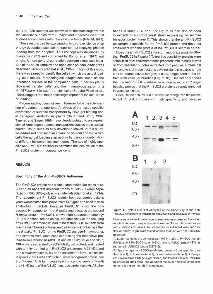
The PmSUC2 protein has a calculated molecular mass of 54kD and an apparent molecular mass of ~35 kD when sepa-rated on 10% SDS-polyacrylamide gels (Gahrtz et al., 1994).The recombinant PmSUC2 protein from transgenic baker'syeast was isolated from preparative SDS gels and used to raiseantibodies in rabbits. Because PmSUC2 is not the onlysucrose-H+ symporter from P. major and because the secondP. major protein, PmSUCI, shows high sequence homology(59.9% identical amino acids), the specificity of the resultinganti-PmSUC2 antiserum had to be tested. SDS extracts fromplasma membranes of transgenic yeast cells expressing eitherthe P. major PmSUCI or the PmSUC2 sucrose-H+ symporter,and extracts from yeast cells expressing the respective pro-teins from Arabidopsis (AtSUCI and AtSUC2; Sauer and Stolz,1994), were separated by SDS-PAGE, gel blotted, and treatedwith affinity-purified anti-PmSUC2 antiserum. A 35-kD bandand a much weaker 70-kD band (the dimeric form), which cor-respond to the PmSUC2 protein, were recognized only in lane3 of Figure 1A. A faint cross-reaction can be seen only withthe 43-kD band of the AtSUCI sucrose carrier (lane 4). All other
bands in lanes 2, 4, and 5 of Figure 1A can also be seenin extracts of a control yeast strain expressing no sucrosetransport protein (lane 1). This shows that the anti-PmSUC2antiserum is specific for the PmSUC2 protein and does notcross-react with the protein of the PmSUCI sucrose carrier.
Does the anti-PmSUC2 antiserum recognize proteins otherthan PmSUC2 in P. major ? To test this possibility, proteins weresolubilized from total membranes prepared from P. major leavesor from vascular bundles extracted from petioles. Protein gelblot analysis of these fractions gave no signals in extracts fromsink or source leaves but gave a clear, single band in the ex-tract from vascular bundles (Figure 1B). This not only showsthat the anti-PmSUC2 antiserum is monospecific in P. majorbut also proves that the PmSUC2 protein is strongly enrichedin vascular tissue.
Because the anti-PmSUC2 antiserum recognized the recom-binant PmSUC2 protein with high specificity and because
1 2 3 4 5
1 2 3
kD68-
43-
29-
BkD68-
43-
29-
Figure 1. Protein Gel Blot Analyses of the Specificity of the Anti-PmSUC2 Antiserum in Transgenic Yeast Cells and in Leaves of P. major.Plasma membranes from transgenic yeast strains expressing four differ-ent plant sucrose transporters, as shown in (A), or total membranesfrom P. major sink leaves, source leaves, or extracted vascular bun-dles, as shown in (B), were tested for their reaction with anti-PmSUC2antiserum.(A) Lane 1 contains the control (strain MGY1); lane 2, PmSUCI (strainMGY6); lane 3, PmSUC2 (strain MGY2); lane 4, AtSUCI (strain NSY611);and lane 5, A1SUC2 (strain NSY612).(B) Ten micrograms of SDS-solubilized proteins from vascular bun-dles (lane 1), sink leaves (lane 2), or source leaves (lane 3) of P. majorwas separated on SDS gels, gel blotted, and treated with anti-PmSUC2antiserum (diluted 1:16). The apparent molecular masses of the sizemarkers are given at left in kilodaltons.

Localization of PmSUC2 1547
Figure 2. A Vascular Bundle and a se-cc Complex from P. major Petioles.Petiole sections were photographed under the light microscope, as shown in (A), or the electron microscope, as shown in (B).(A) An overview of a vascular bundle from a petiole. Scale bar = 50 urn.(B) A typical se-cc complex surrounded by five phloem parenchyma cells; the dense cytoplasm of the smaller companion cell is rich in mitochon-dria. Scale bar = 2 urn.
cross-reactions could be detected with neither the secondP. major sucrose transporter nor any other membrane proteinof P. major, the serum was used for immunohistochemical anal-ysis of PmSUC2 localization in P. major.
Localization of the PmSUC2 Protein
The cross-section of a typical vascular bundle from petiolesof P. major is presented in Figure 2A. The large xylem vesselsin the center are sandwiched between two layers of phloem(bicollateral bundle) that can be identified as groups of smallercells above and below the xylem. Typical pairs of sieve tubesand smaller companion cells are found only in these parts ofthe vascular bundle. The darker cells at the top and the bot-tom of the bundle represent sclerenchymatic fibers that arepartly lignified but still alive. At higher magnification (Figure2B), the dense cytoplasm of the companion cells and the com-paratively empty sieve tubes can be distinguished easily.Typically, the companion cells are smaller in diameter thanare the sieve elements. We wanted to determine in which celltype(s) of the phloem the PmSUC2 gene was expressed—thephloem parenchyma, or one or both cell types of the se-cccomplex?
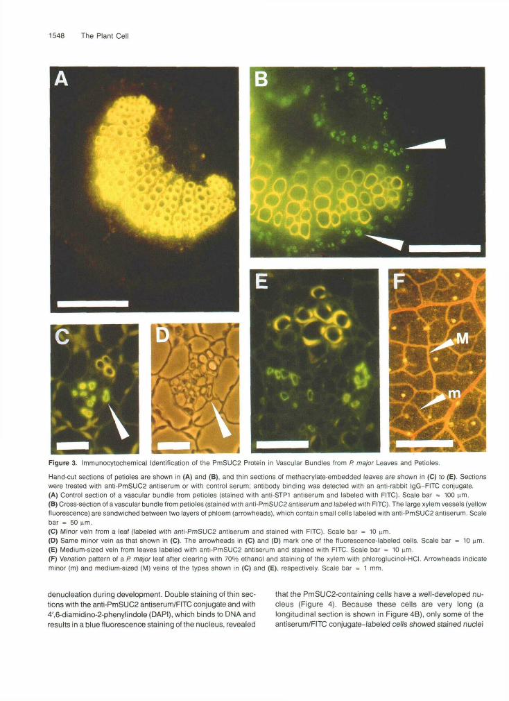
Sections from petioles of P. major were incubated with theanti-PmSUC2 antiserum (Figure 3B) or with the anti-STP1(sugar transport protein 1; Stolz et al., 1994) antiserum (con-trol; Figure 3A) and decorated with a fluorescein isothiocyanate(FITC) isomer 1-conjugated secondary antibody. Fluorescencemicroscopy of labeled sections shows a green, anti-PmSUC2
antibody-dependent fluorescence in defined regions on bothsides of the xylem (Figure 3B). This localization of the fluores-cence label is in perfect agreement with the bicollateralorganization of the P. major vascular bundles in the petioles,indicating that the cells labeled with the anti-PmSUC2 antise-rum are located within the phloem. A second anti-PmSUC2antiserum, which was raised independently in a second rab-bit, gave identical results. Controls in which the anti-PmSUC2antibody was either omitted or replaced by a control antise-rum raised against the Arabidopsis STP1 monosaccharide-H+
symporter showed no FITC labeling (Figure 3A). The yellowstaining of the xylem vessels is due to phenolic compoundsin the cell walls of the respective cells.
In contrast to the large vascular bundles of the petioles, theminor and medium-sized veins of P. major leaves revealed acollateral organization with only one layer of phloem below asmall number of xylem elements. Figures 3C to 3E showsections of minor and medium-sized veins stained with anti-PmSUC2 antiserum and labeled with FITC. Again, thePmSUC2 protein is seen only in the phloem portion. Figure3F shows the venation pattern of a P. major leaf with the sametypes of vein shown in cross-section in Figures 3C (minor vein)and 3E (medium-sized vein).
Characterization of the PmSUC2-Expressing Cells
For a more precise characterization of the cell type, it wasnecessary to look at additional properties of the labeledcells. One of the characteristics typical of sieve elements is
1548 The Plant Cell
Figure 3. Immunocytochemical Identification of the PmSUC2 Protein in Vascular Bundles from P. major Leaves and Petioles.
Hand-cut sections of petioles are shown in (A) and (B), and thin sections of methacrylate-embedded leaves are shown in (C) to (E). Sectionswere treated with anti-PmSUC2 antiserum or with control serum; antibody binding was detected with an anti-rabbit IgG-FITC conjugate.(A) Control section of a vascular bundle from petioles (stained with anti-STP1 antiserum and labeled with FITC). Scale bar = 100 urn.(B) Cross-section of a vascular bundle from petioles (stained with anti-PmSUC2 antiserum and labeled with FITC). The large xylem vessels (yellowfluorescence) are sandwiched between two layers of phloem (arrowheads), which contain small cells labeled with anti-PmSUC2 antiserum. Scalebar = 50 urn.(C) Minor vein from a leaf (labeled with anti-PmSUC2 antiserum and stained with FITC). Scale bar = 10 urn.(D) Same minor vein as that shown in (C). The arrowheads in (C) and (D) mark one of the fluorescence-labeled cells. Scale bar = 10 urn.(E) Medium-sized vein from leaves labeled with anti-PmSUC2 antiserum and stained with FITC. Scale bar = 10 urn.(F) Venation pattern of a P. major leaf after clearing with 70% ethanol and staining of the xylem with phloroglucinol-HCI. Arrowheads indicateminor (m) and medium-sized (M) veins of the types shown in (C) and (E), respectively. Scale bar = 1 mm.
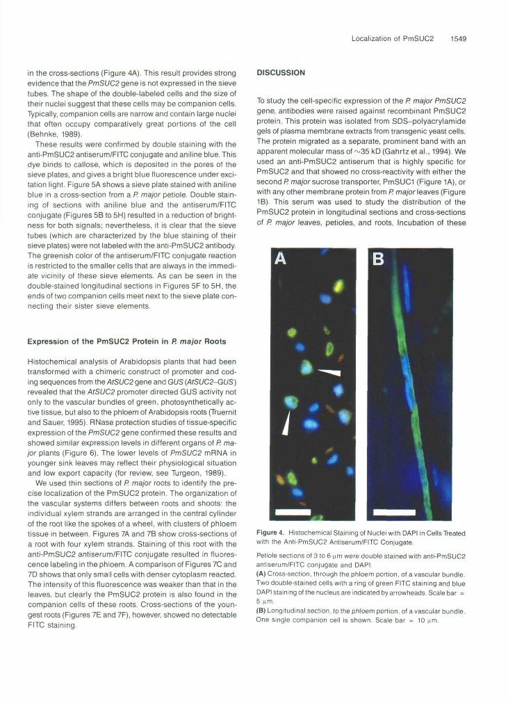
denucleation during development. Double staining of thin sec-tions with the anti-PmSUC2 antiserum/FITC conjugate and with4',6-diamidino-2-phenylindole (DAPI), which binds to DNA andresults in a blue fluorescence staining of the nucleus, revealed
that the PmSUC2-containing cells have a well-developed nu-cleus (Figure 4). Because these cells are very long (alongitudinal section is shown in Figure 4B), only some of theantiserum/FITC conjugate-labeled cells showed stained nuclei
Localization of PmSUC2 1549
in the cross-sections (Figure 4A). This result provides strongevidence that the PmSUC2 gene is not expressed in the sievetubes. The shape of the double-labeled cells and the size oftheir nuclei suggest that these cells may be companion cells.Typically, companion cells are narrow and contain large nucleithat often occupy comparatively great portions of the cell(Behnke, 1989).
These results were confirmed by double staining with theanti-PmSUC2 antiserum/FITC conjugate and aniline blue. Thisdye binds to callose, which is deposited in the pores of thesieve plates, and gives a bright blue fluorescence under exci-tation light. Figure 5A shows a sieve plate stained with anilineblue in a cross-section from a P. major petiole. Double stain-ing of sections with aniline blue and the antiserum/FITCconjugate (Figures 5B to 5H) resulted in a reduction of bright-ness for both signals; nevertheless, it is clear that the sievetubes (which are characterized by the blue staining of theirsieve plates) were not labeled with the anti-PmSUC2 antibody.The greenish color of the antiserum/FITC conjugate reactionis restricted to the smaller cells that are always in the immedi-ate vicinity of these sieve elements. As can be seen in thedouble-stained longitudinal sections in Figures 5F to 5H, theends of two companion cells meet next to the sieve plate con-necting their sister sieve elements.
Expression of the PmSUC2 Protein in R major Roots
Histochemical analysis of Arabidopsis plants that had beentransformed with a chimeric construct of promoter and cod-ing sequences from the AtSUC2 gene and GUS (AtSUC2-GUS)revealed that the AtSUC2 promoter directed GUS activity notonly to the vascular bundles of green, photosynthetically ac-tive tissue, but also to the phloem of Arabidopsis roots (Truernitand Sauer, 1995). RNase protection studies of tissue-specificexpression of the PmSUC2 gene confirmed these results andshowed similar expression levels in different organs of P. ma-jor plants (Figure 6). The lower levels of PmSUC2 mRNA inyounger sink leaves may reflect their physiological situationand low export capacity (for review, see Turgeon, 1989).
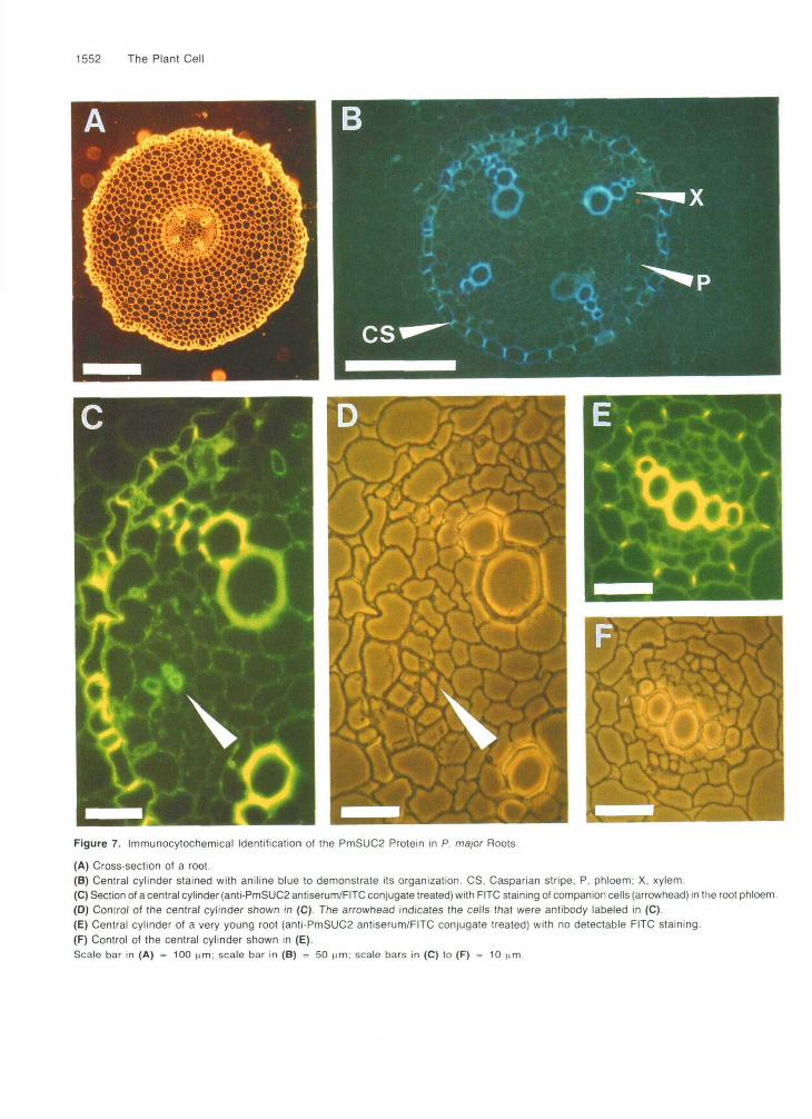
We used thin sections of P. major roots to identify the pre-cise localization of the PmSUC2 protein. The organization ofthe vascular systems differs between roots and shoots: theindividual xylem strands are arranged in the central cylinderof the root like the spokes of a wheel, with clusters of phloemtissue in between. Figures 7A and 7B show cross-sections ofa root with four xylem strands. Staining of this root with theanti-PmSUC2 antiserum/FITC conjugate resulted in fluores-cence labeling in the phloem. A comparison of Figures 7C and7D shows that only small cells with denser cytoplasm reacted.The intensity of this fluorescence was weaker than that in theleaves, but clearly the PmSUC2 protein is also found in thecompanion cells of these roots. Cross-sections of the youn-gest roots (Figures 7E and 7F), however, showed no detectableFITC staining.
DISCUSSION
To study the cell-specific expression of the R major PmSUC2gene, antibodies were raised against recombinant PmSUC2protein. This protein was isolated from SDS-polyacrylamidegels of plasma membrane extracts from transgenic yeast cells.The protein migrated as a separate, prominent band with anapparent molecular mass of ~35 kD (Gahrtz et al., 1994). Weused an anti-PmSUC2 antiserum that is highly specific forPmSUC2 and that showed no cross-reactivity with either thesecond R major sucrose transporter, PmSUCI (Figure 1 A), orwith any other membrane protein from P. major leaves (Figure1B). This serum was used to study the distribution of thePmSUC2 protein in longitudinal sections and cross-sectionsof P. major leaves, petioles, and roots. Incubation of these
Figure 4. Histochemical Staining of Nuclei with DAPI in Cells Treatedwith the Anti-PmSUC2 Antiserum/FITC Conjugate.
Petiole sections of 3 to 6 urn were double stained with anti-PmSUC2antiserum/FITC conjugate and DAPI.(A) Cross-section, through the phloem portion, of a vascular bundle.Two double-stained cells with a ring of green FITC staining and blueDAPI staining of the nucleus are indicated by arrowheads. Scale bar =5 urn.(B) Longitudinal section, to the phloem portion, of a vascular bundle.One single companion cell is shown. Scale bar = 10 ^im.

1550 The Plant Cell
Figure 5. Double Staining of Sieve Plates with Aniline Blue and of Companion Cells with the Anti-PmSUC2 Antiserum/FITC Conjugate.
(A) Sieve plate from a petiole cross-section stained with aniline blue only.(B) to (E) Sieve plates (blue dots) and companion cells (green rings) from petiole cross-sections double stained with aniline blue and the anti-PmSUC2 antiserum/FITC conjugate. Scale bars in (A) to (E) = 5 urn.(F) to (H) Sieve plates and companion cells from petiole longitudinal sections double stained with aniline blue and the anti-PmSUC2 antise-rum/FITC conjugate, respectively. Arrowheads indicate the ends of companion cells next to the respective sieve plate. Scale bars = 10 iam.
sections with the anti-PmSUC2 antiserum and with the FITC-labeled second antibody revealed a specific localization of thePmSUC2 protein in one single cell type within the phloem.The additional use of classic histochemical techniques for lightmicroscopy showed that the antiserum/FITC conjugate-labeled
cells are longitudinally extended and relatively narrow, witha well-developed nucleus. In petioles and major veins, thediameter of these cells is smaller than the diameter of the ad-jacent sieve tubes. However, in minor veins, these cells haveat least the same diameter as the adjacent sieve elements.

The sieve tubes themselves, whose sieve plates were stainedwith aniline blue, did not have detectable amounts of thePmSUC2 protein.
The se-cc complex originates from one cambial or procam-bial derivative; typically, the last, usually unequal division ofthis cell gives rise to a larger sieve element and a narrow com-panion cell. The companion cell may then undergo a numberof anticlinal divisions or perform the same longitudinal exten-sion as its sister sieve element. In any case, the outermostends of a companion cell will be just at the ends of the respec-tive sieve tube. This can be seen in Figure 5E, in which thefluorescence of the antiserum/FITC conjugate-labeled com-panion cells ends at the sieve plate connecting their sieveelements.
The gene for the PmSUC2 sucrose-H+ symporter is ex-pressed in the companion cells of the minor and medium-sizedveins of the leaves that collect the assimilates and that arethe primary sites of sucrose loading. PmSUC2 is also ex-pressed in the companion cells of the largest vascular bundlesof leaves and petioles that are primarily responsible for theexport of sucrose and other assimilates from the leaves. Toa lesser extent, these cells are responsible for sucrose load-ing. A direct comparison of antibody/FITC conjugate-labeledthin sections from veins of different sizes revealed that thehighest intensity of FITC fluorescence is seen reproducibly inthe minor veins. This suggests that more PmSUC2 protein issynthesized in the companion cells of these minor veins, whichare the sites of maximal phloem loading, than in the compan-ion cells of the larger exporting veins.
The identification of PmSUC2 mRNA in root tissue and thelocalization of PmSUC2 protein in companion cells of the rootphloem confirm results obtained earlier from Arabidopsis plantstransformed with an AtSUC2-GUS fusion (Truernit and Sauer,1995). Therefore, the function of PmSUC2 cannot be restrictedto the loading of newly synthesized sucrose into the phloemof source leaf minor veins. PmSUC2 also may be involved inone of the following two reactions: First, the high concentra-tion of sucrose within the sieve tubes causes a permanentpassive leak of sucrose out of the sieve tubes. During the long-distance transport from the source leaf to the different sinks,a dramatic loss of assimilate could occur and cause reducedflux rates within the sieve tubes. To avoid this loss of sucrose
Localization of PmSUC2 1551
and to maintain an optimal mass flow within the sieve tubes,it may be necessary to reimport the lost sucrose into the se-cccomplex. Such retrieval mechanisms have been discussedpreviously as the primary or even exclusive function of plantsugar transporters (Maynard and Lucas, 1982). Second, thePmSUC2 protein may catalyze not only energy-dependentphloem loading but, under appropriate conditions in the sinks,also the export of sucrose from the se-cc complex.
In this study, we addressed how sucrose enters the sievetubes of P. major. Sucrose is loaded from the apoplast into thecompanion cells of the phloem; from there, sucrose diffusessymplastically through the plasmodesmata of the se-cc com-plex into the sieve elements. Companion cells, which are richin mitochondria, do not supply the sieve tubes with energyfor sucrose uptake; rather, they use this energy themselves.The energy could be provided from sucrose hydrolysis by acompanion cell-specific sucrose synthase (Nolte and Koch,1993). A plasma membrane, H+-ATPase, which is expressedpredominantly in transfer cells and which generates the PMFnecessary for sucrose loading, has previously been describedin broad bean (Bouche-Pillon et al., 1994).
The PmSUC2 protein studied by heterologous expressionin yeast is extremely pH sensitive and shows increased trans-port rates with decreasing extracellular pH values (Gahrtz etal., 1994). The respective H+-ATPase in P. major may there-fore not only supply the PMF needed for sucrose transportbut also regulate the transport rates of the PmSUC2 proteinby influencing the extracellular pH. It has been shown recently(Buckhout, 1994) that changes in the extracellular pH stronglyaffect the Km value of a sucrose carrier from sugar beet, sug-gesting that such changes may in fact represent a regulatorymechanism. However, in contrast with these results, Bush(1990) proposed a model in which, according to the proposedpH optimum, the proton binding site of the sucrose transporteris always occupied under physiological conditions. Accord-ing to this model, changes in the extracellular pH would notinfluence the activity of the transporter, and only changes inthe extracellular sucrose concentration or in the membranepotential could affect the rates of phloem loading.
METHODS
si so r s f
Figure 6. RNase Protection Analysis of the Organ-Specific Expres-sion of the PmSUC2 Gene.
Twenty micrograms of total RNA from each organ was used in eachlane, si, sink leaves; so, source leaves; r, roots; s, stems; f, flowers
Strains
Plants (Plantago major) were grown in the greenhouse. The specific-ity of the anti-PmSUC2 antiserum was tested with different strains ofbaker's yeast. All were transformants of strain DBY2617 (MATa, his4-539, Iys2-801, ura3-52, suc2-438; Kaiser and Botstein, 1986). The strainsincluded MGY2 (Gahrtz et al., 1994) and MGY6, which express theR major sucrose-H* symporters PmSUC2 and PmSUCl, respectively,and strains NSY611 and NSY612, which express the Arabidopsis suc-rose-H* symporters AtSUCI and AtSUC2, respectively. Strain MGY1,which harbors the PmSUC2 construct of strain MGY2 in an antisenseorientation (Gahrtz et al., 1994), was used as a control strain. Forconstruction of strains MGY1, MGY2, NSY611, and NSY612, the yeast-
1552 The Plant Cell
Figure 7. Immunocytochemical Identification of the PmSUC2 Protein in P. major Roots.
(A) Cross-section ot a root.(B) Central cylinder stained with aniline blue to demonstrate its organization. CS, Casparian stripe; P. phloem; X, xylem.(C) Section of a central cylinder (anti-PmSUC2 antiserum/FITC conjugate treated) with FITC staining of companion cells (arrowhead) in the root phloem.(D) Control of the central cylinder shown in (C). The arrowhead indicates the cells that were antibody labeled in (C).(E) Central cylinder of a very young root (anti-PmSUC2 antiserum/FITC conjugate treated) with no detectable FITC staining.(F) Control of the central cylinder shown in (E).Scale bar in (A) = 100 (im; scale bar in (B) = 50 tim; scale bars in (C) to (F) = 10 nm.
Localization of PmSUC2 1553
Escherichia colishuttle vector NEV-E was used (Sauer and Stolz, 1994). For construction of strain MGY6, afull-length cDNA clone of the F! ma- jor PmSUCl sucrose-H+ symporter (EMBL accession number X84379) was cloned into the unique Notl site of the yeast-E colishut- tle vector NEV-N (Sauer and Stolz, 1994).
buffer (50 mM Tris-HCI, pH 7.5, 150 mM NaCI, 1% skim milk powder), and incubated in blocking buffer for 1 hr; hand-cut sections were not treated with acetone. After overnight incubation with affinity-purified anti-PmSUCP antiserum (diluted 1:2 in blocking buffer), the coverslips were washed three times with blocking buffer containing 0.1% Tri- ton X-100 and incubated for 1 hr with the anti-rabbit IgG-fluorescein isothiocyanate (FITC) isomer 1 conjugate (Sigma; diluted 1:300 in block- ing buffer with 0.1% Triton X-100). After three final washes with blocking buffer containing 0.1% Triton X-100, the coverslips were rinsed with water and mounted in FITC-Guard (Testoc Inc., Chicago, IL). Photo- graphs were taken with a fluorescence phase microscope (Standard 16; Carl Zeiss, Oberkochen, Germany) with an excitation light of 450 to 490 nm.
For double staining of the PmSUC2 protein with the antiserumlFlTC conjugate and of nuclei with 4’,6-diamidincd-phenylindole (DAPI; Serva, Heidelberg, Germany), sections were treated as described above. Af- ter the three final washing steps, slides were incubated for 1 hr at room temperature in DAPl(0.2 pglmL). DAPI fluorescence was detected with an excitation light of 365 nm.
For double staining of the PmSUC2 protein with the antiserumlFITC conjugate and of Sieve plates with aniline blue (Water Blue; Fluka, Buchs, Switzerland), the slides were incubated for 5 min in aniline blue (0.5% in 50 mM NalK-PO, buffer, pH 7.2). Aniline blue fluores- cence was detected with an excitation light of 365 nm.
Preparation of Yeast Plasma Membranes and of Total Membranes from R major Tissue
Plasma membranes were isolated from yeast cells as described pre- viously by Stolz et al. (1994). Total membranes from P major leaves or isolated vascular bundles were isolated as described by Stolz et al. (1994).
Preparation of the Anti-PmSUCP Antiserum, SDS-PAGE, and Protein Gel Blotting
The recombinant PmSUC2 protein was isolated from the yeast strain MGY2 (Gahrtz et al., 1994). Plasma membranes of MGY2 cells were isolated (Stolz et al., 1994), plasma membrane proteins were solubi- lized with SDS, and the proteins were separated by preparative SDS-PAGE (Laemmli, 1970). The recombinant PmSUC2 protein was visualized, excised, and eluted from the gel as described by Sauer and Stadler (1993). From 1 mg of SDS-solubilized yeast plasma mem- brane proteins, 30 to 40 pg of purified PmSUC2 protein was obtained. Three to four portions of -10 pg were injected into rabbits at intervals
RNA lsolation and RNase Protection Analysis
of -3 weeks. Crude anti-PmSUCP antisera were affinity purified by adsorption
to the recombinant PmSUC2 protein, which had been immobilized on nitrocellulose filters (Sauer and Stadler, 1993). Residual unspecific antibodies were removed (Sauer and Stadler, 1993) by adsorption to immobilized plasma membrane proteins from yeast strain MGYl (Gahrtz et al., 1994). The quality of the purified anti-PmSUCP antiserum was tested on protein gel blots (Dunn, 1986). Antibody binding was detected with the chemiluminescence Western blot detection kit from Amer- sham, according to the manufacturer’s protocol.
Preparation and Fixation of R major Sections for Light Microscopy
P majortissue was collected, and RNA was isolated as described pre- viously (Sauer et al., 1990). RNase protection assays were performed with a radiolabeled 214-bp fmSUC2 antisense RNA. RNA was sepa- rated on 10% polyacrylamide gels.
Electron Microscopy
For fixation, longitudinal hand-cut sections (10 x 3 x 0.5 mm) from petioles of F! major were immediately immersed in a formaldehyde- glutaraldehyde mixture (Karnovsky, 1965) in 100 mM sodium cacodyl- ate buffer, pH 7.3. After rinsing with the same buffer, postfixation with 1% OsO,, dehydration through an acetone series, and embedding with Spurr’s resin, the ends of the sections (2 mm) were discarded, and middle portions were oriented for transverse sectioning and poly- merized in at 6OOC for 36 hr, Semithin sections (1 pm thick) were stained with 0.5% crystal violet for light microscopy (Figure 2A). Ultra- thin sections (50 nm thick) were treated with uranyl acetate and lead citrate and observed with an electron microscope (CM 10; Philips, Eind- hoven, The Netherlands).
Fragments frOm IeaVeS, petiOleS, and rOOtS Of F! major p h t S Were briefly degased in 1 mL Of MoPs buffer, PH 6.9 (50 mM 2-[N-morPholinol- ProPanesulfonic acid/NaOH, PH 6.9, 5 mM EGTA, 2 mM MgClz). The MOpS buffer was replaced by 1 mL Of fiXatiOn SOhJtiOn (ethanol-form- aldehyde [37%]-acidic acid, 60:40:2 [vlv]; Schlüter, 1976), and the tiSSUe was fixed at room temperature for 1 hr. After washes with 70 and 100% ethanol, the tissue was embedded into methacrylate (Baskin et al., 1992) as described previously (Stadler et al., 1995). Semithin sections (3 to 6 pm) were made with an ultramicrotome(Reichert, Vienna, Aus- tria), and sections were put on poly-L-lysine-coated coverslips. Handcut sections (shown in Figures 3A and 38) were made with a razor blade, fixed as described above, and washed with Mops buffer, pH 6.9.
ACKNoWLEDGMENTS
We thank Heinz-Dietmar Behnke (University of Heidelberg) and Jürgen Stolz (University of Erlangen-Nürnberg) for helpful discussions and Günther Peissig (University of Regensburg) for growing the F! major plants. This work was supported by the Deutsche Forschungsgemein- schaft (Grant No. SFB 43/C5) and a grant from the Bundesministerium für Bildung, Wissenschaft, Forschung und Technologie (No. BE021- 0310331~).
Staining of Sections with Fluorescent Dyes
For removal of methacrylate from the thin sections, coverslips were incubated for 30 sec in 100% acetone, washed three times with blocking
1554 The Plant Cell
Received May 22, 1995; accepted August 11, 1995.
REFERENCES
Baskin, T.I., Busby, C.H., Fowke, L.C., Sammut, M., and Gubler, F. (1992). lmprovements in immunostaining samples embedded in methacrylate: Localization of microtubules and other antigens throughout developing organs in plants of diverse taxa. Planta 187, 405-413.
Behnke, H.-D. (1989). Structure of the phloem. In Transport of Pho- toassimilates, D.A. Baker and J. Milburn, eds (Harlow, UK: Longman Scientific and Technical), pp. 79-137.
Bouche-Pillon, S., Fleurat-Lessard, P., Fromont, J.-C., Serrano, R., and Bonnemain, J.-L. (1994). lmmunolocalization of the plasma membrane H+-ATPase in minor veins of Vicia faba in relation to phloem loading. Plant Physiol. 105, 691-697.
Buckhout, T.J. (1989). Sucrose transport in isolated plasma membrane vesicles from sugar beet. Planta 178, 393-399.
Buckhout, T.J. (1994). Kinetic analysis of the plasma membrane suc- rose-H+ symporter from sugar beet (Beta vulgaris L.) leaves. Plant Physiol. 106, 991-998.
Bush, D.R. (1989). Proton-coupled sucrose transport in plasmalemma vesicles isolated from sugar beet (Beta vulgaris L. cv. Great West- ern) leaves. Plant Physiol. 89, 1318-1323.
Bush, D.R. (1990). Electrogenicity, pH-dependence, and stoichiome- try of the proton sucrose symport. Plant Physiol. 93, 1590-1596.
Delrot, S. (1989). Loading of photoassimilates. In Transport of Pho- toassimilates, D.A. Baker and J.A. Milburn, eds (Harlow, UK: Longman Scientific and Technical), pp. 167-205.
Dunn, S.D. (1986). Effects of the modification of transfer buffer com- position on the renaturation of proteins in gels on the recognition of proteins on Western blots by monoclonal antibodies. Anal. Bio- chem. 157, 144-153.
Gahrtz, M., Stolz, J., and Sauer, N. (1994). A phloem specific suc- rose-H+ symporter from Plantago major L. supports the model of apoplastic phloem loading. Plant J. 6, 697-706.
Gamalei, Y.V. (1989). Structure and function of leaf minor veins in trees and herbs: A taxonomic review. Trees 3, 96-110.
Giaquinta, R.T. (1977). Possible role of pH-gradient and membrane ATPase in the loading of sucrose into the sieve tubes. Nature 267, 369-370.
Hayes, P.M., Offler, C.E., and Patrick, J.W. (1985). Cellular structures, plasma membrane surface areas and plasmodesmatal frequencies of the stem of Phaseolus vulgaris L. in relation to radial photosyn- thate transfer. Ann. Bot. 56, 125-138.
Kaiser, C.A., and Botstein, D. (1986). Secretion-defective mutations in the signal sequence for Saccharomyces cerevisiae invertase. MOI. Cell. Biol. 6, 2382-2391.
Karnovsky, M.J. (1965). A formaldehyde-glutaraldehyde fixative of high osmolality for use in electron microscopy. J. Cell Biol. 27, 137A-138A.
Komor, E., Rotter, M., and Tanner, W. (1977). A proton-cotransport system in a higher plant: Sucrose transport in Ricinus communis. Plant Sci. Lett. 9, 153-162.
Laemmli, U.K. (1970). Cleavage of structural proteins during the as- sembly of the head of bacteriophage T4. Nature 227, 680-685.
Maniatis, T., Fritsch, E.F., and Sambrook, J. (1982). Molecular Clon- ing: A Laboratory Manual. (Cold Spring Harbor, NY: Cold Spring Harbor Laboratory Press).
Maynard, J.W., and Lucas, W.J. (1982). Sucrose and glucose uptake into Beta vulgaris leaf tissues: A case for general (apoplastic) re- trieval systems. Plant Physiol. 70, 1436-1443.
Nolte, K.D., and Koch, K.E. (1993). Companion-cell specific local- ization of sucrose synthase in zones of phloem loading and unloading. Plant Physiol. 101, 899-905.
Riesmeier, J.W., Willmitzer, L., and Frommer, W.B. (1992). lsolation and characterization of a sucrose carrier cDNA from spinach by func- tional expression in yeast. EMBO J. 11, 4705-4713.
Riesmeier, J.W., Hirner, B., and Frommer, W.B. (1993). Potato su- crose transporter expression in minor veins indicates a role in phloem loading. Plant Cell 5, 1591-1598.
Riesmeier, J.W., Willmitzer, L., and Frommer, W.B. (1994). Evidence for an essential role of the sucrose transporter in phloem loading and assimilate partitioning. EMBO J. 13, 1-7.
Ritschl, A. (1929). Weitere Untersuchungen iiber das gegenseitige Mengenverhaltnis der Kohlehydrate im Laubblatt unter verschiede- nen Aussenbedingungen. Bot. Archiv. 26, 349-384
Sauer, N., and Stadler, R. (1993). A sink specific H+/monosaccharide co-transporter from Nicotiana tabacum: Cloning and heterologous expression in baker's yeast. Plant J. 4, 601-610.
Sauer, N., and Stolz, J. (1994). SUCI and SUC2: Two sucrose trans- porters from Arabidopsis thaliana; expression and characterization in baker's yeast and identification of the histidine tagged protein. Plant J. 6, 67-77.
Sauer, N., Friedlander, K., and Griml-Wicke, U. (1990). Primary struc- ture, genomic organization and heterologous expression of a glucose transporter from Arabidopsis thaliana. EMBO J. 9, 3045-3050.
Schliiter, W. (1976). Mikroskopie. (Stuttgart, Germany: Aulis Verlag). Stadler, R., Wolf, K., Hilgarth, C., Tanner, W., and Sauer, N. (1995).
Subcellular localization of the inducible Chlorella HUPI monosac- charide-H+ symporter and cloning of a coinduced galactose-H+ symporter. Plant Physiol. 107, 33-41.
Stolz, J., Stadler, R., Opekarová, M., and Sauer, N. (1994). Func- tional reconstitution of the solubilized Arabidopsis thaliana STPl monosaccharide-H+ symporter in lipid vesicles and purification of the histidine tagged protein from transgenic Saccharomyces cerevisiae. Plant J. 6, 225-233.
Truernit, E., and Sauer, N. (1995). The promoter of the Arabidopsis thaliana SUCP sucrose-H+ symporter gene directs expression of !3-glucuronidase to the phloem: Evidence for phloem loading and unloading by SUC2. Planta 196, 564-570.
Turgeon, R. (1989). The sink-source transition in leaves. Annu. Rev. Plant MOI. Biol. 40, 119-138.
van Bel, A.J.E., and Kempen, R. (1990). Symplastic isolation of the sieve element companion cell complex in the phloem of Ricinus com- munis and Salix alba stems. Planta 183, 69-76.
van Bel, A.J.E., Amerlaan, A., and van Dijk, A.A. (1994). A three- step screening procedure to identify the mode of phloem loading in intact leaves: Evidence for symplasmic and apoplasmic phloem loading associated with the type of companion cell. Planta 192,31-39.
van der Schoot, C., and van Bel, A.J.E. (1990). Mapping membrane potentials and dye coupling in internodal tissues of tomato (Sola- num lycopersicum L.). Planta 182, 9-21.
DOI 10.1105/tpc.7.10.1545 1995;7;1545-1554Plant Cell
R. Stadler, J. Brandner, A. Schulz, M. Gahrtz and N. SauerCompanion Cells.
Phloem Loading by the PmSUC2 Sucrose Carrier from Plantago major Occurs into
This information is current as of April 12, 2020
Permissions 98X
https://www.copyright.com/ccc/openurl.do?sid=pd_hw1532298X&issn=1532298X&WT.mc_id=pd_hw15322
eTOCs http://www.plantcell.org/cgi/alerts/ctmain
Sign up for eTOCs at:
CiteTrack Alerts http://www.plantcell.org/cgi/alerts/ctmain
Sign up for CiteTrack Alerts at:
Subscription Information http://www.aspb.org/publications/subscriptions.cfm
is available at:Plant Physiology and The Plant CellSubscription Information for
ADVANCING THE SCIENCE OF PLANT BIOLOGY © American Society of Plant Biologists