pharynx - naaccr.org · regionalnodes pos 01 regionalnodes ex 25 eod primary tumor 800 eod lymph...
TRANSCRIPT
Pharynx 2018 10/4/18
NAACCR 2018‐2019 Webinar Series 1
PharynxNAACCR 2018‐2019 WEBINAR SERIES
1
Q&APlease submit all questions concerning the webinar content through the Q&A panel.
If you have participants watching this webinar at your site, please collect their names and emails
We will be distributing a Q&A document in about one week. This document will fully answer questions asked during the webinar and will contain any corrections that we may discover after the webinar.
2

Pharynx 2018 10/4/18
NAACCR 2018‐2019 Webinar Series 2
Fabulous Prizes
3
Guest SpeakerWilson Apollo, Radiation Therapist and CTR
4
Pharynx 2018 10/4/18
NAACCR 2018‐2019 Webinar Series 3
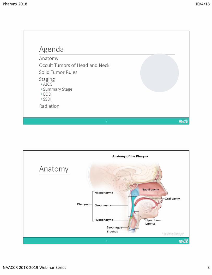
AgendaAnatomy
Occult Tumors of Head and Neck
Solid Tumor Rules
Staging◦ AJCC◦ Summary Stage◦ EOD◦ SSDI
Radiation
5
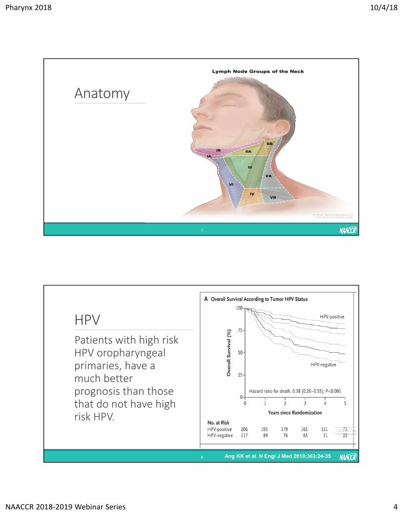
Anatomy
6
Pharynx 2018 10/4/18
NAACCR 2018‐2019 Webinar Series 4
Anatomy
7
HPV
8 Ang KK et al. N Engl J Med 2010;363:24-35
Patients with high risk HPV oropharyngeal primaries, have a much better prognosis than those that do not have high risk HPV.
Pharynx 2018 10/4/18
NAACCR 2018‐2019 Webinar Series 5
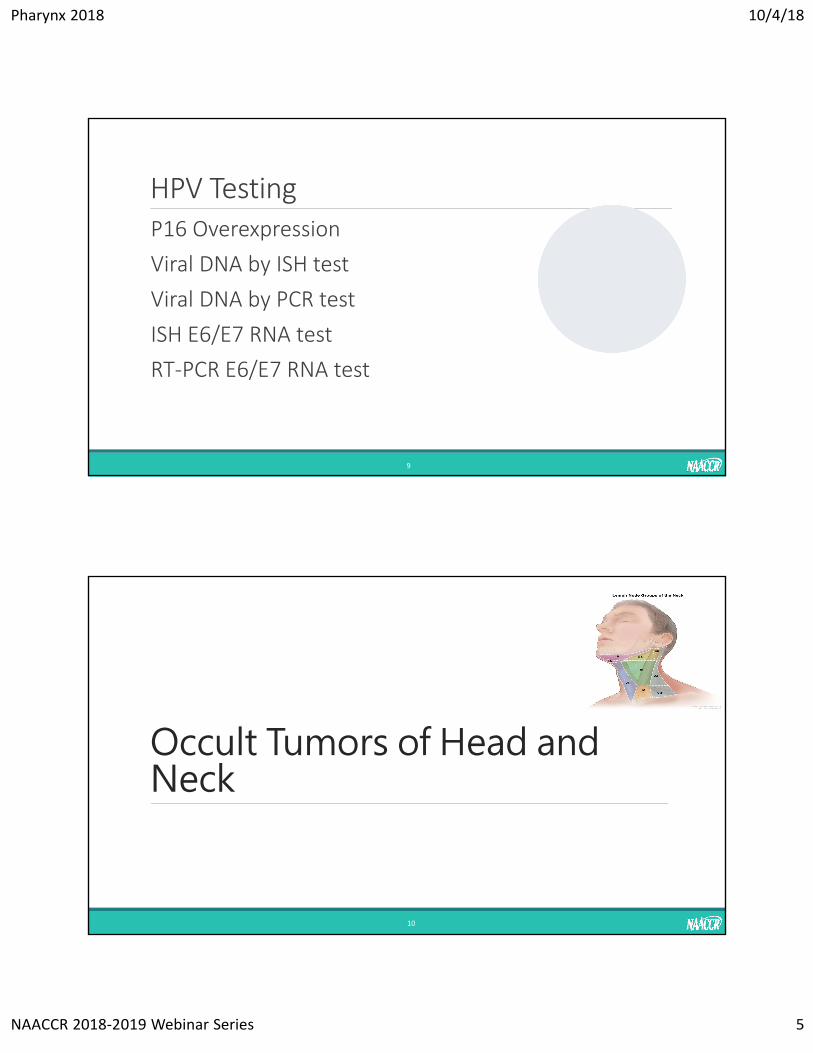
HPV TestingP16 Overexpression
Viral DNA by ISH test
Viral DNA by PCR test
ISH E6/E7 RNA test
RT‐PCR E6/E7 RNA test
9
Occult Tumors of Head and Neck
10
Pharynx 2018 10/4/18
NAACCR 2018‐2019 Webinar Series 6
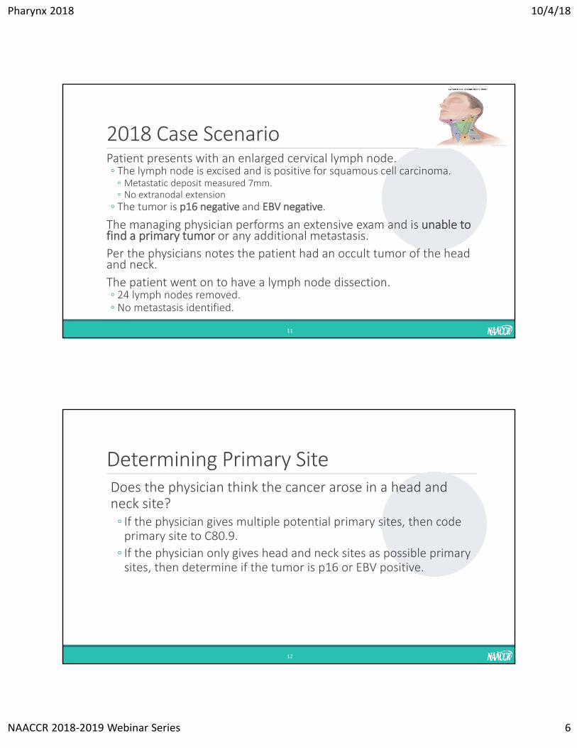
2018 Case ScenarioPatient presents with an enlarged cervical lymph node. ◦ The lymph node is excised and is positive for squamous cell carcinoma. ◦ Metastatic deposit measured 7mm.◦ No extranodal extension
◦ The tumor is p16 negative and EBV negative.
The managing physician performs an extensive exam and is unable to find a primary tumor or any additional metastasis.
Per the physicians notes the patient had an occult tumor of the head and neck.
The patient went on to have a lymph node dissection. ◦ 24 lymph nodes removed. ◦ No metastasis identified.
11
Determining Primary SiteDoes the physician think the cancer arose in a head and neck site?◦ If the physician gives multiple potential primary sites, then code primary site to C80.9.
◦ If the physician only gives head and neck sites as possible primary sites, then determine if the tumor is p16 or EBV positive.
12
Pharynx 2018 10/4/18
NAACCR 2018‐2019 Webinar Series 7
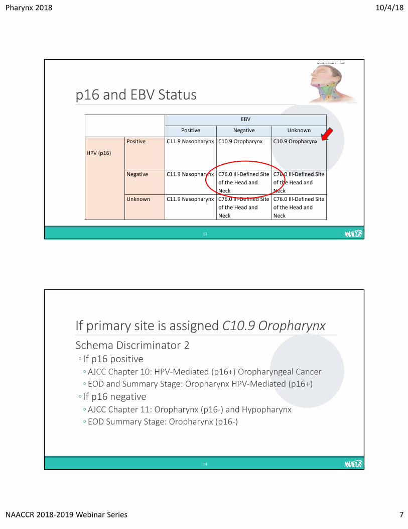
p16 and EBV Status
EBV
Positive Negative Unknown
HPV (p16)
Positive C11.9 Nasopharynx C10.9 Oropharynx C10.9 Oropharynx
Negative C11.9 Nasopharynx C76.0 Ill‐Defined Site
of the Head and
Neck
C76.0 Ill‐Defined Site
of the Head and
Neck
Unknown C11.9 Nasopharynx C76.0 Ill‐Defined Site
of the Head and
Neck
C76.0 Ill‐Defined Site
of the Head and
Neck
13
If primary site is assigned C10.9 Oropharynx
Schema Discriminator 2◦ If p16 positive ◦AJCC Chapter 10: HPV‐Mediated (p16+) Oropharyngeal Cancer
◦ EOD and Summary Stage: Oropharynx HPV‐Mediated (p16+)
◦ If p16 negative◦AJCC Chapter 11: Oropharynx (p16‐) and Hypopharynx
◦ EOD Summary Stage: Oropharynx (p16‐)
14
Pharynx 2018 10/4/18
NAACCR 2018‐2019 Webinar Series 8
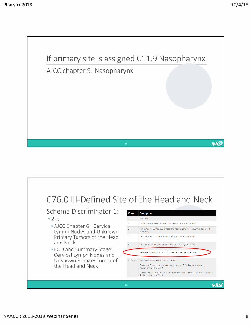
If primary site is assigned C11.9 Nasopharynx
AJCC chapter 9: Nasopharynx
15
C76.0 Ill‐Defined Site of the Head and NeckSchema Discriminator 1:◦2‐5 ◦AJCC Chapter 6: Cervical Lymph Nodes and Unknown Primary Tumors of the Head and Neck
◦ EOD and Summary Stage: Cervical Lymph Nodes and Unknown Primary Tumor of the Head and Neck
16
Pharynx 2018 10/4/18
NAACCR 2018‐2019 Webinar Series 9
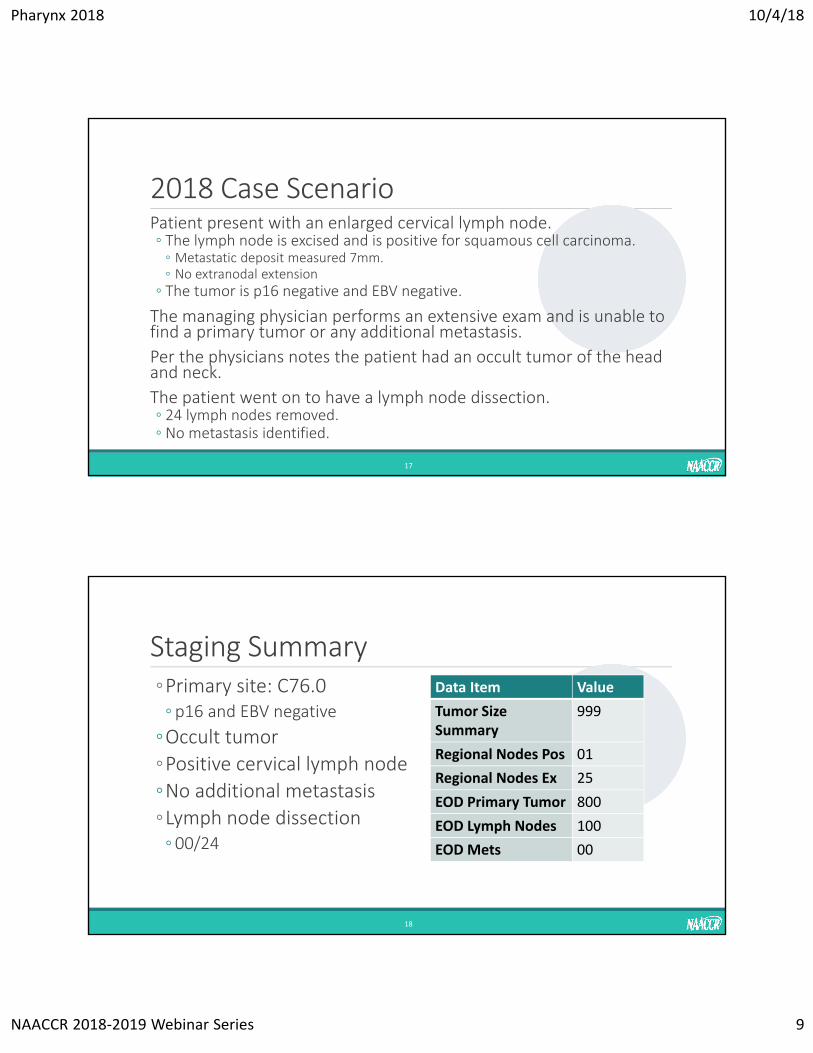
2018 Case ScenarioPatient present with an enlarged cervical lymph node. ◦ The lymph node is excised and is positive for squamous cell carcinoma. ◦ Metastatic deposit measured 7mm.◦ No extranodal extension
◦ The tumor is p16 negative and EBV negative.
The managing physician performs an extensive exam and is unable to find a primary tumor or any additional metastasis.
Per the physicians notes the patient had an occult tumor of the head and neck.
The patient went on to have a lymph node dissection. ◦ 24 lymph nodes removed. ◦ No metastasis identified.
17
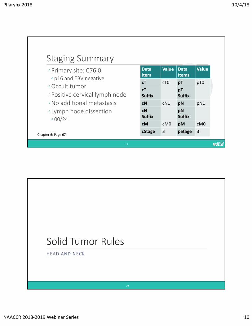
Staging Summary ◦Primary site: C76.0◦ p16 and EBV negative
◦Occult tumor
◦Positive cervical lymph node
◦No additional metastasis
◦Lymph node dissection◦ 00/24
18
Data Item Value
Tumor Size Summary
999
Regional Nodes Pos 01
Regional Nodes Ex 25
EOD Primary Tumor 800
EOD Lymph Nodes 100
EOD Mets 00
Pharynx 2018 10/4/18
NAACCR 2018‐2019 Webinar Series 10
Staging Summary ◦Primary site: C76.0◦ p16 and EBV negative
◦Occult tumor
◦Positive cervical lymph node
◦No additional metastasis
◦Lymph node dissection◦ 00/24
19
DataItem
Value Data Items
Value
cT cT0 pT pT0
cTSuffix
pT Suffix
cN cN1 pN pN1
cN Suffix
pNSuffix
cM cM0 pM cM0
cStage 3 pStage 3Chapter 6: Page 67
Solid Tumor RulesHEAD AND NECK
20
Pharynx 2018 10/4/18
NAACCR 2018‐2019 Webinar Series 11
21
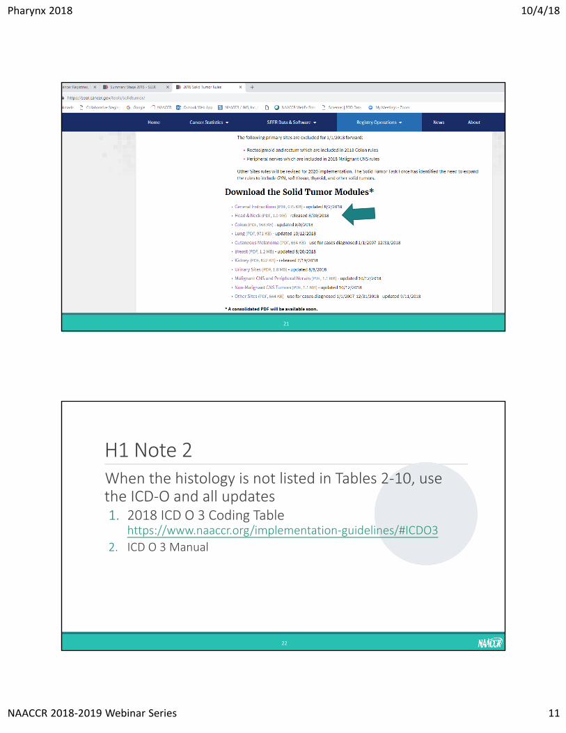
H1 Note 2When the histology is not listed in Tables 2‐10, use the ICD‐O and all updates1. 2018 ICD O 3 Coding Table
https://www.naaccr.org/implementation‐guidelines/#ICDO3
2. ICD O 3 Manual
22
Pharynx 2018 10/4/18
NAACCR 2018‐2019 Webinar Series 12
New Histologies Coding ClarificationSquamous cell carcinoma HPV‐negative 8086
Squamous cell carcinoma HPV‐positive 8085◦Do not use a p16 test to code 8085 or 8086. ◦HPV testing must be positive by viral detection tests in order to code histology as 8085.
Per the 2018 SEER Manual◦HPV‐type 16 refers to virus type and is different from p16 overexpression (p16+).
◦HPV status is determined by tests designed to detect viral DNA or RNA. Tests based on ISH, PCR, RT‐PCR technologies detect the viral DNA or RNA; whereas, the test for p16 expression, a surrogate marker for HPV, is IHC.
23
Pop QuizWhat histology would be coded to the following:◦Final diagnosis from path report is “squamous cell carcinoma”. Separate report shows tumor is p16+◦ 8070 Squamous cell carcinoma
◦Final diagnosis is “squamous cell carcinoma, HPV positive”◦ 8085 Squamous cell carcinoma, HPV positive
◦Final diagnosis is “squamous cell carcinoma”. A separate report shows HPV positive for viral DNA by ISH test◦ 8085 Squamous cell carcinoma, HPV positive
24
Pharynx 2018 10/4/18
NAACCR 2018‐2019 Webinar Series 13
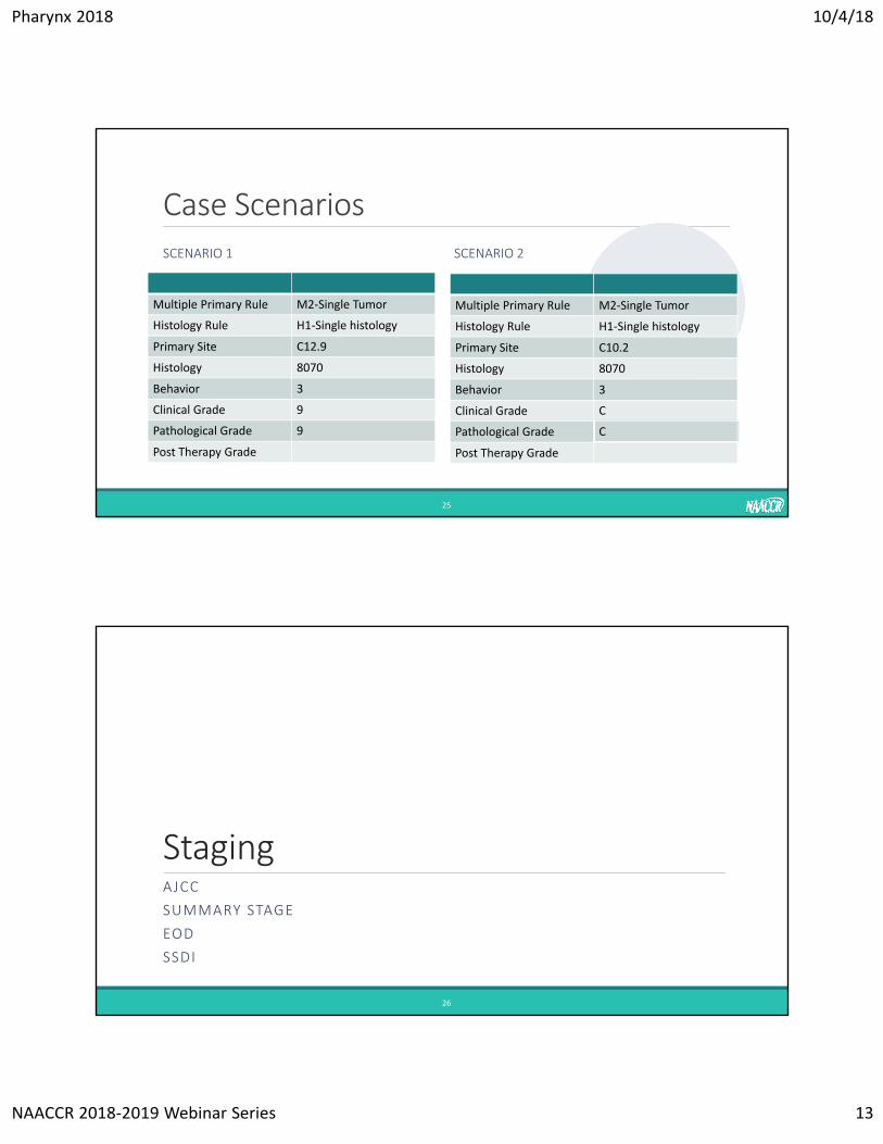
Case ScenariosSCENARIO 1 SCENARIO 2
25
Multiple Primary Rule M2‐Single Tumor
Histology Rule H1‐Single histology
Primary Site C12.9
Histology 8070
Behavior 3
Clinical Grade 9
Pathological Grade 9
Post Therapy Grade
Multiple Primary Rule M2‐Single Tumor
Histology Rule H1‐Single histology
Primary Site C10.2
Histology 8070
Behavior 3
Clinical Grade C
Pathological Grade C
Post Therapy Grade
StagingAJCC
SUMMARY STAGE
EOD
SSDI
26
Pharynx 2018 10/4/18
NAACCR 2018‐2019 Webinar Series 14
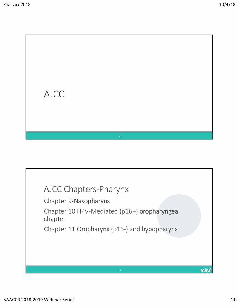
AJCC
27
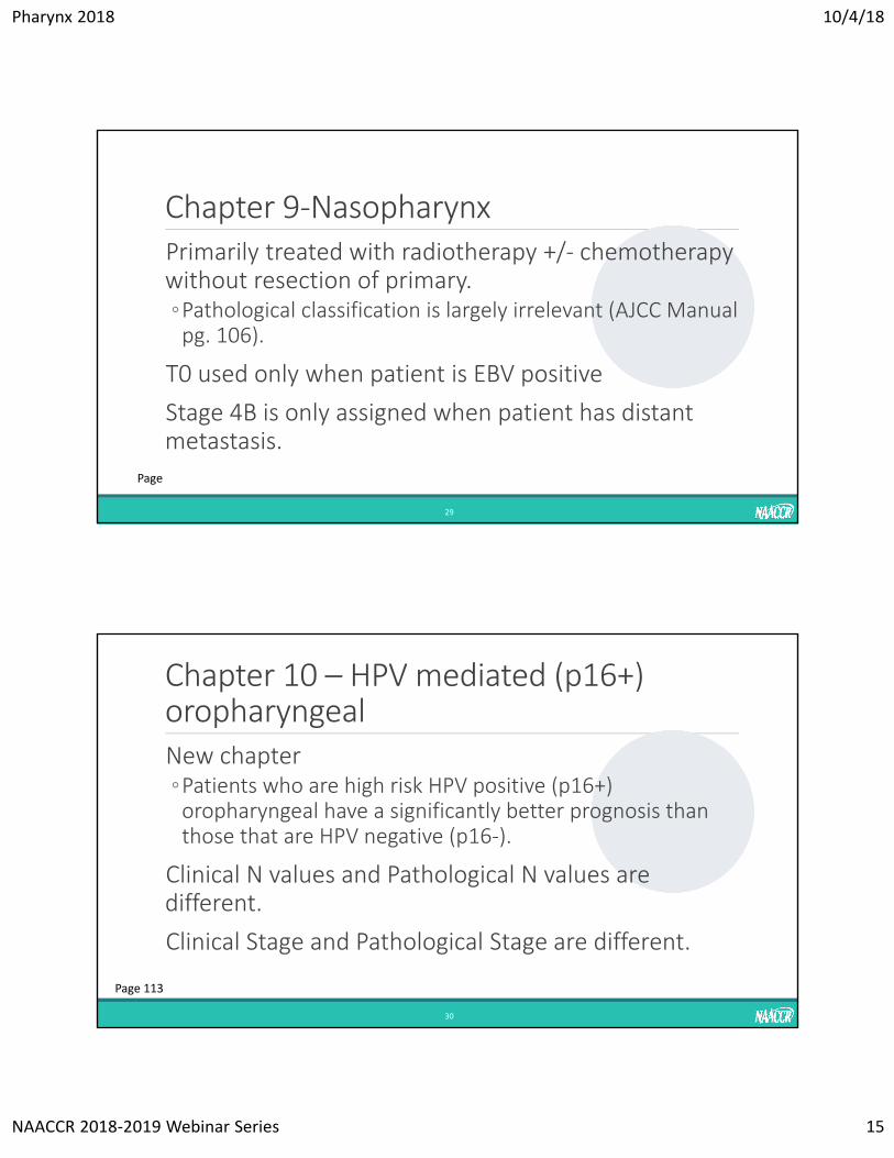
AJCC Chapters‐PharynxChapter 9‐Nasopharynx
Chapter 10 HPV‐Mediated (p16+) oropharyngeal chapter
Chapter 11 Oropharynx (p16‐) and hypopharynx
28
Pharynx 2018 10/4/18
NAACCR 2018‐2019 Webinar Series 15
Chapter 9‐NasopharynxPrimarily treated with radiotherapy +/‐ chemotherapy without resection of primary.◦Pathological classification is largely irrelevant (AJCC Manual pg. 106).
T0 used only when patient is EBV positive
Stage 4B is only assigned when patient has distant metastasis.
29
Page
Chapter 10 – HPV mediated (p16+) oropharyngeal New chapter◦Patients who are high risk HPV positive (p16+) oropharyngeal have a significantly better prognosis than those that are HPV negative (p16‐).
Clinical N values and Pathological N values are different.
Clinical Stage and Pathological Stage are different.
30
Page 113
Pharynx 2018 10/4/18
NAACCR 2018‐2019 Webinar Series 16
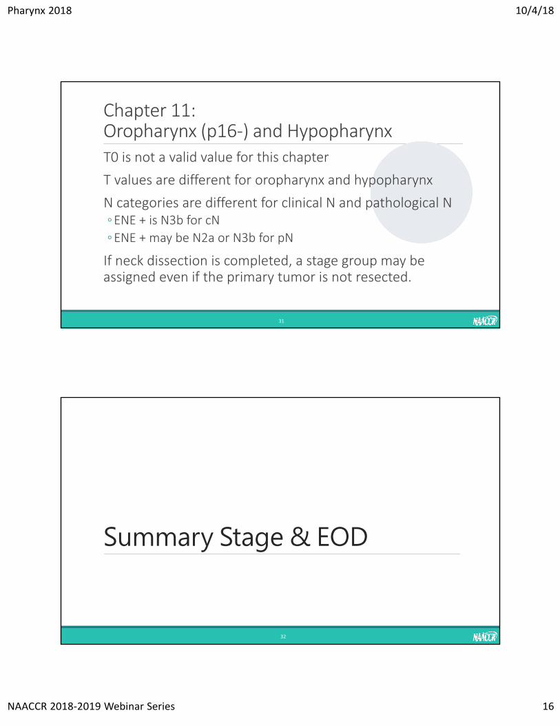
Chapter 11: Oropharynx (p16‐) and HypopharynxT0 is not a valid value for this chapter
T values are different for oropharynx and hypopharynx
N categories are different for clinical N and pathological N◦ENE + is N3b for cN
◦ENE + may be N2a or N3b for pN
If neck dissection is completed, a stage group may be assigned even if the primary tumor is not resected.
31
Summary Stage & EOD
32
Pharynx 2018 10/4/18
NAACCR 2018‐2019 Webinar Series 17
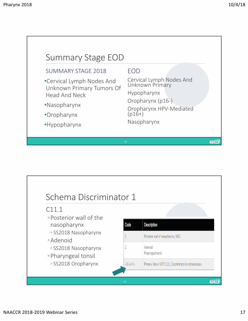
Summary Stage EOD
SUMMARY STAGE 2018
•Cervical Lymph Nodes And Unknown Primary Tumors Of Head And Neck
•Nasopharynx
•Oropharynx
•Hypopharynx
EODCervical Lymph Nodes And Unknown Primary
Hypopharynx
Oropharynx (p16‐)
Oropharynx HPV‐Mediated (p16+)
Nasopharynx
33
Schema Discriminator 1C11.1 ◦Posterior wall of the nasopharynx◦ SS2018 Nasopharynx
◦Adenoid◦ SS2018 Nasopharynx
◦Pharyngeal tonsil◦ SS2018 Oropharynx
34
Pharynx 2018 10/4/18
NAACCR 2018‐2019 Webinar Series 18
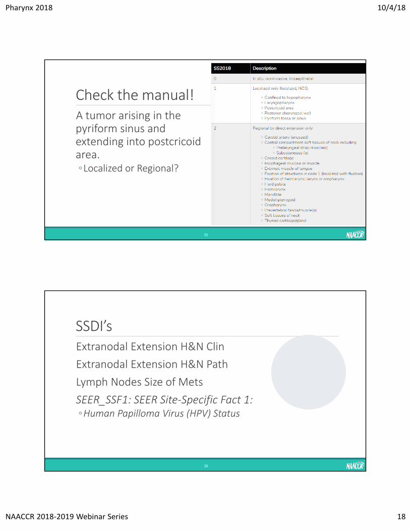
Check the manual!A tumor arising in the pyriform sinus and extending into postcricoid area.◦Localized or Regional?
35
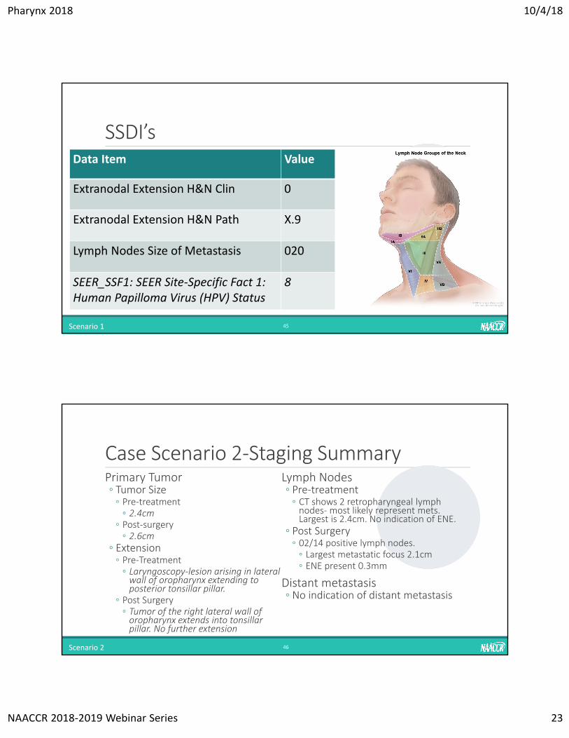
SSDI’s
36
Extranodal Extension H&N Clin
Extranodal Extension H&N Path
Lymph Nodes Size of Mets
SEER_SSF1: SEER Site‐Specific Fact 1: ◦Human Papilloma Virus (HPV) Status
Pharynx 2018 10/4/18
NAACCR 2018‐2019 Webinar Series 19
Extranodal Extension H&N Clinical•Imaging alone is not enough to determine or exclude ENE.•Code 0 when lymph nodes are determined to be positive and physical examination does not indicate any signs of extranodal extension.•Clinical ENE is described in the AJCC 8th edition as "Unambiguous evidence of gross ENE on clinical examination• (e.g., invasion of skin, infiltration of musculature, tethering to adjacent structures, or cranial nerve, brachial plexus, sympathetic trunk, or phrenic nerve invasion with dysfunction)“
•The terms 'fixed' or 'matted' are used to describe lymph nodes.
37
Extranodal Extension H&N PathologicalCode the status of ENE assessed on histopathologic examination of surgically resected involved regional lymph node(s). ◦Do not code ENE from a lymph node biopsy (FNA, core, incisional, excisional, sentinel).
◦Do not code ENE for any distant lymph node
Definitions of ENE subtypes and rules:◦Microscopic ENE [ENE (mi)] is defined as less than or equal to 2 mm.◦Major ENE [ENE (ma)] is defined as greater than 2 mm.◦ Both ENE (mi) and ENE (ma) qualify as ENE (+) for definition of pN.
38
Pharynx 2018 10/4/18
NAACCR 2018‐2019 Webinar Series 20
Lymph Nodes Size of MetastasisRecord the size of the largest metastatic lymph node◦ If the same involved node (or same level) is examined both clinically and pathologically, record the size of the node from the pathology report, even if it is smaller.◦ Example: Clinical evaluation shows 1.5 cm (15 mm) Level II lymph node, pathological examination shows Level II 1.3 cm (13 mm) metastatic deposit. Code 13.0.
◦ If the largest involved node is not examined pathologically, use the clinical node size
39
SEER_SSF1: SEER Site‐Specific Fact 1: Human Papilloma Virus (HPV) StatusRequired for SEER Registries only◦There are several methods for determination of HPV status. The most frequently used test is IHC for p16 expression which is surrogate marker for HPV infection.◦Do not record the results of IHC p16 expression in this field.◦ The rest of the tests (based on ISH, PCR, RT‐PCR technologies) detect the viral DNA or RNA.
◦ This data item is only for HPV status determined by tests designed to detect viral DNA or RNA.
◦ Leave this field blank if tests not done.
40
Pharynx 2018 10/4/18
NAACCR 2018‐2019 Webinar Series 21
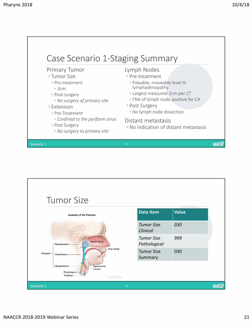
Case Scenario 1‐Staging SummaryPrimary Tumor◦ Tumor Size◦ Pre‐treatment◦ 3cm
◦ Post‐surgery◦ No surgery of primary site
◦ Extension◦ Pre‐Treatment◦ Confined to the pyriform sinus
◦ Post Surgery◦ No surgery to primary site
Lymph Nodes◦ Pre‐treatment◦ Palpable, moveable level III lymphadenopathy.
◦ Largest measured 2cm per CT◦ FNA of lymph node positive for CA
◦ Post Surgery◦ No lymph node dissection
Distant metastasis◦No indication of distant metastasis
41Scenario 1
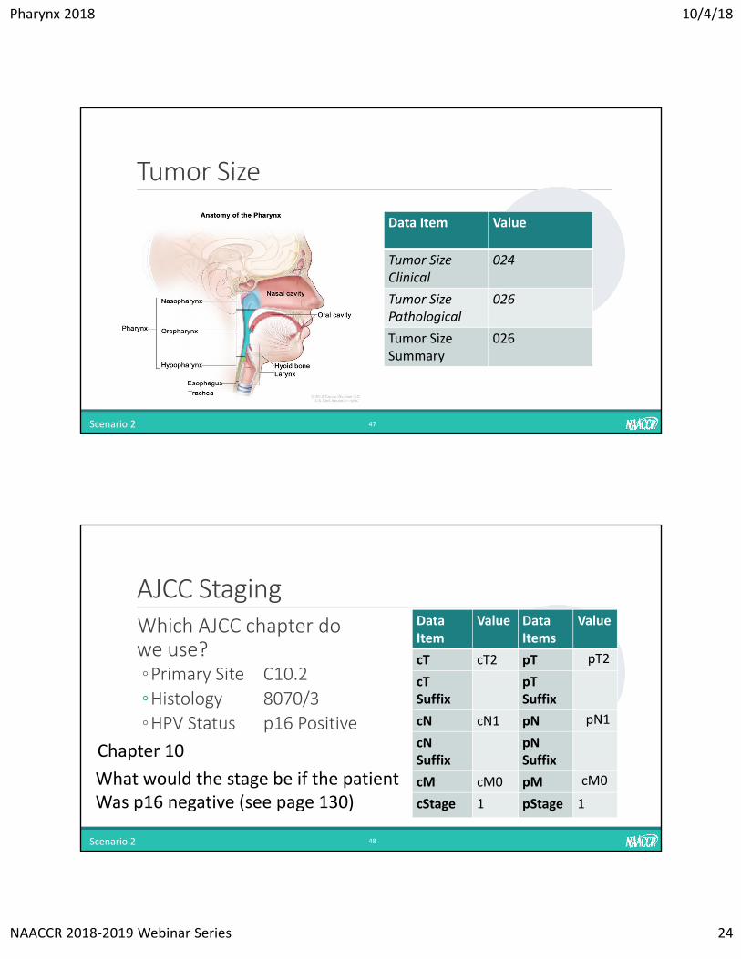
Tumor Size
42
Data Item Value
Tumor Size Clinical
030
Tumor Size Pathological
999
Tumor Size Summary
030
Scenario 1
Pharynx 2018 10/4/18
NAACCR 2018‐2019 Webinar Series 22
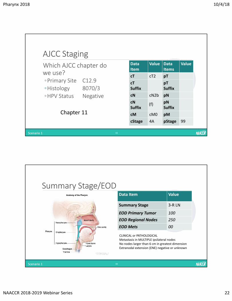
AJCC Staging
Scenario 1 43
DataItem
Value Data Items
Value
cT cT2 pT
cTSuffix
pTSuffix
cN cN2b pN
cN Suffix
pNSuffix
cM cM0 pM
cStage 4A pStage 99
Which AJCC chapter do we use?◦Primary Site C12.9
◦Histology 8070/3
◦HPV Status Negative
Chapter 11
(f)
Summary Stage/EOD
Scenario 1 44
Data Item Value
Summary Stage 3‐R LN
EOD Primary Tumor 100
EOD Regional Nodes 250
EOD Mets 00
CLINICAL or PATHOLOGICALMetastasis in MULTIPLE ipsilateral nodesNo nodes larger than 6 cm in greatest dimensionExtranodal extension (ENE) negative or unknown
Pharynx 2018 10/4/18
NAACCR 2018‐2019 Webinar Series 23
SSDI’s
45Scenario 1
Data Item Value
Extranodal Extension H&N Clin 0
Extranodal Extension H&N Path X.9
Lymph Nodes Size of Metastasis 020
SEER_SSF1: SEER Site‐Specific Fact 1: Human Papilloma Virus (HPV) Status
8
Case Scenario 2‐Staging SummaryPrimary Tumor◦ Tumor Size◦ Pre‐treatment◦ 2.4cm
◦ Post‐surgery◦ 2.6cm
◦ Extension◦ Pre‐Treatment◦ Laryngoscopy‐lesion arising in lateral wall of oropharynx extending to posterior tonsillar pillar.
◦ Post Surgery◦ Tumor of the right lateral wall of oropharynx extends into tonsillar pillar. No further extension
Lymph Nodes◦ Pre‐treatment◦ CT shows 2 retropharyngeal lymph nodes‐ most likely represent mets. Largest is 2.4cm. No indication of ENE.
◦ Post Surgery◦ 02/14 positive lymph nodes. ◦ Largest metastatic focus 2.1cm◦ ENE present 0.3mm
Distant metastasis◦ No indication of distant metastasis
46Scenario 2
Pharynx 2018 10/4/18
NAACCR 2018‐2019 Webinar Series 24
Tumor Size
47Scenario 2
Data Item Value
Tumor Size Clinical
024
Tumor Size Pathological
026
Tumor Size Summary
026
AJCC Staging
Scenario 2 48
DataItem
Value Data Items
Value
cT cT2 pT
cTSuffix
pTSuffix
cN cN1 pN
cN Suffix
pNSuffix
cM cM0 pM
cStage 1 pStage 1
Which AJCC chapter do we use?◦Primary Site C10.2
◦Histology 8070/3
◦HPV Status p16 Positive
Chapter 10
What would the stage be if the patientWas p16 negative (see page 130)
pT2
pN1
cM0
Pharynx 2018 10/4/18
NAACCR 2018‐2019 Webinar Series 25
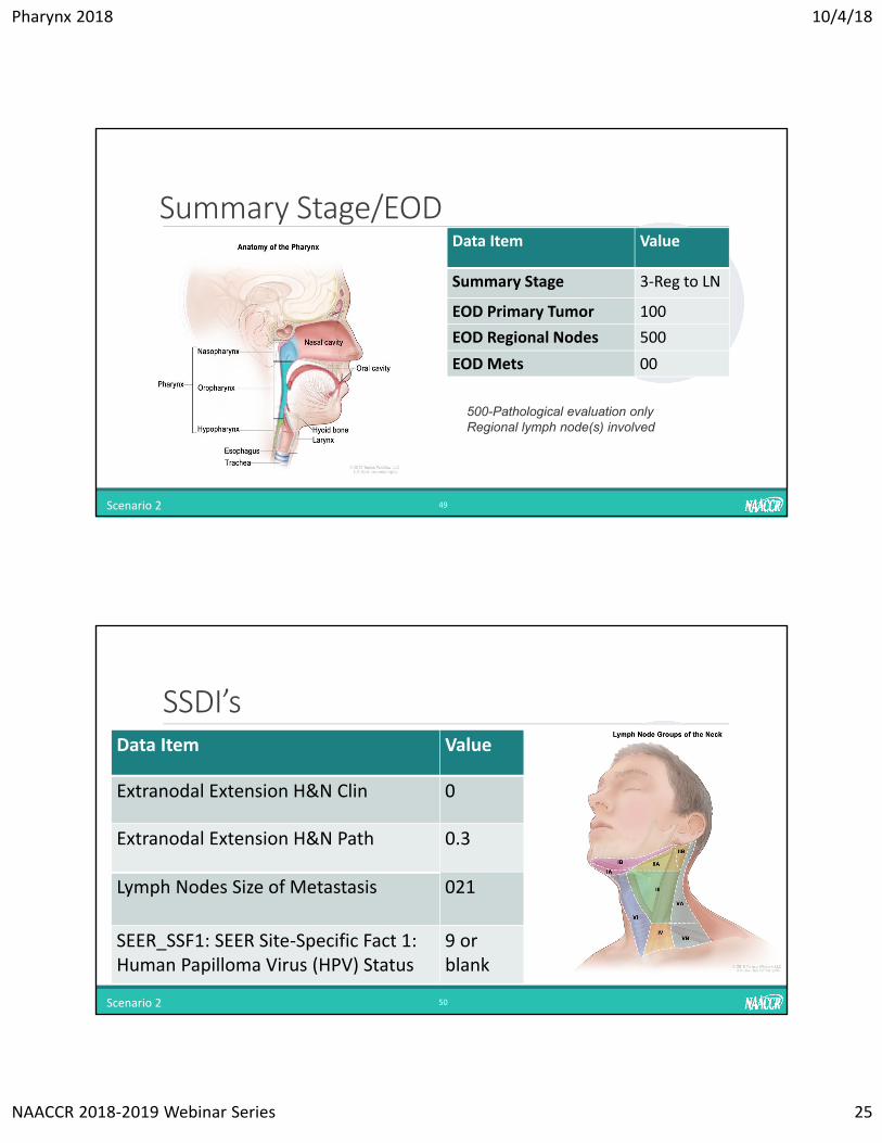
Summary Stage/EOD
Scenario 2 49
Data Item Value
Summary Stage 3‐Reg to LN
EOD Primary Tumor 100
EOD Regional Nodes 500
EOD Mets 00
500-Pathological evaluation onlyRegional lymph node(s) involved
SSDI’s
50Scenario 2
Data Item Value
Extranodal Extension H&N Clin 0
Extranodal Extension H&N Path 0.3
Lymph Nodes Size of Metastasis 021
SEER_SSF1: SEER Site‐Specific Fact 1: Human Papilloma Virus (HPV) Status
9 or blank
Pharynx 2018 10/4/18
NAACCR 2018‐2019 Webinar Series 26
Questions?
51
The Role of Radiation Therapy inthe Management of Pharyngeal CancerWILSON APOLLO, MS, CTR, RTT
52

Pharynx 2018 10/4/18
NAACCR 2018‐2019 Webinar Series 27
Questions?
53
Fabulous Prize Winners
5454
Pharynx 2018 10/4/18
NAACCR 2018‐2019 Webinar Series 28
Coming UP…Collecting Cancer Data: Breast
• 12/06/2018
Collecting Cancer Data: Testis• 01/10/2019
CE Certificate Quiz/SurveyPhrase
Link
https://www.surveygizmo.com/s3/4656348/Pharynx‐2018