pharmacy 04.10¡ntranszport ea...to transport across. transmembrane proteins •protein has...
TRANSCRIPT

Membrane transportPharmacy
04.10.2017
Dr. Szilvia Barkó

Cell Membranes

Cell Membrane Functions
• Protection
• Communication
• Import and and export of molecules
• Movement of the cell

General Structure
A lipid bilayer that contains 2 sheets of lipids interdispersed with proteins.

Fluid-Mosaic Model
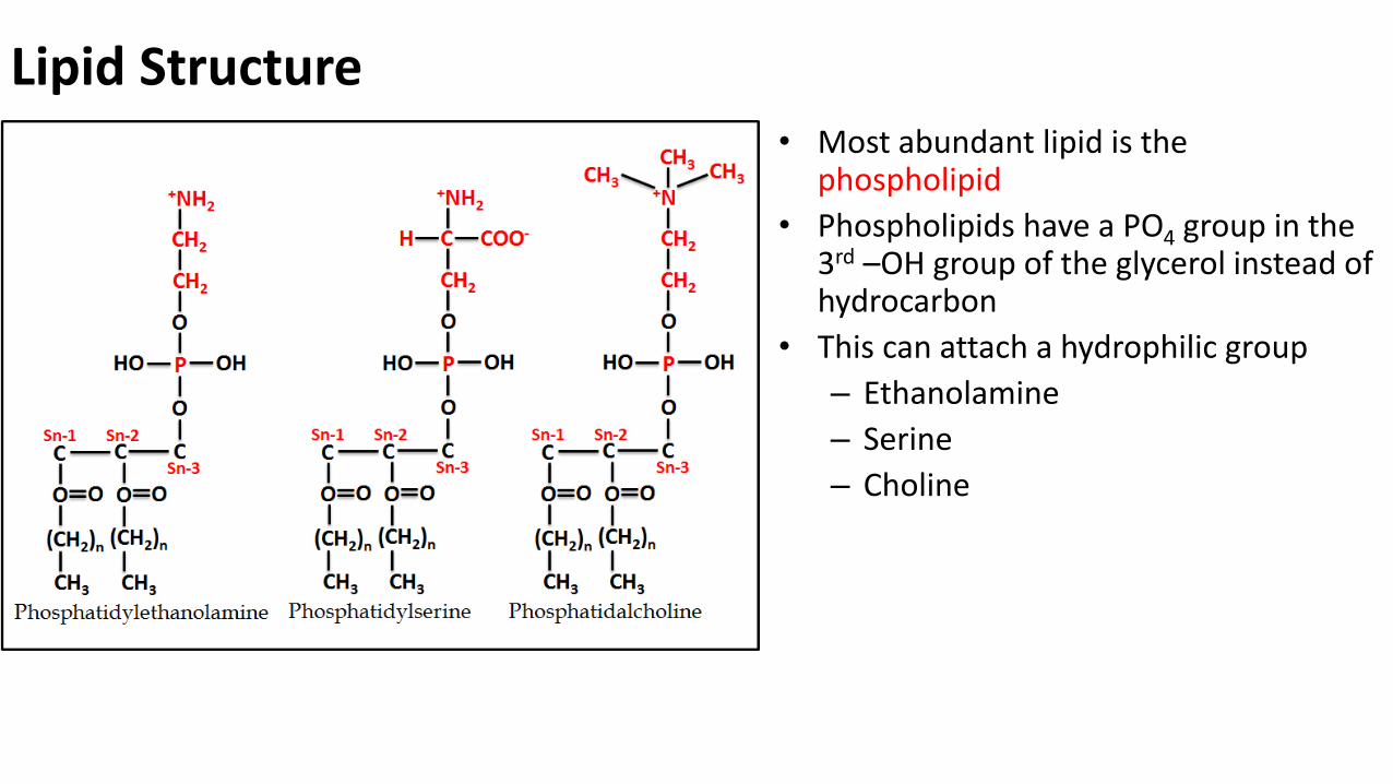
• Most abundant lipid is the phospholipid
• Phospholipids have a PO4 group in the 3rd –OH group of the glycerol instead of hydrocarbon
• This can attach a hydrophilic group
– Ethanolamine
– Serine
– Choline
Lipid Structure
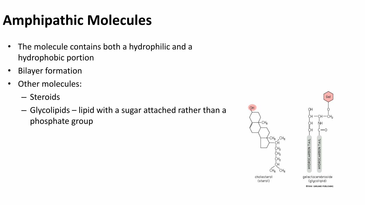
Amphipathic Molecules
• The molecule contains both a hydrophilic and a hydrophobic portion
• Bilayer formation
• Other molecules:
– Steroids
– Glycolipids – lipid with a sugar attached rather than a phosphate group
Membrane Fluidity• Enables the membrane proteins to diffuse rapidly
• Simple means of distributing lipids and proteins
• Allows membranes to fuse with one another
• Evenly distributed during daughter cell formation

Membrane Fluidity
• Hydrocarbon tail determines the fluidity of the membrane just as it does in fats and oils
• 2 components are important
– Length of hydrocarbon chain
• 14 to 24 C but usually 18 to 20 C per tail
– Level of unsaturation (# of C=C bonds)
Each C=C bond causes a kink or bend in the tailCholesterol is added to areas that have lots of unsaturated lipids to help fill in the gaps between the tailsHelps to stiffen and stabilize the bilayer
Less fluidLess permeable

Amphipathic molecules in the membrane
• The hydrophilic head molecules interact with the aqueous solution
• The hydrophobic tails will interact with each other
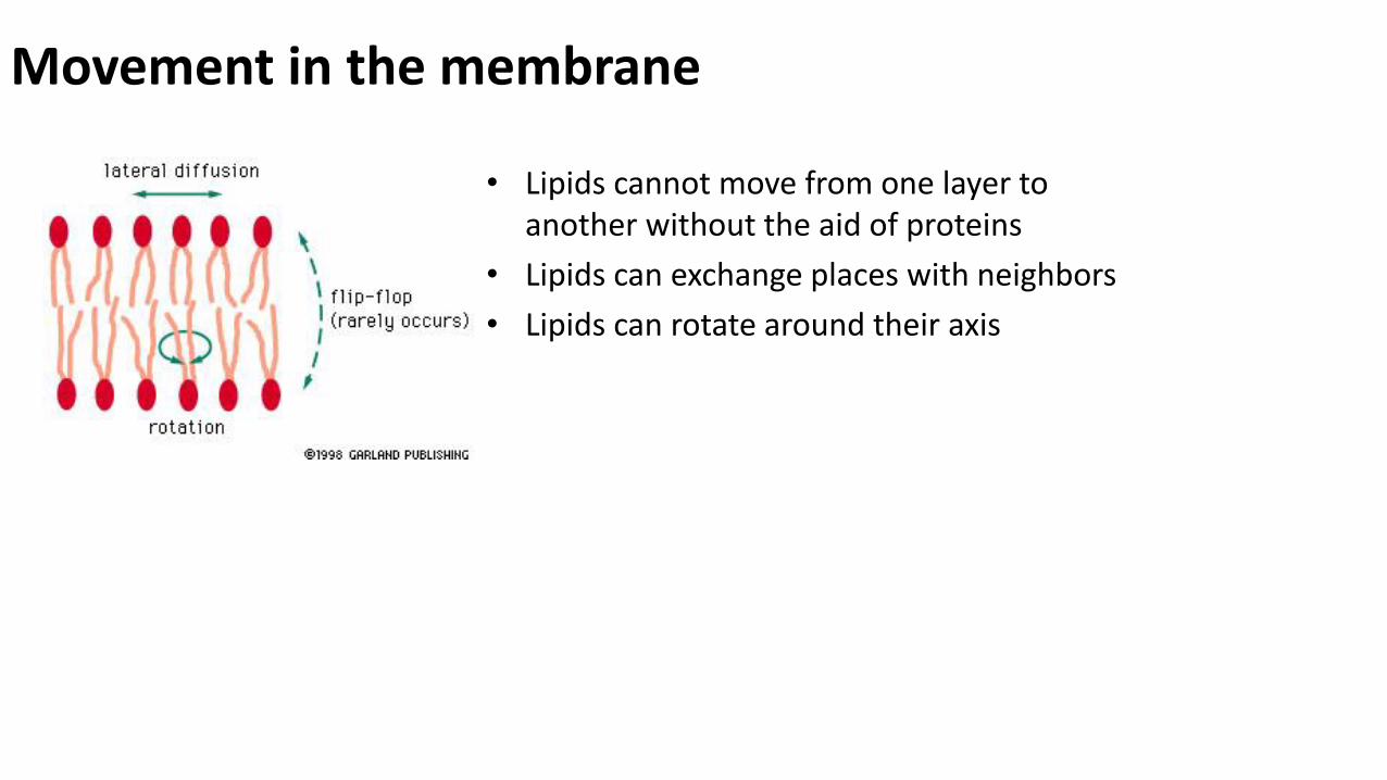
Movement in the membrane
• Lipids cannot move from one layer to another without the aid of proteins
• Lipids can exchange places with neighbors
• Lipids can rotate around their axis

Membranes are Asymmetrical
• Inner surface is different from the outer surface
– Types of lipids in each layer
• Proteins in the bilayer have a specific orientation due to its function

New Membrane
• New lipids are added on one side of the membrane
• Enzyme called flippase used to put the lipid in the other half of the bilayer
– Flippase may be selective for the type of lipids that it puts on either surface

Membranes as Barriers
• Because of the hydrophobic interior of the bilayer
• Membrane is impermeable to ions and large charged molecules and require special membrane proteins to transport across

Transmembrane Proteins
• Protein has hydrophilic and hydrophobic portions
– Hydrophilic will interact with the aqueous solutions on either surface
– Hydrophobic will be in contact with the hydrophobic interior of the bilayer
• Also called integral membrane proteins

PROTEINS CAN MOVE IN THE MEMBRANE, TOO!
Plasma Membrane Proteins: different functions

Diffusion through the cell membrane

Carrier proteinSolute
A carrier protein alternates between two conformations, moving a
solute across the membrane as the shape of the protein changes.
The protein can transport the solute in either direction, with the net
movement being down the concentration gradient of the solute.
Transmembrane transport proteinsallow selective transport of hydrophilic molecules & ions
1. carrier protein

EXTRACELLULAR
FLUID
Channel proteinSolute
CYTOPLASM
A channel protein (purple) has a channel through which
water molecules or a specific solute can pass.
(a)
Transmembrane transport proteinsallow selective transport of hydrophilic molecules & ions
2. channel protein
Note: channel proteins mediate only passive transport

Coupled Transporters

Example1: Glucose transporter GluT1 : (carrier-mediated facilitated diffusion-uniport)
Glucose + ATP glucose-6-phosphate + ADPhexokinase

Example2: Na-glucose cotransport(carrier-mediated facilitated diffusion-synport)
• Mostly Na+ and an other molecule (charged or neutral)• Direction: from the extracellular space inside• Driving force: gradient of Na + toward to inside• The concentration of the transported molecule is going to be higher in the cell than
out (secondary active transport)

Example3: Anion exchange protein 1(carrier-mediated facilitated diffusion-antiport)
At the region of tissues capillaries:• CO2: free diffusion inside the
erythrocyte• The carbonic anhydrase converts
CO2 to H2CO3
• H2CO3 dissociates to H+ and HCO3-
• AE1 changes HCO3- to Cl-
At the region of lungs capillaries: • same antiporter reverse function

The sodium-potassium pump
PP i
EXTRACELLULAR
FLUID
Na+ binding stimulates
phosphorylation by ATP.
2
Na+
Cytoplasmic Na+ binds to
the sodium-potassium pump.
1
K+ is released and Na+
sites are receptive again;
the cycle repeats.
3Phosphorylation causes the
protein to change its conformation, expelling Na+ to
the outside.
4
Extracellular K+ binds to the
protein, triggering release of the
Phosphate group.
6Loss of the phosphate
restores the protein’s
original conformation.
5
CYTOPLASM
[Na+] low
[K+] high
Na+
Na+
Na+
Na+
Na+
PATP
Na+
Na+
Na+
P
ADP
K+
K+
K+
K+K+
K+
[Na+] high
[K+] low

Simple diffusion Facilitated diffusion
Active transport
No protein channel carrier protein protein
carrier protein
HIGH to low conc HIGH to low conc low to HIGH conc
favorable favorable UnfavorableAdd energy
ATP
Passive transport

Carbohydrates on Cell Surface
• Many of the plasma membrane proteins have sugars attached to them
– Short oligosaccharides – glycoproteins
– Long polysaccharides - proteoglycans
• Sugars on the surface make up the glycocalyx
– Keeps cells moist and slippery
– Used as cell recognition (lectins) and adhesion molecules

Glycocalyx – Cell Coat