pharmacologyonline 1: 23-56 (2011) omoregie et al. in...
TRANSCRIPT

Pharmacologyonline 1: 23-56 (2011) Omoregie et al.
22
IN VITRO ANTIOXIDANT ACTIVITY AND THE EFFECT OF METHANOLIC EXTRACTS OF SOME LOCAL PLANTS
ON NUTRITIONALLY STRESSED RATS.
E.S. Omoregie1*, A.U. Osagie2, E.O. Iruolaje1
1Department of Biochemistry, Faculty of Life Sciences, University of Benin, Benin City, Nigeria.
2 Department of Biochemistry, Faculty of Basic and Applied Sciences,
Benson Idahosa University, Benin City, Nigeria.
Summary
In this study, the in vitro and in vivo antioxidant activities of the methanolic extracts from six locally consumed plants in Nigeria were comparatively evaluated. The study plants include V. amygdalina (bitter leaf); C. rubens (‘ebolo’); A. hybridus (‘tete’); J. tanjorensis (‘hospital too far’, jatropha); G. africana (‘okazi’ ) and T. triangulare (water leaf). Results from the in vitro study indicated that the DPPH radical scavenging activity was highest (p<0.05) in ebolo and water
* Corresponding Author: Dr (Mrs) Ehimwenma Sheena Omoregie
Email: [email protected]; Tel. No.: +234 (0) 8023397020.

Pharmacologyonline 1: 23-56 (2011) Omoregie et al.
23
leaf; the proanthocyanidin content was significantly high (p<0.05) in ebolo and tete; ‘hospital too far’ (HTF) extract recorded the highest phenol content; the flavonoid content was significantly increased (p<0.05) in HTF and water leaf extracts. The effects of the extracts were examined in vivo in male albino rats. The animals were divided into two groups with each group having seven subgroups. One of the groups was induced with nutritional oxidative stress, for six (6) weeks, by using a low protein diet (containing 2% protein) – PDD and PDD + oral treatment with the various leaves extracts (i.e PDD + ebolo, PDD + jatropha, PDD + tete, PDD + waterleaf, PDD + bitterleaf and PDD + okazi leaves extracts); the other group of animals were placed on the normal diet (20% protein) – ND (control) and ND + oral treatment with the leaves extracts as before. Results showed that the rats fed the PDD had significantly reduced (p<0.05) superoxide dismutase (SOD), catalase (CAT), vitamin E, vitamin C and increased lipid peroxidation levels in their liver and kidney tissues when compared with the control. However, the supplementation of the PDD diet with the various extracts resulted in significantly increased (p<0.05) levels of SOD, CAT, vitamin E, vitamin C and reduced lipid peroxidation relative to the PDD group. Likewise, the feeding of the normal rats with the extracts resulted in normal levels (p>0.05) of these parameters when compared with the control. The results suggest that the various plant leaves possess varied degrees of potent antioxidant activity both in vitro and in vivo. Keywords: Nutritional oxidative stress, Reactive oxygen species, Antioxidant, Protein deficient diet.

Pharmacologyonline 1: 23-56 (2011) Omoregie et al.
24
Introduction
For decades, the screening of medicinal plant materials for their therapeutic values has continued to represent potential sources of new effective medicines. Besides, evidences from epidemiological studies have suggested that high consumption of fruits and vegetables may be linked to reduced risk of developing most oxidative stress – induced diseases such as cancer, diabetes mellitus, protein energy malnutrition (PEM), cataract, infections and other degenerative diseases of aging (1, 2, 3).
Previous scientifically based studies have shown that increased
production of reactive oxygen species (ROS) may be one of the underlying causes of these diseases. Nevertheless, ROS can be generated during normal metabolism in the body; and if not removed may lead to any of the diseases. The long term effects of increased ROS level include damages to important cellular components especially proteins, nucleic acids and polyunsaturated fatty acids in cell membranes and plasma lipoproteins (4, 5, 2, 6, 7).
A number of papers have also reported the antioxidant activities of
phytochemical constituents of medicinal plants (e.g. polyphenols, carotenoids, non – flavonoids, phenolics, vitamins C and E). These phytochemicals act as antioxidants by preventing damages to cell membrane and cellular oxidative processes that may give rise to diseases (8, 2, 3). For instance, natural polyphenols from plant vegetables have been found to exert their beneficial effect by removing free radicals, chelating metal catalyst, activating antioxidant enzymes, etc (9, 10).
In recent times, antioxidants from plant sources have received a lot of
attention and are even preferred to synthetic ones especially due to their potential health benefits, availability, affordability and in many cases, reduced toxicity (11,12,13). This study reports the in vitro and in vivo antioxidant

Pharmacologyonline 1: 23-56 (2011) Omoregie et al.
25
activities of six locally consumed vegetables, namely, Vernonia amygdalina (bitterleaf); Crassocephalum rubens (ebolo); Jatropha tanjorensis (“catholic vegetable”); Amaranthus hybridus (tete); Talinum triangulare (water leaf); Gnetum Africana (okazi). The plants leaves were selected based on their popular use as food and medicine from the south-western region of Nigeria. Nutritional oxidative stress was induced in rats by using a low protein diet.
Materials and Methods In vitro Antioxidant Activity Collection and Identification of Plant Materials
Fresh leaves of J. tanjorensis, T. triangulare, G. Africana, A. hybridus and V. amygdalina were collected during the rainy season (between April – May, 2009) from a cultivated farm in Ugbowo, Edo State, Nigeria. The Crassocephalum rubens leaves were however collected freshly from a farmland at Ikare, Ekiti State, Nigeria. All the plant leaves were identified and authenticated by a Botanist from the Department of Botany, University of Benin, Benin City, Nigeria. A voucher specimen of each plant was thereafter deposited in the herbarium of the Department of Pharmacognosy, University of Benin, Benin City, Nigeria.

Pharmacologyonline 1: 23-56 (2011) Omoregie et al.
26
The botanical names, family, common names, and the uses of these plants are presented as follows: Family Botanical Name Names
Common Names
Uses
Gnetaceae Gnetum africanum
Okazi Leaves are used to make soups. The leaves and seeds are employed traditionally in the treatment of enlarged spleen, sore throat, pain at child birth, snake poisoning, diabetes mellitus, worm expellant (14).
Amaranthaceae Amaranthus hybridus
Tete, aliefo inine
The leaves are commonly used in making soups and medicinally as a blood booster (14, 15).
Asteraceae Crassocephalum rubens
Ebolo, oromidoko
Its mucilaginous leaves are used in dried or fresh form in varieties of dishes and medicinally as laxatives, purgatives, constipation, stomach aches, colitis etc (15, 16).
Compositae Vernonia amygdalina Del.
Bitterleaf, ewuro It possesses

Pharmacologyonline 1: 23-56 (2011) Omoregie et al.
27
characteristic astringent bitter taste. It is used as vegetable in soups and in the treatment of malaria, diabetes mellitus, veneral disease, wounds, diarrhea, hepatitis, beer making and cancer (17, 18, 19).
Euphorbiaceae Jatropha tanjorensis
‘Hospital too far’, ‘catholic vegetable’, ‘yana Ipaja’ Jatropha
The plant leaves are used for soups. The medicinal uses include, antianaemic, haemostatic, treatment of skin disease, soap making, etc (12, 3, 20).
Portulaceae Talinum triangulare
Waterleaf, gbure
It serves as soups. It is rich in vitamins and minerals. It is however, employed locally as a laxative, purgative (15).

Pharmacologyonline 1: 23-56 (2011) Omoregie et al.
28
Preparation of Plant Extracts Methanolic extracts of each plant material was prepared according to
previous method of Ayoola et al (2006) (11). 100g of the washed, air dried powdered leaves of the plants were extracted with methanol at room temperature for 48 hours and stirring at interval. The methanolic extracts obtained were concentrated to dryness at 40oC by using a rotary evaporator under reduced pressure. The dried extracts were weighed and then stored at 4oC for subsequent analysis. Estimation of Diphenyl-2-picryl-hydrazyl (DPPH) Radical Scavenging Activity
The free radical scavenging capacity of the plant extracts against 1, 1 – diphenyl – 2 – picrylhydrazyl (DPPH) free radical was measured using the modified method of Szabo et al (2007) (21). The reaction mixtures contained 1.5 x 10⁻⁷M methanolic solution of DPPH and various concentrations of the test substances and were kept in dark for 50 min. Optical density (OD) of the samples was measured at 517 nm against a blank and % inhibition values were calculated from the absorbance values. Estimation of Total Phenolic Content
Total phenolic content of the leaves was estimated based on the Folin – Ciocalteau’s method as modified by Singleton et al (1999) (22). Gallic acid served as the reference standard and different concentrations of the standard and extracts were prepared in methanol. Then 0.5ml of the sample was mixed with 2.5 ml of ten-fold diluted Folin-Ciocalteau’s reagent and 2ml of 7.5% sodium carbonate. The mixture was allowed to stand for 30mins at room temperature before the absorbance was read at 760nm. The total phenolic contents were expressed as gallic acid equivalents (GAE) (11).

Pharmacologyonline 1: 23-56 (2011) Omoregie et al.
29
Estimation of Proanthocyanidin Content Proanthocyanidin content of the leaves was estimated according to the
method of Ayoola et al (2006) (11). Different concentrations of the extracts and standard catechin were prepared in methanol. 0.5ml of HCL was added and allowed to stand for 15mins. The absorbance at 500nm was measured and the final results were expressed as catechin equivalent. Determination of Flavonoid Content
Total flavonoids in the leaves were determined using the method of Miliauskas et al (2004). To 2ml sample and standard rutin, 2ml of 2% AlCl3 in ethanol was added. The absorbance value of the resulting mixture was measured at 420nm after one hour of incubation at room temperature. The total flavonoids contents of the extracts were calculated as rutin equivalents (11).
In vivo Animal Study Animals and Preparation of Experimental Diet
84 male albino rats of the Wister strain weighing between (100-120)g were obtained from the animal breeding unit of the Department of Biochemistry, University of Lagos, Nigeria. The experimental diet was locally sourced and prepared as previously reported by Omoregie and Osagie (2002), (2007); (2009) (24, 12, 3). Garri served as a source of carbohydrate, processed soya beans served as protein source and fat and oil were obtained from groundnut oil. The mineral and vitamins mixture were from Centrum® multivitamin tablets (see Table 2 for diet preparation).

Pharmacologyonline 1: 23-56 (2011) Omoregie et al.
30
Table 2: Composition of the Experimental Diet Dietary Components Normal Diet
(ND) % Protein Deficient Diet
(LPD)%
Carbohydrate (garri)1 Protein (defatted soya beans) 1 Fat (palm oil) 1 Vitamin mix2 Mineral salt mix2
63
21
8
4
4
82 2 8 4
4
1Obtained from New Benin market, a local market in Benin City, Nigeria 2 Contained all the vitamins and minerals (in recommended daily allowance). The vitamin and mineral mixture was obtained from ABC Plus® Capsule, a product of Hollard and Barrett Ltd., Nuneaton, Warwickshire, USA (24, 12, 3). Feeding Experiment
The experimental animals were divided into two (2) groups with each group having seven (7) subgroups containing six (6) animals each. One group was induced with nutritional oxidative stress, for six (6) weeks, by using protein deficient diet (PDD). Animals in this group were treated as PDD and PDD + oral treatment with the various leaves extracts (i.e. PDD + Ebolo, PDD + Jatropha, PDD + Tete, PDD + Waterleaf, PDD + Bitterleaf and PDD + Okazi leaves extracts). The second group of animals was placed on the normal diet (ND) with adequate protein and was treated as the ND (positive control) and ND + the various extracts (ND + Ebolo, ND + Jatropha, ND + Tete, ND + Waterleaf, ND + Bitterleaf and ND + Okazi extracts) as before. The extracts

Pharmacologyonline 1: 23-56 (2011) Omoregie et al.
31
were administered to each animal orally and once daily at a safe dose of 300 mg/body weight. The feeding experiment lasted for six (6) weeks during which the animals were allowed access to food and water ad libitum. Food intake, faecal output and body weights were recorded weekly.
All experimental protocols were performed within internationally
accepted guidelines for animals use and care (according to NIN guide for Laboratory Animals Welfare) with the approval of the Local Ethics Committee of the University of Benin, Benin City, Nigeria.
At the completion of the feeding period, animals were fasted overnight
and sacrificed by decapitation. Blood was collected and the serum obtained from the whole blood was stored at 40C. The tissues (liver and kidney) were removed at once, blotted dry, weighed and stored as before. Preparation of Liver and Kidney Homogenates
1g of the liver and kidney tissues were homogenized in 10ml of ice-cold physiological saline to obtain 10% (w/v) homogenates. The resulting homogenates were centrifuged at 5,000g for 10min and the supernatants obtained were used for determination of superoxide dismutase (SOD), catalase (CAT), vitamin C, vitamin E and lipid peroxidation. Biochemical Assays
Superoxide dismutase (SOD) activity was assayed in the tissues according to the method of Misra and Fridovich (1972) (25), based on the rapid auto-oxidation of adrenaline due to the presence of superoxide anions. This is measured spetrophotometrically at 420nm and SOD concentration is expressed as Units / g tissue.

Pharmacologyonline 1: 23-56 (2011) Omoregie et al.
32
Catalase activity in the tissues was determined as residual H2O2 after incubation with the enzyme (26).
Estimation of lipid peroxidation in the tissues involved the
determination of thiobarbituric acid reactive substances (TBARS), which are indicators of membrane lipid peroxidation. Values for TBARS were reported as malondialdehyde (MDA) and quantified using a Molar extinction coefficient of 1.5 × 10⁵ M cm−1 and expressed as mmole MDA g−1 of tissue (27).
The vitamin C content of the tissues was determined according to the
method of Roe and Kuether, (1943) (28). Estimation of vitamin E was based on the method of Desai (1984) (29). The total protein was determined by the Biuret method (30). Statistical Analysis
Data were expressed as means ± standard error of mean (SEM). One-way analysis of variance (ANOVA) was performed to test for differences between the groups mean. Significant differences between the means were determined by Duncan’s multiple range test and P values < 0.05 were regarded as significant (31).
Results
The in vitro antioxidative activities as well as the effects of the methanolic extracts of some local plants in nutritionally stressed rats were evaluated in this study. Results from the in vitro antioxidant study are depicted in figures 1 to 4. Figures 5 to 10 represent results from the animal study.

Pharmacologyonline 1: 23-56 (2011) Omoregie et al.
33
The DPPH scavenging activity of the various extracts against standard
vitamin E are shown in figure 1. The scavenging activities of the extracts were comparable to that of vitamin E. However, ebolo and waterleaf extracts showed the highest radical scavenging ability with percentage inhibition of 77.6% and 69.5%, respectively, which was even higher than that of the standard vitamin E (67.2%).The DPPH scavenging capacity of all the extracts was dose dependent.
Figure 2 shows the proanthocyanidin content of the extracts against
standard catechin. Among the extracts, Ebolo and tete had considerably higher level of proanthocyanidin than the standard catechin.
Figure 3 represents the total phenolic content of the extracts. The
phenolic content of the extracts was considerably low when compared with the standard gallic acid. However, Jatropha recorded the highest phenolic content among the extracts.
The total flavonoid content of the various extracts is depicted in figure
4. This was particularly high for jatropha and waterleaf extracts as against the other extracts but was close to that of standard rutin.
Figure 5 represents the food intake, faecal output and weight gain / loss
of the extracts treated rats. From the result, the protein deficient diet (PDD) fed rats showed preliminary signs of protein energy malnutrition (PEM) with characteristically reduced (p<0.05) food intake (anorexia), faecal output and increased weight loss. The degree of anorexia was however reduced in the PDD + extracts treated rats except for the okazi extract treated rats which showed very high weight loss when compared with the other groups. The normal diet + extracts treated rats had normal appetite for food as observed in

Pharmacologyonline 1: 23-56 (2011) Omoregie et al.
34
the significantly increased (p<0.05) food intake, faecal output and weight gain when compared with control (ND).
The effect of the various extracts on hepatic and kidney catalase (CAT)
and superoxide dismutase (SOD) activities are represented in figures 6 and 7, respectively. These enzymes were considerably reduced (p<0.05) in the PDD treated rats more in the liver than the kidney when compared with the control. However, oral supplementation of the PDD diet with the various extracts resulted in increased levels of these enzymes when compared with that of the PDD treated rats. The only exception was the tete (PDD+TE) treated group in which the hepatic catalase activity remained as low as that of the PDD group irrespective of the treatment with the extract.
The hepatic and kidney malonaldehyde (MDA) level of the rats are
shown in figure 8. The MDA level was considerably reduced (p<0.05) or inhibited in all the extracts treated rats as against the control. Whereas, the PDD group showed characteristic evidence of lipid peroxidation with high MDA level relative to the control.
Figures 9 and 10 represent hepatic and kidney vitamin E and vitamin C
levels in the rats. The results showed significantly increased vitamin E and C levels in the extracts treated malnourished rats, except for the PDD+WL group which had relatively low vitamin E levels, when compared with the PDD fed rats. However, the vitamins levels still remained lower than that of the controls.
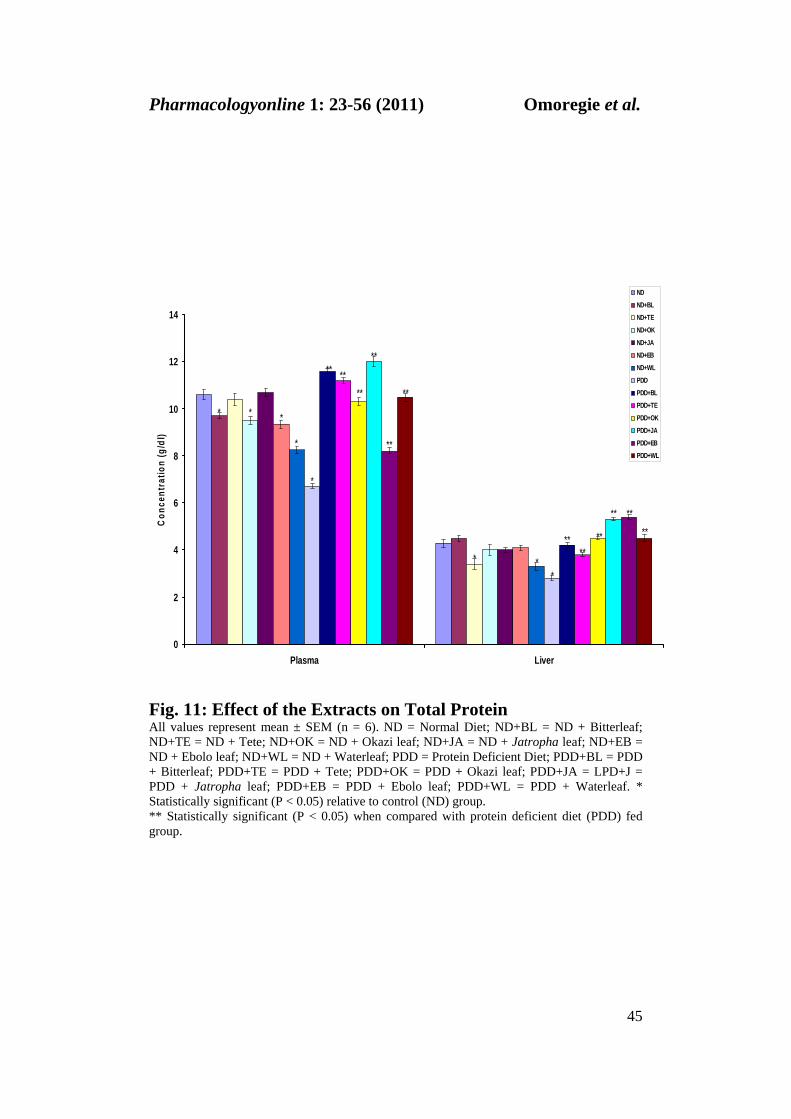
Figure 11 depicts the total protein concentration in the plasma and
kidney of the rats. The protein concentration in the liver and plasma were significantly increased (p<0.05) in the extracts-treated malnourished rats relative to the PDD fed rats. A similar trend was seen in the extracts-treated normal diet fed rats except for the ebolo, waterleaf, okazi and bitterleaf groups

Pharmacologyonline 1: 23-56 (2011) Omoregie et al.
35
which had significantly (p<0.05) reduced plasma total protein levels when compared with the controls.
0
10
20
30
40
50
60
70
80
90
Vitamin E Ebolo Bitterleaf Okazi Waterleaf Tete Jatropha
Inhi
bitio
n of
DPP
H Sc
aven
ging
Abi
lity
(%)
Fig. 1. DPPH Scavenging Activity of the Extracts
All values represent the mean ± SEM of triplicate readings (n = 3).

Pharmacologyonline 1: 23-56 (2011) Omoregie et al.
36
0
0.1
0.2
0.3
0.4
0.5
0.6
Catechin Ebolo Bitterleaf Okazi Waterleaf Tete Jatropha
Proa
ntho
cyan
idin
Con
tent
(mg
cate
chin
equ
ival
ent)
Fig. 2: Proanthocyanidin Content of the Extracts Data represent the mean ± SEM of triplicate readings (n = 3)

Pharmacologyonline 1: 23-56 (2011) Omoregie et al.
37
0.00
0.50
1.00
1.50
2.00
2.50
3.00
Gallic Acid Ebolo Bitterleaf Okazi Waterleaf Tete Jatropha
Tota
l Phe
nol C
onte
nt (m
g ga
llic
acid
equ
ival
ent)
Fig. 3: Total Phenol Content of the Extracts Data represent the mean ± SEM of triplicate readings (n = 3)

Pharmacologyonline 1: 23-56 (2011) Omoregie et al.
38
0.00
0.50
1.00
1.50
2.00
2.50
3.00
Rutin Ebolo Bitterleaf Okazi Waterleaf Tete Jatropha
Tota
l Fla
vono
id (m
g ru
tin e
quiv
alen
t)
Fig. 4: Total Flavonoid Content of the Extracts Data represent the mean ± SEM of triplicate readings (n=3)

Pharmacologyonline 1: 23-56 (2011) Omoregie et al.
39
-15
-10
-5
0
5
10
15
20
25
30
Food Intake Weight Faecal Output
g / d
ay
NDND+BLND+TEND+OKND+JAND+EBND+WLPDDPDD+BLPDD+TEPDD+OKPDD+JAPDD+EBPDD+WL
*
**
**
*
**
****
**
**
**
*
*
** *
*
****
**
****
*** * * * *
* **** ** **
** **
Fig. 5: Food Intake, Weight Gain / Loss and Faecal Output Data represent mean ± SEM (n = 6). ND = Normal Diet; ND+BL = ND + Bitterleaf; ND+TE = ND + Tete; ND+OK = ND + Okazi leaf; ND+JA = ND + Jatropha leaf; ND+EB = ND + Ebolo leaf; ND+WL = ND + Waterleaf; PDD = Protein Deficient Diet; PDD+BL = PDD + Bitterleaf; PDD+TE = PDD + Tete; PDD+OK = PDD + Okazi leaf; PDD+JA = LPD+J = PDD + Jatropha leaf; PDD+EB = PDD + Ebolo leaf; PDD+WL = PDD + Waterleaf. *Statistically significant (P < 0.05) relative to control (ND) group. ** Statistically significant (P < 0.05) when compared with protein deficient diet (PDD) fed group.

Pharmacologyonline 1: 23-56 (2011) Omoregie et al.
40
0
5
10
15
20
25
30
Liver Kidney
cata
lase
act
ivity
(mm
oles
/mg
tissu
e)
NDND+BLND+TEND+OKND+JAND+EBND+WLPDDPDD+BLPDD+TEPDD+OKPDD+JAPDD+EBPDD+WL
*
* *
*
*
**
**
**
**
**
*
*
****
**** ** **
Fig. 6: Effect of the Extracts on Catalase Activity Values represent mean ± SEM (n = 6). ND = Normal Diet; ND+BL = ND + Bitterleaf; ND+TE = ND + Tete; ND+OK = ND + Okazi leaf; ND+JA = ND + Jatropha leaf; ND+EB = ND + Ebolo leaf; ND+WL = ND + Waterleaf; PDD = Protein Deficient Diet; PDD+BL = PDD + Bitterleaf; PDD+TE = PDD + Tete; PDD+OK = PDD + Okazi leaf; PDD+JA = LPD+J = PDD + Jatropha leaf; PDD+EB = PDD + Ebolo leaf; PDD+WL = PDD + Waterleaf. Catalase activities are expressed as mmoles / mg tissue. * Statistically significant (P < 0.05) relative to control (ND) group. ** Statistically significant (P < 0.05) when compared with protein deficient diet (PDD) fed group.

Pharmacologyonline 1: 23-56 (2011) Omoregie et al.
41
0
10
20
30
40
50
60
Liver Kidney
SOD
act
ivity
(Uni
ts /
g tis
sue)
NDND+BLND+TEND+OKND+JAND+EBND+WLPDDPDD+BLPDD+TEPDD+OKPDD+JAPDD+EBPDD+WL
* *
*
****
**
**
**
* *
*
*
** ** **
** **
**
**
Fig. 7: Effect of the Extracts on Superoxide Dismutase (SOD) Activity Values represent mean ± SEM (n = 6). ND = Normal Diet; ND+BL = ND + Bitterleaf; ND+TE = ND + Tete; ND+OK = ND + Okazi leaf; ND+JA = ND + Jatropha leaf; ND+EB = ND + Ebolo leaf; ND+WL = ND + Waterleaf; PDD = Protein Deficient Diet; PDD+BL = PDD + Bitterleaf; PDD+TE = PDD + Tete; PDD+OK = PDD + Okazi leaf; PDD+JA = LPD+J = PDD + Jatropha leaf; PDD+EB = PDD + Ebolo leaf; PDD+WL = PDD + Waterleaf. SOD activities are expressed as units / g tissue. * Statistically significant (P < 0.05) relative to control (ND) group. ** Statistically significant (P < 0.05) when compared with protein deficient diet (PDD) fed group.

Pharmacologyonline 1: 23-56 (2011) Omoregie et al.
42
0
0.05
0.1
0.15
0.2
0.25
0.3
0.35
0.4
Liver Kidney
MD
A C
once
ntra
tion
(mm
ol /
g tis
sue)
NDND+BLND+TEND+OKND+JAND+EBND+WLPDDPDD+BLPDD+TEPDD+OKPDD+JAPDD+EBPDD+WL
*
**
*
*
****
**
** **
**
* * *
*
**
*
** **
**
**
Fig. 8: Effect of the Extracts on Membrane Lipid Peroxidation Level Values represent mean ± SEM (n = 6). ND = Normal Diet; ND+BL = ND + Bitterleaf; ND+TE = ND + Tete; ND+OK = ND + Okazi leaf; ND+JA = ND + Jatropha leaf; ND+EB = ND + Ebolo leaf; ND+WL = ND + Waterleaf; PDD = Protein Deficient Diet; PDD+BL = PDD + Bitterleaf; PDD+TE = PDD + Tete; PDD+OK = PDD + Okazi leaf; PDD+JA = LPD+J = PDD + Jatropha leaf; PDD+EB = PDD + Ebolo leaf; PDD+WL = PDD + Waterleaf. Lipid peroxidation levels are expressed as mmole MDA / g tissue.*Statistically significant (P < 0.05) relative to control (ND) group. ** Statistically significant (P < 0.05) when compared with protein deficient diet (PDD) fed group.

Pharmacologyonline 1: 23-56 (2011) Omoregie et al.
43
0
5
10
15
20
25
30
35
Liver Kidney
Con
cent
ratio
n (m
g/dl
)
NDND+BLND+TEND+OK
ND+JAND+EBND+WLPDD
PDD+BLPDD+TEPDD+OK
PDD+JAPDD+EBPDD+WL
*
*
* * *
*
****
** ** **
**
*
**
**
*
*
**
**
**
** **
Fig. 9: Effect of the Extracts on Vitamin E Concentration Values represent mean ± SEM (n = 6). ND = Normal Diet; ND+BL = ND + Bitterleaf; ND+TE = ND + Tete; ND+OK = ND + Okazi leaf; ND+JA = ND + Jatropha leaf; ND+EB = ND + Ebolo leaf; ND+WL = ND + Waterleaf; PDD = Protein Deficient Diet; PDD+BL = PDD + Bitterleaf; PDD+TE = PDD + Tete; PDD+OK = PDD + Okazi leaf; PDD+JA = LPD+J = PDD + Jatropha leaf; PDD+EB = PDD + Ebolo leaf; PDD+WL = PDD + Waterleaf. * Statistically significant (P < 0.05) relative to control (ND) group. ** Statistically significant (P < 0.05) when compared with protein deficient diet (PDD) fed group.

Pharmacologyonline 1: 23-56 (2011) Omoregie et al.
44
0
5
10
15
20
25
30
35
40
45
Liver Kidney
Con
cent
ratio
n (m
g/dl
)
NDND+BLND+TEND+OK
ND+JAND+EBND+WL
PDDPDD+BLPDD+TEPDD+OK
PDD+JAPDD+WL
**
**
*
*
** **
****
***
*
*
*
*
*
** **
**
**
**
Fig. 10: Effect of the Extracts on Vitamin C Concentration Data represent mean ± SEM (n = 6). ND = Normal Diet; ND+BL = ND + Bitterleaf; ND+TE = ND + Tete; ND+OK = ND + Okazi leaf; ND+JA = ND + Jatropha leaf; ND+EB = ND + Ebolo leaf; ND+WL = ND + Waterleaf; PDD = Protein Deficient Diet; PDD+BL = PDD + Bitterleaf; PDD+TE = PDD + Tete; PDD+OK = PDD + Okazi leaf; PDD+JA = LPD+J = PDD + Jatropha leaf; PDD+EB = PDD + Ebolo leaf; PDD+WL = PDD + Waterleaf. * Statistically significant (P < 0.05) relative to control (ND) group. ** Statistically significant (P < 0.05) when compared with protein deficient diet (PDD) fed group.
Pharmacologyonline 1: 23-56 (2011) Omoregie et al.
45
0
2
4
6
8
10
12
14
Plasma Liver
Con
cent
ratio
n (g
/dl)
NDND+BLND+TEND+OK
ND+JAND+EBND+WL
PDDPDD+BLPDD+TEPDD+OK
PDD+JAPDD+EBPDD+WL
* *
*
*
*
**
**
**
**
** **
* * *
**
**
**
**
** **
Fig. 11: Effect of the Extracts on Total Protein All values represent mean ± SEM (n = 6). ND = Normal Diet; ND+BL = ND + Bitterleaf; ND+TE = ND + Tete; ND+OK = ND + Okazi leaf; ND+JA = ND + Jatropha leaf; ND+EB = ND + Ebolo leaf; ND+WL = ND + Waterleaf; PDD = Protein Deficient Diet; PDD+BL = PDD + Bitterleaf; PDD+TE = PDD + Tete; PDD+OK = PDD + Okazi leaf; PDD+JA = LPD+J = PDD + Jatropha leaf; PDD+EB = PDD + Ebolo leaf; PDD+WL = PDD + Waterleaf. * Statistically significant (P < 0.05) relative to control (ND) group. ** Statistically significant (P < 0.05) when compared with protein deficient diet (PDD) fed group.

Pharmacologyonline 1: 23-56 (2011) Omoregie et al.
46
Discussion
In this study, six (6) commonly consumed plants in Nigeria were evaluated for their in vitro antioxidant activities. The antioxidant activities of the various extracts were also assessed in vivo in rats exposed to nutritionally challenged diets for six (6) weeks.
In vitro Antioxidant Study The DPPH test provided information on the reactivity of test compounds with a stable free radical. Due to its odd electron, 2, 2 – diphenyl – picryl – hydrazyl (DPPH) radical gives a strong absorption band at 517nm in visible spectroscopy (deep violet colour). As the electron becomes paired off in the presence of a free radical scavenger, the absorption vanishes; the resulting decolorization is stoichiometric with respect to the number of electrons taken up. Thus, the efficacies of antioxidants are often associated with their ability to scavenge stable, highly reactive, free radicals. This may be useful in treatment of radical related pathological damage. These highly reactive free radicals have been implicated in the pathology of different number of diseases in humans such as diabetes mellitus, artherosclerosis, cancer, Parkinson’s disease and other neurodegenerative disorders. It is also reported that antioxidant compounds such as phenols and other phytochemicals play a vital role in removing free radicals and in inhibition of lipid peroxidation (32, 33, 34).
In the present study, all the plants extracts exhibited comparable DPPH free radical scavenging ability in a dose-dependent manner. Among the extracts, ebolo and waterleaf had significantly increased (p<0.05) free radical scavenging ability higher than that of standard vitamin E. The scavenging ability of the extracts may be a reflection of the total activities of various components present in these extracts (35, 36,). Indeed, several studies have reported that the antioxidant activity of most plants with therapeutic properties

Pharmacologyonline 1: 23-56 (2011) Omoregie et al.
47
may be due to the presence of natural substances mainly phenolic compounds (37, 36).
Plant phenolics which occur widely in the plant kingdom, especially in
fruits and vegetables, constitute one of the major groups of compounds acting as primary antioxidants or free radical terminators (11, 38). There is a strong relationship between total phenolic content and total antioxidant activity in selected fruits, vegetables and grain products. The antioxidant capacity of phenolic compounds is mainly attributed to their redox properties which allow them to act as reducing agents, hydrogen donors, singlet oxygen quenchers or metal chelators (39). In this study, the phenolic content of the extracts was varied and lower than that of the standard gallic acid. However, the Jatropha extract was found to have higher level of phenol compared with the other plants extracts. Waterleaf on the other hand, had the lowest phenol content.
The reduced phenolic content as found in the extracts, compared with
that of the standard gallic acid, may be attributed to poor specificity of the Folin – Ciocalteau method employed. In addition, phenolic compounds, depending on the number of phenolic groups respond differently to the Folin-Ciocalteau reagent (22, 40). Nevertheless, the antioxidant properties of a single compound within a group can vary remarkably, so that the same levels of phenolics do not necessarily correspond to the same antioxidant response (41, 39).
The flavonoid content of the extracts was in the descending order:
Ebolo > Waterleaf > Jatropha > Okazi > Tete > Bitterleaf. Flavonoids may account for part of the benefits associated with the consumption of fruits and vegetables. They have been reported to interfere with the activities of the enzymes involved in ROS generation, quenching of free radicals, chelating transition metals and rendering them redox inactive in the Fenton reaction (40, 34).

Pharmacologyonline 1: 23-56 (2011) Omoregie et al.
48
Proanthocyanidins are polymeric flavan-3-ols. They are the second
most abundant group of natural phenolics (42). They are also effective antioxidants which provide several health benefits, including the prevention of cancer, urinary tract infection and cardiovascular diseases as well as the inhibition of LDL oxidation and platelet aggregation (43, 44). The results from this study showed that all the extracts had considerably increased proanthocyanidin content with the highest and least values observed in ebolo and bitterleaf, respectively. In vivo Animal Study From the results, early physical features of PEM were observed particularly in the PDD fed rats which include reduced food intake (anorexia), weight loss, reduced faecal output, hair loss, and failure to thrive. These features were however less obvious in the PDD + extracts treated rats. These findings agree with previous literature on induction of PEM using a low protein diet (24, 12, 3). Nutritional oxidative stress in PEM describes an inbalance between the pro-oxidant load and the antioxidant defense system in the body (45). In the malnourished state, there is a characteristic depression of the free radical defense mechanism including alterations in the activities of antioxidant enzymes, vitamins and minerals (46, 3). Similarly, in this study, the animals placed on the PDD diet for six (6) weeks showed appreciably reduced activities of the antioxidant enzymes SOD, CAT, vitamins E and C as well as high level of malonaldehyde (an index of lipid peroxidation) in the liver and the kidney. This might be possible because in the malnourished state, the concentration of antioxidant enzymes, vitamins, essential polyunsaturated fatty acids and mineral elements are compromised, exposing the body’s own antioxidant defense system to damage by ROS (12, 3).

Pharmacologyonline 1: 23-56 (2011) Omoregie et al.
49
Malonaldehyde is the major oxidation product of peroxidized PUFAs and increased MDA level is an important indicator of lipid peroxidation. Catalase on the other hand, is an enzymatic antioxidant widely distributed in all animal tissues including the red blood cell and liver. Catalase decomposes H2O2 and helps protect the tissues from highly reactive hydroxyl radicals. SOD, another antioxidant enzyme, removes superoxide radical by converting it to H2O2 (33). The enhanced oxidative stress in the PDD fed rats was however significantly reduced (p<0.05) in PDD + extracts; and in ND + extracts treated rats. The PDD fed rats had considerably reduced total protein levels in the liver and the plasma when compared with the control. From past literatures, reduced concentration of protein is one of the important factors of anaemia in PEM. They affect the bone marrow erythroid activity and decrease haemoglobin content (12). The extract treatment of the PDD fed rats resulted in little or no changes in their protein concentration (p<0.05) when compared with the control.
The present study involved a comparative evaluation of the antioxidative activity of methanolic extracts from various plants both in vitro and in vivo. The overall antioxidative capacity of the extracts suggest a positive correlation as well as synergistic effects with respect to the DPPH scavenging activity, total phenol, flavonoid and proanthocyanindin components of the extracts. This indicates that the components are more likely to contribute to the antioxidant potential of the extracts (7). But there were wide degrees of variations, between the plant extracts especially in their effectiveness as antioxidants. This was particularly obvious in okazi and bitterleaf extracts which showed no correlation between their in vitro and in vivo antioxidant activities. It is possible that the in vivo antioxidant capacity observed in these extracts was not solely from the phenolic contents alone, but could be due to

Pharmacologyonline 1: 23-56 (2011) Omoregie et al.
50
the presence of some other phytochemicals such as ascorbic acid, tocopherol and pigments or the synergistic effects among them (47).
Furthermore, other factors may be responsible for this variation such
as, the extracting solvent, the isolation procedures employed, the purity of active compounds, seasonal changes and the test system employed. All these factors may influence the synthetic pathways of the active compounds in the plant extracts (48, 49, 39, 36). Extracts from Jatropha tanjorensis, Crassocephylum rubens and Talinum triangulare exhibited the most potent antioxidant activity both in vitro and in vivo.
The plants leaves may be considered as potential sources of natural
antioxidants able to be used with a therapeutic or industrial aim as alternative for the synthetic products which are known for their multiple disadvantages. This study therefore confirms local claims on the efficacy of the plants leaves and may provide effective intervention for free radical mediated diseases. Indeed, our ancestors made soups and medicine from these plants leaves and fed them to vulnerable groups in the population including lactating mothers and feverish individuals.
References
1. Dani C, Pasquali MAB, Oliveira MR, et al. Protective effects of purple grape juice on carbon tetrachloride – induced oxidative stress in brains of adult Wistar rats. J. Med. Foods 2008; 11(1 suppl): 55 – 61
2. Wasson GR, Mckelvey-Martin VJ, Downes SC. The use of the comet
assay in the study of human nutrition and cancer. Mutagenesis 2008; 23(3 suppl): 153 – 162

Pharmacologyonline 1: 23-56 (2011) Omoregie et al.
51
3. Omoregie ES, Osagie AU. Effect of Jatropha tanjorensis leaves supplement on the activities of some antioxidant enzymes, vitamins and lipid peroxidation in rats. J Food Biochem 2010; DOI: 10.1111/j.1745 – 4514.2010.00392.x (in press)
4. Vertuani S, Angusti A, Manfredini S. “The antioxidants and
proanthocyanidins network: An overview”. Curr. Pharm. Des 2004; 10(14 suppl): 1677 – 1694.
5. Bende DA. The antioxidant paradox: Damages and defence.
Biochemistry 2006; 28(5 suppl): 9-12
6. Martin KR, Appel CL. Polyphenols as dietary supplements: A double-edged sword. Nutr Dietary Suppl 2001; 2: 1 – 12.
7. Tarawneh KA, Irshaid F, Jaran AS, et al. Evaluation of antibacterial
and antioxidant activities of methanolic extracts of some medicinal plants in northern part of Jordan. J. Biol. Sci 2010; 10(4 suppl): 325 – 332.
8. Oboh G, Rocha JBT. Water extractable phytochemicals from Capsicum
pubescent (tree pepper) inhibit lipid peroxidation induced by different pro-oxidant agents in brain: In vitro. Eur. Food Res. Technol 2008; 226: 707 – 713.
9. Amic D, Davidovic-Amic D, Beslo D, Trinajstic N. Structure – radical
scavenging activity relationship of flavonoids. Croatia Chem Acta 2003; 76: 55 – 61.
10. Oboh G, Raddatz H, Henle T. Characterization of the antioxidant
properties of hydrophilic and lipophilic extracts of Jute (Corchorus olitorius) leaf. Int J Food Sci Nutr 2009; 1: 1 – 11.

Pharmacologyonline 1: 23-56 (2011) Omoregie et al.
52
11. Ayoola GA, Sofowora T, Odukoya O, Coker HAB. Phytochemical screening and free radical scavenging activity of some Nigerian medicinal plant. J Pharm Sci 2006; 8: 133 – 137.
12. Omoregie ES, Osagie AU. Phytochemical screening and anti-anemic
effect of Jatropha tanjorensis leaf in protein malnourished rats. Plant Arch 2007; 7: 509 – 516.
13. Oyewole IO, Magaji Z, Awoyinka OA. Biochemical and toxicological
studies of aqueous extract of Tithonia diversifolia (Hemsl.) leaves in Wister albino rats. J Med Plants Res 2007; 1(2 suppl): 030 – 033.
14. Ekpo AS. Determination of chemical composition of Gnetum
africanum seeds. Pak J Nutr 2007; 6(1 suppl): 40 – 43.
15. Oguntona T. Green leafy vegetables. In: Osagie AU, Eka OU, eds. Nutritional Quality of Plants Food, Benin City, Nigeria, Ambik Press, 1998: 120 – 133.
16. Grubben GJH. Amaranthus cruentus L. Plant resources of tropical
Africa. In: Grubben GJH, Denton OA, eds.Vegetables. PROTA Foundation, Leiden: Backhuys Publishers. 2004: 67 – 72.
17. Akah PA, Ekekwe RK. Ethnopharmacology of some Asteraceae family
used in Nigerian traditional medicine. Fitoterpia 1995; 66: 351 – 355.
18. Hamill FA, Apio S, Murbiru NK, et al. Traditional herbal drugs of Southern Ugandan. J Ethnopharmacol 2000; 70: 281 – 300.
19. Kambizi L, Afolayan AJ. An ethnobotanical study of plants used for
the treatment of sexually transmitted diseases (njovhera) in Guruve district, Zimbabwe. J Ethnopharmacol 2001; 77: 5 – 9.
Pharmacologyonline 1: 23-56 (2011) Omoregie et al.
53
20. Oduola T, Avwioro OG, Ayanniyi TB. Suitability of the leaf extract of Jatropha gossypifolia as an anticoagulant for biochemical and haematological analysis. Afr J Biotechnol 2005; 4(7 suppl): 679 – 681.
21. Szabo MR, Lditoiu C, Chambre D, Lupes AX. Improved DPPH
determination for antioxidant activity spectrophotometric assay. Chem Pap 2007; 61: 214 – 216. DOI: 10.2478/s 11696-007-0022-7.
22. Singleton VL, Orthefer R, Lamuela-Raventos RM. Analysis of total
phenols and other oxidative substrates and antioxidants by means of Folin-Ciocalteau reagent. Methods in Enzymology 299 (Oxidants and Antioxidants, Part A). San Diego, California, Academic Press 1999: 152 – 178.
23. Miliauskas G, Vensketonis PR, Van-Bec TA. Screening of radical
scavenging of some medicinal and aromatic plant extracts. Food Chem., 2004; 85: 231 – 237.
24. Omoregie ES, Osagie AU. Effect of Telfairia occidentalis leaf on some
hematological parameters and lipid profile in protein malnourished rats. Nig. J. Biochem Mol Biol 2002; 17: 41 – 45.
25. Misra H, Fridovich I. The role of superoxide dismutase anion in the
antioxidation of epinephrine and a simple assay for superoxide dismutase. J Biol Chem 1972; 247: 3170.
26. Kaplan JH, Grooves JN. Liver and blood cell catalase activity of
tumour-bearing mice. Cancer Res 1992; 32: 1190 – 1194.
27. Gutteridge JMC, Wilkins S. Copper – dependent hydroxyl radical damage to ascorbic acid. FEBS Lett 1982; 137: 327 – 329.

Pharmacologyonline 1: 23-56 (2011) Omoregie et al.
54
28. Roe JH, Kuether CA. The determination of ascorbic acid in whole blood and urine through the 2, 4 – dinitrophenylhydrazine derivative of dehydroascorbic acid. J Biol Chem 1943; 147: 399.
29. Desai ID. Vitamin E analysis method for animal tissues. Methods
Enzymol 1984; 1105: 138 - 143.
30. Henry RJ, Sobel C, Beckman, S. Determination of serum protein by biuret reaction. Anal Chem 1957; 92(149 suppl): 1 – 5.
31. Sokal RR, Rohlf FJ. Biometryl. In The Principle and Practice of
Statistics in Biological Research, 3rd Ed., Freeman, W.H. and Co., New York. 1995: 887.
32. Oke JM, Hamburger MO. Screening of some Nigerian medicinal plants
for antioxidants activity using 2, 2, Diphenyl – picryl – hydrazyl radical. Afr J Biomed Res 2002; 5: 77 – 79.
33. Krishnaraju AV, Rao CV, Rao TVN, et al. In vitro and in vivo
antioxidant activity of Aphanamixis polystachya bark. Am J Infect Diseases, 2009; 5(2 suppl): 60 – 67.
34. Aiyegoro OA, Okoh AI. Phytochemical screening and polyphenolic antioxidant activity of aqueous crude leaf extract of Helichrysum pedunculatum. Int J Mol Sci 2009; 10: 4990 – 5001.
35. Wang H, Zhao M, Yang B, et al. Identification of polyphenols in
tobacco leaf and their antioxidant and antimicrobial activities. Food Chem 2008; 107: 1399 – 1406.
36. Rached W, Benamar H, Bennaceur M. Screening of the antioxidant
potential of some Algerian indigenous plants. J Biol Sci 2010; 10: 316 – 324.

Pharmacologyonline 1: 23-56 (2011) Omoregie et al.
55
37. Virgili F, Scaccini C, Packer L, Rimbach G. Cardiovascular disease and nutritional phenolics. In: Pokomy J, Yannishlieva N, Rimbach G, eds. Antioxidants in Food, Woodhead Publishing Ltd., Cambridge, 2001: 87 – 99.
38. Maisuthisakul P, Suttajit M, Pongsawatmanit R. Assessment of
phenolic content and free radical scavenging capacity of some Thai indigenous plants. Food Chem 2007; 100: 1409 – 1418.
39. Demiray S, Pintado ME, Castro PML. Evaluation of phenolic profiles
and antioxidant activity of Turkish medicinal plants: Tilia argentea, Crataegifolium leaves and Polygonum bistorta roots. World Acad Sci Engineering and Tech 2009; 54: 312 – 317.
40. Wong SP, Leong LP, Koh JHW. Antioxidant activities of aqueous
extracts of selected plants. J Food Chem 2006; 99(4 suppl): 775 – 783.
41. Parejo I, Viladomt F, Bastida J, et al.“Comparison between the radical scavenging activity and antioxidant activity of 6 distilled and non-distilled Meditarranean herbs and aromatic plants”. J Agric Food Chem 2002; 50: 6882 – 6890.
42. Prior RL. Fruits and vegetables in the prevention of cellular oxidative
damage. Am J Clin Nutr 2004; 78: 570S – 578S.
43. Howard LR, Talcott ST, Brenes CH, Villalon B. Changes in phytochemical and antioxidant activity of selected pepper cultivars (Capsicum species) as influenced by maturity. J Agric Food Chem 2000; 48: 1713 – 1720.

Pharmacologyonline 1: 23-56 (2011) Omoregie et al.
56
44. Bors W, Michel C, Stettmaier K. Electron paramagnetic resonance studies of radical species of proanthocyanins and gallate esters. Arch Biochem Biophys 2000; 374: 347 – 355.
45. Sies H, Stahl W, Sevanian A. Nutritional dietary and postprandial
oxidative stress. J Nutr 2005; 135: 969 – 972.
46. Shaaban SY, Nassar MF, Ibrahim SA, Mahmoud SE. Impact of nutritional rehabilitation on enzymatic antioxidant levels in protein malnutrition. East Mediterr Health J 2002; 8: 2 – 3.
47. Sengul M, Yildiz H, Gungor N, et al. Total phenolic content,
antioxidant and antimicrobial activities of some medicinal plants. Pak J Pharm Sci 2009; 22(1 suppl): 102 – 106.
48. Gardeli C, Papageorgion V, Mallouchos A, et al. Essential oil
composition of Pistacia lentiscus L. and Myrtus communis L.: Evaluation of antioxidant capacity of methanolic extracts. Food Chem 2008; 107: 1120 – 1130.
49. Tsai PJ, Wu SC, Cheng YK. Role of polyphenols in antioxidant capacity of napiergrass from different growing seasons. Food Chem 2008; 106: 27 – 32.