pharmacoanalytical assays of erwinia asparaginase (erwinase
TRANSCRIPT

Abstract. Background: Asparaginases are the cornerstonetherapy of many successful combination regimens for thetreatment of acute lymphoblastic leukemia (ALL), the mostcommon malignancy in children and adolescents. Currently, twoasparaginase formulations are available in the US, nativeEscherichia coli asparaginase (ASNase) and pegaspargase. Athird formulation native Erwinia asparaginase (Erwinase, ERW)has recently been made available under a licensing exception forpersonal use. We report here the development and validationprocess of ERW pharmacoanalytical assays and the results in afew patients. Materials and Methods: We developed andsystematically validated the ERW enzyme activity and ERWconcentration, anti-ERW antibody and related assays.Pharmacokinetic and pharmacodynamic (PK-PD) studies wereperformed in a limited number of patients who received 6,000IU/m2 x 3 per week x 2 courses, and 4 patients who received25,000 IU/m2 x 3 per week x 2 courses of ERW. Results: Thelinearity and range of the Erwinase calibration lines for thepharmacoanalytical assays were excellent. The accuracy andprecision were better than the FDA limit allows for oncologybiological products (<30%) coefficient of variation (%CV) andrelated parameters in the quantification of ERW concentration.The validation of these parameters was equal to or better than
during the assay development. PK-PD analyses of ERW in a fewpatients yielded an average half-life of elimination of 15.8±1.64hours. There was an excellent PD response post ERWadministration resulting in an ERW concentration-dependentasparagine (ASN, <0.5 ̪) and glutamine (GLN, <50 ̪)deamination. Pharmacodynamic correlations demonstrated that0.1 to 0.2 IU/ml of ERW in serum were sufficient for 90% GLNand/or ASN deamination for up to 2 weeks. No anti-ERWantibody [Ab(+)] was seen among those few patients. None ofthe other 5 patients had an adverse event. Based on these posthoc results, simulations on various doses and schedules of thisdrug have been made. Conclusion: The pharmacoanalyticalassays were excellent tools to evaluate the PK and PD data ofERW in pediatric patients with HR ALL. However, this initialPK-PD evidence needs further validation in future clinical trials.Insights into the PD contributions of ERW in anti-E. coliASNase Ab(+) patients will guide us in optimal design and useof ERW as part of combination chemotherapy regimens infuture clinical trials.
The discovery that asparaginase (Escherichia coli L-asparagine amidohydrolase, ASNase) possesses anti-cancerproperties began in the 1950's in the United Kingdom (1-6). Asparaginases represent a key drug, which is used intreatment of haematopoietic malignancies, in particular inacute lymphoblastic leukemia (ALL) and non-Hodgkin'slymphoma, and act by depriving tumor cells of the aminoacid asparagine (ASN) (7, 8). In most cases, repeatedadministration of ASNase is required for effective therapy,but this can be restricted by undesirable effects caused byimmune responses and anti-asparaginase antibody [Ab(+)]production. Despite several recent insightful reviews
2561
Correspondence to: Dr. Vassilios I. Avramis, Childrens Hospital ofLos Angeles, Hematology/Oncology, 4650 Sunset Blvd, Mail 57, LosAngeles, CA 90026, U.S.A. e-mail: [email protected]
Key Words: ALL, Erwinia asparaginase, pharmacokinetics,pharmacodynamics, non-linear mixed effects model (NONMEM),treatment outcome.
ANTICANCER RESEARCH 27: 2561-2572 (2007)
Pharmacoanalytical Assays of Erwinia Asparaginase (Erwinase) and Pharmacokinetic Results in High-riskAcute Lymphoblastic Leukemia (HR ALL) Patients: Simulations of Erwinase Population PK-PD Models
VASSILIOS I. AVRAMIS1,2, SAGRARIO MARTÍN-ARAGÓN1,3, EARL V. AVRAMIS1,4 and BARBARA L. ASSELIN5
1Division of Hematology/Oncology, Department of Pediatrics, Childrens Hospital Los Angeles, USC Keck School of Medicine, Los Angeles, CA 90027;
2University of Southern California Keck School of Medicine, Los Angeles, CA 90033, U.S.A.;3Pharmacology Department, School of Pharmacy, Complutense University of Madrid, 28040 Madrid, Spain;
4UC Davis, Davis, College of Environmental Toxicology, Davis, CA 95616;5Pediatric Hematology/Oncology, University of Rochester Medical Center, Rochester, NY 14642, U.S.A.
0250-7005/2007 $2.00+.40

detailing the history and mechanisms of action of ASNasesagainst ALL, important questions still remain (9-15). Theprimary mechanism of action of ASNase is to deaminateASN and, to a lesser extent, GLN, in serum.
Rapid depletion of ASN and GLN in the patients' serumensures optimal leukemic blast-kill. In contrast, a gradualASN or GLN depletion may allow the leukemic blasts toadapt and survive (16-18). Repeated or prolonged treatmentwith ASNases can lead to an Ab(+) response and this mayneutralize the enzyme activity and prevent the protein fromremaining in the circulation (10-12, 19-20). Ultimately, thiscan lead to a diminished therapeutic effect with the risk ofinfusion reactions (12, 16). More insidiously, there is adanger that antibodies will not only neutralize the foreignenzyme, but will also cross-react with a vital component innormal tissues with subsequent host reactions. Immediate(Type I) and delayed (Type II) immune hypersensitivityreactions are further immunological concerns (12).Therefore, the aim for the use of biological oncologyproducts (enzymes) in cancer therapy is to allow repeatedtreatments without compromising their efficacy or the safetyof patients. Potentially this can be achieved byunderstanding and controlling the immune response and byselecting the appropriate ASNase enzyme accordingly (16).However, a significant proportion of patients treated withASNase develop allergic reactions or undergo neutralizationof the enzyme so they could require treatment cessation oralteration of the formulation and the dosage, respectively.Thus, there is a need for another structurally unrelatedASNase to manage patients sensitized to E. coli ASNase.Many investigators have determined the adverse effects thatanti-ASNase Ab(+) have had on treatment outcomes inALL patients (10, 12, 20). In the presence of Ab(+), theASNase enzymatic activity and the percentage of ASN andGLN depletion become non-existent (12, 16). Once Ab(+)appears, it neutralizes the antigen with loss of its specificenzymatic activity and the subsequent rebounding of ASNand GLN to control concentrations in serum.
The toxicities of ASNases are serious. Liver and pancreasdysfunction are presented as coagulopathies andpancreatitis, with CNS dysfunction seen less frequently (12,21). Hypersensitivity is the most common dose-limitingtoxicity seen with the E. coli ASNase formulations, whichshare immunological cross-reactivity (16). The severity ofclinical symptoms of allergy, ranging from localizederythema at the injection site to systemic anaphylaxis, arereported in up to two thirds of patients receiving intensiveschedules of the native form (5, 7-10, 16, 21). Patients withallergic reactions usually have neutralizing IgG Ab andother subtypes of immunoglobulins in their serum. In themajority of cases (85%-90%) these Ab(+) interfere with thetherapeutic effect of ASNase by neutralizing enzymaticactivity and/or increasing the rate of enzyme clearance (10,
19-21, 22). Evaluations of Ab(+) in relapsed pediatricpatients suggest that development of a high anti-ASNaseAb(+) titer is associated with inferior treatment response(s)(16, 19-20). In CCG-1961, occurrence of ASNase allergywas not associated with inferior outcome, while thepresence of “silent hypersensitivity” as defined by thepresence of anti-ASNase Ab(+) without clinical history ofallergy symptoms was identified in a subset of patients withpoor outcome (16).
Erwinase® (ERW, crisantaspase) (EC 3.5.1.1.) is an L-asparaginase isolated from Erwinia chrysanthemi whichdoes not cross-react with the Ab(+) against the E. coliASNase formulations. ERW is available in the U.S. forpersonal use from OPi SA (Lyon, France). Clinical studiesneed to be conducted in order to determine the optimaldose and schedule of the ERW formulation, its populationpharmacology, toxicities and possible impact on event-freesurvival (EFS) in HR ALL pediatric patients treated withthis ASNase on a personal use. In patients with obviousclinical allergy symptoms who were also Ab(+) to E. coliformulations (anamnestic response), ERW doses wereused equally substituting native E. coli ASNase dosing ina limited number of patients (16, 23). Hence, ERW wassafely and successfully administered to these few patientson a compassionate basis (16). The assays for quantifyingERW were developed in order to evaluate these patients.We present here their development and quality controlwith the pertinent accuracy and precision and in-studyvalidation. More recently, 5 other patients received ERWin a more intensive dosing schedule upon clinical allergyto E. coli formulations. Interesting and useful PK-PD datafor ERW are available from these patients (22). Based onthese limited clinical studies, pharmacokinetic (PK) andpharmacodynamic (PD) population modeling (NONMEM)has been used to provide new insights into the optimaldose and formulation of ERW that may be used in futureclinical trials.
Patients and Methods
Patients and treatment. Between May 2006 to the present, patientswith newly diagnosed ALL and HR features were enrolled onCCG studies receiving E. coli pegaspargase after the appropriateCOG IRB review and the individual informed consent. Patientswho developed clinical allergy to native ASNase or pegaspargasetreatment, if symptoms persisted, were switched to ERWtreatment available since May 2006 under a special licensingexception for personal use. ERW was administered at a dose of6,000 IU/m2 and then increased to 10,000 IU/m2 and wassubstituted for each dose of E. coli-ASNase in patients withclinical allergy with CHLA IRB approval (16, 23). More recently,and based on the compensation for the shorter half-life of thisASNase, high doses and schedules, 4 patients received ERW at25,000 IU/m2 x 3 doses per week x 2 weeks per course oftreatment (150,000 IU/m2/ treatment).
ANTICANCER RESEARCH 27: 2561-2572 (2007)
2562
ASNase formulations. All preparations of ASNase were given byintramuscular (i.m.) injection. Both native E. coli and ERWformulations are lyophilized crystalline powders. Doses were 6,000IU/m2 for each dose of native E. coli-ASNase (ELSPAR, Merck &Co., Inc. West Point, PA, USA).
Assay development HPLC amino acid determinations. The same serum specimen wasused for all three ERW assays and the amino acid quantificationsby HPLC. Amino acid levels for ASN, GLN, ASP, and GLU weredetermined using a pre-column derivatization and reversed-phasehigh-performance (C18, ÌC18 reverse phase column in line with apre-column mini-column filter) liquid chromatography (HPLC)method as reported previously (10, 16, 21). Briefly, the serumspecimens were derivatized in batches of 24 specimens as describedelsewhere (10, 24) in the presence of an external standard using aUV detector. The elution schema consisted of a complex isocraticelution for 10 minutes followed by a gradient elution from 0%methanol into 50% methanol in sodium acetate, pH 6.50,controlled by a computer-specific program which collected the UVchanges and produced a peak and area under the curve (AUC)analysis for each amino acid (Waters, Inc., Bedford, Mass). Thecalibration lines of the peak ratio or AUC ratio of each amino acidof interest (ASN, GLN, ASP and GLU) to the external standardwere linear (R2=0.99), with all mean data points being includedwithin the 95% confidence interval (95% CI) range (10, 16). Whenthe peak ratio for any amino acid of interest varied more than the±5% assay error from its own AUC ratio (sample HPLC assay) forany given serum sample, a repeat HPLC quantification assay wasperformed with each serum specimen derivatized individually(validation assay) (10). This validation procedure corrected theerror and thus the precision of the assay. Derivatized standardamino acid solutions were made and used over the period of 12-24 months (the stability of the derivatized amino acid solutions waswithin ±5% of the original quantification over this time period).A single standard solution of the known concentrations of theamino acid mixture was run every day after a background HPLCrun to obtain a flat base-line and before any unknown/experimentalsamples were assayed manually. We found that computercontrolled sample auto-injection did not assist with the accuracynor the precision of this assay. Similarly, quality controls wereperformed of the total calibration line every few months and thelinearity and parallelism of these lines were excellent (10, 16). Thelowest linear point of quantification (LOQ) of low amino acidconcentrations in serum was 0.01 ÌM and the highest varied from50 to 1000 ÌM for each of those amino acids, depending on theirphysiological range in human serum specimens. However, thelowest quantification concentration or the level defined as beingbelow the limit of quantification (BLQ) within the ±5% error(lower sigmoidal point of the curve) was 0.001 ÌM (10). This rangeof a log10 between BLQ and LOQ gives great accuracy for thedetermination of the low, but significant, amino acidconcentrations and any values below, which are very lowconcentrations, hence, they are rarely used for PD analyses.
ERW enzymatic activity assay. Quantification assays have beendeveloped with quality assurance and quality control for ERWenzymatic activity, anti-ERW antibody, and Erwinase proteinquantification in human sera in a similar manner to native E. coliof PEG-ASNase (10, 16, 23). Briefly, serum ERW activity is
measured using an enzymatic reaction that converts L-ASN to L-aspartate and ammonia in the presence of ASNase. Pre-ASNase(control) and post-treatment-ASNase serum specimens from ALLpatients were collected during various phases of therapy and placedimmediately in an ice-water bath (0ÆC) to prevent ex vivo aminoacid deamination. Serum samples were shipped on dry-ice andstored at –80ÆC until analysis. Any samples not shipped perprotocol requirements were strictly forbidden from these assays.
The linearity of the calibration lines, the lower LOQ, the inter-and intra-batch accuracy and precision of these assays will bereported. The results from these assays have been used for theevaluation of Erwinase PK-PD and are much better than the USFDA requirements for Biological Oncology Products.
The calibration line for quantification of ERW from 0.0125 to0.60 IU/ml was generated by regression of the mean +/– SDEVdata by logistic function of a standard line equation: y = a + bx or its appropriate semi-log equivalent line equation. The linearportion of the sigmoidal relationship between ERW concentrationand optical density (OD 405 nm) was determined as being from0.025 to 0.6 IU/ml (23). For this purpose serial dilutions of thebiological oncology product were made from the stock solution –pharmaceutical preparation that the patients received.
The intra-assay precision (intra-batch error) expressed aspercent of coefficient of variation (%CV - or relative error) andvariance (SDEV2 or Û2) were calculated from the relative responsevalues of the various drug concentrations in these assays. Asummary of the %CV and/or Û2 is shown below. Other statisticalevaluation derivatives of SDEV of each ERW concentration havebeen evaluated (Fisher information =1/variance =1/Û2), but due totheir rarity they have not been used here. Because of the low valueof SDEV, both %CV and variance were relatively low. Moreimportantly, when variance was plotted vs. the concentrations ofthe ERW assayed, a near horizontal line (very low slope) wasobtained signifying the great accuracy and precision of thesepharmacoanalytical assays. The in-study validations for the ERWactivity assay were also monitored by “spiked” standards andcalibration lines every time these experiments were used toquantify ERW in human serum for many assays. Recently, we hadthe opportunity to redevelop these assays for the new ERWformulation in the U.S. The results of these new assays aresuperimposable on the “old” ones so that the sample assays can becompared with a high accuracy and precision.
Anti-Erwinase antibody ELISA assay. Anti-Erwinase antibody titerswere measured using a direct antibody-capture enzyme-linkedimmuno-sorbent assay (ELISA), which is a multi-step serum assayin a similar manner to that of native E. coli of PEG-ASNase (9)with minor modifications for the accommodation of the newcomputerized equipment (10, 16, 23). This assay uses theVECTASTAIN ABC kit, which was obtained from VectorLaboratories, Burlingame, CA, USA. Briefly, anti-ERW Ab titerswere measured using an antibody-capture enzyme-linkedimmunosorbent assay (ELISA). In the case of ERW, a rabbit anti-ERW Ab(+) was developed and it was used for the initial anti-ERW assay development. Eventually a human anti-ERW Abbecame available and it was used for these assays (16, 23). Thelinearity of the calibration lines of the anti-ERW Ab(+) assay wasapplied as described above. Pre-ASNase (control) and post-treatment-ASNase serum specimens from ALL patients werecollected during various phases of therapy and placed immediately
Avramis et al: Pharmacoanalytical Assays of Erwinase
2563

in an ice-water bath (0ÆC) and shipped on dry-ice and stored at–80ÆC prior to ELISA assay. The linear portion of the sigmoidalrelationship between ERW concentration and optical density(OD490 nm) was determined from 3.125% (LOQ) to 100% of 1:100dilution of anti-ERW Ab(+) and up to 200% by using the 1:50 anti-ERW Ab(+) dilution.
The intra-assay precision (intra-batch error) expressed as percentof coefficient of variation (%CV - or relative error) and variance(SDEV2 or Û2) were calculated from the relative response values ofthe various drug concentrations in these assays. A summary of the%CV and/or Û2 is shown below. More importantly, when variancewas plotted vs. the dilutions of anti-ERW Ab(+) assayed a line witha very low slope was obtained, signifying its accuracy and precisionthroughout the range of Ab(+) determination. Similarly, Ab(+)assay validation was monitored by Ab(+) standards and calibrationlines every time these experiments were used to assay ERW inhuman patient serum for many assays. Recently, we had theopportunity to redevelop these assays for the new ERW formulationin the U.S. The results of these new assays are superimposable onthe “old” ones so that the sample assays can be compared with highaccuracy and precision.
Erwinase protein assay (VECTASTAIN). The protein assay foreither ERW or pegaspargase antigen (protein) was based on thereported method by Asselin et al., (9) with minor modifications forthe currently used equipment (16, 23). This assay uses an indirectantibody-capture enzyme-linked immuno-sorbent assay (ELISA).In this assay, the antibody against the specific antigen (protein) isplated first and then the antibody–antigen reactions take place in amulti-step serum assay in a similar manner to native E. coli ofPEG-ASNase based on the Vectastain ABC kit (VectorLaboratories). The linearity of the calibration lines of the anti-ERW Ab(+) assay was as described above (16, 23). Pre-ERW(control) and post-treatment-ERW serum specimens from ALLpatients were collected during various phases of therapy and placedimmediately in an ice-water bath (0ÆC) and shipped on dry-ice andstored at –80ÆC prior to ELISA assay. In reality, the same serumspecimen is used for all three Erwinase assays and the amino acidquantifications by HPLC.
Pharmacokinetics and pharmacodynamics of Erwinase in pediatricpatients with obvious clinical allergy to E. coli formulations. Thereare no published detailed PK or PD studies of ERW in pediatricALL patients in the US. We present here the PK and PD datafrom 5 pediatric ALL patients. Although these are a limitednumber of patients, the results obtained are superimposable on theones obtained from the CCG-1961 trial (23). Hence, due to theirstrong clinical significance after a much higher ERW dose, webelieve that these evaluations should be reported here so that theycan be performed in a new clinical study.
Statistical analyses. Population pharmacokinetic parameters weredetermined using the limited sampling technique after multipledoses of ERW in a similar manner to the one reported previously(10, 12, 16). A one-compartment, open-model populationpharmacokinetic analysis was performed on serum ERW activityvalues after each dosing using the nonlinear mixed effects model(NONMEM) computer program, designed to fit general statisticalregression-type models, and allowing for estimation of averagepopulation values of PK parameters, as well as estimation of inter-
and intra-individual variabilities, as described previously (10, 12,25). Percentage of ASN and GLN deamination was expressed as(amino acid pretreatment level – amino acid post-treatment level)/(amino acid pretreatment level) x100%. Statistical comparisons ofPD parameters used non-paired t-tests. Finally, correlationsbetween ERW activity and ASN and GLN deamination weresought, expressed as % of pre-treatment control. The populationmaximum input of ASN (Imax) was estimated.
Results
The %CV for the ASNase assays were 10.28% for theAb(+) ELISA assay, 4.72% for the ERW activity, and 8.45% for the HPLC assay. These parameters for ERW are verysimilar to the ones obtained for pegaspargase, which havebeen reviewed by the FDA who licensed it (Oncaspar™) asfront-line in both pediatric and adult ALL patients (July2006).
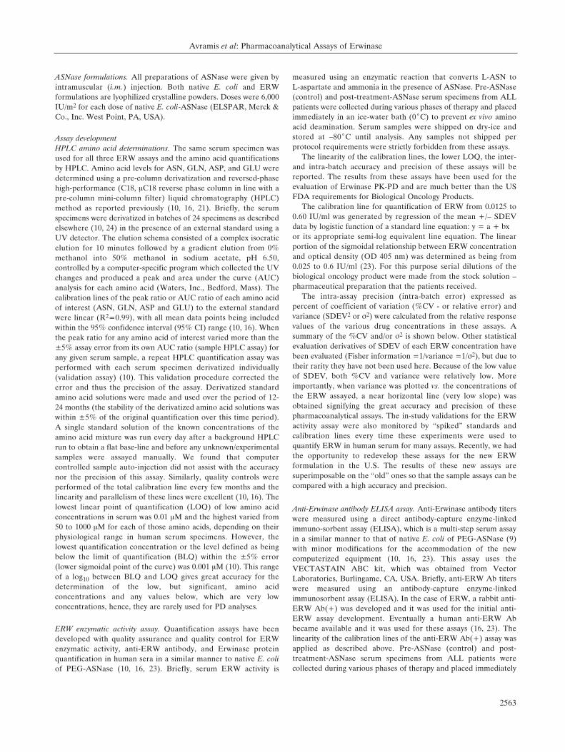
Erwinase activity assay. The standard curve of ERW, andhence, the range of ERW quantification, from 0.025-0.6IU/ml were found to be linearly correlated with the opticaldensity (OD 405 nm) ranging from 0.2 to 1.5 with anR2=0.999 (Figure 1). The negative control human serumOD 405 nm averaged 0.002 OD units or it was 2 orders ofmagnitude lower than the lowest linear OD value of ERW(0.025 IU/ml). This major difference allows us to clearlydetermine the lowest possible EWR concentrationquantification and bifurcate them from the “true” negativecontrol.
Validation at a similar (OD 450 nm) wavelength showedan equal linearity and characteristics, but at lower OD units.The intra-batch and inter-batch precision of the ERWactivity assay in the range of the drug concentrations fromthe calibration curve are shown in Table I. The variance ismode-dependent on the SDEV of each ERW drugconcentration determination, so it varies more than the%CV, which is a relationship between SDEV and the meanof each ERW drug concentration. The excellent accuracy ofthe ERW assay is shown in Table II.
Significant sigmoidicity was seen in calibration curves when9 different ERW activity concentrations were used, 2 at lowerplus one at higher ERW concentrations (0.0075-1.2 IU/ml,data not shown). However, the linear portion of thecalibration line ranged between the ERW concentrationsshown in Figure 1. The relative error (SDEV) was >12.5%in the lowest ERW concentration (0.025 IU/ml) and with%CV for the higher ERW concentrations in the range of 3%-5.7%. The QC samples of intra-day and inter-day accuracyfor those ERW and native E. coli ASNase concentrations(performed the same days as the ERW assays) are shown inTable II. The % relative errors are similar for both ERW andnative ASNase activity determination. Additional calibrationlines representing many assays on separate dates (days-
ANTICANCER RESEARCH 27: 2561-2572 (2007)
2564
months) were conducted. The parallelism of the validation ofthe ERW calibration lines was excellent and is shown inFigure 1. Parallelism is conceptually similar to the pre-studyassay validation. It should be noted that parallelism is nottypically evaluated during a method development, but ratheris used as a validation tool over an extended period of time orthroughout the duration of the pharmacoanalytical assays ofmany months (for the duration of a clinical study).
The lower limit of quantification (LOQ) for ERW in humanserum was 0.025 IU/ml. At this low concentration(s) there wasrelative small SDEV and low %CV (relative Error) and hence,variance. Although the LOQ was slightly higher in ERW thanPEG-ASNase in human specimens this method yielded similarLOQ values to those of native ASNase, which is a similarBiologic Oncology Product ASNase formulation with 8-hourstability post-rehydration from the lyophilized powder.
Anti-Erwinase antibody [Ab(+)] assay. The standard line forthe determination of anti-ERW Ab(+) was established from1:1000 dilution (100%) to as low as 1:32000 dilution
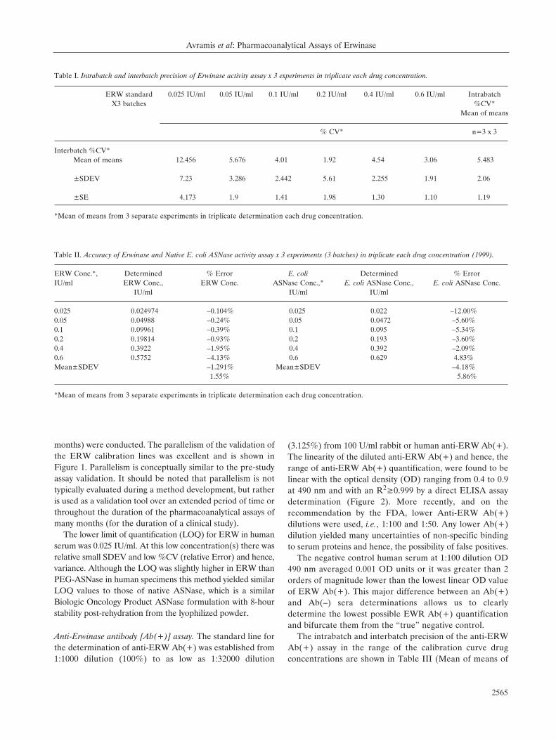
(3.125%) from 100 U/ml rabbit or human anti-ERW Ab(+).The linearity of the diluted anti-ERW Ab(+) and hence, therange of anti-ERW Ab(+) quantification, were found to belinear with the optical density (OD) ranging from 0.4 to 0.9at 490 nm and with an R2≥0.999 by a direct ELISA assaydetermination (Figure 2). More recently, and on therecommendation by the FDA, lower Anti-ERW Ab(+)dilutions were used, i.e., 1:100 and 1:50. Any lower Ab(+)dilution yielded many uncertainties of non-specific bindingto serum proteins and hence, the possibility of false positives.
The negative control human serum at 1:100 dilution OD490 nm averaged 0.001 OD units or it was greater than 2orders of magnitude lower than the lowest linear OD valueof ERW Ab(+). This major difference between an Ab(+)and Ab(–) sera determinations allows us to clearlydetermine the lowest possible EWR Ab(+) quantificationand bifurcate them from the “true” negative control.
The intrabatch and interbatch precision of the anti-ERWAb(+) assay in the range of the calibration curve drugconcentrations are shown in Table III (Mean of means of
Avramis et al: Pharmacoanalytical Assays of Erwinase
2565
Table I. Intrabatch and interbatch precision of Erwinase activity assay x 3 experiments in triplicate each drug concentration.
ERW standard 0.025 IU/ml 0.05 IU/ml 0.1 IU/ml 0.2 IU/ml 0.4 IU/ml 0.6 IU/ml IntrabatchX3 batches %CV*
Mean of means
% CV* n=3 x 3
Interbatch %CV*Mean of means 12.456 5.676 4.01 1.92 4.54 3.06 5.483
±SDEV 7.23 3.286 2.442 5.61 2.255 1.91 2.06
±SE 4.173 1.9 1.41 1.98 1.30 1.10 1.19
*Mean of means from 3 separate experiments in triplicate determination each drug concentration.
Table II. Accuracy of Erwinase and Native E. coli ASNase activity assay x 3 experiments (3 batches) in triplicate each drug concentration (1999).
ERW Conc.*, Determined % Error E. coli Determined % ErrorIU/ml ERW Conc., ERW Conc. ASNase Conc.,* E. coli ASNase Conc., E. coli ASNase Conc.
IU/ml IU/ml IU/ml
0.025 0.024974 –0.104% 0.025 0.022 –12.00%0.05 0.04988 –0.24% 0.05 0.0472 –5.60%0.1 0.09961 –0.39% 0.1 0.095 –5.34%0.2 0.19814 –0.93% 0.2 0.193 –3.60%0.4 0.3922 –1.95% 0.4 0.392 –2.09%0.6 0.5752 –4.13% 0.6 0.629 4.83%Mean±SDEV –1.291% Mean±SDEV –4.18%
1.55% 5.86%
*Mean of means from 3 separate experiments in triplicate determination each drug concentration.
the %CV from 3 separate experiments in triplicatedetermination for each ERW concentration (±SDEV and±SE of %CV). The Relative Error (%CV) was >12.5% inthe lowest range of anti-ERW Ab(+) dilutions. The QCsamples of intra-day and inter-day accuracy for those anti-ERW and anti-native E. coli ASNase Ab(+) dilutions(performed on the same days as the ERW assays) are shown
ANTICANCER RESEARCH 27: 2561-2572 (2007)
2566
Figure 1. Calibration line showing the relationship between the Erwinaseconcentration average (black circles as “mean of means”, and individualn=3 determinations) ±SDEV ±95% confidence intervals (%CI) dilutedin human serum and optical density OD 405 nm response for Erwinasestandard solutions. There is a remarkably low variability as the SDEV iscontained within the size of each symbol. The n=3 individual calibrationlines and their mean demonstrate the parallelism and superimposability ofthese calibration lines of Erwinase. All symbols are the mean of n=3±SDEV ±95% CI. Parallelism is typically evaluated during in-studyvalidation and it represents the variability (relative error) of these assaysthat are generated during a study. The negative control OD 405 nm is alsoshown as being extremely low and highly statistically different from thelowest linear limit of quantification (LOQ) of Erwinase. This along withthe excellent parallelism shown here allows us to be very confident in that,when a patient's serum sample was assayed has an extremely high accuracywith low error of a false positive.
Figure 2. Calibration line showing the relationship between the averagehuman anti-Erwinase antibody [Ab(+)] at 1:100 (1:50 dilutions for 200%)dilutions in human serum (n=3 in triplicate or n=9) ±SDEV ±95%confidence intervals (%CI) and optical density OD 490 nm response withthe new Erwinase antigen standard solutions. The Negative control OD 490nm is also shown as being extremely low and highly statistically differentfrom the lowest linear limit of quantification (LOQ) of anti-ERW Ab(+).This along with the excellent parallelism shown here allows us to be veryconfident in that, when a patient's serum sample was assayed has anextremely high accuracy with low error of a false Ab(+) positive.
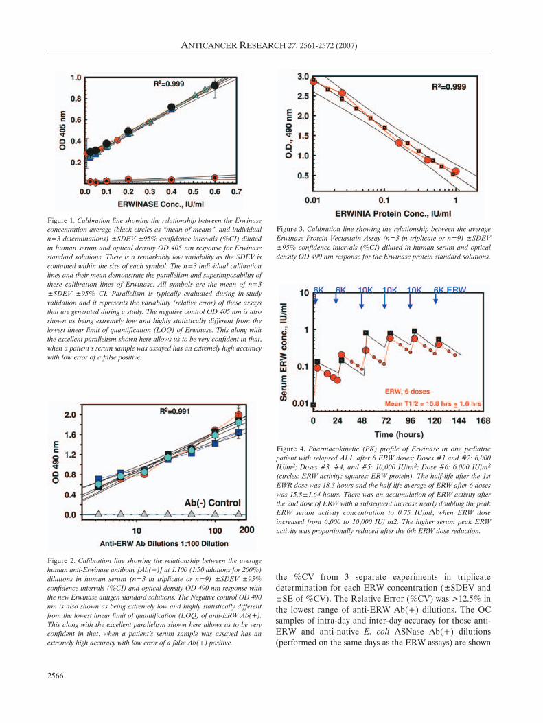
Figure 3. Calibration line showing the relationship between the averageErwinase Protein Vectastain Assay (n=3 in triplicate or n=9) ±SDEV±95% confidence intervals (%CI) diluted in human serum and opticaldensity OD 490 nm response for the Erwinase protein standard solutions.
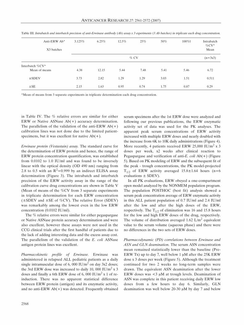
Figure 4. Pharmacokinetic (PK) profile of Erwinase in one pediatricpatient with relapsed ALL after 6 ERW doses; Doses #1 and #2: 6,000IU/m2; Doses #3, #4, and #5: 10,000 IU/m2; Dose #6: 6,000 IU/m2
(circles: ERW activity; squares: ERW protein). The half-life after the 1stEWR dose was 18.3 hours and the half-life average of ERW after 6 doseswas 15.8±1.64 hours. There was an accumulation of ERW activity afterthe 2nd dose of ERW with a subsequent increase nearly doubling the peakERW serum activity concentration to 0.75 IU/ml, when ERW doseincreased from 6,000 to 10,000 IU/ m2. The higher serum peak ERWactivity was proportionally reduced after the 6th ERW dose reduction.

Avramis et al: Pharmacoanalytical Assays of Erwinase
2567
Figure 5. Pharmacokinetic (PK) profile of Erwinase in 4 pediatric patientswith ALL front line who had a clinical reaction to E. coli Pegaspargase after2 ERW doses of 25, 000 IU/m2; These doses continued x 3 per week x 2weeks. The serum ERW concentration vs. time data have been examined asif they were from ONE patient with NONMEM analyses yielding a higherpeak of 2.4 IU/ml and with a POST-HOC analysis for a half-life after the1st EWR dose of 15.61 hours in n = 4 patients. There is an excellentconcordance between the half-life in these patients after the 25 K/m2 dosesand the average of ERW after 6 doses of 15.8±1.64 hours (Figure 4). Therewas no apparent peak accumulation of ERW activity after the 2nd dose.The higher serum peak ERW activity was proportionally reduced. The barsindicate the pharmacodynamic ERW effect on ASN at the assayed timepoints of 0, 4, 8, 24, 48 and 52 hours. All post-ERW administration ASNconcentrations were well below 1 ÌM and most of them were below 0.1 ÌM.The Imax for ASN was 1E-10 nmoles/min/ml. The GLN deamination atthe same time points was well below 70 ÌM.
Figure 6. The pharmacodynamic relationship between Erwinase serumenzymatic activity and its substrates, ASN and GLN deamination post-ERW administrations in sera from the 5 patients treated with the higherdoses. Serum ASN declined from a population concentration mean of42±9 ÌM to less than 1 ÌM for post-administration of Q2 days pediatricpatients with HR ALL. There is a casual dose-serum peak- ASNdeamination in these few patients (Figures 4 and 5). Similarly, GLN wasdeaminated from a population concentration mean of 320 ÌM to less than50 ÌM and it attempted to rebound to less than 75 ÌM. The results areshown as a sigmoid pharmacodynamic relationship (parabolic) betweenthe population mean of deaminated ASN and GLN levels expressed as %of pre-treatment controls versus log10 of ASNase enzymatic activity. Thesigmoid relationship is of the 3rd order of regression and it is the upperportion of the sigmoid fit according to Michaelis-Menten population PK-PD modeling. The highest GLN % deamination achieved was98%±1.1% and for ASN 94%±4%. These PD results may indicate thatGLN may have to be deaminated to a greater degree in serum by ERW sothat it cannot be used as a substrate for the de novo biosynthesis of ASNin the tissues with the catalytic aid of asparagine synthetase. Of importanceis that the “time” does not appear in these relationships. However, “time”is embedded in the concentrations of ERW enzymatic activity, which wasobtained from data presented in Figures 4 and 5. Minimal enzymaticactivity of 0.1 to 0.2 IU/ml was needed for optimal ASN deamination ofapproximately 90% compared to baseline.
Figure 7. Simulations of the ERW 25K x 3 doses per week x 3 weeks (9ERW doses = 225K/m2) and the ERW 25K x2doses per week x 3 weeks (8ERW doses = 200K/m2) under NONMEM population PK-PD modeling.Under intermediate Imax of ASN representing the vast majority of highrisk pediatric ALL patients showed that the ERW 25K x2 doses per weekx 3 weeks regimen is much more beneficial in its ability to deaminate ASN<1 ÌM up to Day 28, whereas the ERW 25K x3 doses per week x 3 weeksdeaminated ASN to <1 ÌM only up to day 21-22.
in Table IV. The % relative errors are similar for eitherERW or Native ASNase Ab(+) accuracy determination.The parallelism of the validation of the anti-ERW Ab(+)calibration lines was not done due to the limited patient-specimens, but it was excellent for native Ab(+).
Erwinase protein (Vestastain) assay. The standard curve forthe determination of ERW protein and hence, the range ofERW protein concentration quantification, was establishedfrom 0.0102 to 1.0 IU/ml and was found to be inverselylinear with the optical density (OD 490 nm) ranging from2.8 to 0.5 with an R2=0.999 by an indirect ELISA assaydetermination (Figure 3). The intrabatch and interbatchprecision of the ERW activity assay in the range of thecalibration curve drug concentrations are shown in Table V(Mean of means of the %CV from 3 separate experimentsin triplicate determination for each ERW concentration(±SDEV and ±SE of %CV). The relative Error (SDEV)was remarkably among the lowest even in the low ERWconcentration (0.0102 IU/ml).
The % relative errors were similar for either pegaspargaseor Native ASNase protein accuracy determination and werealso excellent, however these assays were not used in theCCG clinical trials after the first handful of patients due tothe lack of adding interesting data and the excess assay cost.The parallelism of the validation of the E. coli ASNaseantigen protein lines was excellent.
Pharmacokinetic profile of Erwinase. Erwinase wasadministered in relapsed ALL pediatric patients as a dailysingle intramuscular dose of 6, 000 IU/m2 on day 3x2 doses;the 3rd ERW dose was increased to daily 10, 000 IU/m2 x 3doses and finally a 6th ERW dose of 6, 000 IU/m2 x 1 of re-induction. There was no apparent statistical differencebetween ERW protein (antigen) and its enzymatic activity,and no anti-ERW Ab(+) was detected. Frequently obtained
serum specimens after the 1st ERW dose were analyzed andfollowing our previous publications, the ERW enzymaticactivity set of data was used for the PK analyses. Theapparent peak serum concentrations of ERW activityincreased with multiple ERW doses and nearly doubled withthe increase from 6K to 10K daily administrations (Figure 4).More recently, 4 patients received ERW 25,000 IU/m2 x 3doses per week, x2 weeks after clinical reaction toPegaspargase and verification of anti-E. coli Ab(+) (Figure5). Based on PK modeling of ERW and the subsequent fit ofthe peak - trough concentrations, the PK model-projectedT1/2 of ERW activity averaged 15.8±1.64 hours (n=6evaluations ± SDEV).
In all PK evaluations, ERW obeyed a one-compartmentopen model analyzed by the NONMEM population program.The population POSTHOC (best fit) analysis showed aserum peak concentration average of ERW enzymatic activityin this ALL patient population of 0.7 IU/ml and 2.4 IU/mlafter the low and after the high doses of the ERW,respectively. The T1/2 of elimination was 16 and 15.8 hoursfor the low and high ERW doses of the drug, respectively.The volume of distribution averaged 1.62 L/m2 equivalentvalue to the serum volume (aqueous phase) and there wereno differences in the two sets of ERW doses.
Pharmacodynamic (PD) correlations between Erwinase andASN and GLN deamination. The serum ASN concentrationmean remained statistically lower than the baseline (Pre-ERW Tx) up to day 7, well below 1 ÌM after the 25K ERWdose x 3 doses per week (Figure 5). Although the treatmentcontinued for two 2 weeks no long-term samples weredrawn. The equivalent ASN deamination after the lowerERW doses was <3 ÌM at trough levels. Deamination ofASN was complete in this patient receiving daily ERW lowdoses from a few hours to day 6. Similarly, GLNdeamination was well below 20-30 ÌM by day 7 and below
ANTICANCER RESEARCH 27: 2561-2572 (2007)
2568
Table III. Intrabatch and interbatch precision of anti-Erwinase antibody (Ab) assay x 3 experiments (3 Ab batches) in triplicate each drug concentration.
Anti-ERW Ab* 3.125% 6.25% 12.5% 25% 50% 100%l Intrabatch %CV*
X3 batches Mean
% CV (n=3x3)
Interbatch %CV*Mean of means 4.38 12.15 5.44 7.48 5.41 5.46 6.72
±SDEV 3.73 2.82 1.29 1.29 3.03 1.51 0.511
±SE 2.15 1.63 0.95 0.74 1.75 0.87 0.29
*Mean of means from 3 separate experiments in triplicate determination each drug concentration.
55 ÌM after the two ERW doses. Due to the limited numberof patients no graphs have been attached.
However, based on these results and on the experiencewith the E. coli ASNase formulations, a population PDmodel was constructed which took into consideration theserum enzymatic ERW activity and serum deaminated ASN.Based on the Michaelis-Menten equation the modelpredicted at “steady-state” that serum asparagine could bedetermined up to Day 28 post- 3 weeks of ERWadministration. It should be emphasized that despite thenumber of observations, neither Native E. coli norPegaspargase achieved as low population GLN deaminationas post-ERW. This may indicate that the presence of eitherlower GLN in serum of these few Ab(+) to E. coli ASNaseHR ALL patients may validate the greater glutaminaseaffinity that the ERW enzyme is known to possess, hence itsgreater PD effect on GLN, which in turn, may deaminateASN more efficiently.
More importantly, the population average ERWenzymatic activity was correlated with the serum ASN orGLN deamination, expressed as percentage of pre-treatmentcontrol, yielding the upper portion of a sigmoid curve perMichaelis-Menten relationship (Figure 6). The highest GLN% deamination achieved was 98%±1.1% and for ASN93%±4% at 1 IU/ml ERW concentration. At the higherERW doses with their reciprocal higher ERW peak andtrough levels these % deamination values became evenbetter. This apparent pharmacodynamic correlation betweenthe serum deamination and ERW activity showed that aminimal enzymatic activity of 0.1 to 0.2 IU/ml was neededfor optimal ASN deamination of >90%-95% compared toPre-Tx baseline. As seen in Figure 5, the majority of patientshad an enzymatic activity of ≥0.2 IU/ml up to day 2 after asingle ERW drug administration and 0.1 IU/ml up to day 3
after a single ERW dose, thus providing a minimum optimalERW concentration when the drug was administered Q48hours x 3 doses per week. The collective population Imax ofASN was evaluated by NONMEM in these patients at 1E-10nmoles/min/ml, with apparent Km estimations of 14 and 456ÌM for ASN and GLN, respectively.
No emergence of anti-ERW Ab(+) appeared nor wasthere any association with immediate disappearance ofERW enzymatic activity and rebound of serum ASN andmultiple doses of ERW were not associated with any clinicalmanifestations of hypersensitivity reactions.
Simulations of Erwinase. Based on the similarity of the PKparameters from 3 different ERW doses we decided to usethe population Post-hoc (average) PK-PD values toconstruct various simulated (SIMs) ERW dose and scheduleregimens. These regimens were examined under variousinput (maximum Input - Imax) (25) for ASN values rangingfrom 1E-8 to 1E-4 nmoles/min/ml and with increasing
Avramis et al: Pharmacoanalytical Assays of Erwinase
2569
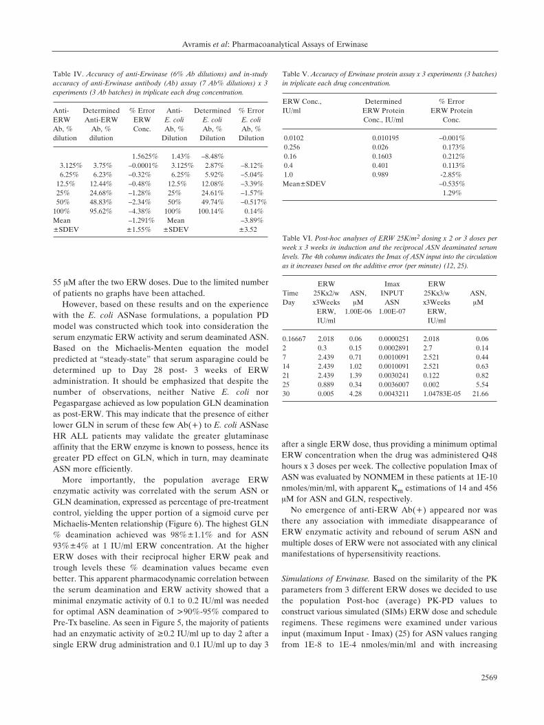
Table IV. Accuracy of anti-Erwinase (6% Ab dilutions) and in-studyaccuracy of anti-Erwinase antibody (Ab) assay (7 Ab% dilutions) x 3experiments (3 Ab batches) in triplicate each drug concentration.
Anti- Determined % Error Anti- Determined % ErrorERW Anti-ERW ERW E. coli E. coli E. coliAb, % Ab, % Conc. Ab, % Ab, % Ab, % dilution dilution Dilution Dilution Dilution
1.5625% 1.43% –8.48%3.125% 3.75% –0.0001% 3.125% 2.87% –8.12%6.25% 6.23% –0.32% 6.25% 5.92% –5.04%
12.5% 12.44% –0.48% 12.5% 12.08% –3.39%25% 24.68% –1.28% 25% 24.61% –1.57%50% 48.83% –2.34% 50% 49.74% –0.517%
100% 95.62% –4.38% 100% 100.14% 0.14%Mean –1.291% Mean –3.89%±SDEV ±1.55% ±SDEV ±3.52
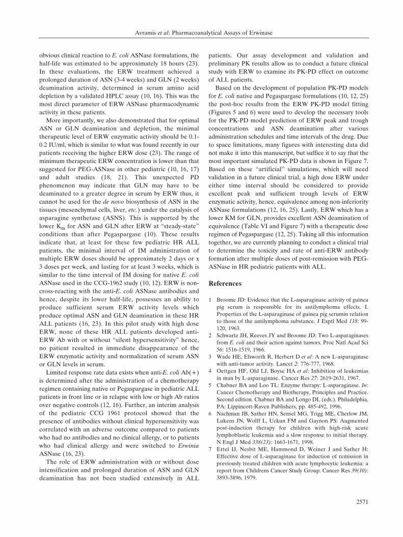
Table V. Accuracy of Erwinase protein assay x 3 experiments (3 batches)in triplicate each drug concentration.
ERW Conc., Determined % ErrorIU/ml ERW Protein ERW Protein
Conc., IU/ml Conc.
0.0102 0.010195 –0.001%0.256 0.026 0.173%0.16 0.1603 0.212%0.4 0.401 0.113%1.0 0.989 -2.85%Mean±SDEV –0.535%
1.29%
Table VI. Post-hoc analyses of ERW 25K/m2 dosing x 2 or 3 doses perweek x 3 weeks in induction and the reciprocal ASN deaminated serumlevels. The 4th column indicates the Imax of ASN input into the circulationas it increases based on the additive error (per minute) (12, 25).
ERW Imax ERW Time 25Kx2/w ASN, INPUT 25Kx3/w ASN, Day x3Weeks ÌM ASN x3Weeks ÌM
ERW, 1.00E-06 1.00E-07 ERW, IU/ml IU/ml
0.16667 2.018 0.06 0.0000251 2.018 0.062 0.3 0.15 0.0002891 2.7 0.147 2.439 0.71 0.0010091 2.521 0.4414 2.439 1.02 0.0010091 2.521 0.6321 2.439 1.39 0.0030241 0.122 0.8225 0.889 0.34 0.0036007 0.002 5.5430 0.005 4.28 0.0043211 1.04783E-05 21.66

Additive Errors. The ERW 6K x 3 doses per week x 3 weeks(like native E. coli dosing) was not very effective inachieving trough levels at or above 0.1 IU/ml of activity witha reciprocal ASN trough level well above 6 ÌM. The ERW10K x 3 doses per week x 3 weeks was much better theprevious dose regimen by achieving ASN less than 1 ÌM forup to 21-22 days. Then, the ERW 25K x 3 doses per week x3 weeks (9 ERW doses = 225K/m2) and the ERW 25Kx2doses per week x 3 weeks (8 ERW doses = 200 K/m2)were evaluated. A remarkable result was produced underlow, intermediate, or high Imax of ASN. SIMs showed thatthe ERW 25K x2 doses per week x 3 weeks regimen is muchmore beneficial in its ability to deaminate ASN <1 ÌM upto Day 28, whereas the ERW 25K x3 doses per week x 3weeks deaminated ASN to <1 ÌM only up to day 21-22(Figure 7).
Discussion
Asparaginases are included in most treatment protocols forboth pediatric and adult patients with newly diagnosed ALL(1-12). For the first time, this report provides thepharmacokinetic and pharmacodynamic data from theintramuscular administration of ERW ASNase in pediatricALL, who were reactive to native E. coli and pegaspargasein the clinic. These elemental PK data are similar to thosewhich have been reported earlier (16, 23). It should also beemphasized that in our limited study ERW was given onlyon a compassionate basis, and only during re-inductiontherapy.
The primary focus of this manuscript is the developmentand validation of the immunoassays necessary to support PKstudies with accuracy and precision. By thoroughly definingand examining each part of these pharmacoanalytical assays,they become the first step in the resulting validation (10,24). These assay quality controls should be concise and thevalidation process straightforward. In order to evaluate theparameters of precision and accuracy prior to conductinglimited ERW PK and PD studies, we developed andsystematically validated the ERW pharmaco-analyticalassays. These assays were produced in a handful of ERWbatches and their results were near-identical to those for theE. coli both native and Pegaspargase (10, 16, 23). Thestandard curve (line) of Erwinase enzymatic activity andhence, the range of ERW quantification, from 0.025 - 0.6IU/ml were found to be linear with the optical density (OD405 nm) ranging from 0.2 to 1.5 with an R2=0.99. Theactual lowest limit of ERW activity based on theextrapolation of this calibration line is 0.01 IU/ml (Figure1). However, this low level will most likely not be used inour clinical trial for ERW drug concentration calculation.When the new ERW calibration lines were added, Mean ofMeans from 3 batches, 2 sent by OPi and one from a
patient's ERW dose to the “old” calibration Mean of Meansline, again, excellent linearity and parallelism weredetermined. All means are included within the 95%Confidence Intervals (95% CI) (Tables I and II).
Moreover, the robustness of an assay is based on itsvariability per drug concentration determined. To this effectwe have examined the % coefficient of variation (%CV)and its Variance (Û2) per drug concentration and throughout the ERW calibration range. Since some older theorystates that variance is a better measurement of thevariability of an assay than %CV this parameter was alsoestimated for the “old” and the new ERW calibrationassays. There are near-horizontal lines produced of either%CV or Û2 vs. the ERW concentrations (data not shown)indicating that this assay has a great accuracy and precisionthought out its calibration line range. Both accuracy andprecision are excellent for ERW enzymatic activity (TablesI and II). Thus, in answering the practical question - Howcredible is the assay? We can reply that % CV is good, butVariance (Û2) as estimated per ERW Drug concentration ismuch better, especially when it does not change very much(No statistically significant difference) with each increasingERW drug concentration determination.
As described above, a typical assay development andvalidation will include at least 3-6 precision and accuracydeterminations in multiple drug concentrations (4-6 or moreper standard line) to define the overall consistency and QCof the biologic assay. QC includes (but are not required bythe FDA regulations) stability of the drug during the assay,specificity, selectivity, lower limit of linearity and range andlower limit of quantification (LOQ), not to mention,parallelism and In-Study validation once the clinical trial isin progress for these assays. Each calibration line shouldevaluate the negative control with non-zero points and forall drug concentrations, so that the accuracy of the drug canbe assessed. The validation standards should re-define therange of the assay and no values below the LOQ may beseen or used. Overall the imunoassays that we havedeveloped and presented here (Figures 1-3 and Tables I-V),are highly sensitive and selective that have been used toquantify ERW protein, its activity and its antibody in abiologic matrix of assays for the early PK-PD analyses inpediatric patients. It should be emphasized that the ERWprotein assay although useful it provides very littleadditional information of significance and based on its cost,it is not yielding a high return of data information for itscost (Figure 4).
Asselin et al., reported the first clinical PK evaluation ofERW in pediatric ALL patients in the early 1990's. Thesestudies reported a half-life for ERW of 16 hours (9).However, after multiple IM injections of ERW to 24patients of 6,000 or 10,000 IU/m2 treated per CCG-1961protocol in newly diagnosed HR ALL children who had an
ANTICANCER RESEARCH 27: 2561-2572 (2007)
2570
obvious clinical reaction to E. coli ASNase formulations, thehalf-life was estimated to be approximately 18 hours (23).In these evaluations, the ERW treatment achieved aprolonged duration of ASN (3-4 weeks) and GLN (2 weeks)deamination activity, determined in serum amino aciddepletion by a validated HPLC assay (10, 16). This was themost direct parameter of ERW ASNase pharmacodynamicactivity in these patients.
More importantly, we also demonstrated that for optimalASN or GLN deamination and depletion, the minimaltherapeutic level of ERW enzymatic activity should be 0.1-0.2 IU/ml, which is similar to what was found recently in ourpatients receiving the higher ERW dose (23). The range ofminimum therapeutic ERW concentration is lower than thatsuggested for PEG-ASNase in other pediatric (10, 16, 17)and adult studies (18, 21). This unexpected PDphenomenon may indicate that GLN may have to bedeaminated to a greater degree in serum by ERW thus, itcannot be used for the de novo biosynthesis of ASN in thetissues (mesenchymal cells, liver, etc.) under the catalysis ofasparagine synthetase (ASNS). This is supported by thelower Km for ASN and GLN after ERW at “steady-state”conditions than after Pegaspargase (10). These resultsindicate that, at least for these few pediatric HR ALLpatients, the minimal interval of IM administration ofmultiple ERW doses should be approximately 2 days or x3 doses per week, and lasting for at least 3 weeks, which issimilar to the time interval of IM dosing for native E. coliASNase used in the CCG-1962 study (10, 12). ERW is non-cross-reacting with the anti-E. coli ASNase antibodies andhence, despite its lower half-life, possesses an ability toproduce sufficient serum ERW activity levels whichproduce optimal ASN and GLN deamination in these HRALL patients (16, 23). In this pilot study with high doseERW, none of these HR ALL patients developed anti-ERW Ab with or without “silent hypersensitivity” hence,no patient resulted in immediate disappearance of theERW enzymatic activity and normalization of serum ASNor GLN levels in serum.
Limited response rate data exists when anti-E. coli Ab(+)is determined after the administration of a chemotherapyregimen containing native or Pegaspargase in pediatric ALLpatients in front line or in relapse with low or high Ab ratiosover negative controls (12, 16). Further, an interim analysisof the pediatric CCG 1961 protocol showed that thepresence of antibodies without clinical hypersensitivity wascorrelated with an adverse outcome compared to patientswho had no antibodies and no clinical allergy, or to patientswho had clinical allergy and were switched to ErwiniaASNase (16, 23).
The role of ERW administration with or without doseintensification and prolonged duration of ASN and GLNdeamination has not been studied extensively in ALL
patients. Our assay development and validation andpreliminary PK results allow us to conduct a future clinicalstudy with ERW to examine its PK-PD effect on outcomeof ALL patients.
Based on the development of population PK-PD modelsfor E. coli native and Pegaspargase formulations (10, 12, 25)the post-hoc results from the ERW PK-PD model fitting(Figures 5 and 6) were used to develop the necessary toolsfor the PK-PD model prediction of ERW peak and troughconcentrations and ASN deamination after variousadministration schedules and time intervals of the drug. Dueto space limitations, many figures with interesting data didnot make it into this manuscript, but suffice it to say that themost important simulated PK-PD data is shown in Figure 7.Based on these “artificial” simulations, which will needvalidation in a future clinical trial, a high dose ERW undereither time interval should be considered to provideexcellent peak and sufficient trough levels of ERWenzymatic activity, hence, equivalence among non-inferiorityASNase formulations (12, 16, 25). Lastly, ERW which has alower KM for GLN, provides excellent ASN deamination ofequivalence (Table VI and Figure 7) with a therapeutic doseregimen of Pegaspargase (12, 25). Taking all this informationtogether, we are currently planning to conduct a clinical trialto determine the toxicity and rate of anti-ERW antibodyformation after multiple doses of post-remission with PEG-ASNase in HR pediatric patients with ALL.
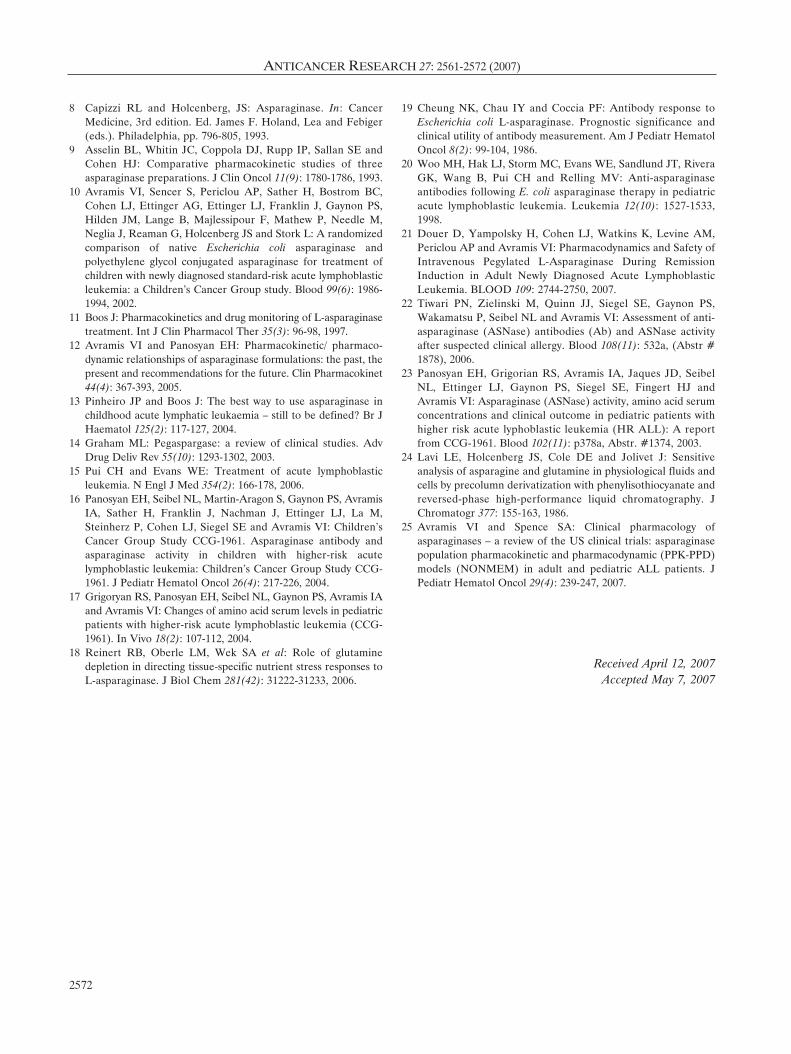
References
1 Broome JD: Evidence that the L-asparaginase activity of guineapig serum is responsible for its antilymphoma effects. I.Properties of the L-asparaginase of guinea pig serumin relationto those of the antilymphoma substance. J Exptl Med 118: 99-120, 1963.
2 Schwartz JH, Reeves JY and Broome JD: Two L-asparaginasesfrom E. coli and their action against tumors. Proc Natl Acad Sci56: 1516-1519, 1966.
3 Wade HE, Elsworth R, Herbert D et al: A new L-asparaginasewith anti-tumor activity. Lancet 2: 776-777, 1968.
4 Oettgen HF, Old LJ, Boyse HA et al: Inhibition of leukemiasin man by L-asparaginase. Cancer Res 27: 2619-2631, 1967.
5 Chabner BA and Loo TL: Enzyme therapy: L-asparaginase. In:Cancer Chemotherapy and Biotherapy, Principles and Practice.Second edition. Chabner BA and Longo DL (eds.). Philadelphia,PA: Lippincott-Raven Publishers, pp. 485-492, 1996.
6 Nachman JB, Sather HN, Sensel MG, Trigg ME, Cherlow JM,Lukens JN, Wolff L, Uckun FM and Gaynon PS: Augmentedpost-induction therapy for children with high-risk acutelymphoblastic leukemia and a slow response to initial therapy.N Engl J Med 338(23): 1663-1671, 1998.
7 Ertel IJ, Nesbit ME, Hammond D, Weiner J and Sather H:Effective dose of L-asparaginase for induction of remission inpreviously treated children with acute lymphocytic leukemia: areport from Childrens Cancer Study Group. Cancer Res 39(10):3893-3896, 1979.
Avramis et al: Pharmacoanalytical Assays of Erwinase
2571
8 Capizzi RL and Holcenberg, JS: Asparaginase. In: CancerMedicine, 3rd edition. Ed. James F. Holand, Lea and Febiger(eds.). Philadelphia, pp. 796-805, 1993.
9 Asselin BL, Whitin JC, Coppola DJ, Rupp IP, Sallan SE andCohen HJ: Comparative pharmacokinetic studies of threeasparaginase preparations. J Clin Oncol 11(9): 1780-1786, 1993.
10 Avramis VI, Sencer S, Periclou AP, Sather H, Bostrom BC,Cohen LJ, Ettinger AG, Ettinger LJ, Franklin J, Gaynon PS,Hilden JM, Lange B, Majlessipour F, Mathew P, Needle M,Neglia J, Reaman G, Holcenberg JS and Stork L: A randomizedcomparison of native Escherichia coli asparaginase andpolyethylene glycol conjugated asparaginase for treatment ofchildren with newly diagnosed standard-risk acute lymphoblasticleukemia: a Children's Cancer Group study. Blood 99(6): 1986-1994, 2002.
11 Boos J: Pharmacokinetics and drug monitoring of L-asparaginasetreatment. Int J Clin Pharmacol Ther 35(3): 96-98, 1997.
12 Avramis VI and Panosyan EH: Pharmacokinetic/ pharmaco-dynamic relationships of asparaginase formulations: the past, thepresent and recommendations for the future. Clin Pharmacokinet44(4): 367-393, 2005.
13 Pinheiro JP and Boos J: The best way to use asparaginase inchildhood acute lymphatic leukaemia – still to be defined? Br JHaematol 125(2): 117-127, 2004.
14 Graham ML: Pegaspargase: a review of clinical studies. AdvDrug Deliv Rev 55(10): 1293-1302, 2003.
15 Pui CH and Evans WE: Treatment of acute lymphoblasticleukemia. N Engl J Med 354(2): 166-178, 2006.
16 Panosyan EH, Seibel NL, Martin-Aragon S, Gaynon PS, AvramisIA, Sather H, Franklin J, Nachman J, Ettinger LJ, La M,Steinherz P, Cohen LJ, Siegel SE and Avramis VI: Children'sCancer Group Study CCG-1961. Asparaginase antibody andasparaginase activity in children with higher-risk acutelymphoblastic leukemia: Children's Cancer Group Study CCG-1961. J Pediatr Hematol Oncol 26(4): 217-226, 2004.
17 Grigoryan RS, Panosyan EH, Seibel NL, Gaynon PS, Avramis IAand Avramis VI: Changes of amino acid serum levels in pediatricpatients with higher-risk acute lymphoblastic leukemia (CCG-1961). In Vivo 18(2): 107-112, 2004.
18 Reinert RB, Oberle LM, Wek SA et al: Role of glutaminedepletion in directing tissue-specific nutrient stress responses toL-asparaginase. J Biol Chem 281(42): 31222-31233, 2006.
19 Cheung NK, Chau IY and Coccia PF: Antibody response toEscherichia coli L-asparaginase. Prognostic significance andclinical utility of antibody measurement. Am J Pediatr HematolOncol 8(2): 99-104, 1986.
20 Woo MH, Hak LJ, Storm MC, Evans WE, Sandlund JT, RiveraGK, Wang B, Pui CH and Relling MV: Anti-asparaginaseantibodies following E. coli asparaginase therapy in pediatricacute lymphoblastic leukemia. Leukemia 12(10): 1527-1533,1998.
21 Douer D, Yampolsky H, Cohen LJ, Watkins K, Levine AM,Periclou AP and Avramis VI: Pharmacodynamics and Safety ofIntravenous Pegylated L-Asparaginase During RemissionInduction in Adult Newly Diagnosed Acute LymphoblasticLeukemia. BLOOD 109: 2744-2750, 2007.
22 Tiwari PN, Zielinski M, Quinn JJ, Siegel SE, Gaynon PS,Wakamatsu P, Seibel NL and Avramis VI: Assessment of anti-asparaginase (ASNase) antibodies (Ab) and ASNase activityafter suspected clinical allergy. Blood 108(11): 532a, (Abstr #1878), 2006.
23 Panosyan EH, Grigorian RS, Avramis IA, Jaques JD, SeibelNL, Ettinger LJ, Gaynon PS, Siegel SE, Fingert HJ andAvramis VI: Asparaginase (ASNase) activity, amino acid serumconcentrations and clinical outcome in pediatric patients withhigher risk acute lyphoblastic leukemia (HR ALL): A reportfrom CCG-1961. Blood 102(11): p378a, Abstr. #1374, 2003.
24 Lavi LE, Holcenberg JS, Cole DE and Jolivet J: Sensitiveanalysis of asparagine and glutamine in physiological fluids andcells by precolumn derivatization with phenylisothiocyanate andreversed-phase high-performance liquid chromatography. JChromatogr 377: 155-163, 1986.
25 Avramis VI and Spence SA: Clinical pharmacology ofasparaginases – a review of the US clinical trials: asparaginasepopulation pharmacokinetic and pharmacodynamic (PPK-PPD)models (NONMEM) in adult and pediatric ALL patients. JPediatr Hematol Oncol 29(4): 239-247, 2007.
Received April 12, 2007Accepted May 7, 2007
ANTICANCER RESEARCH 27: 2561-2572 (2007)
2572