pf fn l dpditiprofessor of neurology and pediatrics chief ... · chief, child neurology ... case 1...
TRANSCRIPT

Robert GreenwoodRobert GreenwoodP f f N l d P di t iProfessor of Neurology and Pediatrics
Chief, Child NeurologyUniversity of North Carolina School ofUniversity of North Carolina School of
Medicine

CSICSIASHEVILLE
Case Studies In Epilepsy
ASHEVILLECase Studies In Epilepsy

Slides Postedhttp//neuron.med.unc.edu/neurology

Invisible EvidenceInvisible EvidenceCSI 76 4 7 404 13 N 03CSI 76. 4- 7 404 13 Nov 03
Case 1 Infant with Seizures
Full term infant born meconium stained, low Apgar scores and found to have low
l t l t H t d i t l i t iplatelets. He stayed in neonatal intensive care for three days. Development was slow. He did not sit until 9 months of age

Case 1 Seizures
Onset 4months of age. The seizures were described by mother as episodes of quick j k th t th h d ld f ll d hijerks so that the head would fall and his upper extremities would stiffen. The
i i l t t ti f lseizures came in clusters at times of sleep transition. Generalized clonic seizures began at 8 months of age

Case 1 EEG
EEG- 8 months old- bitemporal and generalized epileptiform dischargesVideo EEG at 10 months video EEG monitoringVideo EEG at 10 months- video EEG monitoring which showed high amplitude generalized spike and polyspike waves and right and left temporaland polyspike waves and right and left temporal focal slowing greatest on the right.Video EEG at 16 months- Electro-clinical infantile spasms and Left temporal/parietal spikes awake, multifocal epileptiform discharges asleep.
Video EEGVideo-EEG
Case 1Questions
What is the cause and how to find it?What treatment should be used for the seizures?

Case 1Imaging
3T MRI- Normal
Case 1 Laboratory Testing
A i id li ht l ti f it lliAmino acids- slight elevation of citrulline, proline and phenlalanine. Nondiagnositic.O i id i d th l l iOrganic acids- increased methylmalonic acid. Rule out B12 deficiency. Acylcarnitine profile normalAcylcarnitine profile-normalCSF for neurotransmitters could not be obtainedobtained Karyotype- normal male karyotype

Case 1 Treatment
Phenobarbital ineffectivePhenobarbital- ineffectiveTopiramate, leviteracetam and clonazepam -Initial reduction of seizure frequency followed by y ygradually increasing seizures. Zonisamide caused vomiting and extreme restlessness.restlessness.Lamotrigine - no improvement when added to medications above.P d i l S i t d d did tPrednisolone- Seizures stopped and did not return after taper off.

Case 1 Further Genetic Testing
3 C SC 1Heterozygous variant 345 T>C in SCN 1A gene
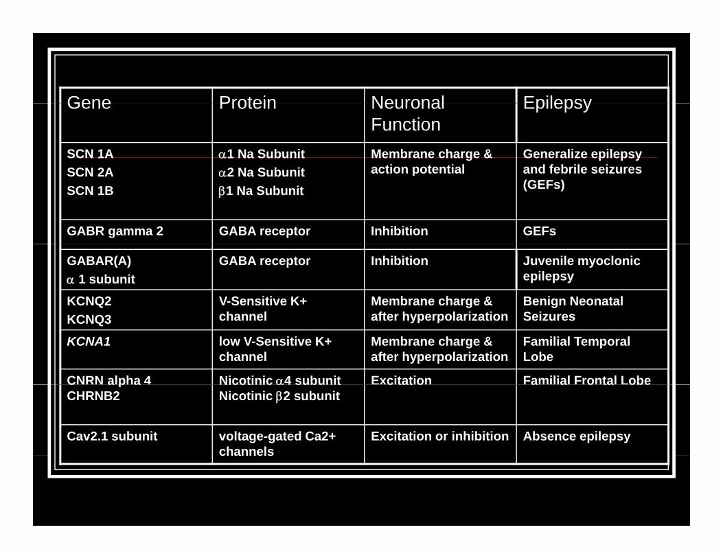
Gene Protein Neuronal EpilepsyGene Protein Neuronal Function
Epilepsy
SCN 1A SCN 2A
α1 Na Subunit α2 Na Subunit
Membrane charge & action potential
Generalize epilepsy and febrile seizures SCN 2A
SCN 1Bα2 Na Subunitβ1 Na Subunit
p(GEFs)
GABR gamma 2 GABA receptor Inhibition GEFs
GABAR(A)α 1 subunit
GABA receptor Inhibition Juvenile myoclonic epilepsy
KCNQ2 V-Sensitive K+ channel
Membrane charge & after hyperpolarization
Benign Neonatal SeizuresKCNQ3 channel after hyperpolarization Seizures
KCNA1 low V-Sensitive K+ channel
Membrane charge & after hyperpolarization
Familial Temporal Lobe
CNRN alpha 4 Nicotinic α4 subunit Excitation Familial Frontal Lobe C a p aCHRNB2
Nicotinic α4 subunit Nicotinic β2 subunit
c tat o a a o ta obe
Cav2.1 subunit voltage-gated Ca2+ channels
Excitation or inhibition Absence epilepsychannels
Th U lThe Unusual SuspectSuspect
CSI 135 6 18 618 30 M 06CSI 135. 6-18 618 30 Mar 06
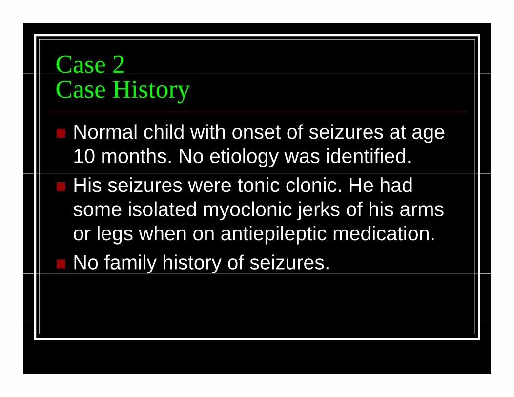
Case 2 Case History
Normal child with onset of seizures at age 10 months. No etiology was identified. His seizures were tonic clonic. He had some isolated myoclonic jerks of his arms or legs when on antiepileptic medication. No family history of seizures.
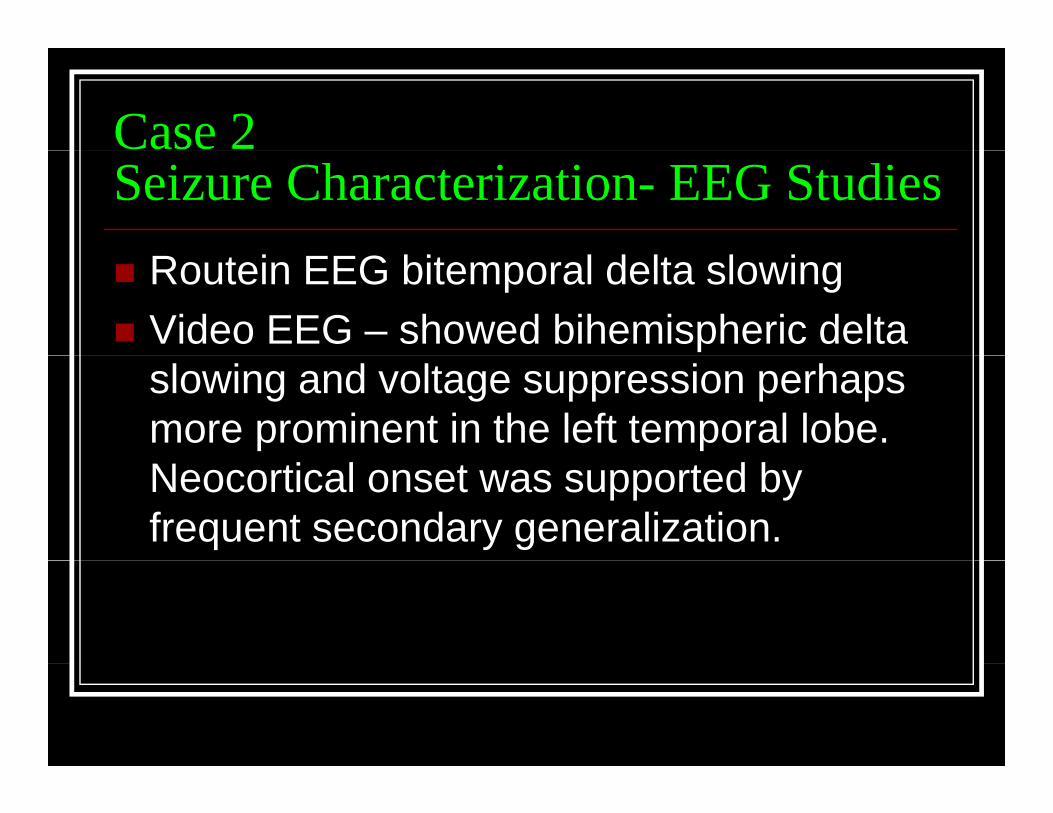
Case 2 Seizure Characterization- EEG Studies
Routein EEG bitemporal delta slowingVideo EEG – showed bihemispheric delta slowing and voltage suppression perhaps more prominent in the left temporal lobe. Neocortical onset was supported by frequent secondary generalization.
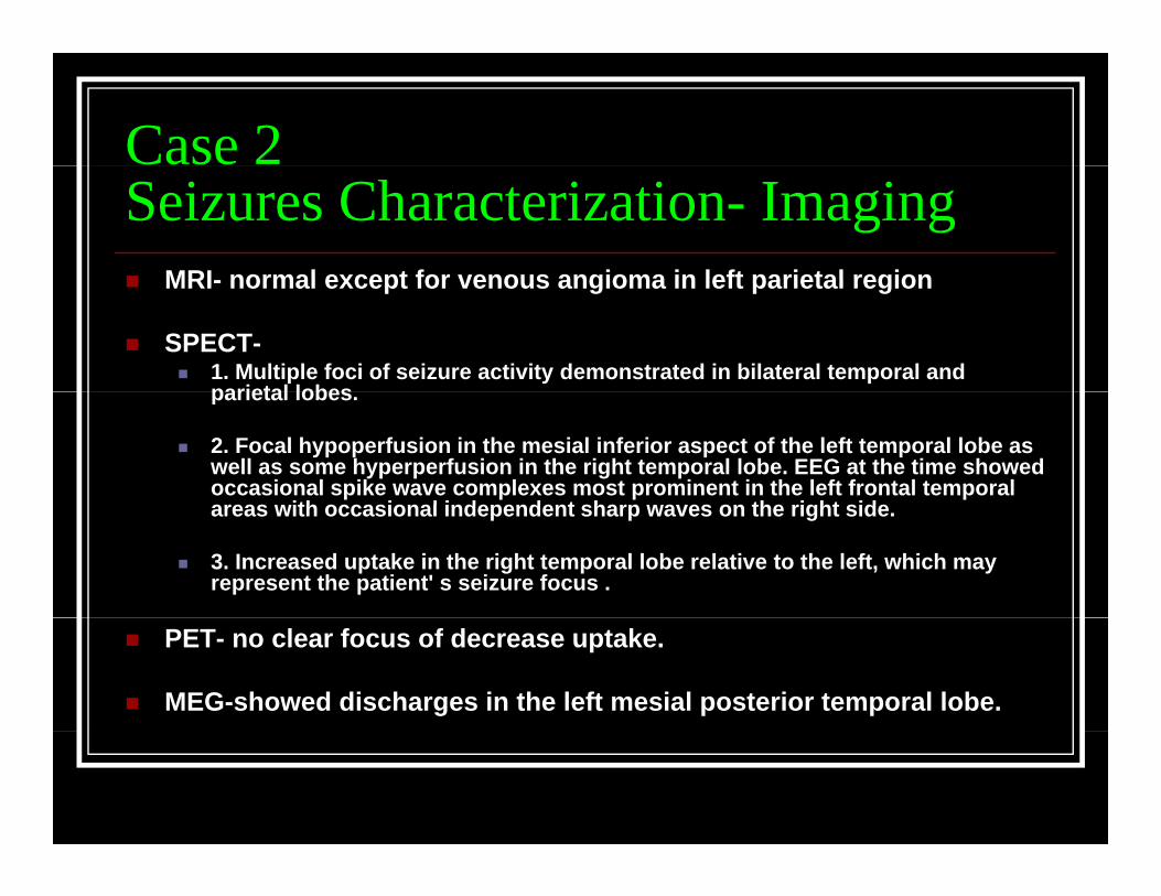
Case 2 Seizures Characterization- Imaging
MRI normal except for venous angioma in left parietal regionMRI- normal except for venous angioma in left parietal region
SPECT-1. Multiple foci of seizure activity demonstrated in bilateral temporal and parietal lobesparietal lobes.
2. Focal hypoperfusion in the mesial inferior aspect of the left temporal lobe as well as some hyperperfusion in the right temporal lobe. EEG at the time showed occasional spike wave complexes most prominent in the left frontal temporal areas ith occasional independent sharp a es on the right sideareas with occasional independent sharp waves on the right side.
3. Increased uptake in the right temporal lobe relative to the left, which may represent the patient' s seizure focus .
PET- no clear focus of decrease uptake.
MEG-showed discharges in the left mesial posterior temporal lobe.
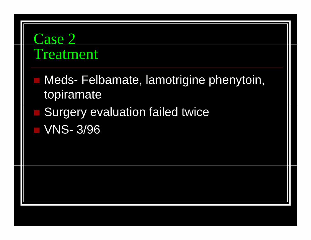
Case 2Treatment
Meds- Felbamate, lamotrigine phenytoin, topiramateSurgery evaluation failed twiceVNS- 3/96
Case 2Question
What does he have?What is the cause?

Case 2Reflex Epilepsy
One year after VNS- VideoFacial twitching only when he was reading and
ll hil di l dusually while reading aloud.
Five years after VNSFacial twitching while speaking
Onset
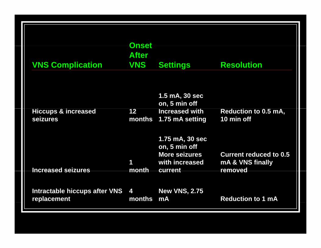
VNS Complication
Onset After VNS Settings Resolution
1.5 mA, 30 sec on, 5 min off
Hiccups & increased seizures
12 months
Increased with 1.75 mA setting
Reduction to 0.5 mA, 10 min off
1 75 mA 30 sec
Increased seizures1 month
1.75 mA, 30 sec on, 5 min offMore seizures with increased current
Current reduced to 0.5 mA & VNS finally removedIncreased seizures month current removed
Intractable hiccups after VNS replacement
4 months
New VNS, 2.75 mA Reduction to 1 mA

A d Th ThAnd Then There Were NoneWere None
CSI 32 2 9 209 22 N 01CSI 32. 2- 9 209 22 Nov 01
Last LaughLast LaughCSI 66 3 20 320 24 A 03CSI 66. 3-20 320 24 Apr 03

Case 3History
A 6-year-old presented for evaluation of precocious puberty and spells.Product of a full-term normal pregnancy and uncomplicated delivery.Good health and normal milestones until 5 years of age.

Case 3SpellsEpisodes of inappropriate laughter, hyperactivity, and giddiness, followed by
f ti hill dprofuse sweating, severe chills, and retching. and ending in abdominal pain and
itivomitingOccurred daily and lasted 1-4 hoursNo loss of awareness

Case 3EEGs
Two EEGs were normal A typical spell lasting 2 hours was captured on video EEG monitoring. The EEG did not show any seizure activity.

Case 3Questions
What are the spells and what is causing them?How can we treat them?
Case 3Imaging Studies
MRI - 6-mm mass in the tuber cinereum consistent with a hamartoma PET- No abnormality noted

Case 3Hormone and Stimulating HormoneHormone and Stimulating Hormone Changes
I t l
Time
Interval from Sz Onset
Prolac.ng/ml
ACTHpg/ml
TSHuU/ml
LHmlU/ml
FSHmlU/ml
900 Baseline 7 47 2.8 2.9 7
1700 Sz onset
1715 15min 32 84 2.9 2.1 4.31730 30min 34 58 2.9 3 4.51750 50min 0 3 13 2 1 3 2 3 91750 50min 0.3 13 2.1 3.2 3.91900 120min 18 NA 1.8 1.9 3.3
Case 3Treatment
GnRH analogue (Lupron) 15-mg IM injection every 28 days Her seizures improved in the first few weeks and by 8 weeks she was seizure free
Viva Las VegasViva Las Vegas93 5 1 501 23 S 0493. 5- 1 501 23 Sep 04

JackpotJackpotCSI 75 4 6 407 6 N 03CSI 75. 4- 6 407 6 Nov 03
Case 4History
Gestation, labor and delivery were unremarkable except for transient neonatal j dijaundice.Development proceeded normally until onset of infantile spasms at 4 months of age. After this development slowed.

Case 4History Continued
Admitted to Brenner Children’s Hospital where the diagnosis of infantile spasms was made and an evaluation for the etiology was performedevaluation for the etiology was performed. He was initially treated with phenobarbital and then placed on ACTH for 28 days.then placed on ACTH for 28 days. He was treated with zonisamide after the ACTH but when the seizures increased again he was ggiven another course of ACTH
Case 4History Continued
H b R Elt f dHe was seen by Roy Elterman for a second opinion and he confirmed the diagnosis of infantile spasms and recommended furtherinfantile spasms and recommended further work up and a trial of vigabatrin. When first seen at UNC at 9 months old heWhen first seen at UNC at 9 months old he was on ACTH 80 units subcutaneously q.a.m., Zonegran 100 mg b.i.d., Vitamin B6 q , g g ,100 mg p.o. t.i.d. and Phenobarbital 90 mg p.o. q.h.s.

Case 4Seizures and EEG at 9 Months
SeizuresClusters of head drops several times each day
EEG showed an abnormal background pattern consistent with a modified hysarrhythmia pattern.

Case 4Imaging
MRI- diffuse atrophyMRS-
Long echo spectra - normal. Short echo spectra - elevation of glutamine and glutamate containing compounds seen in the frontal lobes bilaterally of uncertain significancy.
Case 4Laboratory Tests
N l l t t i i d iNormal venous lactate, urine organic and amino acids, acylcarnitine profile, carbohydrate deficient transferrin, CSF protein, CSF glucose, CSF amino , p , g ,acids, CSF lactate,, and karyotype. Negative TORCH titers. VLCFA- slight increase in the C24/22 ratio which could have been secondary to liver dysfunction or another metabolic disorder affecting liver function.another metabolic disorder affecting liver function.CSF neurotransmitters were sent.

Case 4Treatment
He was discharged on tapering doses of ACTH and phenobarbitalHe was started on another treatment and when seen three months later he had no seizures and was developing normally.
Case 4Question
What was the treatment that stopped the seizures?
Case 4Folinic acid (Leucovorin)

Vitamin Responsive EpilepsyBiotinidase deficiencyPyridoxine-dependent epilepsyPyridoxal phosphate dependent epilepsyFolinic acid-responsive seizuresFolinic acid responsive seizures

Case St dies InCase Studies In EpilepsyEpilepsy

Torres OA, Miller VS, Buist NM, Hyland K. Folinic acid responsive neonatal seizures. J Child N l 1999 14 529 32Child Neurol 1999; 14: 529-32.

Waiting For ReliefWaiting For Relief

OverloadOverloadCSI 26 2 3 203 11 O t 01CSI 26. 2- 3 203 11 Oct 01
Crash and BurnCrash and BurnCSI 63 3 17 317 13 M 03CSI 63. 3-17 317 13 Mar 03

Fallen IdolsFallen IdolsCSI 158 7 17 717 22 F b 07CSI 158. 7-17 717 22 Feb 07