peter thomas · susanna stea metal implant allergy and ... · clinical management of joint...
TRANSCRIPT

Clinical Management of Joint Arthroplasty
Metal Implant Allergy and Immuno-Allergological Compatibility Aspects of Ceramic Materials
Peter Thomas · Susanna Stea


Metal Implant Allergy and Immuno-AllergologicalCompatibility Aspects of Ceramic Materials


P. Thomas, G. Roider, A. Beraudi, D. de Pasquale,B. Bordini, S. Catalani, S. Stea, A. Toni, S. Usbeck,and B. Summer
Metal Implant Allergy and Immuno-AllergologicalCompatibility Aspects of Ceramic Materials
123

Prof. Dr. Peter ThomasClinic for Dermatology and AllergologyLudwig-Maximilians-University MunichFrauenlobstraße 9–11D-80337 Munich
Sylvia UsbeckCeramTec GmbHCeramTec Platz 1–9D-73207 Plochingen
Dr. Susanna SteaMedical Technology LaboratoryRizzoli Orthopaedic InstituteVia di Barbiano, 1/10I-40136 Bologna, Italy
ISBN 978-3-662-47439-6 ISBN 978-3-662-47440-2 (eBook) DOI 10.1007/978-3-662-47440-2
Springer Medizin© Springer-Verlag Berlin Heidelberg 2015This work is subject to copyright. All rights are reserved by the Publisher, whether the whole or part of the material is concerned, specifically the rights of translation, reprinting, reuse of illustrations, recitation, broadcasting, reproduction on microfilms or in any other physical way, and transmission or information storage and retrieval, electronic adaptation, computer software, or by similar or dissimilar methodology now known or hereafter developed. Exempted from this legal reservation are brief excerpts in connection with reviews or scholarly analysis or material supplied specifically for the purpose of being entered and executed on a computer system, for exclusive use by the purchaser of the work. Duplication of this publication or parts thereof is permitted only under the provisions of the Copyright Law of the Publisher’s location, in its current version, and permission for use must always be obtained from Springer. Permis-sions for use may be obtained through RightsLink at the Copyright Clearance Center. Violations are liable to prosecution under the respective Copyright Law.
The use of general descriptive names, registered names, trademarks, service marks, etc. in this publication does not imply, even in the absence of a specific statement, that such names are exempt from the relevant protective laws and regulations and therefore free for general use. While the advice and information in this book are believed to be true and accurate at the date of publication, neither the authors nor the editors nor the publisher can accept any legal responsibility for any errors or omissions that may be made. The publisher makes no warranty, express or implied, with respect to the material contained herein.
Planning: Carola HerzbergCover Design: deblik BerlinCover Illustration (collage): © CeramTec GmbH, © FotoliaProduction: Fotosatz-Service Köhler GmbH – Reinhold Schöberl, Würzburg
Printed on acid-free paper
Springer Medizin is brand of SpringerSpringer is part of Springer Science+Business Media (www.springer.com)
see website:www.biolox.com/clinical-guides/

V
Preface
The growing number of endoprostheses reflects not only the demographical changes with a rising proportion of the elderly within the population but also the growing number of young people getting implants.
Besides optimizing mechanical properties, tissue integration and tribology of metal implants, biocompatibility of implant materials with respect to long-term efficacy will become more significant. Nickel, chromium and cobalt or bone cement components might trigger contact allergies and thus in turn may lead to implant incompatibility. This possibility highlights potential local and systemic adverse reactions of released implant components .
Ceramic implants are often used in patients with hypersensitivities. Trial results of two research groups that studied immuno-allergological compatibility of ceramic materials will be presented in this clinical guide.
The guide presents background information and recommendations on how to proceed in patients with suspected implant allergies since the diagnostic criteria of implant-associated allergies have not yet been defined.
Patient history and clinical findings, in addition to test results must be con-sidered in context.
For the first time, a clinical algorithm has been developed for procedures in clinical practice when an implant allergy is suspected. The algorithm is pre-sented as a laminated insert. The flow chart contains additional information on the histopathological particle algorithm, according to Krenn.
Enhancement of clinical-allergological diagnostic tools and interdisciplinary research approaches will help improve future patient care.


VII
Author
Peter Thomas, MD, PhD, professor of dermatology and allergologyPeter Thomas was born on March 5, 1956, in Augsburg-Göggingen,
Germany, and completed his study in medicine at the Ludwig-
Maximilians-University in Munich (Germany). He spent two years
in the USA as a research fellow in the Department of Immunology
at the Johns Hopkins University in Baltimore, Maryland, and the
La Jolla Institute for Allergy and Immunology in California.
Thomas specialized in allergology, environmental medicine and
problem-based learning (Harvard Medical School). For the last
15 years, Thomas has dedicated his research to metal implant hyper-
sensitivity. Along with his team, he offers special consultation hours
on implant hypersensitivity which more than 1,000 patients with
allergic reactions to implants or suspected allergies have attended.
Peter Thomas
4 is an internationally renown allergologist/dermatologist
4 works with an interdisciplinary team
4 is an expert allergologist for the German Society of Ortho paedics
and Orthopaedic Surgery (DGOOC) and German Society of
Dental, Oral and Orthodontic Implantology (DGI)
4 offers special consultation for patients with implant allergies
4 provides professional training of med school students and
medical personnel
4 reviews articles for ten medical journals and several research
foundations
4 developed a diagnostic algorithm for patients with suspected
implant allergies
4 published more than 100 original scientific papers
4 contributed articles to 42 textbooks
4 has won three research awards

VIII Author
Along with Marc Thomsen, MD, PhD, Thomas leads the Implant
Allergy Working Group of the DGOOC for which he also acts as an
expert allergologist. Thomas is a member of seven medical societies.
He also is the lead author of the interdisciplinary statement on
implant allergy published in 2008 by the DGOOC, the German
Contact Dermatitis Research Group (DKG) and the German Society
for Allergology and Clinical Immunology (DGAKI).

IXAuthor
Susanna Stea, PhD, biologist and researcherSusanna Stea was born on March 9, 1959, in Bologna (Italy). She
studied biology at the University of Bologna, Italy, and graduated
in 1981. Four years later, she earned her degree as a specialist in
biochemistry and clinical chemistry at the University of Parma, Italy.
Her work experience includes assignments at the Rizzoli Ortho-
pedic Institute, the Biocompatibility and the Medical Technology
Laboratories. Today, she is responsible for the biological unit at the
Medical Technology Lab in Bologna, where problems related to
tissue reaction to orthopedic prostheses and mechanobiology of
bone and bone cells are studied. The biology unit, headed by Stea,
isolates wear particles of the synovial fluid from patients with
prostheses and identifies the amount of metal ions in the hair of
prosthesis patients. Further research is dedicated to bone histo-
morphometry, cytokines in the synovial fluid of prosthesis
patients, histology of periprosthetic soft tissue, and in-vitro evalua-
tion of bone homeostasis. Innovative technologies in search of
prevention, diagnosis, treatment, monitoring and rehabilitation of
musculoskeletal diseases are validated and transferred into clinical
practice. The lab closely cooperates with the Department for
Orthopedic Traumatology and Prosthetic Surgery and Revisions of
Hip and Knee Implants.
Since 2009, Stea is in charge of post-marketing surveillance of
orthopedic medical devices. She is further responsible for data col-
lection of the Register of Orthopedic Implants of the Italian region
Emilia-Romagna (RIPO) and runs the Register of Explanted Prosthe-
ses of the Rizzoli Institute (REPO).
Her teaching activities at the University of Bologna include special-
ized courses in biomaterials in prosthetic surgery and biomedical
technology in articular reconstructive surgery.

Table of Content
Metal Implant Allergy and Immuno-Allergological Compatibility Aspects of Ceramic Materials . . . . . . . . . . . . . . . . . . . . 1
P. Thomas, G. Roider, A. Beraudi, D. de Pasquale, B. Bordini, S. Catalani, S. Stea, A. Toni, S. Usbeck, and B. Summer
Level of Evidence: 2b
1 Introduction . . . . . . . . . . . . . . . . . . . . . . . . . . . . . . . . . . . . . . . . . . . . 2
2 Diagnostic Steps in Suspected Implant Allergy . . . . . . . . . . . . . . . . . . . . . 3
2 .1 Testing of Patients Prior to Primary Arthroplasty . . . . . . . . . . . . . . . . . . . . . . 3
2 .2 Testing of Patients with Failed Arthroplasty . . . . . . . . . . . . . . . . . . . . . . . . . 4
2 .3 Patch Testing . . . . . . . . . . . . . . . . . . . . . . . . . . . . . . . . . . . . . . . . . . . . 4
2 .4 Histology . . . . . . . . . . . . . . . . . . . . . . . . . . . . . . . . . . . . . . . . . . . . . . . 5
2 .5 Lymphocyte Transformation Test . . . . . . . . . . . . . . . . . . . . . . . . . . . . . . . . 5 3 Immuno-allergological Properties of Alumina Ceramics In Vitro and In Vivo . . 6
4 Immuno-allergological Compatibility of Ceramic Composite Material . . . . . . 10
4 .1 Ceramic Composite Material and Alumina Ceramics . . . . . . . . . . . . . . . . . . . . 10
4 .2 Evaluation of Ceramic Composite Material Under Physiologic Conditions In Vivo . . 14
5 Use of Alternative Materials in Patients with Allergy to Implant Material
and Adverse Reactions to Metal Debris . . . . . . . . . . . . . . . . . . . . . . . . . . . 16
6 Conclusion . . . . . . . . . . . . . . . . . . . . . . . . . . . . . . . . . . . . . . . . . . . . . 17
References . . . . . . . . . . . . . . . . . . . . . . . . . . . . . . . . . . . . . . . . . . . . . 18

1
Metal Implant Allergy and Immuno-Allergological Compatibility Aspects of Ceramic Materials P. Thomas, G. Roider, A. Beraudi, D. de Pasquale, B. Bordini, S. Catalani, S. Stea, A. Toni, S. Usbeck, and B. Summer
1 Introduction – 2
2 Diagnostic Steps in Suspected Implant Allergy – 32.1 Testing of Patients Prior to Primary Arthroplasty – 32.2 Testing of Patients with Failed Arthroplasty – 42.3 Patch Testing – 42.4 Histology – 52.5 Lymphocyte Transformation Test – 5
3 Immuno-allergological Properties of Alumina Ceramics In Vitro and In Vivo – 6
4 Immuno-allergological Compatibility of Ceramic Composite Material – 10
4.1 Ceramic Composite Material and Alumina Ceramics – 104.2 Evaluation of Ceramic Composite Material
Under Physiologic Conditions In Vivo – 14
5 Use of Alternative Materials in Patients with Allergy to Implant Material and Adverse Reactions to Metal Debris – 16
6 Conclusion – 17
References – 18

Metal Implant Allergy and Immuno-Allergological Compatibility Aspects of Ceramic Materials2
1 Introduction
Contact allergy to nickel, cobalt, or chromium is frequent both in the work area and in private life [1]. In addition to articles of daily life, metal implants have also become a source of metal exposure. These include orthopedic–surgical implants as well as many implanted devices in other medical specialties. Allergic complications after insertion of metallic or-thopedic implants encompass cutaneous changes such as eczema, urticaria, and impaired wound healing, but also extracutaneous reactions such as swelling, effusions, pain, and even loosening [2][3][4][5][6][7]. The aging population will lead to an increased use of metal implants, and consequently also to a growing number of implant failures and subse-quent revision surgeries. In 2011 the numbers of total hip replacement (THR) and total knee replacement (TKR) procedures were reported to be 465,034 and 702,415 in the USA, respectively, and 232,320 and 168,486 in Germany [8]. With the rate of complication-relat-ed revision surgery at approximately 9%, the significance of metal implant allergy in the outcome of arthroplasty is receiving increasing attention.
Since the early reports on patients with cutaneous reactions to metal orthopedic im-plants in the 1960s [9], metal implant hypersensitivity has been a concern for orthopedic surgeons. Over the years a growing number of such reports on dermatitis – but also vascu-litis-like reaction or even urticaria – in association with orthopedic implants were pub-lished [10][11][12][13][14]. The temporal and clinical course of symptoms before and after removal of the implants emphasizes the link between metal hypersensitivity and allergic skin reactions associated with metal implants. Noncutaneous complications such as recur-rent pain, effusion, loosening, and reduced range of motion are not specific for a given etiology. However, especially in patients with knee arthroplasty, such symptoms – after exclusion of infection and mechanical reasons – can be associated with metal allergy [4][15][16]. Predominantly pain – but also recurrent effusions, reduced range of motion, and aseptic loosening – was noted in a series of 200 patients with complications as compared with 100 symptom-free arthroplasty patients. Again, higher rates of metal sensitization were observed in the group with the complications [17]. Cases of failed arthroplasty are more easily linked to allergy if, in addition to metal allergy, peri-implant lymphocytic in-flammation is seen. In a series of 16 patients with failed metal-on-metal (MoM) hip arthro-plasty and peri-implant lymphocytic inflammation, 13 were identified as metal allergic (defined as positive patch test or lymphocyte transformation test or both) [18]. However, the extent to which aseptic loosening, pain, or periarticular fibrosis (“arthrofibrosis”) may be caused by metal allergy is still an open question. Finally, there are also reports on patients with well-performing arthroplasty despite a known metal allergy [19][20]. Thus, in non-cutaneous complications related to arthroplasty a series of elicitors (including mechanical or infectious etiology) have to be excluded first [21]. Accordingly, metal hypersensitivity is often a diagnosis of exclusion and it is considered only after infection, implant malposition, instability, and fractures have been ruled out [22]. Over the years, knowledge on adverse reactions to metallic implants has been growing. Foreign-body-like reactions to metallic and polyethylene wear particles were a focus of interest for many years [23]. Other aspects include: the partly necrotic inflammatory pseudotumor formation to MoM coupling [24];
2 · Diagnostic Steps in Suspected Implant Allergy3
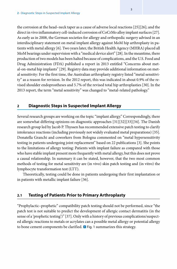
the corrosion at the head–neck taper as a cause of adverse local reactions [25][26]; and the direct in vivo inflammatory cell-induced corrosion of CoCrMo alloy implant surfaces [27]. As early as in 2008, the German societies for allergy and orthopedic surgery advised in an interdisciplinary statement on metal implant allergy against MoM hip arthroplasty in pa-tients with metal allergy [6]. Two years later, the British Health Agency (MHRA) placed all MoM bearings under supervision with a “medical device alert” [28]. In the meantime, there production of two models has been halted because of complications, and the U.S. Food and Drug Administration (FDA) published a report in 2013 entitled “Concerns about met-al-on-metal hip implants” [29]. Registry data may provide additional information on met-al sensitivity: For the first time, the Australian arthroplasty registry listed “metal sensitivi-ty” as a reason for revision. In the 2012 report, this was indicated in about 0.9% of the re-vised shoulder endoprostheses and 5.7% of the revised total hip arthroplasties [30]. In the 2013 report, the term “metal sensitivity” was changed to “metal-related pathology.”
2 Diagnostic Steps in Suspected Implant Allergy
Several research groups are working on the topic “implant allergy.” Correspondingly, there are somewhat differing opinions on diagnostic approaches [31][32][33][34]. The Danish research group led by Jacob P. Thyssen has recommended extensive patch testing to clarify intolerance reactions (including previously not widely evaluated metal preparations) [35]. Donatella Granchi and coworkers from Bologna commented on “metal hypersensitivity testing in patients undergoing joint replacement” based on 22 publications [3]. She points to the limitations of allergy testing: Patients with implant failure as compared with those who have stable implant present more frequently with metal allergy, but this does not prove a causal relationship. In summary it can be stated, however, that the two most common methods of testing for metal sensitivity are (in vivo) skin patch testing and (in vitro) the lymphocyte transformation test (LTT).
Theoretically, testing could be done in patients undergoing their first implantation or in patients with metallic implant failure [36].
2.1 Testing of Patients Prior to Primary Arthroplasty
“Prophylactic–prophetic” compatibility patch testing should not be performed, since “the patch test is not suitable to predict the development of allergic contact dermatitis (in the sense of a ‘prophetic testing’)” [37]. Only with a history of previous complications/suspect-ed allergic reactions to metals or acrylates can a possible metal allergy or potential allergy to bone cement components be clarified. . Fig. 1 summarizes this strategy.

Metal Implant Allergy and Immuno-Allergological Compatibility Aspects of Ceramic Materials4
2.2 Testing of Patients with Failed Arthroplasty
After exclusion of the most frequent differential diagnoses (such as infection) by the super-vising orthopedic surgeon, but also by the dermatologist in the case of skin eruptions (such as psoriasis), the patch test is performed. Histology can help characterize peri-implant in-flammation – in particular to exclude infection. A T-cell metal sensitivity can also be inves-tigated using LTT. This is, however, still restricted to scientific laboratories that evaluate the results critically case by case for clinical relevance [6]. . Fig. 2 presents an algorithm of the diagnostic steps. The proper evaluation of potential allergy begins with a detailed history (e.g., intolerance reaction to metallic items, to acrylate-based dental materials).
2.3 Patch Testing
The standard testing series includes nickel, chromium, and cobalt preparations, which are common implant components. There is still no official consensus on bone cement testing. The bone cement testing series of the Munich allergy group encompasses: gentamicin sulfate, benzoyl peroxide, hydroquinone, 2-hydroxyethylmethacrylate, copper-(II)-sulfate, methyl methacrylate, and N, N-dimethyl-p-toluidine. Additional metal preparations are available, but not yet standardized – and their use should be critically decided case by case [18]. It is useful to also perform a late patch test reading after 1 week. Even when a positive reaction is found, it is mandatory that the clinical relevance of the test results be evaluated.
Before arthroplasty
yes
Allergy yes Allergy no
no
No (prophetic) allergy test
Consider “hypoallergenic”implant material
“Standard” implant material
History of hypersensitivity to metalsor potential bone cement
components
Allergy (patch) test with metalsand bone cement components
. Fig. 1 Flowchart for the evaluation of potential allergy prior to primary arthroplasty (for details, see text)

2 · Diagnostic Steps in Suspected Implant Allergy5
2.4 Histology
A consensus classification [38] describes four types of reactions of periprosthetic mem-branes: a foreign-body-like response (type I), a granulocyte-dominated infectious type (type II), a mixture of type I and II (combined type, type III), and a paucicellular fibrotic reaction (type IV, indifferent type). This has recently been completed by a particle algo-rithm by Veit Krenn et al. showing various reaction patterns also in relation to a compo-sition of particles [39] (see loose insert). Periprosthetic lymphocytic infiltrates might indi-cate hyper-reactivity, but histological characteristics of metal allergy-induced peripros-thetic hypersensitivity are still to be defined. The analysis of the local cytokine pattern might further clarify this issue [40]. Thus, different peri-implant inflammation patterns are described apart from a foreign-body reaction: particle-induced inflammasome activa-tion [38], lymphocytic inflammation [38][40], and differential lymphocytic activation patterns [41].
2.5 Lymphocyte Transformation Test
This scientific test uses the antigen-induced (T cell) proliferation in relation to the baseline proliferation of unstimulated cultures and gives a stimulation index (SI) as a read-out. We have, like other laboratory groups, set the detection limit for sensitization at SI >3 [42] and give interpretation only in view of other diagnostic parameters. If critically evaluated, the LTT is a complementary method, for example, when investigating a suspected allergic drug reaction [43]. However, LTT sensitization does not necessarily mean disease-causing hy-persensitivity [44][45]. Even for the predominant “allergen” nickel, quality assessments of LTT procedures are very rare [44][45]. Accordingly, the Robert Koch Institute does not generally recommend the LTT [44]. On the other hand, several research groups are using LTT to assess “metal sensitization” in arthroplasty patients [31][46]. Future development steps [47] and a follow-up study with evaluation of the clinical relevance of LTT results can lead to LTT optimization.
Take-Home-Message
5 “Prophylactic” patch testing of implant compatibility in patients prior to primary arthroplasty should not be performed 5 Combined evaluation of medical history, clinical findings, patch testing, and histology 5 Patch testing in patients with failed arthroplasty after exclusion of the differential diagnoses 5 Histology is recommended to characterize peri-implant lymphocytic inflammation 5 LLT must be carefully interpreted in the context of additional information

Metal Implant Allergy and Immuno-Allergological Compatibility Aspects of Ceramic Materials6
3 Immuno-allergological Properties of Alumina Ceramics In Vitro and In Vivo
To assess the immuno-allergological properties of alumina ceramic materials (Biolox®-
forte), in vitro and in vivo studies were performed in the Clinic for Dermatology and Al-lergology of the Ludwig Maximilians University, Munich [48]. The aim was to evaluate whether: 4 There was any (allergic) patch test reaction to ceramic material in individuals with other allergies (e.g., also to metals) 4 The presence of ceramic discs would alter in vitro proliferative and cytokine response of human peripheral blood mononuclear cells in a cell culture model
Patch testing was done in a consecutive series of 250 patients visiting the clinic. The patch test was applied on the patient’s back using a European standard series of 30 allergens (Hermal, Reinbek, Germany) supplemented by a sterilized ceramic disc (Al2O3-Biolox®-
forte, CeramTec, Plochingen, Germany). The reactions were evaluated according to stand-ard procedure on days 3 and 4 [37]. Nickel was the most frequent allergen with 17.2% positive reactions. None of the patients reacted to the Biolox®forte discs. The results are summarized in . Tab. 1.
. Fig. 2 Flowchart for the evaluation of potential allergy in arthroplasty with complications (for details, see text)
Arthroplasty with complications
no/suspicion of allergy(after exclusion of infection)
Allergy yes
Allergyno
suggestive ofhyper-
sensitivity
otherpathology positive
yes
Plan treatment accordinglyAllergy diagnostics
Patch test Histology
Integrate history, clinical picture and diagnostic findings to verify potential allergy
Detection of “conventional” elicitors
LTT**) Lymphocyte transformation test can be performed in parallel.Until now for scientific purposes,clinical significance has to be evaluated case by case

73 · Immuno-allergological Properties of Alumina Ceramics In Vitro and In Vivo
. Tab. 1 Patch test reaction frequency in 250 consecutive individuals
Test substance Reactions/250 patients (%)
Nickel(II)-sulfate x 6 H2O 43 17.2
Fragrance mix 38 15.2
Balsam of Peru 33 13.2
Thimerosal 26 10.4
Lanolin alcohol 20 8
Mercury(II)-amid-chloride 17 6.8
p-Phenylenediamine 15 6
Dibromodicyanobutane, phenoxyethanol 11 4.4
Colophonium 10 4
Potassium dichromate 6 2.4
Bufexamac 6 2.4
Neomycin sulfate 5 2
Thiuram mix 5 2
Cobalt(II)-chloride 5 2
(Chlor)-Methylisothiazolinone (Kathon CG in water) 4 1.6
Cocamidopropyl betaine (in water) 4 1.6
Benzocaine 3 1.2
N-Isopropyl-N-phenyl-p-phenylenediamine 3 1.2
Sesquiterpene lactone mix 3 1.2
p-tert-Butylphenol-formaldehyde resin 2 0.8
Formaldehyde 2 0.8
Propolis 2 0.8
Turpentine 2 0.8
Mercapto mix 1 0.4
Epoxide resin 1 0.4
Cetyl stearyl alcohol 1 0.4
Paraben mix 1 0.4
Toluenesulfonamide-formaldehyde resin 1 0.4
Zinc diethyldithiocarbamate – –
Vaselinum album – –
Biolox®forte disc – –

Metal Implant Allergy and Immuno-Allergological Compatibility Aspects of Ceramic Materials8
The in vitro experiments consisted of a lymphocyte proliferation assay and the analysis of the interferon (IFN)-γ and interleukin (IL)-4 cytokine production after stimulation of peripheral blood mononuclear cells (PBMC). For this purpose, heparinized blood of 15 nonallergic and 15 nickel-allergic individuals (22 women, 8 men, 24–70 years) was ob-tained. After isolation of PBMC by density centrifugation, cells were cultured with the pan T-cell mitogen phytohemagglutinin (PHA) at 2.4 µg/ml, NiSO4 at 10-4M, 10-5M, and 10-
6M, Al2O3 ceramic discs, and culture medium alone as control. For the proliferation assay, cells were pulsed after 5 days with 3H thymidine and the proliferation was assessed by in-corporated radioactivity and expressed as SI. IFN-γ and IL-4 production in the superna-tants of stimulated and unstimulated cultures was analyzed by ELISA (antibodies from PharMingen, San Diego, Calif.).
PBMC of nickel-allergic blood donors showed enhanced proliferative response to NiSO4 10-4M. The proliferative response was only marginally influenced by the ceramic Biolox®forte discs (. Fig. 3).
The presence of Biolox®forte discs did not have an overall influence on IL-4 production in the cell cultures (. Fig. 4).
. Fig. 3 Proliferative response (expressed as stimulation index) of peripheral blood mononuclear cells from 15 nickel-allergic and 15 nonallergic blood donors. Statistics: t test. Medium culture medium, NiSO4 nickel sul-fate, PHA phytohemagglutinin, SI stimulation index, +ceramic additional presence of Al2O3 (Biolox®forte) disc
Stim
ulat
ion
inde
x [S
I]
1000 –
100 –
10 –
1 –
0,1 –Medium Medium+
ceramicPHA PHA+
ceramicNiSO4 10-4M
NiSO4 10-4M+ceramic
NiSO4 10-5M
NiSO4 10-5M+ceramic
NiSO4 10-6M
NiSO4 10-6M+ceramic
Ni Patch test pos
Ni Patch test neg

93 · Immuno-allergological Properties of Alumina Ceramics In Vitro and In Vivo
The nonallergic blood donors showed a slight increase in IFN-γ production in the presence of ceramic discs in the medium culture. On the other hand, the IFN-γ production was not altered after stimulation with PHA or NiSO4 (. Fig. 5).
These data show that despite a high rate of delayed-type hypersensitivity reactions to standard allergens, none of the patients had a patch test reaction to Al2O3 ceramic. Also, the cytokine response – which was analyzed by the assessment of IFN-γ and IL-4 produc-tion in vitro – was not influenced by the presence of Al2O3 discs. In summary, the results of this study gave no indication of an impaired or altered immuno-allergological compat-ibility of the Biolox®forte ceramic material.
Take-Home-Message
Alumina Ceramic (Biolox®forte)
5 No patch test reaction 5 No overall influence of cytokine response 5 No indication of impaired or altered immuno-allergological compatibility
. Fig. 4 Interleukin (IL)-4 production (pg/ml) of peripheral blood mononuclear cells from 15 nickel-allergic and 15 nonallergic blood donors. Statistics: t test. Medium culture medium, NiSO4 nickel sulfate, PHA phytohem-agglutinin, SI stimulation index, +ceramic additional presence of Al2O3 (Biolox® forte) disc
IL-4
pro
duct
ion
[pg/
ml]
25 –
20 –
15 –
5 –
10 –
0 –Medium Medium+
ceramicPHA PHA+
ceramicNiSO4 10-4M
NiSO4 10-4M+ceramic
NiSO4 10-5M
NiSO4 10-5M+ceramic
NiSO4 10-6M
NiSO4 10-6M+ceramic
Ni ECT pos
Ni ECT neg

Metal Implant Allergy and Immuno-Allergological Compatibility Aspects of Ceramic Materials10
4 Immuno-allergological Compatibility of Ceramic Composite Material
4.1 Ceramic Composite Material and Alumina Ceramics
A subsequent series of experiments aimed to investigate the immuno-allergological prop-erties of a ceramic composite material (Biolox®delta). This study was done because Biolox®delta contains chromium, which is a potential elicitor of allergic reactions. Thus, the following questions were posed: 4 Will there be a positive (allergic) patch test reaction to chromium-containing ceramic material in individuals with metal allergy? 4 Do human lymphocytes containing cell cultures show enhanced reactivity in the presence of Biolox®delta discs? 4 Is there a release of chromium from Biolox®delta discs when they are kept for 5 days under different “elution” conditions?
. Fig. 5 Interferon (IFN)-γ production (pg/ml) of peripheral blood mononuclear cells from 15 nickel-allergic and 15 nonallergic blood donors. Statistics: t test. Medium culture medium, NiSO4 nickel sulfate, PHA phyto-hemagglutinin, SI stimulation index, +ceramic additional presence of Al2O3 (Biolox® forte) disc
IFN
g pr
oduc
tion
[pg/
ml]
1000 –
100 –
10 –
1 –
0,1 –Medium Medium+
ceramicPHA PHA+
ceramicNiSO4 10-4M
NiSO4 10-4M+ceramic
NiSO4 10-5M
NiSO4 10-5M+ceramic
NiSO4 10-6M
NiSO4 10-6M+ceramic
Ni ECT pos
Ni ECT neg

114 · Immuno-allergological Compatibility of Ceramic Composite Material
Patch testing was done in a consecutive series of 200 patients visiting the clinic. The patch test was applied on the patient’s back using a European standard series of 30 allergens (Hermal, Reinbek, Germany) supplemented by a sterilized ceramic disc (Biolox®delta, CeramTec, Plochingen, Germany). The reactions were evaluated according to a standard procedure on days 3 and 4 [37]. Nickel was the most frequent allergen with 14.5% positive reactions. None of the patients reacted to the Biolox®delta discs. The results are summa-rized in . Tab. 2.
. Tab. 2 Positive patch test reactions in 200 consecutive individuals
Contact allergen Reacting patients (%)
Nickel(II)-sulfate 29 14.5
Cobalt(II)-chloride 15 7.5
Thiomersal 14 7
Fragrance mix 12 6
Balsam of Peru 12 6
Potassium dichromate 10 5
Dibromodicyanobutane, phenoxyethanol 9 4.5
p-Phenylenediamine 8 4
Colophonium 6 3
Neomycin sulfate 6 3
Thiuram mix 6 3
Mercury(II)-amid-chloride 5 2.5
Benzocaine 4 2
Formaldehyde 4 2
Paraben mix 4 2
Lanolin alcohol 4 2
Composite mix 3 1.5
Amerchol L-101 3 1.5
Bufexamac 2 1
Cocamidopropyl betaine 2 1
Mercapto mix 2 1
Propolis 2 1
Biolox®delta discs 0 0

Metal Implant Allergy and Immuno-Allergological Compatibility Aspects of Ceramic Materials12
Take-Home-Message
Biolox®delta
5 No patch test reaction
For the lymphocyte proliferation assay, heparinized blood was obtained from 30 individu-als (7 women, 23 men), of whom 10 were nonallergic and 20 had nickel or chromium al-lergy. After isolation of PBMC by density centrifugation, cells were cultured in two parallel sets of experiments with/without the presence of ceramic discs and the following stimuli: pan T-cell mitogen PHA, NiSO4, CrCl3, CoCl2, and culture medium alone as control. For the proliferation assay, cells were pulsed after 5 days with 3H thymidine and the prolifera-tion was assessed by incorporated radioactivity and expressed as SI. . Fig. 6 shows that the presence of neither the Biolox®forte discs nor the Biolox®delta discs had stimulatory effects.
Take-Home-Message
5 Lymphocyte proliferation: Biolox®delta and Biolox®forte showed no stimulatory effects
. Fig. 6 Lymphocyte proliferative response of metal-allergic and nonallergic blood donors after stimulation with the control stimuli phytohemagglutinin (PHA) and tetanus toxoid (TT) and the metal salts NiSO4, CrCl2, and CoCl3 in different concentrations and the ceramic materials Biolox®delta and forte. Proliferation is given as a stimulation index (SI)
Stim
ulat
ion
inde
x [S
I]
20 –
45 –
20 –
0 –
5 –
10 –
15 –
50 –
25 –
30 –
35 –
PHA TT BioloxDelta
BioloxForte
CoCl310-6 M
CoCl310-7 M
NiSO4 10-5 M
NiSO4 10-4 M
NiSO4 10-6 M
CrCl210-4 M
CrCl210-5 M
CrCl210-6 M
CoCl310-5 M
metal-allergic
non-allergic

134 · Immuno-allergological Compatibility of Ceramic Composite Material
We next assessed the potential chromium release from Biolox®delta discs. In 24-well culture plates, six discs each were kept for 5 days at 37°C in the presence of 1-ml eluent. The eluates were then pooled and the experiment was repeated two more times. The eluents were: 4 Distilled water 4 Artificial sweat 4 Culture medium with 1% human AB serum 4 Culture medium with 10% human AB serum
In a further set of experiments, chromated metallic rings underwent identical elution con-ditions. To assess the potential chromium release, the eluates were first treated by micro-wave digestion with nitric acid. After three-point calibration, atomic absorption spectrom-etry was used to analyze the released chromium (duplicate experiments). As . Fig. 7 shows, the various elution media already contained minute traces of chromium. The different elution conditions did not lead to noteworthy chromium release from both variants of ceramic discs. By contrast, the chromated rings proved to have a high chromium release.
Take-Home-Message
5 No relevant chromium release from Biolox®delta discs
Chro
miu
m c
onte
nt [n
g/m
l] 10 000 –
100 –
0 –
10 –
100 000 –
1000 –
a. d.
medium
10%AB
medium
1%AB
artificial sweat
a. d.
medium
10%AB
medium
1%AB
artificial sweat
a. d.
medium
10%AB
medium
1%AB
artificial sweat
medium
10%AB
artificial sweat
Controls (eluent alone) Biolox Delta Biolox Forte chromatedmetal rings
. Fig. 7 Determination of chromium by atomic absorption spectroscopy in eluates (5-day incubation, 37°C, sterile. Medium 1%AB, elution in RPMI1640 medium supplemented with 1% human AB serum; medium 10%AB, elution in RPMI1640 medium supplemented with 10% human AB serum; a.d. elution in distilled water; artificial sweat, elution in artificial sweat

Metal Implant Allergy and Immuno-Allergological Compatibility Aspects of Ceramic Materials14
4.2 Evaluation of Ceramic Composite Material Under Physiologic Conditions In Vivo
The Bologna orthopedic research group at the Rizzoli Institute conducted a study to as-sess the potential in vivo release of chromium ions from ceramic composite material (Biolox®delta) bearings in arthroplasty patients. For this purpose, aliquots of blood, erythrocytes, and urine were analyzed [49]. Biolox®delta is a ceramic composite material used as an alternative to MoM or metal-on-polyethylene coupling. The included chromium is trivalent chromium strongly bound to the alumina lattice. Since its launch, 3.8 million Biolox®delta ball heads and 1.5 million inserts have been implanted around the world [50]. Because many countries decided to adopt several measures regarding the monitoring of chromium and cobalt ion levels in MoM THA owing to the high failure rates, this study was performed to evaluate the potential release of chromium also from ceramic materials in arthroplasty under physiologic conditions in vivo.
The study participants comprised 20 patients with implanted Biolox®delta THA couplings (THA group; 10 women, 10 men; mean age, 59.9 years; mean body weight, 71 kg) and 21 subjects with no implanted prostheses (control group; 7 women, 14 men; mean age, 57.2 years, mean body weight, 75 kg). Fifteen patients of the THA group had a 32-mm ball head and five a 36-mm ball head. The follow-up was performed between 6 and 63 months after the THA. Blood and urine was taken from all participants and the chromium content was analyzed by inductively coupled plasma mass spectrometry (ICP-MS, ELAN DRCII, Perkin Elmer, Waltham, Mass.) in the blood, serum, isolated erythrocytes, and urine.
The reference values of chromium in the general population (as internal reference range of the laboratory) were: 0.1–5.0 µg/l for blood, 0.1–0.5 µg/l for serum, 0.14–4.58 µg/l for erythrocytes, and <0.05–2.2 µg/l for urine. When comparing the individuals in this study with/without arthroplasty, the mean values of chromium in all samples were not significantly different in the two groups. In both groups, all patients had chromium levels below the upper reference range for the general population. The mean values are given in . Tab. 3 and are shown in . Fig. 8.
In summary, the results of the study show that patch testing with the Biolox®delta THA coupling did not detect elevated chromium levels in vivo. Furthermore, the respective chromium levels in vivo are comparable to healthy individuals without arthroplasty. Thus,
. Tab. 3 Mean values of chromium levels in the four different matrices (adapted from [49], with friendly permission by Hip International)
Group Blood (µg/l)Mean (SD)
Serum (µg/l)Mean (SD)
Erythro-cytes (µg/l)Mean (SD)
Urine (µg/l)Mean (SD)
Urine normalized (µg/g creatinine)Mean (SD)
Patients (n=20) 0.21 (0.09) 0.21 (0.12) 0.13 (0.09) 0.14 (0.18) 0.12 (0.13)
Controls (n=21) 0.22 (0.14) 0.17 (0.08) 0.13 (0.09) 0.10 (0.12) 0.07 (0.08)

154 · Immuno-allergological Compatibility of Ceramic Composite Material
0.8
–
0.6
–
0.4
–
0.2
–
0 –
µg/l
Chro
miu
m c
once
ntra
tion
in w
hole
blo
od5.
0
Patie
nts
Cont
rols
23 o0.
8 –
0.6
–
0.4
–
0.2
–
0 –
µg/l
Chro
miu
m c
once
ntra
tion
in e
ryth
rocy
tes 4.
5
Patie
nts
Cont
rols
28 o
0.8
–
1.0–
0.6
–
0.4
–
0.2
–
0 –
µg/l
Chro
miu
m c
once
ntra
tion
in s
erum
5.0
Patie
nts
Cont
rols
0.8
–
0.6
–
0.4
–
0.2
–
0 –
µg/l
Chro
miu
m c
once
ntra
tion
in u
rine
2.2
Patie
nts
Cont
rols
10 *
41 *
0.8
–
1.0–
0.6
–
0.4
–
0.2
–
0 –
µg/l
Chro
miu
m c
once
ntra
tion
in u
rine
(nor
mal
ized
)
Patie
nts
Cont
rols
10 *
41 *32 o
32 o
.
Fig.
8 B
oxpl
ot re
pres
entin
g th
e di
strib
utio
n of
dat
a co
llect
ed. B
oxes
are
lim
ited
by v
alue
s of
the
25th
and
75t
h pe
rcen
tile.
Hor
izon
tal l
ine
cros
sing
the
box
repr
esen
ts th
e m
edia
n va
lue,
whi
le v
ertic
al li
nes
exte
nd fr
om m
inim
um to
max
imum
val
ues.
The
out
liers
are
sho
wn
with
* o
r °, d
epen
ding
on
the
dist
ance
fr
om th
e 25
th a
nd 7
5th
perc
entil
e (g
reat
er th
an 3
inte
rqua
rtile
diff
eren
ces
or g
reat
er th
an 1
.5) a
nd th
e nu
mbe
r clo
se to
the
sym
bol i
dent
ifies
the
sam
ple.
D
otte
d lin
es re
pres
ent h
ighe
r ref
eren
ce v
alue
. Fro
m [4
9], w
ith fr
iend
ly p
erm
issi
on b
y H
ip In
tern
atio
nal

Metal Implant Allergy and Immuno-Allergological Compatibility Aspects of Ceramic Materials16
it can be concluded that no relevant chromium release from Biolox®delta couplings was observed. The explanation is that because of the chemical structure of Biolox®delta with its strong ionic interatomic bonding a release of chromium ions is unlikely to happen in all four matrices. These results confirm the safety of Biolox®delta materials concerning the release of chromium ions and the possibility of using it as an excellent alternative material to metal-based bearing couples.
Take-Home-Message
Ceramic Composite Material (Biolox®delta)
5 No relevant chromium release 5 Safe with regard to chromium ion release 5 Excellent alternative material to metal-based bearing couplings
5 Use of Alternative Materials in Patients with Allergy to Implant Material and Adverse Reactions to Metal Debris
Implantable metallic devices have become part of therapeutic routine. In some patients with complications, allergy to implant components (metals, bone cement) can be detected. Thus, concerns regarding the risk of allergy-related implant failure have stimulated the interest in alternative “hypoallergenic” materials. There is growing evidence that in patients with metal sensitivity and proven orthopedic dermatitis, peri-implant inflammation, or adverse reactions to metal debris, good results can be achieved by revision with nonmetal-lic components. In addition, surface-coated or zirconium and titanium–niobium primary implants are available as alternatives for allergic patients. The hypersensitivity/immune reactions associated with MoM bearings are only partly defined, but it is agreed that they reflect metal intolerance in such patients. Correspondingly, in patients with complications and adverse reactions to metal debris, the treatment and revision including the use of a non-MoM bearing couple (often ceramic on ceramic) led to resolution of symptoms [51]–[77]. In patients with complications after knee arthroplasty, allergy to metals and bone cement components can be found more frequently than in symptom-free individuals [17]. . Fig. 9 shows a patient with a knee arthroplasty and local dermatitis associated with nickel sensitization.
It is apparent from the literature, despite the low number of clinical studies, that good results and symptom relief can be obtained by revision with “hypoallergenic” materials. Dietrich et al. reported on a series of metal-allergic knee arthroplasty patients, whose symptoms disappeared upon revision with titanium-based arthroplasty [78]. Thomsen and coauthors describe a chromate-allergic patient, in whom local eczema and pain disap-peared when the identical but surface-coated knee arthroplasty was used for revision [79]. Bergschmidt et al. presented a case involving the revision of total knee arthroplasty using a ceramic femoral component. This approach was chosen owing to nickel allergy and con-comitant lymphoplasmacellular peri-implant inflammation [80]. Furthermore, in a case of

176 · Conclusion
posttraumatic osteoarthritis, a TKA with a ceramic femoral component was performed successfully. The patient also suffered from arthrofibrosis together with a proven nickel allergy [81].
In another study, the clinical impact of metal allergy on implant performance was assessed by a combined approach of patch testing, peri-implant histology, and peripros-thetic cytokine assessment. The connection between metal sensitivity and the outcome of the “hypoallergenic” revision implant could be demonstrated by the patients’ markedly improved WOMAC score [40]. It is to be assumed that as the number of patients with re-vision using “hypoallergenic” implants increases, it will provide larger series for follow-up studies and enable a better characterization of implant intolerance reactions.
Take-Home-Message
5 Good results and symptom relief by revision with “hypoallergenic” materials
6 Conclusion
The umbrella term “adverse reaction” encompasses a series of conditions, of which metal implant allergy represents the hypersensitivity type of immune reaction. The diagnosis of “implant allergy” – after exclusion of other problem elicitors such as infection (. Fig. 10)
. Fig. 9 Patient with knee arthroplasty and local dermatitis associated with nickel sensitization

Metal Implant Allergy and Immuno-Allergological Compatibility Aspects of Ceramic Materials18
– results from the synopsis of several diagnostic steps including medical history, clinical findings, patch testing, analysis of peri-implant tissue, and LTT. Alternatives for metal-sen-sitive patients include nonmetallic components, surface-coated, or zirconium and titani-um–niobium primary implants. The immuno-allergological compatibility of ceramic ma-terials in arthroplasty has been shown by clinical patient data and by in vitro and in vivo experiments, including safety assessment in terms of chromium ion release.
References
[1] Schäfer T, Bohler E, Ruhdorfer S, Weigl L, Wessner D, Filipiak B, et al (2001) Epidemiology of contact allergy in adults. Allergy 56(12):1192–1196
[2] Basko-Plluska JL, Thyssen JP, Schalock PC (2011) Cutaneous and systemic hypersensitivity reactions to metal-lic implants. Dermatitis 22(2):65–79
[3] Granchi D, Cenni E, Giunti A, Baldini N (2012) Metal hypersensitivity testing in patients undergoing joint replacement: a systematic review. J Bone Joint Surg Br 94(8):1126–1134
[4] Hallab N, Merritt K, Jacobs JJ (2001) Metal sensitivity in patients with orthopaedic implants. J Bone Joint Surg Am 83A(3):428–436
[5] Hallab NJ, Anderson S, Stafford T, Glant T, Jacobs JJ (2005) Lymphocyte responses in patients with total hip arthroplasty. J Orthop Res 23(2):384–391
[6] Thomas P, Schuh A, Ring J, Thomsen M (2008) Orthopedic surgical implants and allergies: joint statement by the implant allergy working group (AK 20) of the DGOOC (German association of orthopedics and ortho-pedic surgery), DKG (German contact dermatitis research group) and DGAKI (German society for allergology and clinical immunology). Orthopäde 37(1):75–88
[7] Thyssen JP, Johansen JD, Menne T, Liden C, Bruze M, White IR (2010) Hypersensitivity reactions from metallic implants: a future challenge that needs to be addressed. British J Dermatol 162(2):235–236
. Fig. 10 Skin reaction associated with hip arthroplasty: allergy or infection?

19References
[8] Wengler A, Nimptsch U, Mansky T (2014) Hip and knee replacement in Germany and the USA – analysis of individual inpatient data from German and US hospitals for the years 2005 to 2011. Ärzteblatt Int 111 (23–24):9
[9] Foussereau J, Laugier P (1966) Allergic eczemas from metallic foreign bodies. Trans St John’s Hosp Dermatol Soc 52(2):220–225
[10] Carboni GP, Contri P, Pilla G, Vasina PG (1994) Dermatitis associated with orthopaedic prostheses and internal fixations. Contact Dermatitis 31(3):197–198
[11] Kanerva L, Forstrom L (2001) Allergic nickel and chromate hand dermatitis induced by orthopaedic metal implant. Contact Dermatitis 44(2):103–104
[12] Kubba R, Taylor JS, Marks KE (1981) Cutaneous complications of orthopedic implants. A two-year prospective study. Arch Dermatol 117(9):554–560
[13] Rostoker G, Robin J, Binet O, Blamoutier J, Paupe J, Lessana-Leibowitch M, et al (1987) Dermatitis due to orthopaedic implants. A review of the literature and report of three cases. J Bone Joint Surg Am 69(9): 1408–1412
[14] Thomas RH, Rademaker M, Goddard NJ, Munro DD (1987) Severe eczema of the hands due to an orthopaedic plate made of Vitallium. Br Med J (Clin Res Ed) 294(6564):106–107
[15] Eben R, Walk R, Summer B, Maier S, Thomsen M, Thomas P (2009) Implant allergy register – a first report. Orthopäde 38(6):557–562
[16] Granchi D, Cenni E, Tigani D, Trisolino G, Baldini N, Giunti A (2008) Sensitivity to implant materials in patients with total knee arthroplasties. Biomaterials 29(10):1494–1500
[17] Thomas P, Stauner K, Schraml A, Mahler V, Banke IJ, Gollwitzer H, et al (2013) Characteristics of 200 patients with suspected implant allergy compared to 100 symptom-free arthroplasty patients. Orthopäde 42(8): 607–613
[18] Thomas P, Braathen LR, Dorig M, Aubock J, Nestle F, Werfel T, et al (2009) Increased metal allergy in patients with failed metal-on-metal hip arthroplasty and peri-implant T-lymphocytic inflammation. Allergy 64(8):1157–1165
[19] Gawkrodger DJ (1993) Nickel sensitivity and the implantation of orthopaedic prostheses. Contact Dermatitis 28(5):257–259
[20] Thienpont E, Berger Y (2013) No allergic reaction after TKA in a chrome-cobalt-nickel-sensitive patient: case report and review of the literature. Knee Surg, Sports Traumatol, Arthrosc 21(3):636–640
[21] Sharkey PF, Hozack WJ, Rothman RH, Shastri S, Jacoby SM (2002) Insall Award paper. Why are total knee arthroplasties failing today? Clin Orthop Relat Res 404:7–13
[22] Fitzgerald SJ, Trousdale RT (2011) Why knees fail in 2011: patient, surgeon, or device? Orthopedics 34(9):e513–515
[23] Goodman SB, Gibon E, Yao Z (2013) The basic science of periprosthetic osteolysis. Instructional course lectures. 62:201–206
[24] Pandit H, Vlychou M, Whitwell D, Crook D, Luqmani R, Ostlere S, et al (2008) Necrotic granulomatous pseudo-tumours in bilateral resurfacing hip arthoplasties: evidence for a type IV immune response. Virchows Archiv 453(5):529–534
[25] Cooper HJ, Della Valle CJ, Berger RA, Tetreault M, Paprosky WG, Sporer SM, et al (2012) Corrosion at the head-neck taper as a cause for adverse local tissue reactions after total hip arthroplasty. J Bone Joint Surg Am 94(18):1655–1561
[26] Gill IP, Webb J, Sloan K, Beaver RJ (2012) Corrosion at the neck-stem junction as a cause of metal ion release and pseudotumour formation. J Bone Joint Surg Br 94(7):895–900
[27] Gilbert JL, Sivan S, Liu Y, Kocagoz SB, Arnholt CM, Kurtz SM (2014) Direct in vivo inflammatory cell-induced corrosion of CoCrMo alloy orthopedic implant surfaces. J Biomed Materials Res Part A
[28] Medical Device Alert (2012) Ref: MDA/2012/008[29] US Food and Drug Administration (2013) Concerns about metal-on-metal hip implants. FDA[30] Australian Orthopaedic Association National Joint Replacement Registry (2012) Annual report. AOANJR [31] Amini M, Mayes WH, Tzeng A, Tzeng TH, Saleh KJ, Mihalko WM (2014) Evaluation and management of metal
hypersensitivity in total joint arthroplasty: a systematic review. J Long Term Eff Med Implants 24(1):25–36[32] Geier J, Lessmann H, Becker D, Thomas P (2008) Allergy diagnostics in suspected implant intolerance: practi-
cal approach. A position paper of the German Contact Dermatitis Research Group (DKG). Hautarzt 59(7): 594–597

Metal Implant Allergy and Immuno-Allergological Compatibility Aspects of Ceramic Materials20
[33] Schalock PC (2013) Pragmatism and the evaluation of metal hypersensitivity reactions. Dermatitis 24(3): 104–105
[34] Thomas P (2013) Patch testing and hypersensitivity reactions to metallic implants: still many open questions. Dermatitis 24(3):106–107
[35] Thyssen JP, Menne T, Schalock PC, Taylor JS, Maibach HI (2011) Pragmatic approach to the clinical work-up of patients with putative allergic disease to metallic orthopaedic implants before and after surgery. Br J Derma-tol 164(3):473–478
[36] Thomas P, Summer B, Krenn V, Thomsen M (2013) Allergy diagnostics in suspected metal implant intolerance. Orthopäde 42(8):602–606
[37] Schnuch A, Aberer W, Agathos M, Becker D, Brasch J, Elsner P, et al (2008) Performing patch testing with contact allergens. J Dtsch Dermatol Ges 6(9):770–775
[38] Krenn V, Morawietz L, Kienapfel H, Ascherl R, Matziolis G, Hassenpflug J, et al (2013) Revised consensus classi-fication. Histopathological classification of diseases associated with joint endoprostheses. Z Rheumatol 72(4):383–392
[39] Krenn V, Thomas P, Thomsen M, Kretzer JP, Usbeck S, Scheuber L, et al (2014) Histopathological particle algo-rithm : Particle identification in the synovia and the SLIM. Z Rheumatol 73(7):639–649
[40] Thomas PH, C.v.d., Schopf C, Thomsen M, Frommelt L, Schneider J, Flaig M, Krenn V, Mazoochian F, Summer B (2012) Periimplant histology and cytokine pattern in Metal-allergic Knee arthroplasty patients with improve-ment after revision with hypoallergenic materials. Sem Arthroplasty 23(4):268–272
[41] Dapunt U, Giese T, Prior B, Gaida MM, Hansch GM (2014) Infectious versus non-infectious loosening of im-plants: activation of T lymphocytes differentiates between the two entities. Int Orthop 38(6):1291–1296
[42] Summer B, Paul C, Mazoochian F, Rau C, Thomsen M, Banke I, et al (20110) Nickel (Ni) allergic patients with complications to Ni containing joint replacement show preferential IL-17 type reactivity to Ni. Contact Der-matitis 63(1):15–22
[43] Pichler WJ, Tilch J (2004) The lymphocyte transformation test in the diagnosis of drug hypersensitivity. Allergy 59(8):809–820
[44] Eis DW (2008) Qualitätssicherung beim Lymphozytentransformationstest – Addendum zum LTT-Papier der RKI-Kommission Methoden und Qualitätssicherung in der Umweltmedizin. BGG 51:1070–1076
[45] Ständer STP, Rueff F, Summer B (2014) Comparative assessment of nickel sensitization based on medical history, patch test and lymphocyte transformation test. Allergo J Int23:1
[46] Hallab N (2001) Metal sensitivity in patients with orthopedic implants. J Clin Rheumatol 7(4):215–218 [47] Caicedo MS, Solver E, Coleman L, Hallab NJ (2014) Metal sensitivities among TJA patients with post-operative
pain: indications for multi-metal LTT testing. J Long Term Eff Med Implants 24(1):37–44 [48] Thomas P, Barnstorf S, Summer B, editors. Assessment of immuno-allergological properties of ceramic and
metallic compounds in vitro. Bioceramics in joint arthroplasty. Toni A, Willmann G (ed.), Georg Thieme Verlag 2001:68–74
[49] Beraudi A, Stea S, De Pasquale D, Bordini B, Catalani S, Apostoli P, et al (2014) Metal ion release: also a concern for ceramic-on-ceramic couplings? Hip Int 24(4):321–326
[50] CeramTec GmbH (2014) www.ceramtec.com. Cited[51] Algarni AD, Huk OL, Pelmus M (2012) Metallosis-induced iliopsoas bursal cyst causing venous obstruction
and lower-limb swelling after metal-on-metal THA. Orthopedics 35(12):e1811–1814 [52] Cadossi M, Chiarello E, Savarino L, Mazzotti A, Tedesco G, Greco M, et al (2014) Fast growing pseudotumour in a
hairdresser after metal-on-metal hip resurfacing: a case report. Eur Rev Med Pharmacol Sci 18 [1 Suppl]:29–33[53] Campbell P, Shimmin A, Walter L, Solomon M (2008) Metal sensitivity as a cause of groin pain in met-
al-on-metal hip resurfacing. J Arthroplasty 23(7):1080–1085 [54] Cash DJ, Khanduja V (2014) The case for ceramic-on-polyethylene as the preferred bearing for a young adult
hip replacement. Hip Int 24(5):421–427 [55] De Haan R, Campbell PA, Su EP, De Smet KA (2008) Revision of metal-on-metal resurfacing arthroplasty of the
hip: the influence of malpositioning of the components. J Bone Joint Surg Br 90(9):1158–1163 [56] Harvie P, Giele H, Fang C, Ansorge O, Ostlere S, Gibbons M, et al (2008) The treatment of femoral neuropathy
due to pseudotumour caused by metal-on-metal resurfacing arthroplasty. Hip Int 18(4):313–320 [57] Johnson S, Babatunde OM, Lee J, Macaulay WB (2015) Revision of metal-on-metal surfaces in hip arthroplas-
ty. In: Scudery GR (ed) Techniques in revision hip and knee arthroplasty. Elsevier Saunders; 2015: pp. 558–562

21References
[58] Kawakita K, Shibanuma N, Tei K, Nishiyama T, Kuroda R, Kurosaka M (2013) Leg edema due to a mass in the pelvis after a large-diameter metal-on-metal total hip arthroplasty. J Arthroplasty 28(1):197e1–4
[59] Kemp MA, Mitra A, da Costa TM, Spencer RF (2013) Bearing exchange in the management of pseudotumours. Ann R Coll Surg Engl 95(4):266–270
[60] Kosukegawa I, Nagoya S, Kaya M, Sasaki K, Sasaki M, Yamashita T (2011) Revision total hip arthroplasty due to pain from hypersensitivity to cobalt-chromium in total hip arthroplasty. J Arthroplasty 26(6):978 e1–3
[61] Liddle AD, Satchithananda K, Henckel J, Sabah SA, Vipulendran KV, Lewis A, et al (2013) Revision of met-al-on-metal hip arthroplasty in a tertiary center: a prospective study of 39 hips with between 1 and 4 years of follow-up. Acta Orthop 84(3):237–245
[62] Maurer-Ertl W, Friesenbichler J, Liegl-Atzwanger B, Kuerzl G, Windhager R, Leithner A (2011) Noninflammato-ry pseudotumor simulating venous thrombosis after metal-on-metal hip resurfacing. Orthopedics 34(10):e678–681
[63] Rajpura A, Porter ML, Gambhir AK, Freemont AJ, Board TN (2010) Clinical experience of revision of metal on metal hip arthroplasty for aseptic lymphocyte dominated vasculitis associated lesions (ALVAL). Hip Int 21(1):43–51
[64] Sandiford NA, Muirhead-Allwood SK, Skinner JA (2010) Revision of failed hip resurfacing to total hip arthro-plasty rapidly relieves pain and improves function in the early post operative period. J Orthop Surg Res 5:88
[65] Su EP, Su SL (2011) Surface replacement conversion: ensuring an optimal total hip replacement outcome. Sem Arthroplasty 22(2):4
[66] Watters TS, Cardona DM, Menon KS, Vinson EN, Bolognesi MP, Dodd LG (2010) Aseptic lymphocyte-dominat-ed vasculitis-associated lesion: a clinicopathologic review of an underrecognized cause of prosthetic failure. Am J Clin Pathol 134(6):886–693
[67] Amini MH, Mayes WH, Tzeng A, Tzeng TH, Saleh KJ, Mihalko WM (2014) Evaluation and Management of Metal Hypersensitivity in Total Joint Arthroplasty: A Systematic Review. J Long Term Eff Med Implants 24(1):25–36
[68] Bizzotto N, Sandri A, Trivellin G, Magnan B, Micheloni GM, Zamò, Bernardi P, Sbarbati A, Regis D (2014) Chromium-Induced Diffuse Dermatitis and Lymph Node Involvement by Langerhans Cell Histiocytosis After Metal-on-Metal Hip Resurfacing. Br J Deramtol. Nov 10. doi: 10.1111/bjd.13517. [Epub ahead of print]
[69] Cooper HJ, Della CJ, Berger RA, Tetreault M, Paprosky WG, Sporer SM, Jacobs JJ (2012) Corrosion at the Head-Neck Tape ras a Cause for Adverse Local Tissue Reactions After Total Hip Arthroplasty. J Bone Joint Surg Am 94:1655–1661
[70] Garbuz DS, Tanzer M, Greidanus NV, Masri BA, Duncan CP (2010) The John Charnley Award: Metal-on-metal-hip resurfacing versus large-diameter head metal-on-metal total hip arthroplasty: a randomized clinical trial. Clin Orthop Relat Res 468(2):318–325
[71] Gill IPS, Webb J, Sloan K, Beaver RJ (2012) Corrosion at the neck-stem junction as a cause of metal ion release and pseudotumour formation. J Bone Joint Surg Br 94-B:895–900
[72] Kiran M, Boscainos PJ (2015) Adverse Reactions to Metal Debris in Metal-on-Polyethylene Total Hip Arthro-plasty Using a Titanium-Molybdenum-Zirconium-Iron Alloy Stem. J Arthroplasty 30(2):277–281
[73] Mao X, Tay GH, Godbolt DB, Crawford RW (2012) Pseudotumor in a Well-Fixed Metal-on-Polyethylene Uncemented Hip Arthroplasty. J Arthroplasty 27(3):493.e13–e17
[74] Meftah M, Nicolaou N, Rodriguez JA (2010) Metal allergy response to femoral head-neck corrosion after total hip replacement. Curr Orthop Pract 21:530–533
[75] Picardo NE, Al-Khateeb H, Pollock RC (2011) Atypical pseudotumour after metal-on-polyethylene total hip arthroplasty causing deep venous thrombosis. Hip Int 21(06):762–765
[76] Scully WF, Teeny SM (2013) Pseudotumor Associated With Metal-on-Polyethylene Total Hip Arthroplasty. Orthopedics 36(5):e666–e670
[77] Van der Wegen W, Sijbesma T, Hoekstra HJ, Brakel K, Pilot P, Nelissen RGHH (2013) Treatment of pseudo-tumors after metal-on-metal hip resurfacing based on Magnetic Resonance Imaging, metal ion levels and symptoms. J Arthroplasty 29(2):416–421
[78] Dietrich KA, Mazoochian F, Summer B, Reinert M, Ruzicka T, Thomas P (2009) Intolerance reactions to knee arthroplasty in patients with nickel/cobalt allergy and disappearance of symptoms after revision surgery with titanium-based endoprostheses. J Dtsch Dermatol Ges 7(5):410–413

Metal Implant Allergy and Immuno-Allergological Compatibility Aspects of Ceramic Materials22
[79] Thomsen M, Rozak M, Thomas P (2011) Pain in a chromium-allergic patient with total knee arthroplasty: disappearance of symptoms after revision with a special surface-coated TKA–-a case report. Acta Orthop 82(3):386–388
[80] Bergschmidt P, Bader R, Mittelmeier W (2010) Metal hypersensitivity in total knee arthroplasty: revision surgery using a ceramic femoral component – a case report. Knee 19(2):144–1447
[81] Bergschmidt P, Kluess D, Zietz C, Finze S, Bader R, Mittelmeier W (2011) Composite ceramics in total knee arthroplasty: two-year experience in clinical application. Sem Arthroplasty 22(4):264–270


9 7 8 3 6 6 2 4 7 4 3 9 6
isbn 978-3-662-47439-6
Clinical Management of Joint Arthroplasty
The volumes of the Clinical Management Guide series are directed at orthopaedic surgeons who want to acquire information rapidly while saving as much time as possible. As a helpful advisory tool, this Pocket Guide concisely and clearly imparts the current state of knowledge on selected issues of everyday clinical practice and, in doing so, concentrates purely on the essential.
In addition, it also addresses medical practitioners and scientists of adjacent spe-cialist disciplines who are not confronted on a daily basis with problems regarding endoprosthetics but, when required, would like to access important information on a specific topic.
Metal Implant Allergy
The clinical guide is one of a series written for orthopaedic surgeons who seek help in how to proceed when an allergic reaction is suspected to any metal implant component, such as nickel, cobalt, or chromium or to bone cement components. An algorithm is being provided as a laminated insert.