pet-mri: device & clinical applications
TRANSCRIPT
PET/MRI
Device and Clinical Applications
Jiraporn Sriprapaporn
Siriraj Hospital, Bangkok (Last update in December 2016)
Jiraporn_PET/CT Onco 2016
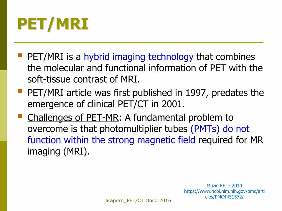
PET/MRI
PET/MRI is a hybrid imaging technology that combines the molecular and functional information of PET with the soft-tissue contrast of MRI.
PET/MRI article was first published in 1997, predates the emergence of clinical PET/CT in 2001.
Challenges of PET-MR: A fundamental problem to overcome is that photomultiplier tubes (PMTs) do not function within the strong magnetic field required for MR imaging (MRI).
Muzic RF Jr 2014 https://www.ncbi.nlm.nih.gov/pmc/arti
cles/PMC4451572/
Jiraporn_PET/CT Onco 2016
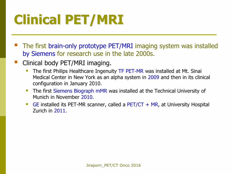
Clinical PET/MRI
The first brain-only prototype PET/MRI imaging system was installed by Siemens for research use in the late 2000s.
Clinical body PET/MRI imaging. The first Philips Healthcare Ingenuity TF PET-MR was installed at Mt. Sinai
Medical Center in New York as an alpha system in 2009 and then in its clinical configuration in January 2010.
The first Siemens Biograph mMR was installed at the Technical University of Munich in November 2010.
GE installed its PET-MR scanner, called a PET/CT + MR, at University Hospital Zurich in 2011.
Jiraporn_PET/CT Onco 2016
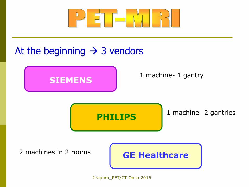
At the beginning 3 vendors
SIEMENS
PHILIPS
GE Healthcare 2 machines in 2 rooms
1 machine- 2 gantries
1 machine- 1 gantry
Jiraporn_PET/CT Onco 2016
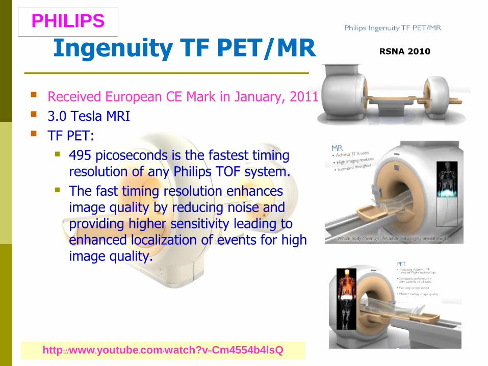
Ingenuity TF PET/MR
Received European CE Mark in January, 2011
3.0 Tesla MRI
TF PET:
495 picoseconds is the fastest timing resolution of any Philips TOF system.
The fast timing resolution enhances image quality by reducing noise and providing higher sensitivity leading to enhanced localization of events for high image quality.
http://www.youtube.com/watch?v=Cm4554b4lsQ
PHILIPS
RSNA 2010
Jiraporn_PET/CT Onco 2016
From Concept to Reality Simon R. Cherry and his colleagues first conceptualized the PET/MRI fusion in 1996 and developed a rudimentary prototype one year later. http://www.radiologytoday.net/archive/rt060208p20.shtml
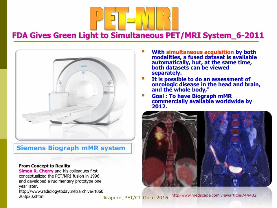
FDA Gives Green Light to Simultaneous PET/MRI System_6-2011
With simultaneous acquisition by both modalities, a fused dataset is available automatically, but, at the same time, both datasets can be viewed separately.
It is possible to do an assessment of oncologic disease in the head and brain, and the whole body,"
Goal : To have Biograph mMR commercially available worldwide by 2012.
Siemens Biograph mMR system
http://www.medscape.com/viewarticle/744402

Jiraporn_PET/CT Onco 2016
SIEMENS:Biograph mMR
The world’s first system to combine MRI & PET in one scanner.
simulataneous scanning the “m” stands for molecular
FDA approval on June 10, 2011
Jiraporn_PET/CT Onco 2016
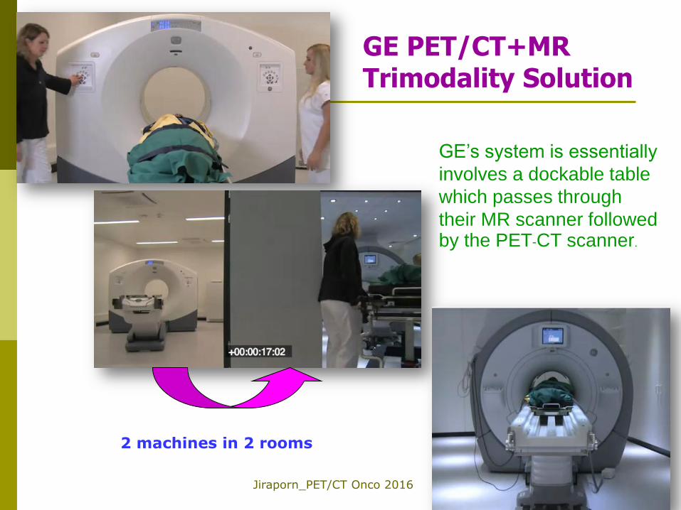
GE PET/CT+MR Trimodality Solution
GE’s system is essentially
involves a dockable table
which passes through
their MR scanner followed by the PET-CT scanner.
2 machines in 2 rooms
Jiraporn_PET/CT Onco 2016
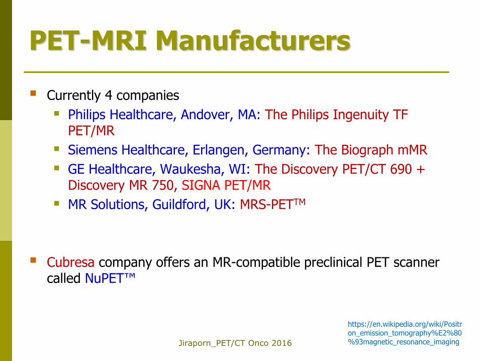
PET-MRI Manufacturers
Currently 4 companies
Philips Healthcare, Andover, MA: The Philips Ingenuity TF PET/MR
Siemens Healthcare, Erlangen, Germany: The Biograph mMR
GE Healthcare, Waukesha, WI: The Discovery PET/CT 690 + Discovery MR 750, SIGNA PET/MR
MR Solutions, Guildford, UK: MRS-PETTM
Cubresa company offers an MR-compatible preclinical PET scanner called NuPET™
https://en.wikipedia.org/wiki/Positron_emission_tomography%E2%80%93magnetic_resonance_imaging
Jiraporn_PET/CT Onco 2016
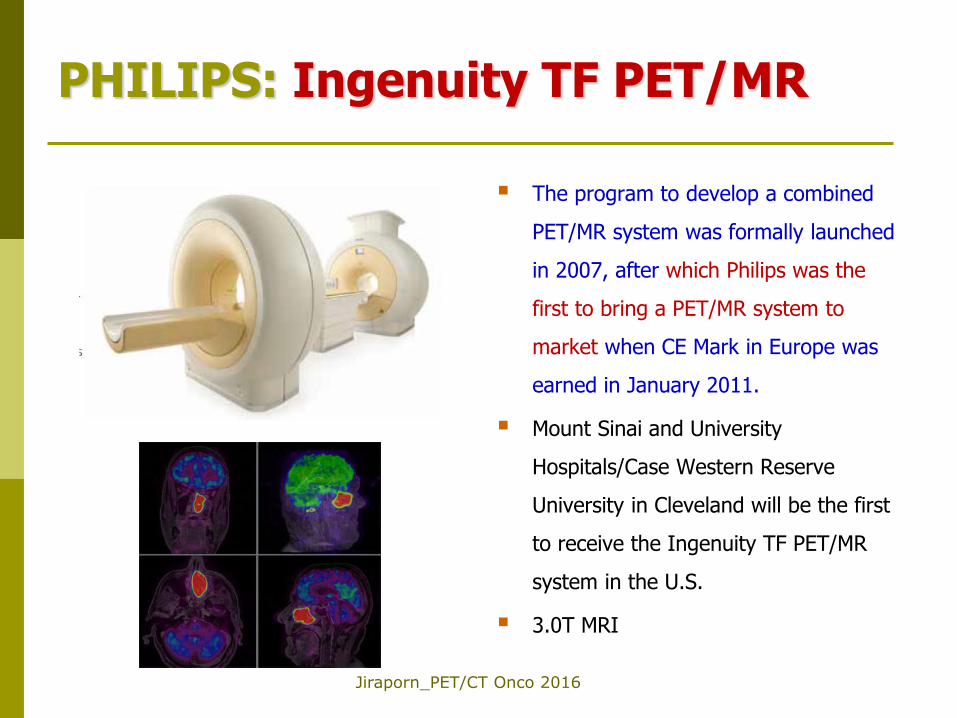
PHILIPS: Ingenuity TF PET/MR
The program to develop a combined
PET/MR system was formally launched
in 2007, after which Philips was the
first to bring a PET/MR system to
market when CE Mark in Europe was
earned in January 2011.
Mount Sinai and University
Hospitals/Case Western Reserve
University in Cleveland will be the first
to receive the Ingenuity TF PET/MR
system in the U.S.
3.0T MRI
Jiraporn_PET/CT Onco 2016
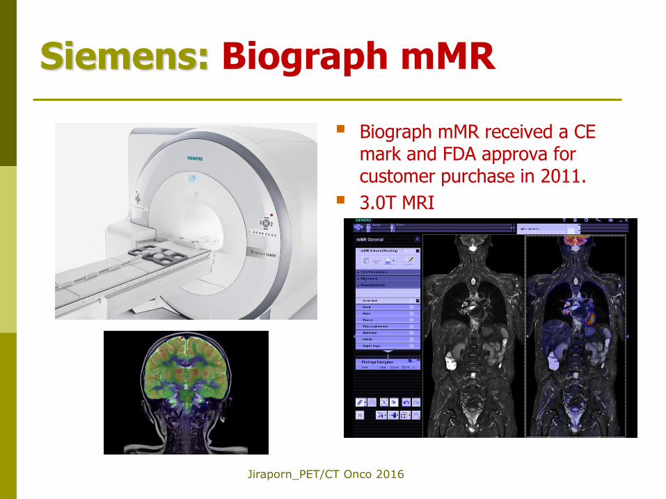
Siemens: Biograph mMR
Biograph mMR received a CE mark and FDA approva for customer purchase in 2011.
3.0T MRI
Jiraporn_PET/CT Onco 2016
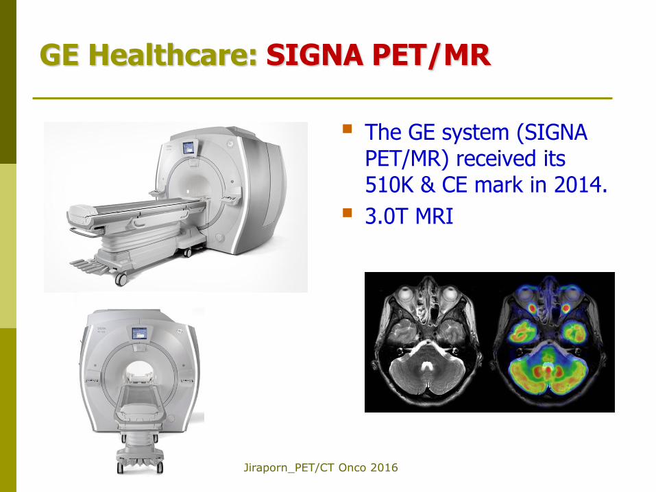
GE Healthcare: SIGNA PET/MR
The GE system (SIGNA PET/MR) received its 510K & CE mark in 2014.
3.0T MRI
Jiraporn_PET/CT Onco 2016
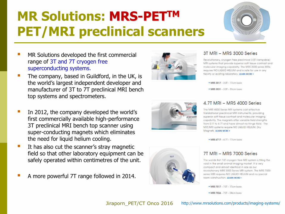
MR Solutions: MRS-PETTM PET/MRI preclinical scanners
MR Solutions developed the first commercial range of 3T and 7T cryogen free superconducting systems.
The company, based in Guildford, in the UK, is the world’s largest independent developer and manufacturer of 3T to 7T preclinical MRI bench top systems and spectrometers.
In 2012, the company developed the world’s first commercially available high-performance 3T preclinical MRI bench top scanner using super-conducting magnets which eliminates the need for liquid helium cooling.
It has also cut the scanner’s stray magnetic field so that other laboratory equipment can be safely operated within centimetres of the unit.
A more powerful 7T range followed in 2014.
http://www.mrsolutions.com/products/imaging-systems/
Jiraporn_PET/CT Onco 2016
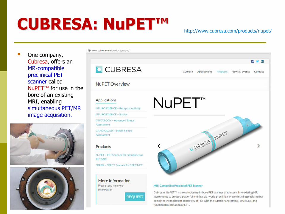
CUBRESA: NuPET™
One company, Cubresa, offers an MR-compatible preclinical PET scanner called NuPET™ for use in the bore of an existing MRI, enabling simultaneous PET/MR image acquisition.
http://www.cubresa.com/products/nupet/
Jiraporn_PET/CT Onco 2016
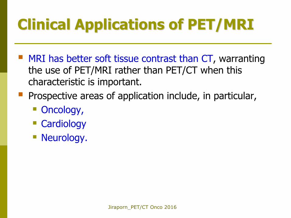
Clinical Applications of PET/MRI
MRI has better soft tissue contrast than CT, warranting the use of PET/MRI rather than PET/CT when this characteristic is important.
Prospective areas of application include, in particular,
Oncology,
Cardiology
Neurology.
Jiraporn_PET/CT Onco 2016
PET/MRI in Oncologic Applications
Pediatric patients and pregnant women
To reduce overall radiation exposure to the patient.
When using PET/MR, the radiation dose from CT is omitted and the actual radiation exposure is limited to the radiation dose from the PET component only (which is substantially minor in comparison to the radiation dose from CT)
Neurologic and head & neck applications
Brain tumors: 11C-methionine or 68Ga-DOTATOC radiopharmaceuticals could be reliably used in PET imaging of intracranial tumors simultaneously with MRI
Head and upper neck cancers : Increased detailed resolution and improved image contrast could be found in the PET images of the PET/MRI system in comparison to the standard PET/CT
The strongest evidence for a clinical indication of PET/MRI exists in the head & neck cancer population.
For local staging the high spatial and contrast resolution of MRI can delineate the tumor extent and lymph node involvement from surrounding normal tissue in the complex head and neck anatomical region.
This may lead to a superior primary tumor staging (T-staging) and regional lymph node staging (N-staging).
Furthermore PET/MRI can be useful for radiation therapy and presurgical treatment planning in head and neck cancer patients.
Partovi S 2014
Jiraporn_PET/CT Onco 2016
PET/MRI in Oncologic Applications
Chest
Superior sulcus tumors: the combination of high spatial resolution MR imaging for the involvement of the brachial plexus and spine
Abdomen and pelvis
Liver lesions
MR has shown a superior sensitivity for the detection of focal liver lesions, especially when <1 cm in size.
PET/MRI an excellent method to screen for metastases (Figure 1) or monitor embolization therapy for liver lesions (PET can provide information on their potentially neoplastic nature)
Liver screening in colorectal cancer reduces patient mortality by 25%.
Pancreatic, biliary and upper gastrointestinal neoplasms
Partovi S 2014
Jiraporn_PET/CT Onco 2016
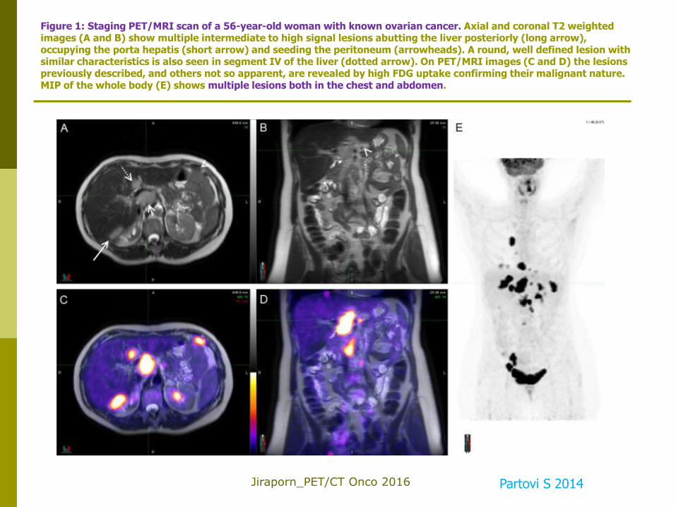
Figure 1: Staging PET/MRI scan of a 56-year-old woman with known ovarian cancer. Axial and coronal T2 weighted images (A and B) show multiple intermediate to high signal lesions abutting the liver posteriorly (long arrow), occupying the porta hepatis (short arrow) and seeding the peritoneum (arrowheads). A round, well defined lesion with similar characteristics is also seen in segment IV of the liver (dotted arrow). On PET/MRI images (C and D) the lesions previously described, and others not so apparent, are revealed by high FDG uptake confirming their malignant nature. MIP of the whole body (E) shows multiple lesions both in the chest and abdomen.
Partovi S 2014
Jiraporn_PET/CT Onco 2016
PET/MRI in Oncologic Applications
Abdomen and pelvis (cont)
Pelvic oncologic diseases such as
Gynecologic malignancies,
Prostate cancer esp. when using C-11 choline, F-18 choline or C-11 acetate
Rectal cancer
Jiraporn_PET/CT Onco 2016
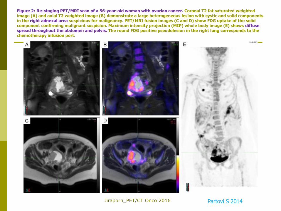
Figure 2: Re-staging PET/MRI scan of a 56-year-old woman with ovarian cancer. Coronal T2 fat saturated weighted image (A) and axial T2 weighted image (B) demonstrate a large heterogeneous lesion with cystic and solid components in the right adnexal area suspicious for malignancy. PET/MRI fusion images (C and D) show FDG uptake of the solid component confirming malignant suspicion. Maximum intensity projection (MIP) whole body image (E) shows diffuse spread throughout the abdomen and pelvis. The round FDG positive pseudolesion in the right lung corresponds to the chemotherapy infusion port.
Partovi S 2014
Jiraporn_PET/CT Onco 2016
PET/MRI in Oncologic Applications
More details on oncologic applications can be found in the article below.
https://www.ncbi.nlm.nih.gov/pmc/articles/PMC4915069/
Jiraporn_PET/CT Onco 2016
PET/MRI in Musculoskeletal Imaging
Neoplastic Conditions
Sarcoma
Osseous metastases
Lymphoma
Multiple myeloma
Non-neoplastic conditions
Infectious and inflammatory conditions
https://www.ncbi.nlm.nih.gov/pmc/articles/PMC4807335/
Jiraporn_PET/CT Onco 2016
PET/MRI for Neurological Applications
Brain Tumors
Dementias/Neurodegeneration
Stroke/Cerebrovascular disorders
Epilepsy
Neurobiology of Brain Activation
Translational Research
https://www.ncbi.nlm.nih.gov/pmc/articles/PMC3806202/

Jiraporn_PET/CT Onco 2016
PET/MRI for Cardiovascular Applications
CAD
Detection of CAD
Myocardial viability imaging
Myocardial infarct
Carotid atherosclerosis
Hereditary, infiltrative and inflammatory cardiovascular diseasessclerosis
Hypertrophic cardiomyopathy (HCM)
Amyloidosis
Cardiac sarcoidosis
https://www.ncbi.nlm.nih.gov/pmc/articles/PMC4929286/
Quant Imaging Med Surg 2016;6(3):297-307