perlecan/hspg2 deficiency alters the pericellular … et...original article perlecan/hspg2...
TRANSCRIPT

Original Article Perlecan/Hspg2 Deficiency Alters the Pericellular Space of the Lacuno-Canalicular System Surrounding Osteocytic Processes in Cortical Bone William R. Thompson1,2, Shannon Modla3, Brian J. Grindel4, Kirk J. Czymmek3,5, Catherine B. Kirn-Safran5, Liyun Wang2,6, Randall L. Duncan2,5,6, Mary C. Farach-Carson4,5 1University of Delaware, Department of Physical Therapy, Newark, DE, 19716 2University of Delaware, Program in Biomechanics and Movement Science, Newark, DE, 19716 3Delaware Biotechnology Institute, Newark, DE, 19716 4Rice University, Department of Biochemistry and Cell Biology, Houston, TX, 77005 5University of Delaware, Department of Biological Sciences, Newark, DE, 19716 6University of Delaware, Department of Mechanical Engineering, Newark, DE, 19716 Corresponding Author: Mary C. Farach-Carson, PhD; Department of Biochemistry and Cell Biology; Rice University, MS-140; 6100 Main Street, Houston, TX 77251-1892 Email: [email protected] Ph.: 713-348-5052 Fax: 713-348-5154 E-mail Addresses: William Thompson: [email protected]; Shannon Modla: [email protected]; Brian Grindel: [email protected]; Kirk Czymmek: [email protected]; Catherine Kirn-Safran: [email protected]; Liyun Wang: [email protected]; Randall Duncan: [email protected]; Mary Farach-Carson: [email protected]
Funding support: This study was supported by the NIH T32 HD07490, Foundation for Physical Therapy Florence Kendall Scholarship, Foundation for Physical Therapy Promotion of Doctoral Studies II training fellowship, and American Physical Therapy Association Section on Geriatrics Adopt-A-Doc Scholarship to WRT, P01 CA098912 to MCFC, P20 RR016458 to MCFC, CKS, and LW. Abstract words/characters: 226/1,782; Manuscript words/characters: 8,118/55,174; Figures (black and white): 3; Figures (color): 3; Tables: 1 Conflict of Interest: All authors have no conflicts of interest. Initial Date Submitted April 30, 2010; Date Revision Submitted July 9, 2010; Date Final Disposition Set August 24, 2010
Journal of Bone and Mineral Research © 2010 American Society for Bone and Mineral Research
DOI 10.1002/jbmr.236

Abstract
Osteocytes project long, slender processes throughout the mineralized matrix
of bone where they connect and communicate with effector cells. The
interconnected cellular projections form the functional lacuno-canalicular system
allowing fluid to pass for cell to cell communication and nutrient and waste
exchange. Prevention of mineralization in the pericellular space of the lacuno-
canalicular pericellular space is crucial for uninhibited interstitial fluid movement.
Factors contributing to the ability of the pericellular space of the lacuno-canalicular
system to remain open and unmineralized are unclear. Immunofluorescence and
immunogold localization by transmission electron microscopy demonstrated
perlecan/HSPG2 signal localized to the osteocyte lacuno-canalicular system of
cortical bone and this proteoglycan was found in the pericellular space of the lacuno-
canalicular system. In this study we examined osteocyte lacuno-canalicular
morphology in mice deficient in the large heparan sulfate proteoglycan
perlecan/HSPG2 in this tissue. Ultrastructural measurements with electron
microscopy of perlecan/HSPG2 deficient mice demonstrated diminished osteocyte
canalicular pericellular area, resulting from a reduction in the total canalicular area.
Additionally, perlecan/HSPG2 deficient mice showed decreased canalicular density
and a reduced number of transverse tethering elements per canaliculus. These data
indicated that perlecan/HSPG2 contributed to the integrity of the osteocyte lacuno-
canalicular system by maintaining the size of the pericellular space, an essential task
to promote uninhibited interstitial fluid movement in this mechanosensitive
environment. This work thus identified a new barrier function for perlecan/HSPG2 in
murine cortical bone.

Keywords: Osteocyte, Perlecan/HSPG2, Mechanosensing, Heparan Sulfate,
Lacuno-Canalicular System, Cortical Bone

Introduction
Bone is a unique, mineralized connective tissue that dynamically adapts its
structure in response to its environment (1). Osteocytes are the most abundant cell
type of bone, accounting for approximately 90% of all bone cells (2) and their primary
function is to sense mechanical load (3). These cells are embedded deep within the
mineralized matrix and connect via long, slender dendrite-like processes encased in
small channels of mineralized matrix called canaliculi (4). Each canaliculus connects
to the cave-like mineralized structure surrounding the osteocyte cell body, which
together make up the functional lacuno-canalicular system (LCS) (4).
The pericellular space between the cell process of the osteocyte and the
mineralized matrix is filled with interstitial fluid and a variety of extracellular matrix
(ECM) molecules (5). The exact role of this space, and the fluid within it, is
incompletely understood; however, this fluid is thought to promote the transport of
waste and nutrients to facilitate metabolism (6). Additionally, fluid flow-induced
shear stress initiates mechanosensory responses from bone cells (5,7).
The dimensions of the LCS have been well characterized by electron
microscopy (8); however, the ability of this system to maintain an open,
unmineralized pericellular space while being deeply embedded in calcified tissue
remains unclear. Inoue and colleagues have demonstrated that matrix
metalloproteinases may help prevent mineral deposition in the LCS (9).
Fluid flow throughout the LCS induces shear stress that stimulates the
dendrite-like processes within canaliculi. The mechanism of action behind this
process is widely debated; however, You and colleagues proposed a strain
amplification model to account for the ability of load-induced fluid shear stress to

mechanically deform the osteocyte cell membrane on the order necessary to initiate
a mechanosensitive response from osteocytes (7). In this model, an
uncharacterized organic matrix filled the pericellular space around the osteocyte
processes. Transverse “tethering elements” were proposed to anchor the cell
processes and center them within the canaliculi (7).
The presence of transverse tethering elements in the LCS, a crucial
component of the proposed model, was further validated by transmission electron
microscopy (TEM) confirming the presence and quantity of the ultrastructural
elements surrounding the osteocyte cell processes. High resolution images showed
transverse tethering elements connecting the osteocyte process to the mineralized
matrix of bone across a pericellular space of approximately 78 nm (8).
Proteoglycans represent a major component of the pericellular material
surrounding osteocyte processes (10) and are thought to comprise the transverse
tethering elements in the LCS (8). The only heparan sulfate proteoglycan with
sufficient size to span the pericellular space of the LCS is perlecan/HSPG2 (PLN)
(11,12). The name perlecan means “string of pearls”, given due to its appearance as
globules separated by rods when imaged by TEM and atomic force microscopy
(AFM) (12,13). PLN is a very large, five domain heparan sulfate proteoglycan with a
core protein of over 4,000 amino acids (14,15). The amino terminus domain I
contains three GAG attachment sites, while the carboxy terminal domain V contains
another variably used putative GAG attachment site (16).
In addition to the size of PLN, various unique features of this molecule
suggest several possible functions in the LCS of osteocytes. PLN is abundantly
secreted into the pericellular space of numerous tissues, particularly near tissue
barriers, and is ideally positioned to mediate signaling events by sequestering growth

factors and binding integrins (12,17-22). PLN and its long heparan sulfate chains
regulate various physiological functions in a variety of tissues where barriers are
required including separating epithelia and stroma, preventing cancer cell invasion
(23,24), maintaining the blood-brain barrier (25), and controlling glomerular filtration
and fluid movement (26-29). Additionally, heparan sulfate inhibits hydroxyapatite
(HAP) formation (30,31).
In addition to recent evidence demonstrating the presence of tethering
elements that span the pericellular space of the LCS, integrins, specifically β3
integrin, are expressed on the membrane of osteocyte processes (32). These
transmembrane proteins have been proposed to form focal attachments directly with
regularly spaced protrusions or “hillocks” of the bone matrix wall within the osteocyte
LCS (32).
Proper maintenance of the pericellular space of the osteocyte LCS is essential
for uninhibited interstitial fluid movement in cortical bone. The purpose of this study
was to determine if the proteoglycan PLN is a component of the transverse tethering
elements, discrete from the direct integrin-“hillock” links, in the pericellular space of
the LCS of osteocytes. In this study, we investigated the concept that PLN is
positioned in the osteocyte LCS where it functions to maintain the structural integrity
of the open fluid-filled, unmineralized pericellular space. In vitro and in vivo cell
systems and a PLN deficient mouse model were used to examine this hypothesis.
Materials and methods
Cell culture
Murine long bone osteocyte cells (MLO-Y4) were a generous gift from Dr.
Lynda Bonewald (University of Missouri-Kansas City, Kansas City, MO). Cells were

cultured in 100 mm tissue culture dishes (Corning, Inc, Corning, NY) coated with rat
tail type I collagen (0.15 mg/ml) (BD Biosciences™, San Jose, CA) as previously
described (33). WiDr human colon carcinoma cells were cultured on T75 cell culture
flasks (Fisher Scientific, Pittsburgh, PA) as previously described (34).
mRNA isolation and PCR assays
MLO-Y4 cells were grown to 80-90% confluence (~6 days in culture) and total
RNA extracts were obtained using the RNeasy kit (Qiagen®, Valencia, CA) with a
typical yield of 400-800 ng/µl. mRNA extracts were DNase treated using the DNA-
free™ DNase kit (Ambion®, Austin, TX) to remove DNA contamination. mRNA was
reverse transcribed using the Omniscript™ reverse transcriptase polymerase chain
reaction (RT-PCR) kit (Qiagen®) according to the manufacturer’s protocol. MLO-Y4
cDNA gene products then were amplified via conventional PCR using GoTaq Green
PCR master mix (Promega, San Luis Obispo, CA) and custom-designed primers
against the murine PLN gene, hspg2. Primers were designed using the PRIMER3
tool in the Biology Workbench online application
(http://seqtool.sdsc.edu/CGI/BW.cgi, University of California, San Diego) from cDNA
sequences published in the NCBI nucleotide database. Sequences were analyzed
for optimal folding, hetero and homo-dimerization patterns using the Integrated DNA
Technologies®, OligoAnalyzer 3.1 tool
(https://www.idtdna.com/analyzer/Applications/OligoAnalyzer/, Coralville, IA) and for
proper folding and dimerization. The NCBI Basic Local Alignment Search Tool
(BLAST) was also used to ensure gene and species specificity
(http://www.ncbi.nlm.nih.gov/BLAST). Primer sequences were as follows, 5’-
CCCACTCTTGGACCCTGATA-3’ and 5’-ATAGCTCCTCCTCTCTGGGC-3’ for
murine hspg2 (NCBI accession # M77174), generating a 94 bp product; 5’-
GATCATTGCTCCTCCTGAGC-3’and 5’-ACATCTGCTGGAAGGTGGAC-3’ for
murine actb (NCBI accession # NM_007393), generating an 83 bp product. Gene
products were visualized on an agarose gel (1.5%, w/v) (Fisher Scientific) using
ethidium bromide (4x10⁻⁷%, v/v) (Fisher Scientific).
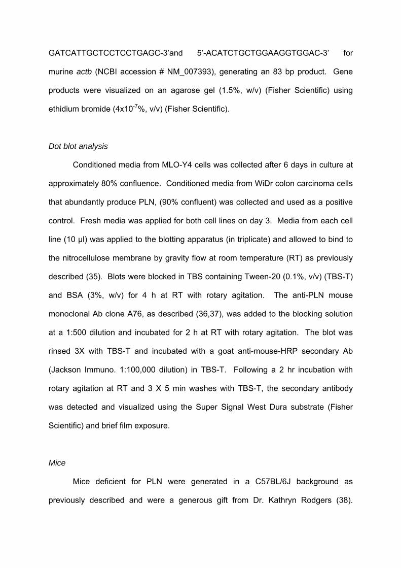
Dot blot analysis
Conditioned media from MLO-Y4 cells was collected after 6 days in culture at
approximately 80% confluence. Conditioned media from WiDr colon carcinoma cells
that abundantly produce PLN, (90% confluent) was collected and used as a positive
control. Fresh media was applied for both cell lines on day 3. Media from each cell
line (10 µl) was applied to the blotting apparatus (in triplicate) and allowed to bind to
the nitrocellulose membrane by gravity flow at room temperature (RT) as previously
described (35). Blots were blocked in TBS containing Tween-20 (0.1%, v/v) (TBS-T)
and BSA (3%, w/v) for 4 h at RT with rotary agitation. The anti-PLN mouse
monoclonal Ab clone A76, as described (36,37), was added to the blocking solution
at a 1:500 dilution and incubated for 2 h at RT with rotary agitation. The blot was
rinsed 3X with TBS-T and incubated with a goat anti-mouse-HRP secondary Ab
(Jackson Immuno. 1:100,000 dilution) in TBS-T. Following a 2 hr incubation with
rotary agitation at RT and 3 X 5 min washes with TBS-T, the secondary antibody
was detected and visualized using the Super Signal West Dura substrate (Fisher
Scientific) and brief film exposure.
Mice
Mice deficient for PLN were generated in a C57BL/6J background as
previously described and were a generous gift from Dr. Kathryn Rodgers (38).

Briefly, retention of the neomycin selection cassette (C1532Yneo) on intron 16
located between exons encoding for PLN domain III-3 structure results in
transcriptional defects leading to decreased PLN secretion that we describe as “PLN
deficiency” in homozygous mutants. These mice exhibit dwarfism and skeletal
defects including pigeon breast, flat face and hip dysplasia characteristic of the
human condition Schwartz Jampel Syndrome (38). These mice have been
previously referred to as “hypomorphic” due to the retention of some wild-type (WT)
function of the gene resulting in a less severe phenotype than a null allele (39,40).
WT and animals heterozygous for the C1532Yneo mutation were reported to be
undistinguishable phenotypically, and both were used as controls. All PLN deficient
mice or control mice were age-matched (8-9 month old), male littermates. All animal
studies were performed with the approval of the University of Delaware IACUC.
Tissue preparation
To assess osteocyte ultrastructure from control and PLN deficient mice, each
femur was surgically dissected, cut into 1 mm segments with a diamond wafering
saw and immersion-fixed with paraformaldehyde (4%, v/v) and glutaraldehyde (2%,
v/v) with ruthenium hexamine trichloride (RHT) (0.7%, w/v) in 0.05 M sodium
cacodylate buffer as described (8). Because of the potential interference by
glutaraldehyde and RHT with antibody interactions and to attain the best morphology
possible in the absence of these additional fixatives, tissues used for immunogold
assays were perfusion-fixed with only paraformaldehyde (4%, v/v) in 0.05 M sodium
cacodylate buffer as described (41). Briefly, mice were fixed by vascular perfusion
using perfluoro-compound FC-75 (13.3%, v/v) (Fisher Scientific), pluronic F-68 polyol
(3.3%, w/v) (Fisher Scientific), paraformaldehyde (4%, v/v) (EMS) and sodium

cacodylate buffer (0.05 M) (EMS). Following administration of fixatives, femur and
tibia were surgically removed and all soft tissue was debrided from the bone. Each
bone was cut into 1 mm cross-sections using a diamond wafering saw, immediately
immersed in fresh fixative containing paraformaldehyde (4%, v/v) and calcium
chloride (0.02 M) in sodium cacodylate buffer (0.05 M) incubated at 4°C overnight.
Bone sections were decalcified in EDTA (10%, w/v) with paraformaldehyde (1%, v/v)
in Tris-HCl buffer (0.1 M, pH 7.4) as described (8).
Cell culture and cortical bone immunofluorescence
Approximately 1,000 MLO-Y4 cells were seeded onto collagen I coated 8-well
chambers (NUNC™, Rochester, NY) and cultured as described previously (33).
When cells were 80-90% confluent media was removed, cells were washed 3X with
TBS and fixed with paraformaldehyde (4%, v/v) (EMS, Hatfield, PA) diluted in TBS
for 45 min at RT. Cells were washed 3X with TBS to remove residual fixative and
incubated for 1 hr in blocking solution consisting of bovine serum albumin (BSA)
(3%, w/v) and normal goat serum (10%, v/v) (Sigma, St. Louis, MO) diluted in TBS.
Decalcified bone sections were infiltrated with sucrose (2.3 M) in phosphate
buffer (PB), immersed in tissue freezing medium (EMS) and stored at -80°C.
Smaller sections (10 µm) were cut using a Leica CM3050 S cryostat microtome and
transferred to adhesive glass slides using the Cryojane tape transfer system
(Instrumedics, Inc, St. Louis, MO). Sections were washed with TBS to remove
residual paraformaldehyde, blocked with BSA (3%, w/v) and normal goat serum (2%,
v/v) in TBS.
Cells or bone sections were incubated with a mixture of four custom-designed
anti-PLN rabbit polyclonal primary antibodies (Strategic Diagnostics Inc., Newark,

DE) generated against human PLN domain IV sequences. Antibodies were diluted
in blocking buffer (1:100) and cells or tissues were incubated with primary antibodies
for 1 hr at RT. Following incubation with the primary Abs, cells (4 X 10 min) or
tissues (8 X 15 min) were washed with blocking solution and incubated with goat
anti-rabbit Alexa Fluor 488® conjugated (Invitrogen™, Carlsbad, CA) secondary Ab
(1:200) and DRAQ5™ nuclear stain (1:1000) (Biostatus, Ltd, Shepshed
Leicestershire, UK) diluted in blocking solution. Samples were washed 4 X 10 min
with TBS, mounted and stored at 4°C until imaged. Negative controls for cultured
cells and bone sections were performed using non-immune IgGs diluted at
concentrations equivalent to primary antibodies or without primary antibodies. All
samples were imaged with a Zeiss LSM 510 VIS confocal microscope using a 40X
C-aprochromat water immersion objective (NA 1.2) (Zeiss, Inc, Thornwood, NY).
Images were captured with an Axiovert 100M using the 488nm, 543nm and 633nm
laser lines for Alexa 488, Alexa 555 and Draq5 respectively.
Ultrastructure imaging by TEM
Sections fixed with paraformaldehyde, glutaraldehyde, and RHT were
dehydrated in an ascending series of ethanol (25%, 50%, 75%, 95%) for 15 min
each, left in ethanol (95%) overnight at 4°C followed by 100% for 2 X 15 min. Quetol
resin (EMS) was used to embed the sections according to the manufacturer’s
protocol. Briefly, a quetol/n-butyl glycidyl ether (NBGE) mixture was added to bone
sections in ascending ratios (1:3, 1:1, 3:1, 100%) for 2 hrs each followed by 100%
quetol overnight and an additional 1 hr incubation with 100% quetol. Resin then was
allowed to polymerize at 60°C for 48 hrs.

Bone sections were cut into ultrathin sections with a diamond knife, stained
with a saturated solution of uranyl acetate in methanol, followed by Reynolds’ lead
citrate and imaged with a Zeiss Libra 120 TEM at 120 kV. Images were acquired
with a Gatan Ultrascan 1000 2k x 2k digital camera.
Immunogold
Immunogold labeling and imaging by TEM was used to determine the position
and localization of PLN with optimal resolution within the LCS of cortical osteocytes.
Tissue sectioning, blocking and incubation with primary Abs recognizing epitopes
primarily in domain IV of PLN was identical to the procedure described above for
immunofluorescence assays. Sections were labeled with goat anti-rabbit F(ab’) ultra
small gold conjugated secondary Ab (1:100) (EMS) and silver enhanced according to
the Aurion ultra-small gold labeling protocol (Aurion, Wageningen, Netherlands).
Negative controls were performed using non-immune IgGs diluted at concentrations
equivalent to primary antibodies or without primary antibodies. Sections were
dehydrated, embedded in quetol resin, cut into ultrathin sections, stained with uranyl
acetate and Reynolds’ lead citrate, and viewed by TEM as described above.
Analysis of canalicular ultrastructure
Canalicular pericellular area, total canalicular area, process area, canalicular
density and the number of tethering elements per canaliculus were analyzed from
sections of control and PLN deficient mouse femoral bone by TEM. Each femur was
cut crosswise into 1 mm sections at midshaft and embedded in resin as described
above for ultrastructural imaging. Sections used for TEM analysis were chosen
randomly to provide a sampling along the femoral diaphysis. Multiple ultrathin

sections were created from each 1 mm section and images obtained from each
ultrathin section were analyzed randomly so as not to create any analytical bias.
Only cross-sectional images of canaliculi were included in the analysis. Pericellular
area was determined using the ImageJ® program (NIH, Bethesda, MD). The total
canalicular area and the area of each osteocyte process first were determined by
outlining each structure using ImageJ®, a method that reported the area of each
outlined region. The pericellular area next was found by subtracting the process
area from the total canalicular area. Canalicular density was assessed by
determining the total number of cross-sectional canaliculi per total bone area.
Longitudinal sections of canaliculi or canaliculi with poorly defined canalicular walls
were omitted from all analyses to improve objectivity by including only cross-
sectional canaliculi with well defined canalicular walls. Tethering elements were
counted for each canaliculus and for the purpose of this study were defined as
projections emanating from the osteocyte process that span the pericellular space
and are in contact with the canalicular wall, as previously illustrated (8). Objects
present in the pericellular space lacking these criteria were not included in the
analysis. Ultrastructural analysis included images from three separate bone sections
of each of three PLN deficient mice and two control mice. Animal genotype was
blinded during the analysis of all canalicular measurements. Significance was
determined by two-tailed Student’s t-test and variance reported as standard errors of
the mean.
Results
Osteocyte ultrastructure

TEM images of cortical mouse osteocytes were obtained to confirm the
presence of the transverse tethering elements in the LCS and to determine the size
relationship between the pericellular space and PLN. Figure 1A showed a single
osteocyte with a prominent nucleus (N) and very little ER and Golgi-like structures in
the cytosol (Cyt), features characteristic of osteocytes. Multiple canalicular
projections (Can) protrude from the cell body, several of which were devoid of a
process or the plane of the section was in varying orientation preventing full
visualization of the process. A single canaliculus with a process (P) centered in it
was seen emanating from the cell body (Fig. 1A). The ultrastructure of the osteocyte
process was well defined in the enlarged image of the canaliculus (Fig. 1B). Distinct
collagen bundles of the mineralized ECM were seen surrounding either side of the
canaliculus. Transverse tethering elements (TE) span the pericellular space (PS)
between the process (P) and the canalicular wall (CW). The osteocyte process was
centered in the canaliculus and the pericellular space was distinctly seen. This
space was previously measured to be ~78 nm, which is consistent with the data
shown here (Fig. 1B). The cartoon image of PLN demonstrated the multi-domain
character of this large heparan sulfate proteoglycan (Fig. 1C). Notably, the four
potential heparan sulfate chains, three on domain one and one on domain five are
shown. A compilation of studies indicate the approximate size of PLN to be 100-200
nm making it the only heparan sulfate proteoglycan of which we are aware that could
span the very large pericellular space of the osteocyte LCS.
PLN localizes along the processes of MLO-Y4 cells and the LCS of osteocytes
PLN displays a unique distribution on the membrane and dendritic processes
of MLO-Y4 osteocyte-like cells (Fig. 2A-B) and in the LCS of cortical osteocytes in
vivo (Fig. 2D-E). PLN is expressed in a unique pattern typical of extracellularly

deposited matrix proteins on the surface of MLO-Y4 cells (Fig. 2A). To maintain
extracellular localization of labeling no detergent or other permeabilizing reagents
were added. The staining pattern demonstrated the ability of PLN to be distributed in
a network-like fashion along and between the cytoplasmic projections of these
osteocyte-like cells. This pattern was clearly delineated in the enlarged image in
panel B where PLN was seen localizing along the processes of several cells
connected to each other (Fig. 2B). Normal rabbit IgG (Fig. 2C, white arrows) and
omission of the primary Ab (data not shown) controls showed absence of signal,
indicating the specificity of the fluorescent signal.
To demonstrate the ability of osteocytes to express PLN in vivo, mouse long
bones were labeled with anti-PLN Abs (Fig. 2D-E). Strong fluorescent signal was
observed along the cell bodies and in the LCS of cortical osteocytes (Fig. 2D). The
enlarged image in panel E showed distinct labeling of numerous canaliculi, most
notably staining was observed along canaliculi that interconnect osteocytes (Fig. 2E,
white arrows). There were various areas where small portions of canaliculi were
stained but the entire length of the process cannot be seen due to the section and/or
confocal plane orientation of single optical sections. Fluorescent labeling was
absent around the cell bodies or within the canaliculi with normal rabbit IgG (Fig. 2F)
or without primary antibody (data not shown) controls.
Expression of PLN in MLO-Y4 cells
MLO-Y4 osteocyte-like cells produce hspg2 transcript (Fig. 3A) and PLN
protein (Fig. 3B). Amplification of hspg2 from MLO-Y4 cDNA demonstrates a strong
band seen next to the actb internal control. Amplification of hspg2 and actb from
total mouse embryo (TME) cDNA was used as a positive control to verify the
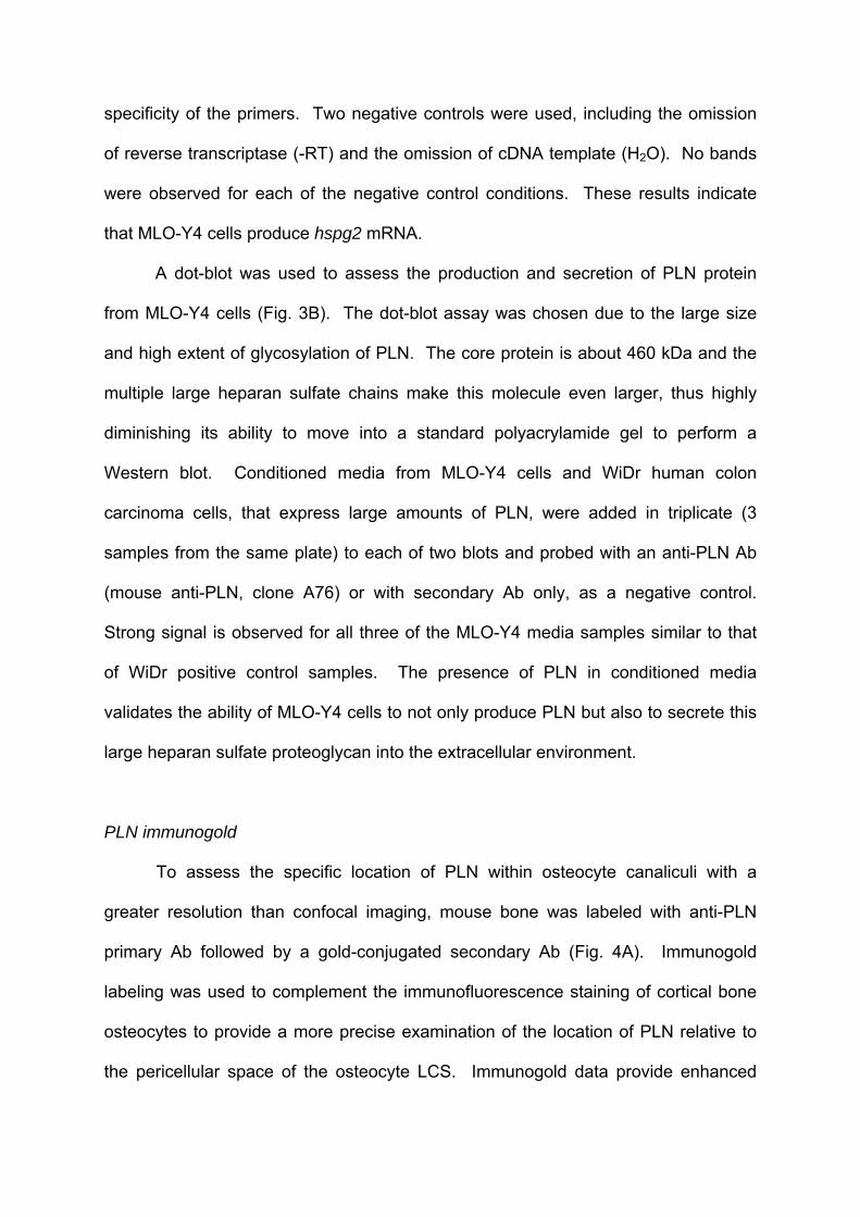
specificity of the primers. Two negative controls were used, including the omission
of reverse transcriptase (-RT) and the omission of cDNA template (H2O). No bands
were observed for each of the negative control conditions. These results indicate
that MLO-Y4 cells produce hspg2 mRNA.
A dot-blot was used to assess the production and secretion of PLN protein
from MLO-Y4 cells (Fig. 3B). The dot-blot assay was chosen due to the large size
and high extent of glycosylation of PLN. The core protein is about 460 kDa and the
multiple large heparan sulfate chains make this molecule even larger, thus highly
diminishing its ability to move into a standard polyacrylamide gel to perform a
Western blot. Conditioned media from MLO-Y4 cells and WiDr human colon
carcinoma cells, that express large amounts of PLN, were added in triplicate (3
samples from the same plate) to each of two blots and probed with an anti-PLN Ab
(mouse anti-PLN, clone A76) or with secondary Ab only, as a negative control.
Strong signal is observed for all three of the MLO-Y4 media samples similar to that
of WiDr positive control samples. The presence of PLN in conditioned media
validates the ability of MLO-Y4 cells to not only produce PLN but also to secrete this
large heparan sulfate proteoglycan into the extracellular environment.
PLN immunogold
To assess the specific location of PLN within osteocyte canaliculi with a
greater resolution than confocal imaging, mouse bone was labeled with anti-PLN
primary Ab followed by a gold-conjugated secondary Ab (Fig. 4A). Immunogold
labeling was used to complement the immunofluorescence staining of cortical bone
osteocytes to provide a more precise examination of the location of PLN relative to
the pericellular space of the osteocyte LCS. Immunogold data provide enhanced
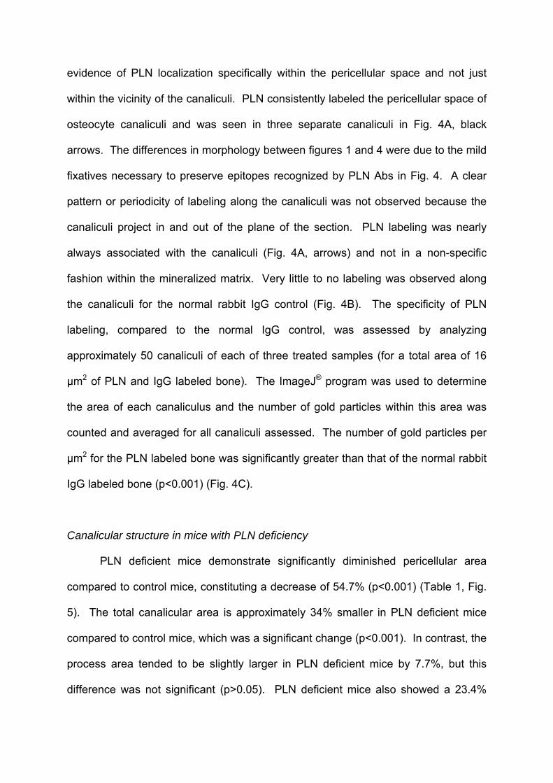
evidence of PLN localization specifically within the pericellular space and not just
within the vicinity of the canaliculi. PLN consistently labeled the pericellular space of
osteocyte canaliculi and was seen in three separate canaliculi in Fig. 4A, black
arrows. The differences in morphology between figures 1 and 4 were due to the mild
fixatives necessary to preserve epitopes recognized by PLN Abs in Fig. 4. A clear
pattern or periodicity of labeling along the canaliculi was not observed because the
canaliculi project in and out of the plane of the section. PLN labeling was nearly
always associated with the canaliculi (Fig. 4A, arrows) and not in a non-specific
fashion within the mineralized matrix. Very little to no labeling was observed along
the canaliculi for the normal rabbit IgG control (Fig. 4B). The specificity of PLN
labeling, compared to the normal IgG control, was assessed by analyzing
approximately 50 canaliculi of each of three treated samples (for a total area of 16
µm2 of PLN and IgG labeled bone). The ImageJ® program was used to determine
the area of each canaliculus and the number of gold particles within this area was
counted and averaged for all canaliculi assessed. The number of gold particles per
µm2 for the PLN labeled bone was significantly greater than that of the normal rabbit
IgG labeled bone (p<0.001) (Fig. 4C).
Canalicular structure in mice with PLN deficiency
PLN deficient mice demonstrate significantly diminished pericellular area
compared to control mice, constituting a decrease of 54.7% (p<0.001) (Table 1, Fig.
5). The total canalicular area is approximately 34% smaller in PLN deficient mice
compared to control mice, which was a significant change (p<0.001). In contrast, the
process area tended to be slightly larger in PLN deficient mice by 7.7%, but this
difference was not significant (p>0.05). PLN deficient mice also showed a 23.4%

decrease in canalicular density in cortical bone and a reduction in the number of
tethering elements within each canaliculus by 35.8%, both values were statistically
significant (p<0.01 and p<0.001 respectively) (Table 1). The appearance of the
tethering elements is shown at high magnification in the insets to Fig. 5. Previous
studies demonstrated approximately a 36% reduction in PLN transcript production in
newborn endochondral skeleton by Northern blot analysis and only trace levels of
PLN protein in long bones by immunostaining (38). Here we report similar findings
of severely diminished fluorescent signal of osteocytes from cortical bone sections
labeled with anti-PLN antibodies (Fig. 6).
Discussion
The cytoplasmic processes extending from osteocytes and the channels in
which they reside have been implicated in several important physiological functions
including mechanosensation, cell-cell communication and waste/nutrient transport.
Previous studies identify the presence of transverse elements spanning the
pericellular space of the LCS (8); however, the molecular identity of these structures
remains speculative. The results presented here identify the presence of the large
heparan sulfate proteoglycan PLN well positioned within the osteocyte LCS to be
part of an ECM complex in the pericellular space where structural integrity is
essential for uninhibited fluid movement. Additionally, PLN deficiency resulted in
significantly altered canalicular ultrastructural morphology, representing a novel
phenotypic alteration involving this unique, multifunctional ECM molecule.
Previous studies identified the presence of transverse tethering elements in
the pericellular space and speculated these elements to be proteoglycans,
glycosaminoglycans, or possibly wall protrusions linked to β3 integrins (8,32).

Although these studies did not take advantage of higher resolution immunogold
labeling to more precisely delineate integrin localization within the LCS, the
distribution of staining correlated with protrusions, demonstrated using TEM,
emanating from the canalicular wall leading to the possibility that integrins may
directly interact matrix wall by way of “hillocks” within the canalicular pericellular
space. The hypothesis that integrins bind directly to the canalicular wall must be
reconciled with previous observations showing an average osteocyte canalicular
pericellular space of 78 nm (8). In the current study we demonstrate distinct
immunofluorescent labeling of PLN along numerous canaliculi in cortical bone.
Higher resolution immunological labeling using immunogold with TEM imaging
revealed that PLN antibodies were localized in the pericellular space of osteocyte
canaliculi. Interestingly, the number of tethering elements in PLN deficient cortical
bone was significantly less than controls. The localization of this molecule and the
reduction of tethering elements in PLN deficient mice strongly suggested that PLN
was part of an ECM complex in the territorial matrix surrounding osteocytic
processes. The tethering elements previously have been proposed to serve as a
means of centering the dendritic processes with the canaliculi and possibly to
function in mechanosensation (7,8,32). While the tethering elements may consist of
one or more molecular entities, the data presented here suggest that PLN is a
component of the tethering complex. Additionally, PLN interacts with β1 and β3
integrins (18,21,42) and is the only heparan sulfate proteoglycan of sufficient size to
span the pericellular space, leading to the possibility that an association between
PLN and integrins may exist within the osteocyte LCS.
Previous studies demonstrate that osteocyte-like MLO-Y4 cells have a
glycocalyx layer rich in hyaluronic acid but display only limited amounts of sulfated

proteoglycans by alcian blue staining, thus contrasting with the data presented here
showing distinct expression of the large heparan sulfate proteoglycan PLN in the
osteocyte LCS (43). Additional in vivo studies provide evidence for the expression of
sulfated proteoglycans in the osteocyte LCS (44-46). Specifically, chondroitin
sulfate-rich proteoglycans discretely line the osteocyte lacunae and canaliculi,
localizing along “fine filamentous structures” within the walls of the LCS in human
alveolar bone (44). Additionally, embryonic chick osteocytes express chondroitin
sulfate, keratan sulfate and dermatan sulfate along the walls of the lacunae and
canaliculi (45). The lack of sulfated proteoglycans previously described in MLO-Y4
cells may stem from differences in conditions related to cell culture versus in vivo
bone samples, as the dense extracellular environment of the in vivo LCS provides
numerous potential binding partners for proteoglycans that may not be available
when the mineralized matrix wall is absent. Moreover, Reilly and colleagues
cultured MLO-Y4 cells for only 2-3 days and it has been proposed that with
additional time in culture sulfated proteoglycans and glycosaminoglycans may
become a component of the osteocyte glycocalyx (43). While hyaluronic acid is a
major component of the osteocyte glycocalyx and contributes to fluid flow induced
prostaglandin E2 (PGE2) release, it has not been implicated as a component of the
tethering elements (43). Sulfated proteoglycans are present alongside hyaluronic
acid in the glycocalyx of endothelial cells (47) providing the potential for interactions
between these two entities within the LCS; however, the ability of PLN to interact
with hyaluronic acid or hyaluronic acid binding protein has not yet been established.
PLN deficient mice have reduced pericellular canalicular area that can be
attributed to diminished total canalicular area and is not due to enlargement of the
cytoplasmic processes. One possible explanation to account for the smaller

pericellular area is the reduced overall size of PLN deficient mice. Reduced skeletal
size of PLN deficient mice may lead to proportionately decreased canaliculi;
however, this does not account for the differences we observe in process area. PLN
deficient mice have osteocyte processes 7.7% larger than control animals, that was
not statistically significant. If the observed differences in pericellular area were
related to skeletal size, a proportionate decrease in the osteocyte process would be
expected; however, this was not seen; in fact the cytoplasmic processes were
slightly larger than in control animals. Therefore, the data support the conclusion
that the deficiency of PLN and its heparan sulfate chains likely allows encroachment
of the mineralized canalicular wall on the osteocyte process. Complimented by the
observation of a reduced number of tethering elements observed by TEM in PLN
deficient mice, these data suggested a role for PLN in the maintenance of the
structural integrity and morphology of the LCS.
Proteoglycans aid in the prevention of tissue mineralization (48-50) and
heparan sulfate is a potent inhibitor of hydroxyapatite (HAP) formation (30).
Although PLN is not ubiquitous, it is found in numerous tissues most commonly at
interfaces where a barrier is required. A lack of PLN is associated with barrier
breakdown leading to pathologies such as cancer cell invasion (23,24), disruption of
the blood brain barrier (25), altered glomerular filtration (26) and failure of the
chondro-osseous junction in bone (36,51). Brown and colleagues demonstrated a
distinct downregulation of PLN at the chondro-osseous junction in murine hind limbs,
with disappearance of PLN coinciding with onset of mineralization (36).
Immunohistochemistry of murine hind limbs reveals very strong PLN signal in
chondrocytes and their matrix, yet virtually no PLN expression was observed in
osteoblasts of trabecular bone or along the periosteal wall (36). Additionally,

Rodgers and colleagues noted increased hypertrophic calcified cartilage in the
developing growth plates of PLN deficient mice (38), consistent with the proposed
mineralization barrier function for PLN and heparan sulfate at the chondro-osseous
junction (36). The presence of PLN within the osteocyte LCS as shown here, and
previous observations of sulfated proteoglycans localized to “fine filamentous
material” in the walls of osteocyte canaliculi (but absent from the mineralized matrix
of mature human alveolar bone) (44), support the hypothesis that while osteoblasts
downregulate PLN expression to allow for proper matrix mineralization, osteocytes
regain the ability to secrete PLN and other glycosaminoglycans. The presence of
PLN within the LCS likely serves as a barrier for mineral formation helping to
maintain the size and structure of the pericellular space. Other potential functions
include facilitating waste exchange, contributing to fluid flow induced
mechanosensation and maintaining osteocyte viability.
Our present study demonstrated a reduction in the density of canaliculi in
cortical bone of PLN deficient mice, suggesting that PLN was able to regulate
osteocyte LCS morphology. Previous studies demonstrated that normal bone is
characterized by high osteocyte connectivity with processes oriented toward the
vasculature (52). In contrast, osteoporotic bone demonstrates decreased
connectivity and a disorientation of the cellular processes. Osteoarthritic bone has
diminished connectivity, but osteocyte process orientation is normal. Additionally,
osteomalacic bone has a highly disorganized LCS with distorted processes (52).
Alterations of osteocyte canalicular density and morphological arrangement is likely
to have an effect on osteocyte function and possibly the mechanical properties of
bone (53). Potential mechanisms involving PLN in the establishment of normal bone
mass will require further investigations.

In summary, our results identified PLN as a constituent of the LCS of murine
cortical bone where it plays a role in preserving the size of the pericellular space in
the LCS, presumably by contributing to the structural integrity of the territorial matrix
surrounding osteocytic processes. Previous studies identified the presence of
transverse tethering elements in the pericellular space and speculated these
elements to be proteoglycans, glycosaminoglycans or possibly integrins (8,32).
Although the data presented here do not definitively identify PLN as the tethering
element, our data indicate PLN to be well positioned in osteocyte canaliculi to
contribute to the specialized matrix environment surrounding osteocyte processes
and that PLN helps maintain the unmineralized pericellular space of the LCS. In this
study, we showed that cortical bone with deficient PLN had fewer tethering elements
per canaliculus and decreased density of canaliculi leading to the possibility that
heparan sulfate in the ECM in the pericellular space influences formation and/or
distribution of the osteocyte processes. These data demonstrate a novel osteocyte
phenotype involving the large heparan sulfate proteoglycan PLN and further
exploration of its role in maintenance of open canalicular structure and osteocyte
function may reveal important molecular and functional insights into this specialized
mechano-regulatory environment.
Acknowledgments
We would like to thank Dr. Lynda Bonewald for the generous gift of the MLO-
Y4 cells, Dr. Kathryn Rodgers for the PLN deficient mice, Drs. James Stave and
Mark Muldoon at SDI for PLN antibodies, Ms. Mary Boggs for assistance with data
analysis and Mr. Jason Koons for his assistance with graphic design. We are
grateful to Ms. Julie Mis and Ms. Sue Seta for help with animal breeding and

dissection respectively.

References
1. Duncan RL, Turner CH 1995 Mechanotransduction and the functional response of bone to mechanical strain. Calcif Tissue Int 57(5):344-58.
2. Parfitt AM 1977 The cellular basis of bone turnover and bone loss: a rebuttal of the osteocytic resorption--bone flow theory. Clin Orthop Relat Res (127):236-47.
3. Tatsumi S, Ishii K, Amizuka N, Li M, Kobayashi T, Kohno K, Ito M, Takeshita S, Ikeda K 2007 Targeted ablation of osteocytes induces osteoporosis with defective mechanotransduction. Cell Metab 5(6):464-75.
4. Noble BS 2008 The osteocyte lineage. Arch Biochem Biophys 473(2):106-11. 5. Cowin SC 2002 Mechanosensation and fluid transport in living bone. J
Musculoskelet Neuronal Interact 2(3):256-60. 6. Piekarski K, Munro M 1977 Transport mechanism operating between blood
supply and osteocytes in long bones. Nature 269(5623):80-2. 7. You L, Cowin SC, Schaffler MB, Weinbaum S 2001 A model for strain
amplification in the actin cytoskeleton of osteocytes due to fluid drag on pericellular matrix. J Biomech 34(11):1375-86.
8. You LD, Weinbaum S, Cowin SC, Schaffler MB 2004 Ultrastructure of the osteocyte process and its pericellular matrix. Anat Rec A Discov Mol Cell Evol Biol 278(2):505-13.
9. Inoue K, Mikuni-Takagaki Y, Oikawa K, Itoh T, Inada M, Noguchi T, Park JS, Onodera T, Krane SM, Noda M, Itohara S 2006 A crucial role for matrix metalloproteinase 2 in osteocytic canalicular formation and bone metabolism. J Biol Chem 281(44):33814-24.
10. Sauren YM, Mieremet RH, Groot CG, Scherft JP 1992 An electron microscopic study on the presence of proteoglycans in the mineralized matrix of rat and human compact lamellar bone. Anat Rec 232(1):36-44.
11. Kirn-Safran C, Farach-Carson MC, Carson DD 2009 Multifunctionality of extracellular and cell surface heparan sulfate proteoglycans. Cell Mol Life Sci.
12. Farach-Carson MC, Carson DD 2007 Perlecan--a multifunctional extracellular proteoglycan scaffold. Glycobiology 17(9):897-905.
13. Noonan DM, Fulle A, Valente P, Cai S, Horigan E, Sasaki M, Yamada Y, Hassell JR 1991 The complete sequence of perlecan, a basement membrane heparan sulfate proteoglycan, reveals extensive similarity with laminin A chain, low density lipoprotein-receptor, and the neural cell adhesion molecule. J Biol Chem 266(34):22939-47.
14. Kallunki P, Tryggvason K 1992 Human basement membrane heparan sulfate proteoglycan core protein: a 467-kD protein containing multiple domains resembling elements of the low density lipoprotein receptor, laminin, neural cell adhesion molecules, and epidermal growth factor. J Cell Biol 116(2):559-71.
15. Noonan DM, Horigan EA, Ledbetter SR, Vogeli G, Sasaki M, Yamada Y, Hassell JR 1988 Identification of cDNA clones encoding different domains of the basement membrane heparan sulfate proteoglycan. J Biol Chem 263(31):16379-87.

16. Tapanadechopone P, Hassell JR, Rigatti B, Couchman JR 1999 Localization of glycosaminoglycan substitution sites on domain V of mouse perlecan. Biochem Biophys Res Commun 265(3):680-90.
17. Iozzo RV 2005 Basement membrane proteoglycans: from cellar to ceiling. Nat Rev Mol Cell Biol 6(8):646-56.
18. Hayashi K, Madri JA, Yurchenco PD 1992 Endothelial cells interact with the core protein of basement membrane perlecan through beta 1 and beta 3 integrins: an adhesion modulated by glycosaminoglycan. J Cell Biol 119(4):945-59.
19. Bix G, Fu J, Gonzalez EM, Macro L, Barker A, Campbell S, Zutter MM, Santoro SA, Kim JK, Hook M, Reed CC, Iozzo RV 2004 Endorepellin causes endothelial cell disassembly of actin cytoskeleton and focal adhesions through alpha2beta1 integrin. J Cell Biol 166(1):97-109.
20. Bix G, Iozzo RA, Woodall B, Burrows M, McQuillan A, Campbell S, Fields GB, Iozzo RV 2007 Endorepellin, the C-terminal angiostatic module of perlecan, enhances collagen-platelet responses via the alpha2beta1-integrin receptor. Blood 109(9):3745-8.
21. Woodall BP, Nystrom A, Iozzo RA, Eble JA, Niland S, Krieg T, Eckes B, Pozzi A, Iozzo RV 2008 Integrin alpha2beta1 is the required receptor for endorepellin angiostatic activity. J Biol Chem 283(4):2335-43.
22. Vincent TL, McLean CJ, Full LE, Peston D, Saklatvala J 2007 FGF-2 is bound to perlecan in the pericellular matrix of articular cartilage, where it acts as a chondrocyte mechanotransducer. Osteoarthritis Cartilage 15(7):752-63.
23. Hasengaowa, Kodama J, Kusumoto T, Shinyo Y, Seki N, Nakamura K, Hongo A, Hiramatsu Y 2005 Loss of basement membrane heparan sulfate expression is associated with tumor progression in endometrial cancer. Eur J Gynaecol Oncol 26(4):403-6.
24. Kodama J, Shinyo Y, Hasengaowa, Kusumoto T, Seki N, Nakamura K, Hongo A, Hiramatsu Y 2005 Loss of basement membrane heparan sulfate expression is associated with pelvic lymph node metastasis in invasive cervical cancer. Oncol Rep 14(1):89-92.
25. Deguchi Y, Okutsu H, Okura T, Yamada S, Kimura R, Yuge T, Furukawa A, Morimoto K, Tachikawa M, Ohtsuki S, Hosoya K, Terasaki T 2002 Internalization of basic fibroblast growth factor at the mouse blood-brain barrier involves perlecan, a heparan sulfate proteoglycan. J Neurochem 83(2):381-9.
26. Morita H, Yoshimura A, Inui K, Ideura T, Watanabe H, Wang L, Soininen R, Tryggvason K 2005 Heparan sulfate of perlecan is involved in glomerular filtration. J Am Soc Nephrol 16(6):1703-10.
27. Kasinath B, Kanwar Y 1993 Molecular and Cellular Aspects of Basement Membranes.
28. Hoh JH 1998 Functional protein domains from the thermally driven motion of polypeptide chains: a proposal. Proteins 32(2):223-8.
29. Chen CH, Hansma HG 2000 Basement membrane macromolecules: insights from atomic force microscopy. J Struct Biol 131(1):44-55.
30. Rees SG, Hughes W, Embery G 2002 Interaction of glucuronic acid and iduronic acid-rich glycosaminoglycans and their modified forms with hydroxyapatite. Biomaterials 23(2):481-9.

31. Yang L, Butcher M, Simon RR, Osip SL, Shaughnessy SG 2005 The effect of heparin on osteoblast differentiation and activity in primary cultures of bovine aortic smooth muscle cells. Atherosclerosis 179(1):79-86.
32. McNamara LM, Majeska RJ, Weinbaum S, Friedrich V, Schaffler MB 2009 Attachment of osteocyte cell processes to the bone matrix. Anat Rec (Hoboken) 292(3):355-63.
33. Kato Y, Windle JJ, Koop BA, Mundy GR, Bonewald LF 1997 Establishment of an osteocyte-like cell line, MLO-Y4. J Bone Miner Res 12(12):2014-23.
34. Iozzo RV 1984 Biosynthesis of heparan sulfate proteoglycan by human colon carcinoma cells and its localization at the cell surface. J Cell Biol 99(2):403-17.
35. Gomes RR, Jr., Joshi SS, Farach-Carson MC, Carson DD 2006 Ribozyme-mediated perlecan knockdown impairs chondrogenic differentiation of C3H10T1/2 fibroblasts. Differentiation 74(1):53-63.
36. Brown AJ, Alicknavitch M, D'Souza SS, Daikoku T, Kirn-Safran CB, Marchetti D, Carson DD, Farach-Carson MC 2008 Heparanase expression and activity influences chondrogenic and osteogenic processes during endochondral bone formation. Bone 43(4):689-99.
37. Whitelock JM, Murdoch AD, Iozzo RV, Underwood PA 1996 The degradation of human endothelial cell-derived perlecan and release of bound basic fibroblast growth factor by stromelysin, collagenase, plasmin, and heparanases. J Biol Chem 271(17):10079-86.
38. Rodgers KD, Sasaki T, Aszodi A, Jacenko O 2007 Reduced perlecan in mice results in chondrodysplasia resembling Schwartz-Jampel syndrome. Hum Mol Genet 16(5):515-28.
39. Arikawa-Hirasawa E, Watanabe H, Takami H, Hassell JR, Yamada Y 1999 Perlecan is essential for cartilage and cephalic development. Nat Genet 23(3):354-8.
40. Costell M, Gustafsson E, Aszodi A, Morgelin M, Bloch W, Hunziker E, Addicks K, Timpl R, Fassler R 1999 Perlecan maintains the integrity of cartilage and some basement membranes. J Cell Biol 147(5):1109-22.
41. Rostgaard J, Qvortrup K 1997 Electron microscopic demonstrations of filamentous molecular sieve plugs in capillary fenestrae. Microvasc Res 53(1):1-13.
42. Brown JC, Sasaki T, Gohring W, Yamada Y, Timpl R 1997 The C-terminal domain V of perlecan promotes beta1 integrin-mediated cell adhesion, binds heparin, nidogen and fibulin-2 and can be modified by glycosaminoglycans. Eur J Biochem 250(1):39-46.
43. Reilly GC, Haut TR, Yellowley CE, Donahue HJ, Jacobs CR 2003 Fluid flow induced PGE2 release by bone cells is reduced by glycocalyx degradation whereas calcium signals are not. Biorheology 40(6):591-603.
44. Smith AJ, Singhrao SK, Newman GR, Waddington RJ, Embery G 1997 A biochemical and immuno-electron microscopical analysis of chondroitin sulphate-rich proteoglycans in human alveolar bone. Histochem J 29(1):1-9.
45. Takagi M, Ono Y, Maeno M, Miyashita K, Omiya K 1997 Immunohistochemical and biochemical characterization of sulphated proteoglycans in embryonic chick bone. J Nihon Univ Sch Dent 39(3):156-63.
46. Murakami T, Hitomi S, Ohtsuka A, Taguchi T 1995 Neurons with perineuronal sulfated proteoglycans in the human visual cortex, with special reference to their reactions to lectins. Arch Histol Cytol 58(3):357-64.

47. Henry CB, Duling BR 1999 Permeation of the luminal capillary glycocalyx is determined by hyaluronan. Am J Physiol 277(2 Pt 2):H508-14.
48. Howell DS, Pita JC 1976 Calcificaiton of growth plate cartilage with special reference to studies on micropuncture fluids. Clin Orthop Relat Res (118):208-29.
49. Chen CC, Boskey AL 1985 Mechanisms of proteoglycan inhibition of hydroxyapatite growth. Calcif Tissue Int 37(4):395-400.
50. Chen CC, Boskey AL, Rosenberg LC 1984 The inhibitory effect of cartilage proteoglycans on hydroxyapatite growth. Calcif Tissue Int 36(3):285-90.
51. French MM, Smith SE, Akanbi K, Sanford T, Hecht J, Farach-Carson MC, Carson DD 1999 Expression of the heparan sulfate proteoglycan, perlecan, during mouse embryogenesis and perlecan chondrogenic activity in vitro. J Cell Biol 145(5):1103-15.
52. Knothe Tate ML, Adamson JR, Tami AE, Bauer TW 2004 The osteocyte. Int J Biochem Cell Biol 36(1):1-8.
53. Zhou X, Novotny JE, Wang L 2009 Anatomic variations of the lacunar-canalicular system influence solute transport in bone. Bone 45(4):704-10.

Figure Legends
Figure 1: The molecular size of PLN is sufficient to span the pericellular space
of the LCS. (A) Electron photomicrograph of a single cortical osteocyte with
cytoplasmic processes (P), empty canaliculi (Can) extending from the cell body and
with a prominent nucleus (N) and very little ER and Golgi-like structures in the
cytosol (Cyt), features characteristic of osteocytes. (B) Electron micrograph of a cell
process where the transverse tethering elements (TE) clearly spans the pericellular
space (PS) between the process (P) and the canalicular wall (CW). (C) Schematic
representation of PLN. The size of this large heparan sulfate proteoglycan is
sufficient to span the pericellular space of the LCS of osteocytes.
Figure 2: PLN localizes along the cytoplasmic processes of MLO-Y4 cells and
the LCS of osteocytes in vivo. MLO-Y4 cells (A-C) were stained with either a mix
of polyclonal PLN Abs (A & B) or with normal rabbit IgG Ab. MLO-Y4 cells display a
unique expression pattern of PLN in a distribution resembling a network or matrix-
like appearance (A) (representative of three separate experiments). PLN is
prominently displayed along the cytoplasmic processes (white arrows) of MLO-Y4
cells (B) and absent in normal IgG controls (C). Cortical mouse osteocytes (D-F)
express PLN abundantly (D) (representative of 2 experiments from 2 animals using 3
bone slices from each animal). Cell bodies and the LCS of osteocytes (white
arrows) show robust PLN signal (E). No signal was observed in osteocytes treated
with normal IgG as a control (F).

Figure 3: MLO-Y4 cells express hspg2 transcript and PLN protein. (A) Hspg2
transcript expression was determined by RT-PCR. Actb transcript served as a load
control and total mouse embryo (TME) RNA as a positive control. Omission of
reverse transcriptase (-RT) and cDNA template (H2O) were used as negative
controls (representative of 3 separate experiments). (B) Production and secretion of
PLN protein in MLO-Y4 cells demonstrated by dot-blot. Conditioned media from
WiDr cells was used as a positive control and omission of primary Ab as a negative
control (performed in triplicate with 3 samples from the same culture dish).
Figure 4: PLN resides within the pericellular space of the LCS. (A) Immunogold
labeling of cortical bone demonstrates the presence of PLN within the pericellular
space of osteocyte canaliculi, in the proper position to possibly serve as a
component of the transverse tethering complex. Normal IgG labeling of the canaliculi
(B) was significantly less than the PLN labeling (A, black arrows) as shown
graphically in panel C. Data were compiled from a total area of 16 µm2 of canalicular
area from approximately 50 canaliculi of anti-PLN or normal IgG treated bone (p <
0.001). Samples are representative of 2 experiments from random femoral bone
sections of 2 animals. Statistical significance determined using two-tailed Student’s
t-test and variance reported as standard errors of the mean.
Figure 5: PLN deficient mice have altered canalicular morphology. (A) PLN
deficient mice demonstrate reduced pericellular area, decreased canalicular density
and fewer tethering elements per canaliculus compared to control mice (B). Inset
images show representative images of a single canaliculus to demonstrate the
tethering elements (arrows within insets). The black arrow in panel A demonstrates

a canaliculus that was omitted from analysis due to an insufficiently defined
canalicular wall. The black arrow in panel B indicates a canaliculus that was omitted
from analysis because it appeared as a longitudinal-section and not in cross-section.
Data are representative of measurements taken from multiple randomly selected
ultrathin bone slices from 3 randomly selected 1 mm femoral bone sections taken
from 3 PLN deficient or 2 control mice.
Figure 6: Cortical bone osteocytes of PLN deficient mice express diminished
levels of PLN. (A) Immunostaining for PLN in femur of mice deficient for PLN
showing decreased signal in cortical osteocytes. (B) High magnification of cortical
osteocytes of PLN deficient mice. (C) Immunostaining for PLN in control mouse
cortical bone demonstrating strong signal around osteocyte cell bodies and
canaliculi. (D) High magnification of cortical osteocytes in control mice showing
strong immunofluorescent signal in osteocyte canaliculi that is absent in PLN
deficient osteocytes (B). Images are representative of 2 experiments from PLN
deficient or control mice using 3 bone sections from each animal.

Fig 1.

Fig 2.

Fig 3.

Fig 4.

Fig 5.

Fig 6.

Table 1. Canalicular ultrastructural measurements1 of 8-9 mo. old male mice Controls PLN Deficient % change vs.
control Pericellular area (µm2) 0.053 (+ 0.002) 0.029 (+ 0.001) -54.7%*** Total canalicular area (µm2)
0.066 (+ 0.002) 0.043 (+ 0.001) -34.8%***
Process area (µm2) 0.013 (+ 0.0006)
0.014 (+ 0.0005) 7.7%
Canalicular density (canaliculi/µm2)
0.124 (+ 0.008) 0.095 (+ 0.005) -23.4%**
Tethering elements/canaliculus
3.223 (+ 0.110) 2.069 (+ 0.069) -35.8%***
** p<0.01, ***p<0.001 1 Values reported represent the average measurements of 293 control canaliculi and 238 canaliculi from PLN deficient mice. Total canalicular area includes both pericellular area plus process area.