peripheral vestibular disorders - ucla health · 1860s worked at large deaf- mute center in paris...
TRANSCRIPT
Peripheral vestibular disorders will affect 1 of 13 people in their lifetime 80% of affected persons seek medical consultation Unclear how many of these are for peripheral vs
central disorders Generally: pts younger than 50 are more likely to
have Peripheral disease vs older than 50 generally have central dysfunction
In the elderly, dizziness is generally a combination of both
After age 75: balance dysfunction = #1 reason for visiting primary care offices

1860s worked at large deaf-mute center in Paris All vertigo thought to be central in origin, he
was the first to describe a inner ear origin Published report of girl with sudden hearing loss
and acute vertigo after injury autopsy showed blood in the inner ear
Ironic: first pt with Ménière’s disease, probably had leukemia, not endolymphatic hydrops
Up to 1940s any peripheral vertigo was called Ménière’s disease, until true pathophysiology was described
Horizontal SCC: horizontal head rotation (yaw)
S-SCC & P-SCC: detect pitching of the head front to back as well as roll from side to side
Utricle/Saccule: linear acceleration; gravity, and motion in straight trajectory
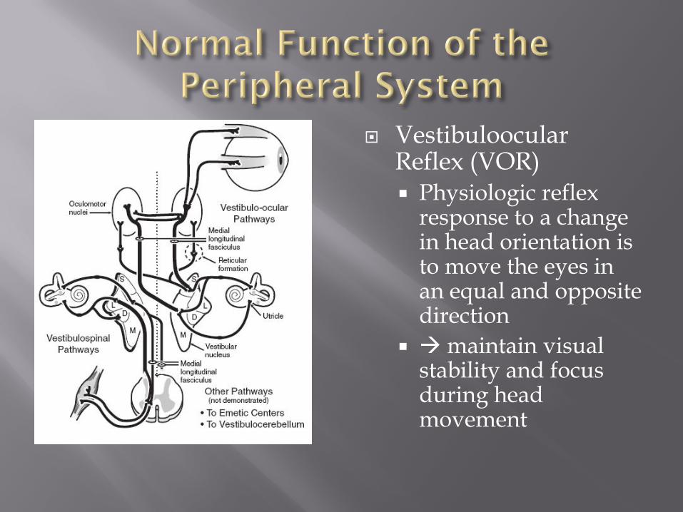
VestibuloocularReflex (VOR) Physiologic reflex
response to a change in head orientation is to move the eyes in an equal and opposite direction
maintain visual stability and focus during head movement
All receptors tonically active Resting, spontaneous continual outflow of action
potentials With head motion, the spontaneous activity is
modulated up or down Ex: turn head to the right increase R-H-SCC
activity and decrease L-H-SCC activity CNS wants balanced input, when input is
assymmetic CNS interprets as head rotation compensatory eye movements and postural adjustments sensation of movement
CNS capable of rebalancing itself Occurs through the ability to compare other sensory,
visual, and kinesthetic inputs to vestibular inputreadjust when these don’t match
Will reset the point of balance Typically takes time, on the order of days
Sudden-onset unilateral dysfunction Infectious: viral/bacterial labyrinthitis/ Ramsey-
Hunt Syndrome Traumatic: T-bone fracture Iatrogenic: labyrinthectomy/ vestibular nerve
section Idiopathic: Meniere's disease Other: ototoxic meds (gentamicin), vascular injury,
labyrinthine fistulae
Acute stage: asymmetry interpreted centrally as movement Vertigo
Oculomotor Nuclei senses reduced tonic activity nystagmus If right labyrinth is altered: slow eye movement is to
the right and the fast phase (nystagmus) is to the left Vestibulospinal and cerebellar activity is altered
sensation of falling Past-pointing, Romberg sign
Nystagmus Immediately after injury it is present in ALL
positions of gaze ↓↓↓ by visual fixation ↑↑↑ when gaze is directed AWAY from injury ↓↓↓ when gaze is directed TOWARD injury Gradually disappears, however rate of recovery is
related to activity level and is decreased with advanced age
Vestibulo-suppressive meds ↓↓↓ rate and extent of recovery
Common Causes: Ototoxicity, meningitic labyrinthitis, bilateral
temporal bone trauma Symmetric decrease in tonic activity from each
labyrinth to the brainstem If loss is simultaneous and symmetrical no
significant vertigo Instead: pt with poor balance, especially at night;
fixed objects jump with any head movement (Oscillopsia)
Cerebellar Compensation can adjust the gain of the VOR typically symptoms resolve within a few days Typically cannot compensate for complete loss pts with
oscillopsia
Symptoms: ↓ vestibular sensitivity to head movement and gravitational position VOR inaccuracy This is necessary for visual acuity during rapid head
movement Patients will complain of visual disturbance and light-
headedness – Blurry vision and disequilibrium

Etiology: CN 8 Neoplasm, degenerative, autoimmune disease
PRESENTS WITH: may not produce severe symptoms because of compensation
by brainstem Gain of VOR—adjusted by lateral cerebellum With vestibular schwannomamay be so gradual, that
vestibular symptoms imperceptible Pts may not present until lesion is large enough to cause
brainstem compression or to compromise blood supply to the peripheral labyrinth cause sudden unilateral dysfunction
TESTING Calorics should still show deceased response on the side of
the lesion when tumor affects the superior vestibular nerve

ETIOLOGY: Aging, ototoxicity, autoimmune disease, syphilis,
and degenerative disorders
PRESENTS WITH: Cause few symptoms because of cerebellar
compensation Symptoms when complete or near complete loss of
vestibular sensitivity Visual disturbance, light-headedness, oscillopsia

ETIOLOGY: Hydrops, benign paroxysmal positional vertigo, and
dehiscence of the SSC PRESENTS WITH:
Intermittent episodes—since they occur episodically, no CNS compensation each attack is a sudden loss of vestibular function

History and physical exam Onset and time course Acute/ progressive/ duration/ frequency
Description of symptoms Spinning/ lightheadedness/ imbalance/ blurry vision
Associated symptoms Hearing loss/ aural fullness/ otorrhea/ otalgia/ facial
paralysis/ headaches/ photophobia/ nausea/ vomiting Precipitating and alleviating factors Head trauma/ Cerebrovascular disease/ autoimmune disease
Status of other sensory systems Vision/ proprioception
Integrity of CNS Integrity of compensatory mechanisms Medication review
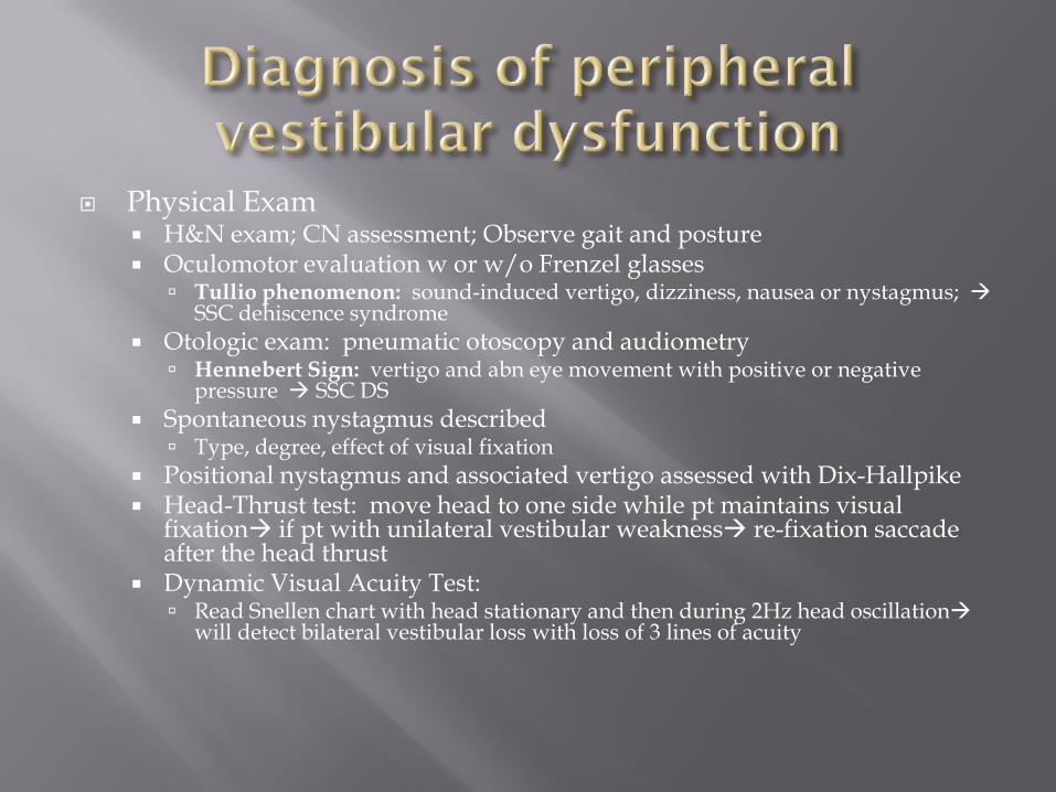
Physical Exam H&N exam; CN assessment; Observe gait and posture Oculomotor evaluation w or w/o Frenzel glasses Tullio phenomenon: sound-induced vertigo, dizziness, nausea or nystagmus;
SSC dehiscence syndrome Otologic exam: pneumatic otoscopy and audiometry Hennebert Sign: vertigo and abn eye movement with positive or negative
pressure SSC DS Spontaneous nystagmus described Type, degree, effect of visual fixation
Positional nystagmus and associated vertigo assessed with Dix-Hallpike Head-Thrust test: move head to one side while pt maintains visual
fixation if pt with unilateral vestibular weakness re-fixation saccade after the head thrust
Dynamic Visual Acuity Test: Read Snellen chart with head stationary and then during 2Hz head oscillation
will detect bilateral vestibular loss with loss of 3 lines of acuity

Combination of magnifying glasses (+20 lenses) and a lighting system –help to detect subtle nystagmus by magnifying the eyes and removes fixation
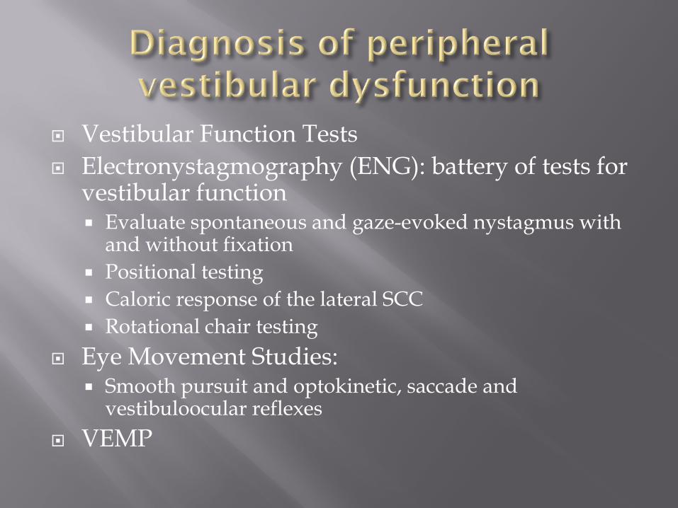
Vestibular Function Tests Electronystagmography (ENG): battery of tests for
vestibular function Evaluate spontaneous and gaze-evoked nystagmus with
and without fixation Positional testing Caloric response of the lateral SCC Rotational chair testing
Eye Movement Studies: Smooth pursuit and optokinetic, saccade and
vestibuloocular reflexes VEMP
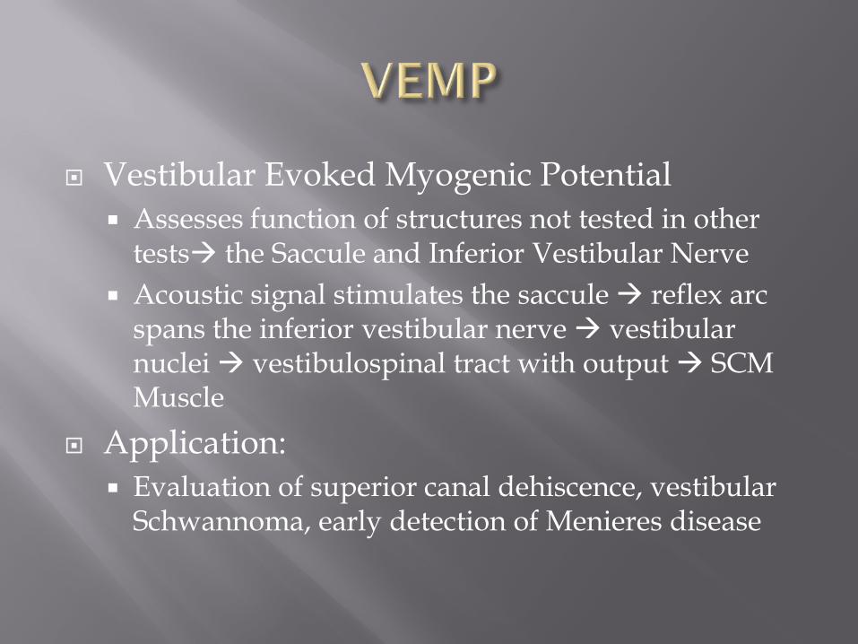
Vestibular Evoked Myogenic Potential Assesses function of structures not tested in other
tests the Saccule and Inferior Vestibular Nerve Acoustic signal stimulates the saccule reflex arc
spans the inferior vestibular nerve vestibular nuclei vestibulospinal tract with output SCM Muscle
Application: Evaluation of superior canal dehiscence, vestibular
Schwannoma, early detection of Menieres disease
If clinical evaluation and ENG suggest middle ear, mastoid, or IAC lesions consider MRI/CT

GOAL: decrease severity of symptoms and restore function Vestibulo-suppressive medication Ablative surgery Chemical labyrinthectomy Rehabilitation
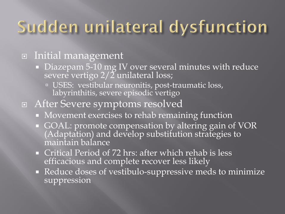
Initial management Diazepam 5-10 mg IV over several minutes with reduce
severe vertigo 2/2 unilateral loss; USES: vestibular neuronitis, post-traumatic loss,
labyrinthitis, severe episodic vertigo After Severe symptoms resolved
Movement exercises to rehab remaining function GOAL: promote compensation by altering gain of VOR
(Adaptation) and develop substitution strategies to maintain balance
Critical Period of 72 hrs: after which rehab is less efficacious and complete recover less likely
Reduce doses of vestibulo-suppressive meds to minimize suppression


Idiopathic endolyphatic hydrops Episodic vertigo + fluctuating SNHL + Tinntus
+ Aural fullness Pathologic Basis: distortion of membranous
labyrinth, 2/2 over-accumulation of endolyphfrom a dysfunction in absorption of the endolyph by the endolyphatic sac Histologic support of this theory

Major SymptomsVertigo
• Recurrent, well-defined episodes of spinning or rotation• Duration from 20 minutes to 24 hours.• Nystagmus associated with attacks• Nausea and vomiting during vertigo spells common• No neurologic symptoms with vertigo
Deafness• Hearing deficits fluctuate• Sensorineural hearing loss• Hearing loss progressive, usually unilateral
Tinnitus• Variable, often low-pitched and louder during attacks• Usually unilateral• Subjective

DiagnosisPossible Meniere's disease
• Episodic vertigo without hearing loss or• Sensorineural hearing loss, fluctuating or fixed, with dysequilibrium, but without
definite episodes• Other causes excluded
Probable Meniere's disease• One definitive episode of vertigo• Hearing loss documented by audiogram at least once• Tinnitus or sense of aural fullness in the presumed affected ear• Other causes excluded
Definite Meniere's disease• Two or more definitive spontaneous episodes of vertigo lasting at least 20
minutes• Audiometrically documented hearing loss on at least one occasion• Tinnitus or sense of aural fullness in the presumed affected ear• Other causes excluded
Certain Meniere's disease• Definite Meniere's disease, plus histopathologic confirmation
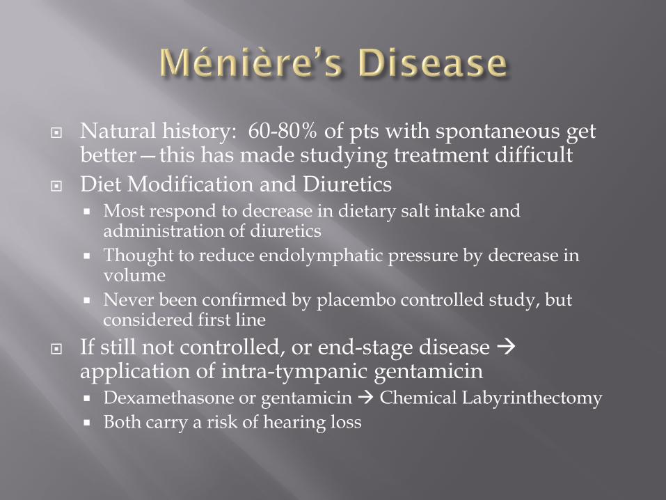
Natural history: 60-80% of pts with spontaneous get better—this has made studying treatment difficult
Diet Modification and Diuretics Most respond to decrease in dietary salt intake and
administration of diuretics Thought to reduce endolymphatic pressure by decrease in
volume Never been confirmed by placembo controlled study, but
considered first line If still not controlled, or end-stage disease
application of intra-tympanic gentamicin Dexamethasone or gentamicin Chemical Labyrinthectomy Both carry a risk of hearing loss

Surgical Intervention Endolyphatic Sac Surgery: double-blind placebo controlled study by Thomsen showed
mastoidectomy alone has same efficacy; controversial Vestibular neurectomy: vertigo control in 80-90%, more invasive and technically challenging
Labyrinthectomy: Most destructive procedure, uniform destruction of hearing and
vestibular function; Ideal candidate—no hearing and failed all conservative therapy High success rate, however used with caution in elderly—poor central
compensation Systemic Streptomycin: Chemical Labryinthectomy
Used for pts with bilateral Ménière's disease, only hearing ear, or poor surgical candidates
Streptomycin given IM BID with daily caloric testing and continued until caloric responses decrease

Due to motion of material in the lumen of the posteior SCC
Most common cause of vertigo—20-40% of cases seen by an otolaryngologist
History: severe vertigo with change in head position, especially rolling over or getting out of bed Most cases no etiologic disorder; Baloh published series
and half patients had no identified cause, the other half, most commonly had history of head injury or vestibular neuritis
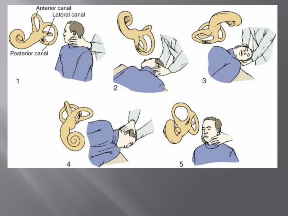
Diagnosis: History + Dix-Hallpike maneuver
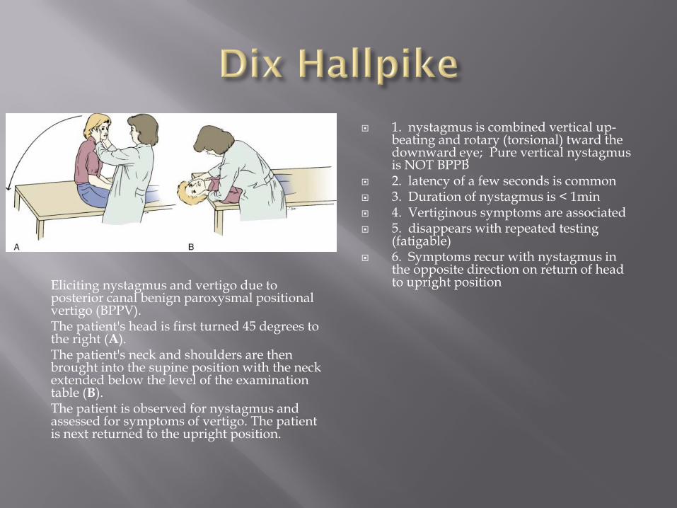
Eliciting nystagmus and vertigo due to posterior canal benign paroxysmal positional vertigo (BPPV). The patient's head is first turned 45 degrees to the right (A). The patient's neck and shoulders are then brought into the supine position with the neck extended below the level of the examination table (B). The patient is observed for nystagmus and assessed for symptoms of vertigo. The patient is next returned to the upright position.
1. nystagmus is combined vertical up-beating and rotary (torsional) tward the downward eye; Pure vertical nystagmus is NOT BPPB
2. latency of a few seconds is common 3. Duration of nystagmus is < 1min 4. Vertiginous symptoms are associated 5. disappears with repeated testing
(fatigable) 6. Symptoms recur with nystagmus in
the opposite direction on return of head to upright position
signs and symptoms of cupular deflection when otoconial debris either adhere to the crista (cupulolithiasis) or become free-floating within a SSC (canalithiasis)
Posterior SCC is most commonly involved Treatment:
Particle repositioning maneuvers are highly successfulEpley (next slide)
Surgery for incapacitating vertigo occlusion of the P-SCC with bone dust (97 cases within the
literature 94 cured) sectioning the posterior canal ampullary nerve is usually
effective series by Gacek of 340 pts 40% hearing loss
Dramatic sudden onset of vertigo, lasts for days, with gradual improvement NOT associated with subjective change in hearing or any
other focal neurologic complaint Cause is almost never identified, neurotropic viruses,
such as herpes or Borellia have been associated Treatment
Supportive and symptomatic Recent placebo-controlled, double blind study with
Methylprednisolone and Valacyclovir: found significantly improved 1 year symptoms with steroids, no effect of the valacyclovir
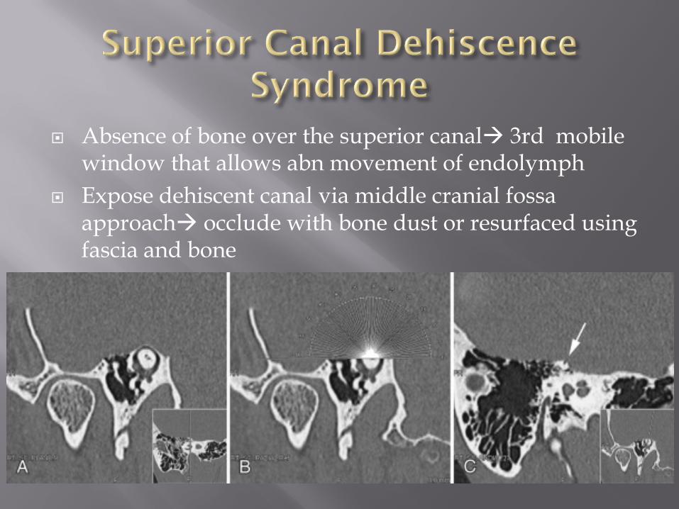
Absence of bone over the superior canal 3rd mobile window that allows abn movement of endolymph
Expose dehiscent canal via middle cranial fossa approach occlude with bone dust or resurfaced using fascia and bone

Interstitial keratitis, low-frequency unilateral HL, and vestibular symptoms (nonreactive syphilis testing)
Ocular and inner ear changes typically occur within 6 months of each other Vertigo: Simialar to Ménière’s Hearing loss: SNHL
Etiology: thought to be autoimmune, responds to steroids
Clinically: pt will report history of URI within 10 days of symptoms
Treatment: systemic corticosteroids, cyclophosphamide and methotrexate
Is highly effective, and can improve symptoms of vertigo of unknown causation and improve general balance conditioning in the elderly
Can include therapy at home, or under supervision of a physical therapist
Techniques: Vestibular adaptation is responsive for long-term
changes in vestibular input Promote altered gain for VOR and VSR by active
head and body movement combined with visual input