peripheral vascular diseases
TRANSCRIPT

Peripheral Vascular Diseases
By- Dr. Armaan Singh

Arteries
are thick-walled vessels that transport 02 and blood via the aorta from the heart to the tissues
3 Layers of Arteries1. inner layer of endothelium (intima) 2. middle layer of connective tissue, smooth muscle and elastic fibers (media)3.outer layer of connective tissue (adventitia)
have smooth muscles that contracts & relaxes to respond changes in blood volume.

Veins are thin-walled vessels that transport deoxygenated blood from the capillaries back to the right side of the heart
3 Layers – intima, media, adventitia
there is little smooth muscle & connective tissue makes the veins more distensible they accumulate large volumes of blood Major veins, particularly in the lower extremities, have one-way valves ---allow blood flow against gravity Valves allow blood to be pumped back to the heart but prevent it from draining back into the periphery

Peripheral Vascular Diseases charac. by a reduction in blood flow and hence 02 through the
peripheral vessels
when the need of the tissues for 02 exceeds the supply, areas of ischemia and necrosis will develop
Factors that can contribute to the development of peripheral vascular disorders :
atherosclerotic changes thrombus formation embolization coagulability of blood hypertension inflammatory process/infection

Arterial Insufficiency
there is a deceased blood flow toward the tissues, producing ischemia pulses one usually diminished or absent sharp, stabbing pain occurs because of the ischemia, particularly with activity there is interference with nutrients and 02 arriving to the tissues, leading to ischemic ulcers and changes in the skin.
Venous Insufficiency
there is deceased return of blood from the tissues to the heart leads to venous congestion and stasis of blood pulses are present lead to edema, skin changes and stasis ulcers
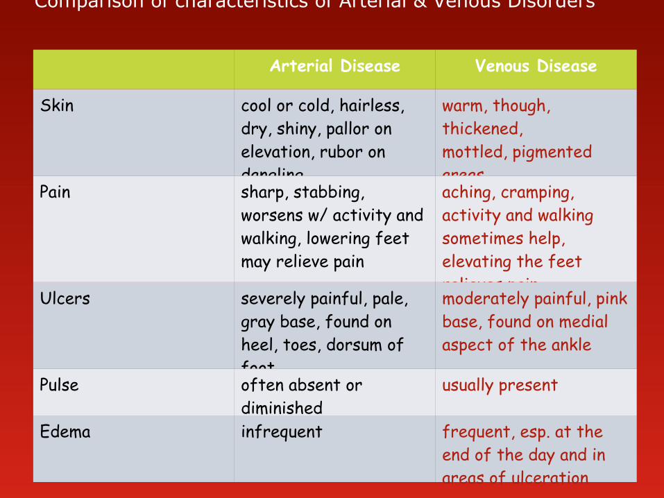
Comparison of characteristics of Arterial & Venous Disorders
Arterial Disease Venous Disease
Skin cool or cold, hairless, dry, shiny, pallor on elevation, rubor on dangling
warm, though, thickened, mottled, pigmented areas
Pain sharp, stabbing, worsens w/ activity and walking, lowering feet may relieve pain
aching, cramping, activity and walking sometimes help, elevating the feet relieves pain
Ulcers severely painful, pale, gray base, found on heel, toes, dorsum of foot
moderately painful, pink base, found on medial aspect of the ankle
Pulse often absent or diminished
usually present
Edema infrequent frequent, esp. at the end of the day and in areas of ulceration

Risk Factors
1. Age (elderly) – blood vessels become less elastic, become
thin walled and calcified – PVR – BP
2. Sex (male)
3. Cigarette smoking nicotine causes vasoconstriction and spasm of the arteries –
circulation to the extremities
C02 inhaled in cigarette smoke reduces 02 transport to tissues
4. Hypertension – cause elastic tissues to be replaced by fibrous collagen tissue arterial wall become less distensible resistance to blood flow BP
5. Hyperlipedimia – atherosclerotic plaque
6. Obesity – places added burden on the heart & blood vessels excess fat contribute to venous congestion

Risk Factors (cont.)
7. Lack of physical activity
Physical activity – promotes muscle contraction venous return to the heart
aids in development of collateral circulation
8. Emotional stress – stimulates sympathetic N.S. - peripheral vasoconstriction BP
9. Diabetes mellitus – changes in glucose & fat metabolism promote the atherosclerotic process
10. Family history of arthrosclerosis

Arteriosclerosis Obliterans is a disorder in which there is an arteriosclerotic
narrowing or obstruction of the inner & middle layer of the artery
most common cause of arterial obstructive disease in the extremities
the lower extremities are involved more than upper extremities
common site of disease – femoral artery, iliac arteries, popliteal arteries
in a diabetic, the disease becomes more progressive, affects the smaller arteries and often involves vessels below the knee


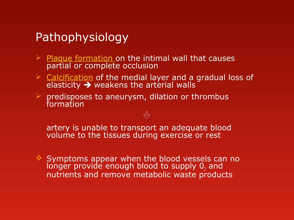
Pathophysiology
Plaque formation on the intimal wall that causes partial or complete occlusion
Calcification of the medial layer and a gradual loss of elasticity weakens the arterial walls
predisposes to aneurysm, dilation or thrombus formation
artery is unable to transport an adequate blood volume to the tissues during exercise or rest
Symptoms appear when the blood vessels can no longer provide enough blood to supply 02 and nutrients and remove metabolic waste products

Clinical Manifestations Intermittent claudication – most common
pain in the extremity that develops in a muscle that has an inadequate blood supply during exercise
the cramping pain disappear w/in 1-2 mins. after stopping the exercise or resting
the femoral artery is often affected – pain in the calf muscle – common symptom
pain at rest is indicative of severe disease gnawing, burning pain, occur more frequently at night
feelings of coldness numbness tingling sensation advanced arteriosclerosis obliterans ischemia may lead to necrosis,
ulceration and gangrene – toes and distal foot
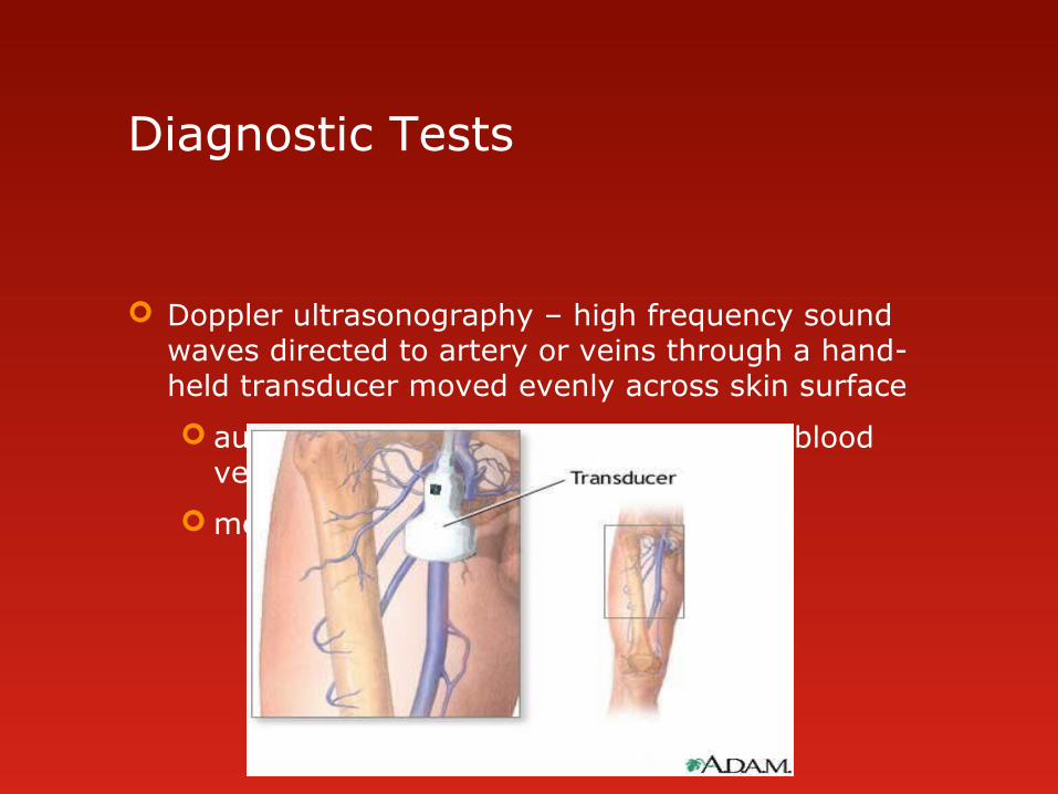
Diagnostic Tests
Doppler ultrasonography – high frequency sound waves directed to artery or veins through a hand-held transducer moved evenly across skin surface
audible tone produced in proportion to blood velocity
measure blood flow through vessels


Management – directed toward prevention of vessel occlusion use of vasodilators
Surgical intervention – in advanced disease – ischemic changes and pain severely impairs activity
Embolectomy removal of a blood clot, done when large arteries are
obstructed Endarterectomy
is removal of a blood clot and stripping of atherosclerotic plaque along with the inner arterial wall.
Arterial by-pass surgery an obstructed arterial segment may be bypassed by using a
prosthetic material (Teflon) or the pt’s. own artery or vein (saphenous vein)

Endarterectomy


Management Percutaneous Transluminal Angioplasty
The balloon tip of the catheter is inflated to provide compression of the plaque
Amputation
with advanced atherosclerosis & gangrene of extremities
toes are the most often amputated part of the body
The surgical goal is the remove the least amt. of tissue possible and create a stump adequate for the fitting of a prosthesis

Assessment
Nursing Interventions prevent further progression of existing disease
Acute care monitor the limb distal to the affected site for changes
in color or temperature
arterial flow – pale & cool (initially) bluish/darker tissue become necrotic & black
activities that cause pain should be avoided
give vasodilators if prescribed – relaxation of vascular smooth muscle decreases the pain
comfort measures – proper body positioning to dec. pressure on affected area

Post – operative care for arterial surgery
pt. is monitored for signs of circulation in the affected limb and interventions done to promote circulation & comfort
1. Assess and report changes in skin color and temperature distal to the surgical site, every 2-4 hrs.
2. assess peripheral pulses sudden absence of pulse may indicate thrombosis
mark location of pulse with a pen to facilitate frequent assessment
use a dapper if pulse in difficult to palpate
1. assess wound for redness, swelling and drainage
2. promote circulation
reposition pt. every 2 hrs.
tell pt. not to cross legs
encourage progressive activity when permitted
1. medication with analgesics to reduce pain

Arterial by-pass surgery
Post-operative care
assess sensation and movement of the limb
monitor extremity for edema
monitor & report signs of complications – increase pain, fever, limitation of movement or paresthesia
avoid sharp flexion in the area of the graft to prevent decreased circulation to the graft.

Thromboangitis Obliterans ( Buerger’s Disease)
characterized by acute inflammatory lesions and occlusive thrombosis of the arteries & veins
has a very strong assoc. with cigarette smoking
commonly occurs in male – bet. 20-40 y.o
may involve the arteries of the upper extremities (wrists)
usually affect the lower leg. toes, feet

Clinical Manifestation intermittent claudication in the arch of the foot
pain during rest – toes
coldness – due to persistent ischemia
paresthesia
pulsation in posterior tibial, dorsalis pedis – weak or absent
extremities are red or cyanotic
ulceration & gangrene are frequent complications – early can occur spontaneously but often follow trauma

Thromboangitis Obliterans

Interventions advise the person to stop smoking
vasodilators
prevent progression of disease
avoid trauma to ischemic tissues
relieve pain
provide emotional support
whiskey or brandy may be of some value during periods of exacerbations vasodilation
advise pt. to avoid mechanical, chemical or thermal injuries to the feet
Amputation of the leg is done only when the ff. occurs:
gangrene extends well into the foot
pain is severe and cannot be controlled
severe infection or toxicity occurs

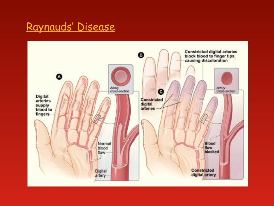
Raynaud’s phenomenon refers to intermittent episodes during which small arteries or
arterioles of L and R arm constrict (spasm) causing changes in skin color and temperature
generally unilateral and may affect only 1 or 2 fingers may occur after trauma, neurogenic lesions, occlusive arterial
disease, connective tissues disease charac. by reduction of blood flow to the fingers manifested by
cutaneous vessel constriction and resulting in blanching (pallor)
Raynauds’ Disease unknown etiology, may be due to immunologic abnormalities common in women 20-40 y.o maybe stimulated by emotional stress, hypersensitivity to cold,
alteration in sympathetic innervation
Raynauds’ Disease

Clinical Manifestations
usually bilateral –(both arms or feet are affected)
during arterial spasm – sluggish blood flow causes pallor, coldness, numbness, cutaneous cyanosis and pain
following the spasm – the involve area becomes intensely reddened with tingling and throbbing sensations
with longstanding or prolonged Raynaud’s disease – ulcerations can develop on the fingertips and toes

Raynauds’ Disease

Medical Management
aimed at prevention person is advised to protect against exposure to cold quit smoking Drug therapy – calcium channel blockers, vascular
smooth muscle relaxants, vasodilators – to promote circulation and reduce pain
sympathectomy ( cutting off of sympathetic nerve fibers) to relieve symptoms in the early stage of
advanced ischemia if ulceration/gangrene occur, the area may need to be
amputated
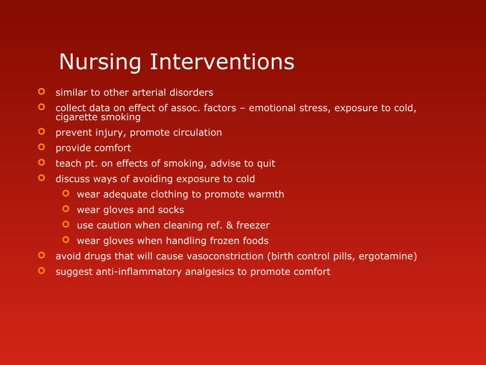
Nursing Interventions similar to other arterial disorders
collect data on effect of assoc. factors – emotional stress, exposure to cold, cigarette smoking
prevent injury, promote circulation
provide comfort
teach pt. on effects of smoking, advise to quit
discuss ways of avoiding exposure to cold
wear adequate clothing to promote warmth
wear gloves and socks
use caution when cleaning ref. & freezer
wear gloves when handling frozen foods
avoid drugs that will cause vasoconstriction (birth control pills, ergotamine)
suggest anti-inflammatory analgesics to promote comfort

Aneurysm
is a localized or diffuse enlargement of an artery at some point along its course
can occur when the vessel becomes weakened from trauma, congenital vascular disease, infection or atherosclerosis
Pathophysiology
enlargement of a segment of an artery the tunica media (middle layer composed of smooth muscle & elastic tissue) is damaged progressive dilation, degeneration risk of rupture
* most common site is the aorta
may develop in any blood vessel
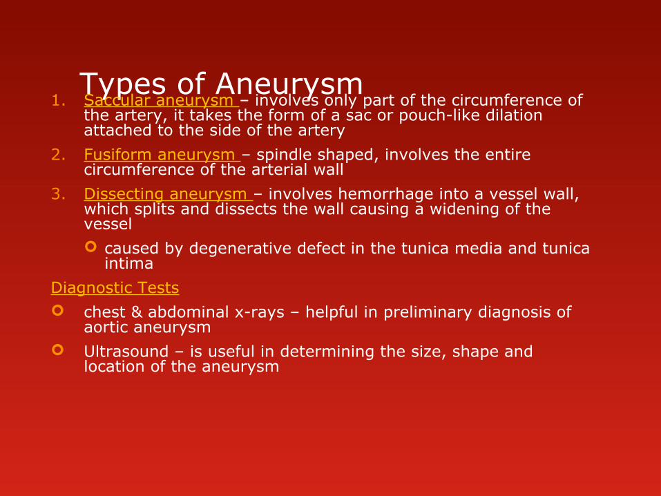
Types of Aneurysm1. Saccular aneurysm – involves only part of the circumference of
the artery, it takes the form of a sac or pouch-like dilation attached to the side of the artery
2. Fusiform aneurysm – spindle shaped, involves the entire circumference of the arterial wall
3. Dissecting aneurysm – involves hemorrhage into a vessel wall, which splits and dissects the wall causing a widening of the vessel caused by degenerative defect in the tunica media and tunica
intima
Diagnostic Tests chest & abdominal x-rays – helpful in preliminary diagnosis of
aortic aneurysm Ultrasound – is useful in determining the size, shape and
location of the aneurysm


Throracic Aortic Aneurysm aneurysm in the thoracic area occur most frequently in hypertensive men bet. 40-70
y.o can develop in the ascending, transverse or
descending aorta
S/Sx chest pain – most frequent; perceived when pt. is in a
supine position cough dyspnea hoarseness dysphagia
related to the pressure of the sac of aneurysm pressing against internal structures
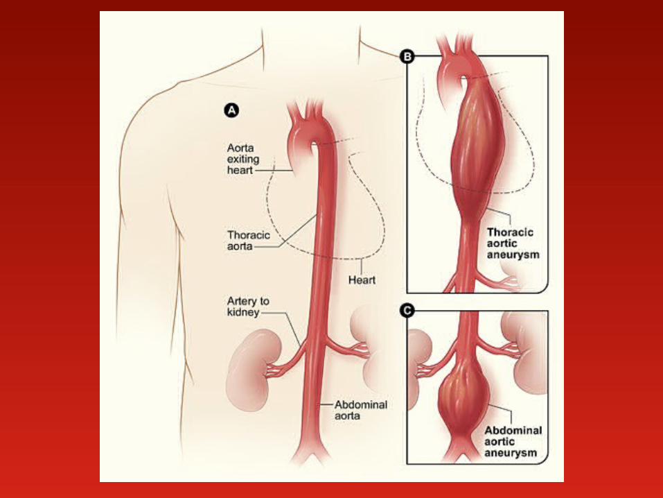
Abdominal Aortic Aneurysm most common site for the formation of an aortic aneurysm
abdominal aorta below the renal arteries
S/Sx: presence of a pulsatile abdominal mass on palpation pain or tenderness in the mid-or upper abdomen the aneurysm may extend to impinge on the renal, iliac, or
mesenteric arteries stasis of blood favors thrombus formation along the wall of the
vessel Rupture of the aneurysm – most feared complication can occur if the aneurysm is large can lead to death
Tx: Surgery – resection of the lesion and replacement with a graft


Arterial Embolism blood clots floating in the circulating arterial blood. the embolus is frequently a fragment of arterioscherotic plaque
loosened from the aorta emboli will tend to lodge in femoral or popliteal arteries, blood flow is
impaired and ischemia develops
Clinical manifestations: S/Sx depends on the size of the embolus, the presence of collateral
circulation and if it is close to a major organ abrupt onset of severe pain from the sudden cessation of circulation muscular weakness and burning, aching pain occur distal pulses are absent and extremity becomes cold, numb and pale symptoms of shock may develop if the embolus blocks a large artery

Medical Management
bed rest
anticoagulants – prolong the clotting time of the blood and are used to prevent clot extension and new clot formation
Ex. 1. heparin – inhibits thrombin action – prevents clotting
IV or SQ, antidote – Protamine sulfate
2. Warfarin sodium – inhibits Vit. K dependent clotting factor
(Coumadin) synthesis, prothrombin activity
- oral (10-15 mg/day) antidote – Vit. K
Fibrinolytics or thrombolytics – are useful for dissolving existing thrombus or clot when rapid dissolution of the clot is required to preserve organ and limb function
Ex. Streptokinase, Urokinase IV side effect - bleeding
Embolectomy – surgical removal of a blood clot, when large arteries are obstructed
must be performed w/in 6-10 hrs. to prevent muscle necrosis and loss of the extremity

Nursing Management
Monitor the pt. during the acute phase for changes in color & temp. of the extremity distal to the clot
assess for increasing pallor, cyanosis, coldness of the skin
indicates vessel occlusion
keep the extremity warm, but do not apply heat, avoid chilling
monitor peripheral pulses – quality – weak/absent
CBR - to prevent further progression of the embolism
keep affected extremity flat or slightly dependent position to promote circulation
monitor anticoagulant or fibrinolytic therapy & assess for signs of bleeding – nose or gum bleeding , petechiae (pinpoint red areas on skin), ecchymosis (bruising) , hematoma formation
monitor urine, stool, emesis and gastric secretions for blood
avoid IM injections, use soft toothbrush, use electric razor rather than razor blade, avoid rectal thermometer

Venous Disorders
alteration in the transport/flow of blood from the capillary back to the heart
changes in smooth muscle and connective tissue make the veins less distensible with limited recoil capacity
valves may malfunction, causing backflow of blood
Virchow’s triad: blood stasis, vessel wall injury, and altered blood coagulation
Thrombophlebitis inflammation of the veins caused by thrombus or blood clot
Factors assoc. with the devt. of Thrombophlebitis venous stasis damage to the vessel wall hypercoagulability of the blood – oral contraceptive use common to hospitalized pts. , undergone major surgery (pelvic or hip
surgery), MI
Pathophysiology develops in both the deep and superficial veins of the lower extremity deep veins – femoral, popliteal, small calf veins superficial veins – saphenous vein Thrombus – form in the veins from accumulation of platelets, fibrin,
WBC and RBC
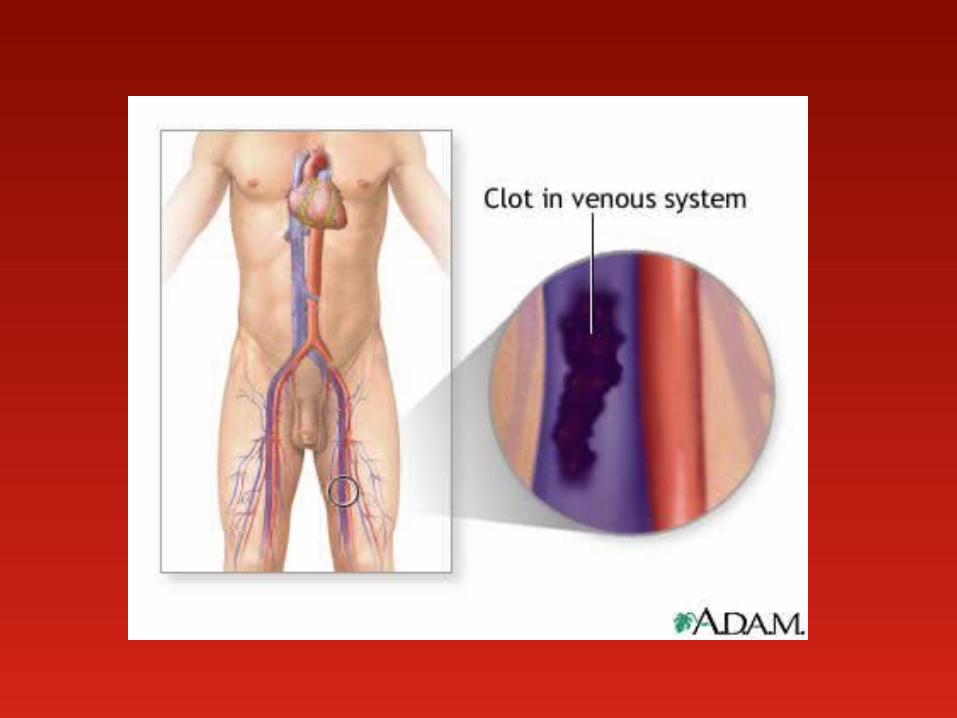

Deep Vein Thrombosis (DVT) tends to occur at bifurcations of the deep veins, which are sites of
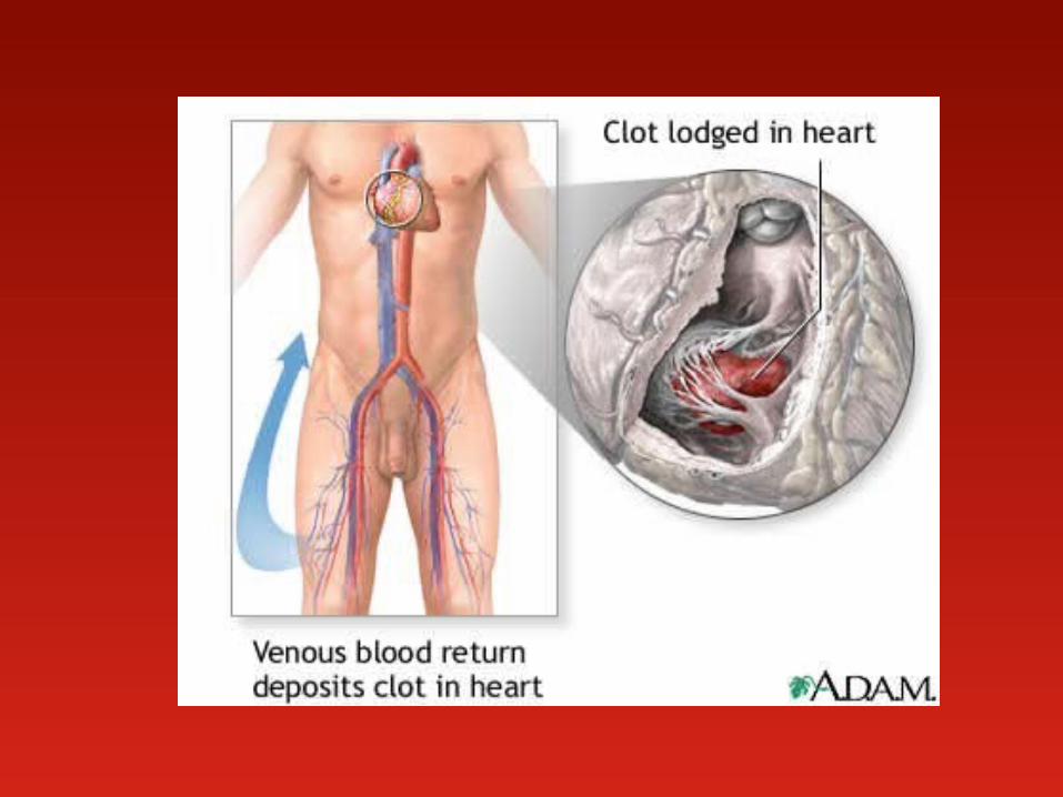
turbulent blood flow a major risk during the acute phase of thrombophlebitis is dislodgment of
the thrombus embolus pulmonary embolus – is a serious complication arising from DVT of the
lower extremities
Clinical Manifestations: pain and edema of extremity – obstruction of venous flow circumference of the thigh or calf (+) Homan’s sign – dorsiflexion of the foot produces calf pain Do not check for the Homan’s sign if DVT is already known to be present
risk of embolus formation * if superficial veins are affected - signs of inflammation may be noted –
redness, warmth, tenderness along the course of the vein, the veins feel hard and thready & sensitive to pressure

Deep Vein Thrombosis (DVT)

Medical Management
Superficial thrombophlebitis
bed rest with legs elevated
apply moist heat
NSAID’s ( Non – steroidal anti-inflammatory drugs) - aspirin
Deep vein thrombosis
requires hospitalization
bed rest w/ legs elevated to 15-20 degrees above heart level ( knees slightly flexed, trunk horizontal (head may be raised) to promote venous return and help prevent further emboli and prevent edema
application of warm moist heat to reduce pain, promotes venous return
elastic stocking or bandage
anticoagulants, initially with IV heparin then coumadin
fibrinolytic to resolve the thrombus
vasodilator if needed to control vessel spasm and improve circulation

Surgery
if the thrombus is recurrent and extensive or if the pt. is at high risk for pulmonary embolism
Thrombectomy – incising the common femoral vein in the groin and extracting the clots
Vena caval interruption – transvenous placement of a grid or umbrella filter in the vena cava to block the passage of emboli
Assessment
characteristic of the pain
onset & duration of symptoms
history of thrombophlebitis or venous disorders
color & temp. of extremity
edema of calf of thigh - use a tape measure, measure both legs for comparison


Nursing InterventionPreventive care prevent long periods of standing or sitting that impair venous
return elevate legs when sitting, dorsiflex feet at regular intervals to
prevent venous pooling if edema occurs, elevate above heart level regular exercise program to promote circulation avoid crossing legs at the knees avoid wearing constrictive clothing such as tight bands around
socks or garters use elastic stocking on affected leg do leg exercises during periods of enforced immobility such as
after surgery

Nursing Management
Acute care
explain purpose of bed rest and leg elevation
use elastic stockings
monitor pt. on anticoagulant & fibrinolytic therapy for signs of bleeding
monitor for signs of pulmonary embolism – sudden onset of chest pain, dyspnea, rapid breathing, tachycardia
Nsg. intervention often surgery of vena caval interruption
assess insertion site – bleeding, hematoma, apply pressure over site and inform physician
keep pt. on bed rest for 1st 24 hrs. then encourage ROM exercises to promote venous return
assist pt. in ambulation when permitted, elevate legs when sitting
keep elastic bandage
avoid rubbing or massaging the affected extremity
give analgesic and anti-inflammatory agents to promote comfort

Chronic Venous Insufficiency Results from obstruction of venous valves
in legs or reflux of blood back through valves
Venous ulceration is serious complication Pharmacological therapy is antibiotics for
infections Debridement to promote healing Topical Therapy may be used with
cleansing and debridement

Venous ulceration
Varicose Veins are abnormally dilated veins with incompetent valves,
occurring most often in the lower extremities
usually affected are woman 30-50 years old.
Causes:
congenital absence of a valve
incompetent valves due to external pressure on the veins from pregnancy, ascites or abdominal tumors
sustained in venous pressure due to CHF, cirrhosis
Prevention
wear elastic stockings during activities that require long standing or when pregnant
moderate exercise, elevation of legs


Pathophysiology
the great and small saphenous veins are most often involved
weakening of the vein wall does not withstand normal pressure
veins dilate , pooling of blood
valves become stretched and incompetent
more accumulation of blood in the veins

Clinical Manifestations Primary varicosities – gradual onset and affect
superficial veins, appearance of dark tortuous veins
S/sx – dull aches, muscle cramps, pressure, heaviness or fatigue arising from reduced blood flow to the tissues
Secondary Varicosities – affect the deep veins
occur due to chronic venous insufficiency or venous thrombosis
S/sx – edema, pain, changes in skin color, ulcerations may occur from venous stasis
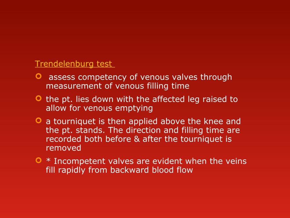
Trendelenburg test
assess competency of venous valves through measurement of venous filling time
the pt. lies down with the affected leg raised to allow for venous emptying
a tourniquet is then applied above the knee and the pt. stands. The direction and filling time are recorded both before & after the tourniquet is removed
* Incompetent valves are evident when the veins fill rapidly from backward blood flow

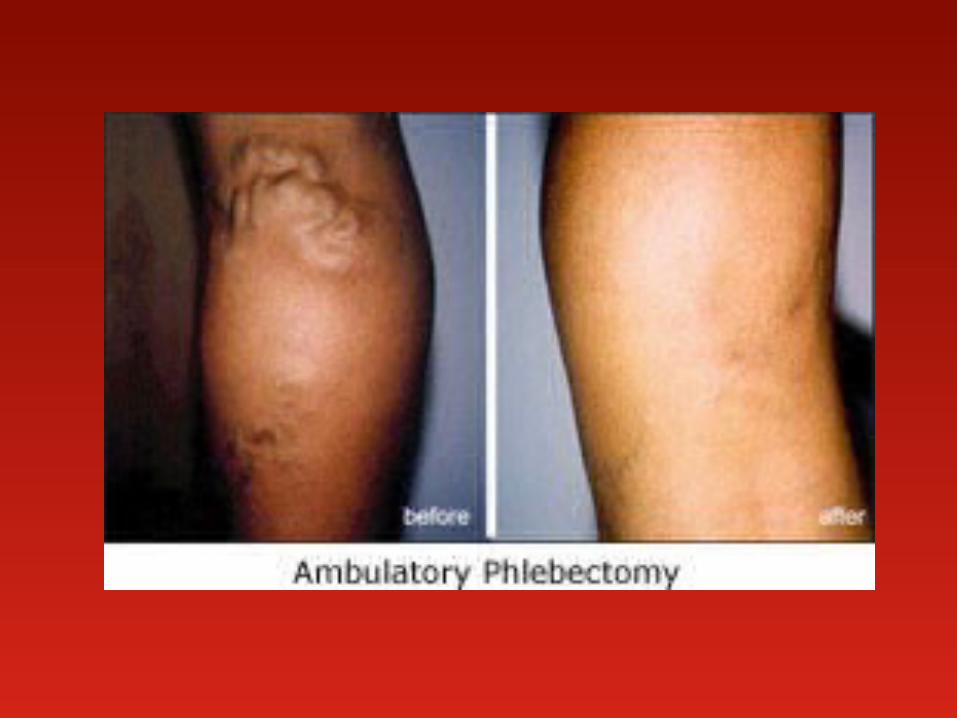
Surgical Intervention indicated or done for prevention or relief of edema, for
recurrent leg ulcers or pain or for cosmetic purposes
Vein ligation and stripping
the great sapheneous vein is ligated (tied) close to the femoral junction
the veins are stripped out through small incisions at the groin, above & below the knee and at the ankles.
sterile dressing are placed over the incisions and an elastic bandage extending from the foot to the groin is firmly applied

Vein ligation and stripping

Nursing care after vein ligation & stripping Monitor for signs of bleeding, esp. on 1st post-op day
if there is bleeding, elevate the leg, apply pressure over the wound and notify the surgeon
Keep pt. flat on bed for first 4 hrs. after surgery, elevate leg to promote venous return when lying or sitting
Medicate 30 mins. before ambulation and assist patient
Keep elastic bandage snug and intact, do not remove bandage