perioperative heat balance - canadian patient safety...
TRANSCRIPT
m REVIEW ARTICLE
Michael M. Todd, M.D., Editor
Anesthesiology2000; 92:578–96© 2000 American Society of Anesthesiologists, Inc.Lippincott Williams & Wilkins, Inc.
Perioperative Heat BalanceDaniel I. Sessler, M.D.*
ANESTHESIA and surgery commonly cause substantial ther-mal perturbations. Hypothermia, the typical alteration, re-sults from a combination of anesthetic-induced impairmentof thermoregulatory control, a cool operating room envi-ronment, and factors unique to surgery that promote ex-cessive heat loss. Available data suggest that inhibition ofnormal thermoregulatory defenses contributes more to hy-pothermia than does cold exposure per se. Furthermore,much core hypothermia results from altered distribution ofbody heat rather than from a systemic imbalance betweenmetabolic heat production and heat loss.
General Features of Heat Production andDistribution
Tissue temperature is directly related to heat contentby the specific heat of tissue, which is approximately
0.83 kcal z kg z 21°C21.1,2 Body temperature perturba-tions are thus a direct consequence of alterations intissue heat content. How heat is generated and distrib-uted within the body is therefore of considerable interest.
Heat ProductionBody tissues produce heat in proportion to their met-
abolic rates.3,4 The First Law of Thermodynamics speci-fies that energy generation by a chemical reaction isdetermined only by the substrates and products of thereaction. Nearly all of this energy is eventually convertedto heat. The major substrates for human metabolism areglucose, protein, and fat; the major products of aerobicmetabolism are generally carbon dioxide and water.Combustion of glucose and protein produces 4.1 kcal/kg, whereas fat releases 9.3 kcal/kg.
The brain and major organs in the trunk are the mostmetabolically active tissues and generate more metabolicheat than does muscle at rest.5–7 In contrast, heat pro-duction by skeletal muscle can, at least briefly, exceedthe basal metabolic rate by a factor of 10. Metabolism isnormally the only internal source of heat, although in-ternal heating can result from drinking warm beverages,microwave radiation, or magnetic resonance gradientcoils.8
Thermal CompartmentsThe human body can very roughly be described as
having a core thermal compartment and a peripheralcompartment.1 The core is defined by well-perfusedtissues in which temperature remains relatively uniform.In other words, distribution of heat within this compart-ment is fast compared with rates at which heat contentnormally changes. As a result, temperatures at varioussites within the core compartment rarely differ by morethan a few tenths of a degree centigrade. Physically, thecore compartment consists of the trunk and head. Ofcourse, the skin and most peripheral tissues of the trunkand head are not technically part of the core; however,
* Professor, Department of Anesthesia, University of California—SanFrancisco; Professor, Ludwig Boltzmann Institute for Clinical Anesthe-sia and Intensive Care; Director Outcomes Research™, Professor, andVice-Chair, Department of Anesthesia and General Intensive Care,University of Vienna.
Received from the Outcomes Research™ Group, Department ofAnesthesia, University of California—San Francisco, San Francisco,California; the Ludwig Boltzmann Institute for Clinical Anesthesia andIntensive Care; and the Department of Anesthesia and General Inten-sive Care, University of Vienna. Submitted for publication June 11,1999. September 22, 1999. Some of the studies presented in thisreview were supported by National Institutes of Health, grantGM58273, Bethesda, Maryland; the Joseph Drown Foundation, LosAngeles, California; the Fonds zur Forderung der wissenschaftlichenForschung, Vienna, Austria; the Burgermeister Fond der Stadt Wien,Vienna, Austria; and the Austrian National Bank Fund, Vienna, Austria.The author does not consult for, accept honoraria from, or own stockor stock options in any company related to this review.
Address correspondence to Dr. Sessler: Department of Anesthesia,University of California—San Francisco, 374 Parnassus Avenue, 3rdFloor, San Francisco, California 94143-0648. Address electronic mail to:[email protected]. On the world wide web: http://or.org
Reprints will not be available.
Key words: Anesthesia; hypothermia; temperature; thermoregulation.
578
Anesthesiology, V 92, No 2, Feb 2000
little accuracy is lost by ignoring this mass. The corecompartment therefore comprises 50–60% of the bodymass.9
Tissues in which temperature is nonhomogeneous andvariable over time define the peripheral thermal com-partment. Physically, this compartment consists of thearms and legs. Temperature of the peripheral compart-ment is usual 2–4°C less than the core temperature inmoderate environments.1 However, this difference canbecome large during extreme thermal10 or physiologic11
circumstances. Lower core-to-peripheral temperaturegradients result when the environment is warm or whenthermoregulatory vasodilation allows facile flow of met-abolic heat (which is largely generated in the core) tothe periphery. In contrast, vasoconstriction constrainsmetabolic heat to the core, increasing the core-to-periph-eral temperature gradient.
There are typically substantial longitudinal tissue tem-perature gradients within the extremities, with distaltissues being several degrees cooler than those posi-tioned more proximally. There are also often substantialradial tissue temperature gradients, which become espe-cially large in extreme thermal environments.10,12 A car-dinal feature of the peripheral compartment is that heatcontent and distribution change substantially over timeand as a function of environmental exposure.13 This is indistinct contrast to the core for which temperature isusually precisely regulated.
In a warm environment, especially in vasodilated sub-jects, tissue temperature in much of the peripheral com-partment can equal core temperature. Some authorsrefer to this situation as an expansion of the core com-partment. However, temperature of these tissues willdecrease and again become nonuniform in a cooler en-vironment. It is thus easier to consider physical size ofthe compartments to be fixed and instead allow temper-ature of the peripheral compartment to vary. It is notwrong to permit flexibility in both the size and thetemperature of the compartments; however, doing soadds unnecessary complexity to the discussion.
Heat Flow and GenerationIn contrast to the rapid distribution of heat within core
tissues, heat flows relatively slowly to peripheral tissues.Core-to-peripheral flow of heat is mediated by blood-borne convection of heat and conduction of heat intoadjacent tissues.14 The convective component can veryroughly be modeled as a large and relatively rapid longi-tudinal flow of heat within the big axial vessels of theextremities. The major factors that influence convective
distribution of heat are peripheral blood flow, counter-current heat exchange between adjacent arteries andveins, and the core-to-peripheral temperature gradient.
The conductive component, in contrast, is a slowerradial flow of heat from relatively warm tissue at thecentral axis to cooler tissues near the skin. Conductiveflow is largely determined by the diffusion coefficient,which is to a great extent a function of tissue character-istics. For example, fat insulates nearly three times aswell as muscle15 and provides substantial insulation.16,17
Hence conductive heat transfer depends mostly on in-trinsic tissue characteristics rather than on thermoregu-latory factors, such as vasomotion.
Deposition of heat into peripheral tissues is augmentedby heat that is produced by local tissue metabolism anddiminished by regional cutaneous heat loss to the envi-ronment. Regional heat transfer and local production isfully described by the bioheat equation.18 However, aprecise solution to this equation requires numerous in-put values that cannot normally be measured. Conse-quently, a number of equations have been developed toquantify various aspects of tissue heat transfer and localheat production in terms of measurable quantities.19,20
Modifications of the bioheat equation have been usedto construct numerous models of tissue heat distribu-tion.20,21 Even simple models perform reasonably wellwithin a restricted set of circumstances, such as cold-water immersion. However, they usually prove less ac-curate during other conditions, prompting refinementsin subsequent generations of the models. As a result,some models have become quite complicated.22
Nonetheless, most models of tissue heat flow havebeen validated only to a limited extent, and many donot include a sophisticated thermoregulatory compo-nent. Ignoring thermoregulatory compensations is ac-ceptable in a situation of extreme thermal stress inwhich it is reasonable to assume that warm or colddefenses are maximally activated. However, failure toinclude thermoregulatory defenses can be a substan-tial limitation in more typical environments in whichactive control—rather than environmental expo-sure—is the major determinant of core temperature.Heat balance models therefore usually have proven tobe less helpful than initially hoped, leaving investiga-tors largely dependent on experimental data. Fortu-nately, detailed measurements are becoming availablein a variety of circumstances.9,12,13,20,23–26
All metabolic heat must eventually be dissipated to theenvironment to maintain thermal steady state. Approxi-mately 95% of this heat traverses the skin surface, with
579
PERIOPERATIVE HEAT BALANCE
Anesthesiology, V 92, No 2, Feb 2000
the remaining small amount being lost via the respira-tory track.27,28 In the absence of sweating, only about10% of cutaneous heat loss is evaporative in adults (al-though the fraction can be considerable in infants, espe-cially premature infants).29 Sweating, however, is re-markably effective and can dissipate 10 times the basalmetabolic rate in a dry, convective environment.30,31
The upper chest and face are most sensitive to temper-ature. However, the common belief that half of bodyheat is lost through the head only applies when the restof the body is covered by an arctic survival suit.32 In-stead, with comparable levels of insulation, heat loss isroughly proportional to surface area over the entire bodyand is not much changed by thermoregulatory vasomo-tion.33,34
General Anesthesia
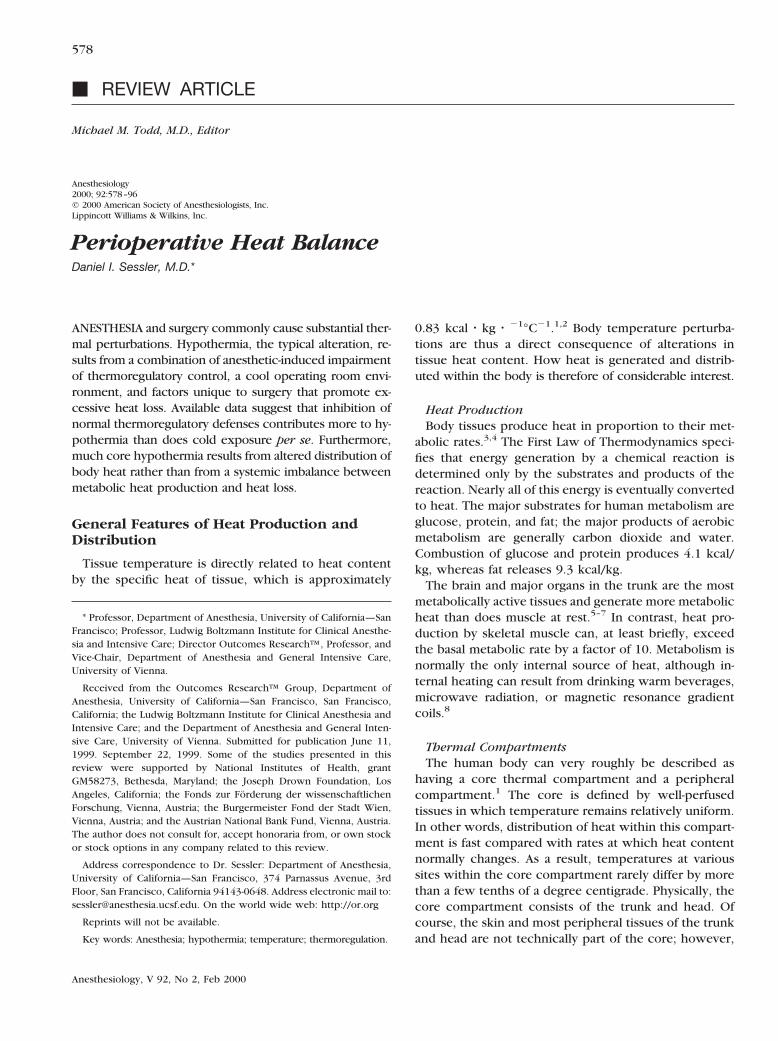
Nearly all patients administered general anesthesia be-come hypothermic, typically by 1–3°C,35 depending onthe type and dose of anesthesia, amount of surgicalexposure,36,37 and ambient temperature.36–38 Hypother-mia develops with a characteristic pattern (fig. 1).1 Coretemperature decreases 1–1.5°C during the first hour.2
This initial hypothermia is followed by 2 or 3 h of a
slower, linear, decrease in core temperature.3 Finally,patients enter a plateau phase during which core tem-perature remains constant. Each segment of this typicalhypothermia curve has a different cause.
RedistributionThe core thermal compartment consists of well-per-
fused tissues of the trunk and head that are maintained ata relatively high temperature. Core temperature, though,poorly represents mean body temperature because pe-ripheral tissues are typically 2–4°C cooler than the trunkand the head.9,26 This normal core-to-peripheral tissuetemperature gradient is maintained by tonic thermoreg-ulatory vasoconstriction of arteriovenous shunts in thefingers and toes.39–42
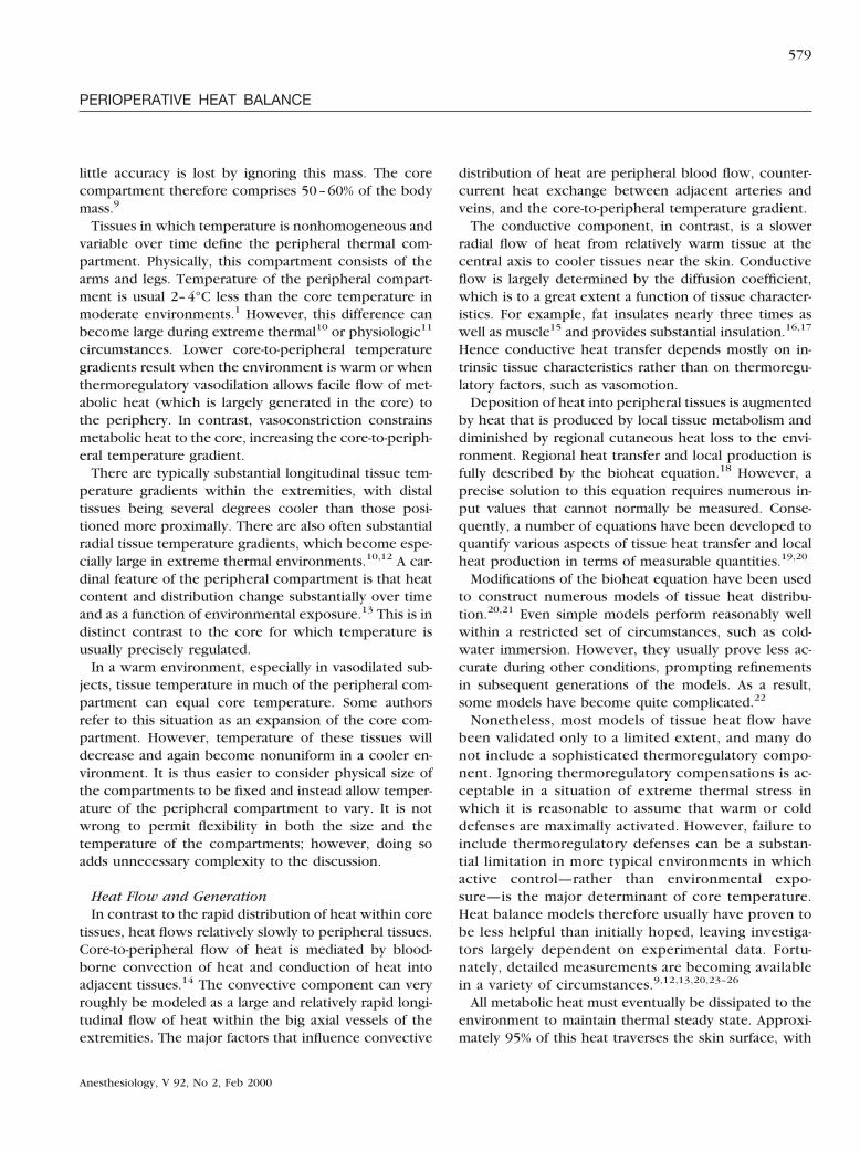
Mechanism. Induction of general anesthesia pro-motes vasodilation via two mechanisms: (1) Generalanesthesia reduces the vasoconstriction threshold towell below core temperature,43–48 thus inhibiting cen-trally medicated thermoregulatory constriction. (2) Mostanesthetics cause direct (peripheral) vasodilation.49 Va-sodilation allows core heat, which is no longer con-strained to the central thermal compartment, to flowdown the temperature gradient into peripheral tissues(fig. 2).50 This internal redistribution of body heat de-creases core temperature and proportionately increasestemperature of peripheral tissues; it does not, however,represent any net exchange of heat to the environment,and body heat content remains constant. Of course anysystemic cooling that occurs simultaneously will aug-ment core hypothermia.
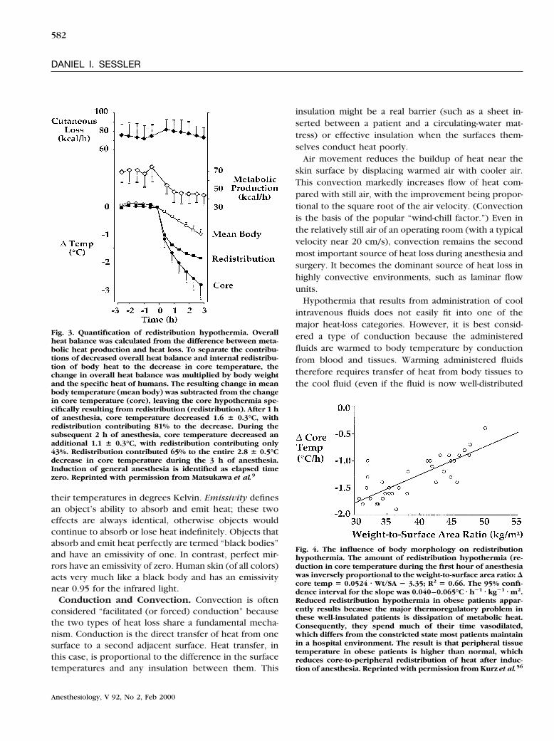
A quantitative study of systemic heat balance and re-gional body heat distribution in volunteers indicated thatafter 1 h of anesthesia, core temperature decreased1.6°C, with redistribution contributing 81% to the de-crease. During the subsequent 2 h of anesthesia, coretemperature decreased an additional 1.1°C, with redis-tribution contributing only 43%. Redistribution thereforecontributed 65% to the total decrease in core tempera-ture during the first 3 h of anesthesia (fig. 3).9 Core-to-peripheral redistribution was therefore the primarycause of hypothermia during the initial phase of anes-thesia—and in this study remained the dominant causeeven after 3 h.
Major Factors Influencing Magnitude. The extentto which induction of general anesthesia induces redis-tribution hypothermia in individual patients depends ona number of factors. Among the most important is thepatient’s initial body heat content. Core temperature,which is precisely regulated, remains essentially normal
Fig. 1. Typical pattern of hypothermia during general anesthe-sia. Hypothermia during anesthesia develops with a character-istic pattern. During the first hour, core temperature usuallydecreases 1–1.5°C. This is followed by a slower, nearly lineardecrease in core temperature. Finally, core temperature reachesa plateau and does not decrease further. Each segment of thishypothermia curve has a different cause. Reprinted with per-mission from Kurz et al.80
580
DANIEL I. SESSLER
Anesthesiology, V 92, No 2, Feb 2000
even in warm environments.51,52 However, body heatcontent increases as peripheral tissues absorb heat; aftera number of hours in a sufficiently warm environment,peripheral tissue temperature approaches core temper-ature.53 Because flow of heat needs a temperature gra-dient, redistribution magnitude is limited when periph-eral and core temperatures are similar.54,55
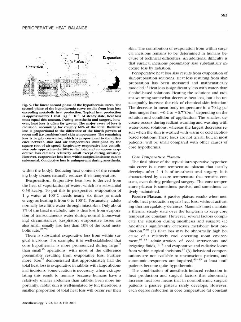
An additional important factor is body morphology.Obese patients redistribute considerably less than thoseof normal weight; conversely, very thin patients redis-tribute more (fig. 4).56 Reduced redistribution hypother-mia in obese patients apparently results because themajor thermoregulatory problem in these well-insulatedpatients is dissipation of metabolic heat. Consequently,they spend much of their time in vasodilation, whichdiffers from the constricted state most patients maintainin a hospital environment. The result is that their periph-eral tissue temperature is higher than normal, whichreduces core-to-peripheral flow of heat after induction ofanesthesia.
The fractional contribution of redistribution to corehypothermia naturally depends on the amount of redis-tribution. However, it also depends on the contributionof systemic heat loss, which in turn depends on numer-ous factors (discussed in Linear Phase section). Factorsthat increase systemic heat imbalance, such as a coolenvironment36–38 or large surgical incision,57 will in-crease the total amount of core hypothermia but de-crease the fraction that results from redistribution.
Linear PhaseThe second portion of the hypothermia curve is a
relatively slow, linear decrease in core temperature. It
results simply from heat loss exceeding metabolic heatproduction. Metabolic rate is reduced by 15–40% byduring general anesthesia.9,58,59 The exact causes for thisreduction are not well-established, but decreased brainmetabolism contributes, as does mechanical ventilation,which spares the diaphragm and chest wall muscles. Tosome extent, the process can be reversed by administra-tion of amino acids59,60 or caffeine.61–63
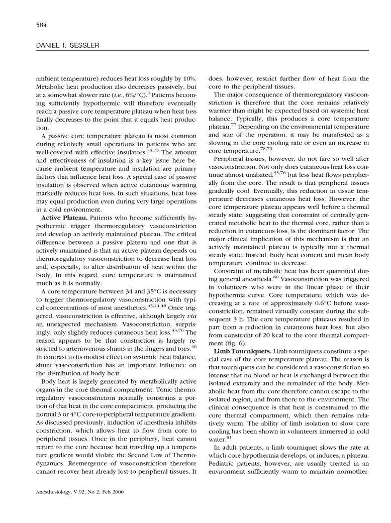
Cutaneous heat loss is mediated by the same fourfundamental mechanisms that modulate heat transferbetween any two substances: radiation, conduction,convection, and evaporation (fig. 5).64 Among the fourheat loss pathways, only conductive loss depends lin-early on the temperature difference between patientsand their environments. Nonetheless, surprisingly littleaccuracy is lost by considering total heat loss from allfour mechanisms to be a linear function of the skin-to-ambient temperature difference over the typical range ofoperating room temperatures.
It is during the linear phase the factors altering heatloss are most apparent. This includes relatively great heatloss in infants and children, more hypothermia duringlarge than small operations, and reduced hypothermiawhen surgery is performed in a warm environment.36–38
This is also the period in which passive insulation andactive intraoperative heating is most effective.65–68
Radiation. Among the four potential heat-loss mech-anisms, radiation usually contributes most.69 Radiation isthe transfer of heat from one surface to another viaphotons and therefore does not depend at all on thetemperature of the intervening air. The amount of heatlost by radiation is a function of the emissivity of the twosurfaces and the difference between the fourth power of
Fig. 2. Cartoon showing redistributionhypothermia after induction of generalanesthesia. Body heat content is not nor-mally distributed evenly. Instead, tonicthermoregulatory vasoconstriction main-tains a core-to-peripheral temperaturegradient. Induction of general anesthesiainhibits vasoconstriction, allowing acore-to-peripheral redistribution of bodyheat. Reprinted with permission fromSessler DI: Temperature monitoring, An-esthesia, 4th edition. Edited by Miller RD.New York, Churchill Livingstone, 1994,pp 1363–82.
581
PERIOPERATIVE HEAT BALANCE
Anesthesiology, V 92, No 2, Feb 2000
their temperatures in degrees Kelvin. Emissivity definesan object’s ability to absorb and emit heat; these twoeffects are always identical, otherwise objects wouldcontinue to absorb or lose heat indefinitely. Objects thatabsorb and emit heat perfectly are termed “black bodies”and have an emissivity of one. In contrast, perfect mir-rors have an emissivity of zero. Human skin (of all colors)acts very much like a black body and has an emissivitynear 0.95 for the infrared light.
Conduction and Convection. Convection is oftenconsidered “facilitated (or forced) conduction” becausethe two types of heat loss share a fundamental mecha-nism. Conduction is the direct transfer of heat from onesurface to a second adjacent surface. Heat transfer, inthis case, is proportional to the difference in the surfacetemperatures and any insulation between them. This
insulation might be a real barrier (such as a sheet in-serted between a patient and a circulating-water mat-tress) or effective insulation when the surfaces them-selves conduct heat poorly.
Air movement reduces the buildup of heat near theskin surface by displacing warmed air with cooler air.This convection markedly increases flow of heat com-pared with still air, with the improvement being propor-tional to the square root of the air velocity. (Convectionis the basis of the popular “wind-chill factor.”) Even inthe relatively still air of an operating room (with a typicalvelocity near 20 cm/s), convection remains the secondmost important source of heat loss during anesthesia andsurgery. It becomes the dominant source of heat loss inhighly convective environments, such as laminar flowunits.
Hypothermia that results from administration of coolintravenous fluids does not easily fit into one of themajor heat-loss categories. However, it is best consid-ered a type of conduction because the administeredfluids are warmed to body temperature by conductionfrom blood and tissues. Warming administered fluidstherefore requires transfer of heat from body tissues tothe cool fluid (even if the fluid is now well-distributed
Fig. 4. The influence of body morphology on redistributionhypothermia. The amount of redistribution hypothermia (re-duction in core temperature during the first hour of anesthesiawas inversely proportional to the weight-to-surface area ratio: Dcore temp 5 0.0524 z Wt/SA 2 3.35; R2 5 0.66. The 95% confi-dence interval for the slope was 0.040–0.065°C z h21 z kg21 z m2.Reduced redistribution hypothermia in obese patients appar-ently results because the major thermoregulatory problem inthese well-insulated patients is dissipation of metabolic heat.Consequently, they spend much of their time vasodilated,which differs from the constricted state most patients maintainin a hospital environment. The result is that peripheral tissuetemperature in obese patients is higher than normal, whichreduces core-to-peripheral redistribution of heat after induc-tion of anesthesia. Reprinted with permission from Kurz et al.56
Fig. 3. Quantification of redistribution hypothermia. Overallheat balance was calculated from the difference between meta-bolic heat production and heat loss. To separate the contribu-tions of decreased overall heat balance and internal redistribu-tion of body heat to the decrease in core temperature, thechange in overall heat balance was multiplied by body weightand the specific heat of humans. The resulting change in meanbody temperature (mean body) was subtracted from the changein core temperature (core), leaving the core hypothermia spe-cifically resulting from redistribution (redistribution). After 1 hof anesthesia, core temperature decreased 1.6 6 0.3°C, withredistribution contributing 81% to the decrease. During thesubsequent 2 h of anesthesia, core temperature decreased anadditional 1.1 6 0.3°C, with redistribution contributing only43%. Redistribution contributed 65% to the entire 2.8 6 0.5°Cdecrease in core temperature during the 3 h of anesthesia.Induction of general anesthesia is identified as elapsed timezero. Reprinted with permission from Matsukawa et al.9
582
DANIEL I. SESSLER
Anesthesiology, V 92, No 2, Feb 2000
within the body). Reducing heat content of the remain-ing body tissues naturally reduces their temperature.
Evaporation. Evaporative heat loss is derived fromthe heat of vaporization of water, which is a substantial0.58 kcal/g. To put this in perspective, evaporation of1 g water at 100°C needs nearly six times as muchenergy as heating it from 0 to 100°C. Fortunately, adultsnormally lose little water through intact skin. Only about5% of the basal metabolic rate is thus lost from evapora-tion of transcutaneous water during normal (nonsweat-ing) circumstances. Respiratory evaporative losses arealso small, usually also less than 10% of the basal meta-bolic rate.27,70
There is substantial evaporative loss from within sur-gical incisions. For example, it is well-established thatcore hypothermia is more pronounced during large37
than small36 operations, with most of the differencepresumably resulting from evaporative loss. Further-more, Roe57 demonstrated that approximately half thetotal heat loss is evaporative in rabbits with large abdom-inal incisions. Some caution is necessary when extrapo-lating this result to humans because humans have arelatively smaller abdomen than rabbits. Even more im-portantly, rabbit skin is well-insulated by fur; therefore, asmaller proportion of total heat loss will occur via their
skin. The contribution of evaporation from within surgi-cal incisions remains to be determined in humans be-cause of technical difficulties. An additional difficulty isthat surgical incisions presumably also substantially in-crease loss by radiation.
Perioperative heat loss also results from evaporation ofskin-preparation solutions. Heat loss resulting from skinpreparation has been measured and mathematicallymodeled.71 Heat loss is significantly less with water- thanalcohol-based solutions. Heating the solutions and radi-ant warming somewhat decrease heat loss, but also un-acceptably increase the risk of chemical skin irritation.The decrease in mean body temperature in a 70-kg pa-tient ranges from 20.2 to 20.7°C/m,2 depending on thesolution and condition of application. The smallest de-crease occurs during radiant warming and washing withwater-based solutions, whereas the largest decreases re-sult when the skin is washed with warm or cold alcohol-based solutions. These loses are not trivial, but, in mostpatients, will be small compared with other causes ofcore hypothermia.
Core Temperature PlateauThe final phase of the typical intraoperative hypother-
mia curve is a core temperature plateau that usuallydevelops after 2–4 h of anesthesia and surgery. It ischaracterized by a core temperature that remains con-stant, even during prolonged surgery. The core temper-ature plateau is sometimes passive, and sometimes ac-tively maintained.
Passive Plateau. A passive plateau results when met-abolic heat production equals heat loss, without activat-ing thermoregulatory defenses. Mammals must maintaina thermal steady state over the long-term to keep coretemperature constant. However, several factors compli-cate the situation during anesthesia and surgery: (1)Anesthesia significantly decreases metabolic heat pro-duction.9,58 (2) Heat loss may be abnormally high be-cause of a relatively cool operating room environ-ment,36–38 administration of cool intravenous andirrigating fluids,72,73 and evaporative and radiative lossesfrom within surgical incisions.57 (3) Behavioral compen-sations are not available to unconscious patients, andautonomic responses are impaired,43–45 at least untilpatients become quite hypothermic.
The combination of anesthetic-induced reduction inheat production and surgical factors that abnormallyincrease heat loss means that in normothermic surgicalpatients a passive plateau rarely develops. However,each degree reduction in core temperature (at constant
Fig. 5. The linear second phase of the hypothermia curve. Thesecond phase of the hypothermia curve results from heat lossexceeding metabolic heat production. Typical heat productionis approximately 1 kcal z kg21 z h21. At steady state, heat lossmust equal this amount. During anesthesia and surgery, how-ever, heat loss is often far greater. The major cause of loss isradiation, accounting for roughly 60% of the total. Radiativeloss is proportional to the difference of the fourth powers ofroom wall (i.e., ambient) and skin temperatures. The remainingloss is largely convective, which is proportional to the differ-ence between skin and air temperatures multiplied by thesquare root of air speed. Respiratory evaporative loss contrib-utes only approximately 10% to the total and cutaneous evap-orative loss remains relatively small except during sweating.However, evaporative loss from within surgical incisions can besubstantial. Conductive loss is unimportant during anesthesia.
583
PERIOPERATIVE HEAT BALANCE
Anesthesiology, V 92, No 2, Feb 2000
ambient temperature) reduces heat loss roughly by 10%.Metabolic heat production also decreases passively, butat a somewhat slower rate (i.e., 6%/°C).4 Patients becom-ing sufficiently hypothermic will therefore eventuallyreach a passive core temperature plateau when heat lossfinally decreases to the point that it equals heat produc-tion.
A passive core temperature plateau is most commonduring relatively small operations in patients who arewell-covered with effective insulators.74,75 The amountand effectiveness of insulation is a key issue here be-cause ambient temperature and insulation are primaryfactors that influence heat loss. A special case of passiveinsulation is observed when active cutaneous warmingmarkedly reduces heat loss. In such situations, heat lossmay equal production even during very large operationsin a cold environment.
Active Plateau. Patients who become sufficiently hy-pothermic trigger thermoregulatory vasoconstrictionand develop an actively maintained plateau. The criticaldifference between a passive plateau and one that isactively maintained is that an active plateau depends onthermoregulatory vasoconstriction to decrease heat lossand, especially, to alter distribution of heat within thebody. In this regard, core temperature is maintainedmuch as it is normally.
A core temperature between 34 and 35°C is necessaryto trigger thermoregulatory vasoconstriction with typi-cal concentrations of most anesthetics.43,44,48 Once trig-gered, vasoconstriction is effective, although largely viaan unexpected mechanism. Vasoconstriction, surpris-ingly, only slightly reduces cutaneous heat loss.33,76 Thereason appears to be that constriction is largely re-stricted to arteriovenous shunts in the fingers and toes.40
In contrast to its modest effect on systemic heat balance,shunt vasoconstriction has an important influence onthe distribution of body heat.
Body heat is largely generated by metabolically activeorgans in the core thermal compartment. Tonic thermo-regulatory vasoconstriction normally constrains a por-tion of that heat in the core compartment, producing thenormal 3 or 4°C core-to-peripheral temperature gradient.As discussed previously, induction of anesthesia inhibitsconstriction, which allows heat to flow from core toperipheral tissues. Once in the periphery, heat cannotreturn to the core because heat traveling up a tempera-ture gradient would violate the Second Law of Thermo-dynamics. Reemergence of vasoconstriction thereforecannot recover heat already lost to peripheral tissues. It
does, however, restrict further flow of heat from thecore to the peripheral tissues.
The major consequence of thermoregulatory vasocon-striction is therefore that the core remains relativelywarmer than might be expected based on systemic heatbalance. Typically, this produces a core temperatureplateau.77 Depending on the environmental temperatureand size of the operation, it may be manifested as aslowing in the core cooling rate or even an increase incore temperature.78,79
Peripheral tissues, however, do not fare so well aftervasoconstriction. Not only does cutaneous heat loss con-tinue almost unabated,33,76 but less heat flows peripher-ally from the core. The result is that peripheral tissuesgradually cool. Eventually, this reduction in tissue tem-perature decreases cutaneous heat loss. However, thecore temperature plateau appears well before a thermalsteady state, suggesting that constraint of centrally gen-erated metabolic heat to the thermal core, rather than areduction in cutaneous loss, is the dominant factor. Themajor clinical implication of this mechanism is that anactively maintained plateau is typically not a thermalsteady state. Instead, body heat content and mean bodytemperature continue to decrease.
Constraint of metabolic heat has been quantified dur-ing general anesthesia.80 Vasoconstriction was triggeredin volunteers who were in the linear phase of theirhypothermia curve. Core temperature, which was de-creasing at a rate of approximately 0.6°C before vaso-constriction, remained virtually constant during the sub-sequent 3 h. The core temperature plateaus resulted inpart from a reduction in cutaneous heat loss, but alsofrom constraint of 20 kcal to the core thermal compart-ment (fig. 6).
Limb Tourniquets. Limb tourniquets constitute a spe-cial case of the core temperature plateau. The reason isthat tourniquets can be considered a vasoconstriction sointense that no blood or heat is exchanged between theisolated extremity and the remainder of the body. Met-abolic heat from the core therefore cannot escape to theisolated region, and from there to the environment. Theclinical consequence is that heat is constrained to thecore thermal compartment, which then remains rela-tively warm. The ability of limb isolation to slow corecooling has been shown in volunteers immersed in coldwater.81
In adult patients, a limb tourniquet slows the rate atwhich core hypothermia develops, or induces, a plateau.Pediatric patients, however, are usually treated in anenvironment sufficiently warm to maintain normother-
584
DANIEL I. SESSLER
Anesthesiology, V 92, No 2, Feb 2000
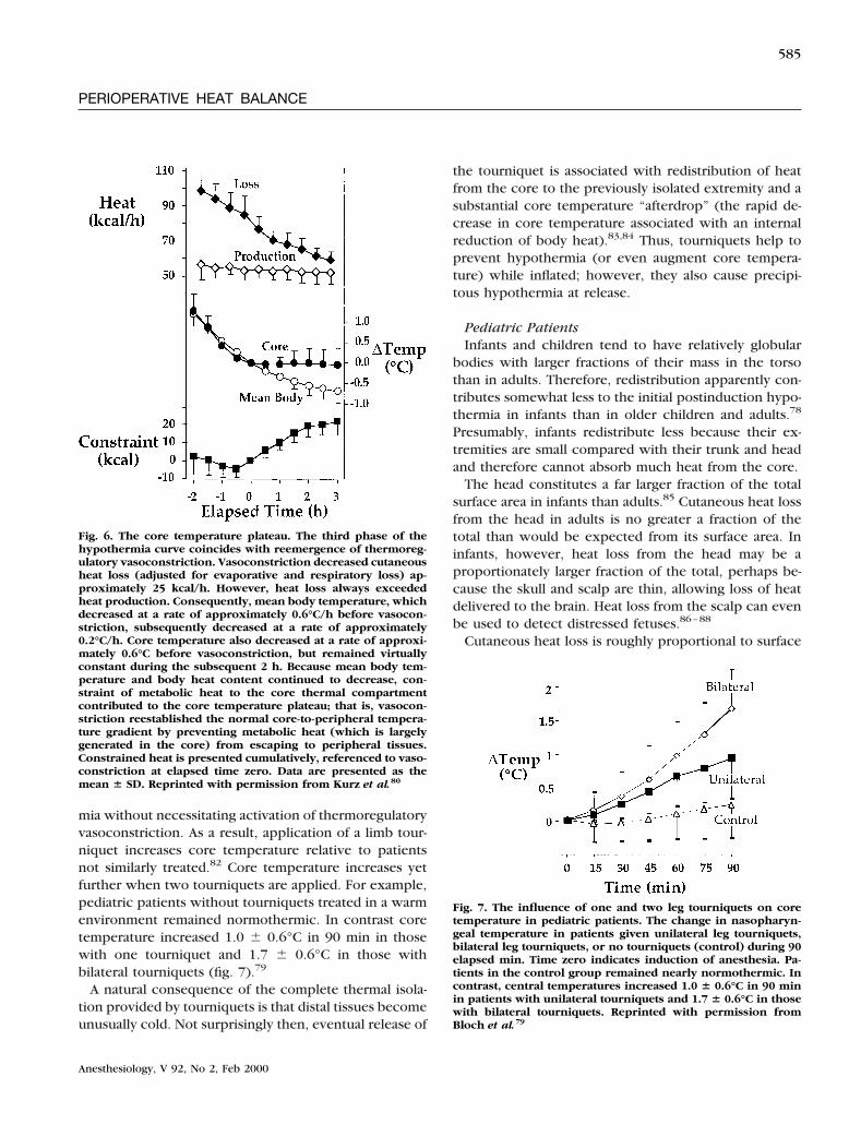
mia without necessitating activation of thermoregulatoryvasoconstriction. As a result, application of a limb tour-niquet increases core temperature relative to patientsnot similarly treated.82 Core temperature increases yetfurther when two tourniquets are applied. For example,pediatric patients without tourniquets treated in a warmenvironment remained normothermic. In contrast coretemperature increased 1.0 6 0.6°C in 90 min in thosewith one tourniquet and 1.7 6 0.6°C in those withbilateral tourniquets (fig. 7).79
A natural consequence of the complete thermal isola-tion provided by tourniquets is that distal tissues becomeunusually cold. Not surprisingly then, eventual release of
the tourniquet is associated with redistribution of heatfrom the core to the previously isolated extremity and asubstantial core temperature “afterdrop” (the rapid de-crease in core temperature associated with an internalreduction of body heat).83,84 Thus, tourniquets help toprevent hypothermia (or even augment core tempera-ture) while inflated; however, they also cause precipi-tous hypothermia at release.
Pediatric PatientsInfants and children tend to have relatively globular
bodies with larger fractions of their mass in the torsothan in adults. Therefore, redistribution apparently con-tributes somewhat less to the initial postinduction hypo-thermia in infants than in older children and adults.78
Presumably, infants redistribute less because their ex-tremities are small compared with their trunk and headand therefore cannot absorb much heat from the core.
The head constitutes a far larger fraction of the totalsurface area in infants than adults.85 Cutaneous heat lossfrom the head in adults is no greater a fraction of thetotal than would be expected from its surface area. Ininfants, however, heat loss from the head may be aproportionately larger fraction of the total, perhaps be-cause the skull and scalp are thin, allowing loss of heatdelivered to the brain. Heat loss from the scalp can evenbe used to detect distressed fetuses.86–88
Cutaneous heat loss is roughly proportional to surface
Fig. 7. The influence of one and two leg tourniquets on coretemperature in pediatric patients. The change in nasopharyn-geal temperature in patients given unilateral leg tourniquets,bilateral leg tourniquets, or no tourniquets (control) during 90elapsed min. Time zero indicates induction of anesthesia. Pa-tients in the control group remained nearly normothermic. Incontrast, central temperatures increased 1.0 6 0.6°C in 90 minin patients with unilateral tourniquets and 1.7 6 0.6°C in thosewith bilateral tourniquets. Reprinted with permission fromBloch et al.79
Fig. 6. The core temperature plateau. The third phase of thehypothermia curve coincides with reemergence of thermoreg-ulatory vasoconstriction. Vasoconstriction decreased cutaneousheat loss (adjusted for evaporative and respiratory loss) ap-proximately 25 kcal/h. However, heat loss always exceededheat production. Consequently, mean body temperature, whichdecreased at a rate of approximately 0.6°C/h before vasocon-striction, subsequently decreased at a rate of approximately0.2°C/h. Core temperature also decreased at a rate of approxi-mately 0.6°C before vasoconstriction, but remained virtuallyconstant during the subsequent 2 h. Because mean body tem-perature and body heat content continued to decrease, con-straint of metabolic heat to the core thermal compartmentcontributed to the core temperature plateau; that is, vasocon-striction reestablished the normal core-to-peripheral tempera-ture gradient by preventing metabolic heat (which is largelygenerated in the core) from escaping to peripheral tissues.Constrained heat is presented cumulatively, referenced to vaso-constriction at elapsed time zero. Data are presented as themean 6 SD. Reprinted with permission from Kurz et al.80
585
PERIOPERATIVE HEAT BALANCE
Anesthesiology, V 92, No 2, Feb 2000
area, whereas metabolic heat production is largely afunction of mass. Consequently, it is relatively easy forinfants and children to lose large amounts of heat via theskin surface.34,89 Intraoperative heat loss in infants cantherefore easily exceed metabolic heat production.90
The linear hypothermia phase is therefore usually rapidin infants and children because of a high surface-area-to-weight ratio. In contrast, respiratory losses are similar tothat in adults.
The threshold for intraoperative vasoconstriction issimilar in adults and pediatric patients.78,91 Once con-stricted, an effective core temperature plateau developsin infants and children.
Neuraxial Anesthesia
It is not widely recognized that neuraxial anesthesiaproduces thermal perturbations that may be as large orlarger than those observed during general anesthesia.The mechanisms are in some ways similar, but also differin numerous important respects.
RedistributionJust as with general anesthesia, redistribution of body
heat is the major initial cause of hypothermia in patientsadministered spinal or epidural anesthesia. Neuraxialanesthesia inhibits thermoregulatory control centrally,92–94
but a far more important effect of major conductionanesthesia is blocking of peripheral sympathetic andmotor nerves, which prevents thermoregulatory vaso-constriction and shivering.95–97
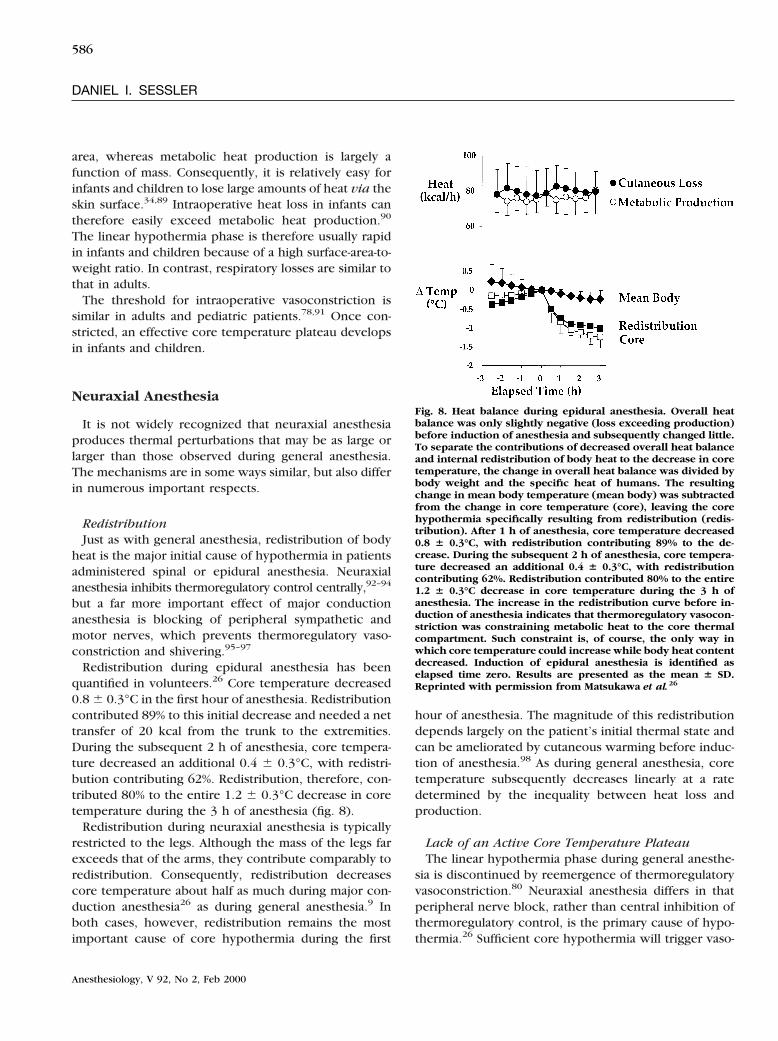
Redistribution during epidural anesthesia has beenquantified in volunteers.26 Core temperature decreased0.8 6 0.3°C in the first hour of anesthesia. Redistributioncontributed 89% to this initial decrease and needed a nettransfer of 20 kcal from the trunk to the extremities.During the subsequent 2 h of anesthesia, core tempera-ture decreased an additional 0.4 6 0.3°C, with redistri-bution contributing 62%. Redistribution, therefore, con-tributed 80% to the entire 1.2 6 0.3°C decrease in coretemperature during the 3 h of anesthesia (fig. 8).
Redistribution during neuraxial anesthesia is typicallyrestricted to the legs. Although the mass of the legs farexceeds that of the arms, they contribute comparably toredistribution. Consequently, redistribution decreasescore temperature about half as much during major con-duction anesthesia26 as during general anesthesia.9 Inboth cases, however, redistribution remains the mostimportant cause of core hypothermia during the first
hour of anesthesia. The magnitude of this redistributiondepends largely on the patient’s initial thermal state andcan be ameliorated by cutaneous warming before induc-tion of anesthesia.98 As during general anesthesia, coretemperature subsequently decreases linearly at a ratedetermined by the inequality between heat loss andproduction.
Lack of an Active Core Temperature PlateauThe linear hypothermia phase during general anesthe-
sia is discontinued by reemergence of thermoregulatoryvasoconstriction.80 Neuraxial anesthesia differs in thatperipheral nerve block, rather than central inhibition ofthermoregulatory control, is the primary cause of hypo-thermia.26 Sufficient core hypothermia will trigger vaso-
Fig. 8. Heat balance during epidural anesthesia. Overall heatbalance was only slightly negative (loss exceeding production)before induction of anesthesia and subsequently changed little.To separate the contributions of decreased overall heat balanceand internal redistribution of body heat to the decrease in coretemperature, the change in overall heat balance was divided bybody weight and the specific heat of humans. The resultingchange in mean body temperature (mean body) was subtractedfrom the change in core temperature (core), leaving the corehypothermia specifically resulting from redistribution (redis-tribution). After 1 h of anesthesia, core temperature decreased0.8 6 0.3°C, with redistribution contributing 89% to the de-crease. During the subsequent 2 h of anesthesia, core tempera-ture decreased an additional 0.4 6 0.3°C, with redistributioncontributing 62%. Redistribution contributed 80% to the entire1.2 6 0.3°C decrease in core temperature during the 3 h ofanesthesia. The increase in the redistribution curve before in-duction of anesthesia indicates that thermoregulatory vasocon-striction was constraining metabolic heat to the core thermalcompartment. Such constraint is, of course, the only way inwhich core temperature could increase while body heat contentdecreased. Induction of epidural anesthesia is identified aselapsed time zero. Results are presented as the mean 6 SD.Reprinted with permission from Matsukawa et al.26
586
DANIEL I. SESSLER
Anesthesiology, V 92, No 2, Feb 2000

constriction and shivering in unblocked regions (i.e., thearms) if thermoregulation is not excessively impaired byold age99 or sedative medications.44,46 However, upperbody shivering is relatively ineffective and often insuffi-cient to prevent further hypothermia. Furthermore, shiv-ering per se is often treated pharmacologically,45,47,100,101
which negates its thermogenic function.The nerve block associated with neuraxial anesthesia
continues throughout anesthesia. Patients thereforehave little ability to vasoconstrict or shiver in the lowerbody during regional anesthesia, no matter what theircentral thermoregulatory status.102 Progressive hypo-thermia therefore is not limited by development of anactive core temperature plateau the way it is duringgeneral anesthesia. Inability to vasoconstrict and shiverin the lower body is usually of little consequence if thesurgical procedure is small because a passive plateauusually develops in well-insulated patients undergoingminor procedures. In contrast, patients undergoing largeoperations with neuraxial anesthesia have the potentialfor serious hypothermia to develop.103
Hypothermia during neuraxial anesthesia is far morecommon than generally appreciated. Several studies in-dicate that hypothermia during neuraxial anesthesia forlarge operations is nearly as common and severe asduring general anesthesia.104,105 An insidious aspect ofthis hypothermia is that neuraxial anesthesia not onlyinhibits autonomic thermoregulatory control, but alsoinhibits behavioral control.106 Patients therefore do notnecessarily feel cold and consequently do not complainto their anesthesiologist. Because temperature monitor-ing remains rare during major conduction anesthesia,107
it is common that neither patients nor their anesthesiol-ogists appreciate the extent of hypothermia.
Neuraxial and General AnesthesiaMost studies that evaluate hypothermia during regional
and general anesthesia conclude that core temperaturedecreases more with general than with regional anesthe-sia.103,104 The mechanisms by which hypothermia de-velop provide some indication of what might be ex-pected during various clinical circumstances. In thisanalysis, we will assume operations of comparable sizeand administration of similar amounts of unwarmed in-travenous fluid.
Redistribution of body heat is the primary cause ofhypothermia during the first hour of neuraxial26 or gen-eral9 anesthesia. However, redistribution decreases coretemperature about twice as much during general asduring regional anesthesia. During relatively short pro-
cedures, hypothermia is likely to be greater in patientsadministered general anesthesia. Patients administeredregional anesthesia will generally start the linear hypo-thermia phase at a relatively high temperature becausethey initially lost less to redistribution. Furthermore,hypothermia may develop at a reduced rate duringneuraxial anesthesia because metabolic heat productionremains near normal.
After long and large operations, the opposite pattern islikely to prevail because in patients administered generalanesthesia a core temperature plateau will develop,whereas those undergoing neuraxial anesthesia oftencontinue to become hypothermic. Whether core hypo-thermia is worse with general or regional anesthesia isthus likely to depend largely on the duration and mag-nitude of surgery.
Combined Neuraxial and General Anesthesia. Thepatients at greatest risk for inadvertent perioperativehypothermia are those in whom regional and generalanesthesia are combined. The reason is that these pa-tients will initially become rapidly hypothermic becauseof redistribution to all four extremities. During the linearphase, they will continue to cool, but at the relativelyhigher rate associated with general anesthesia.
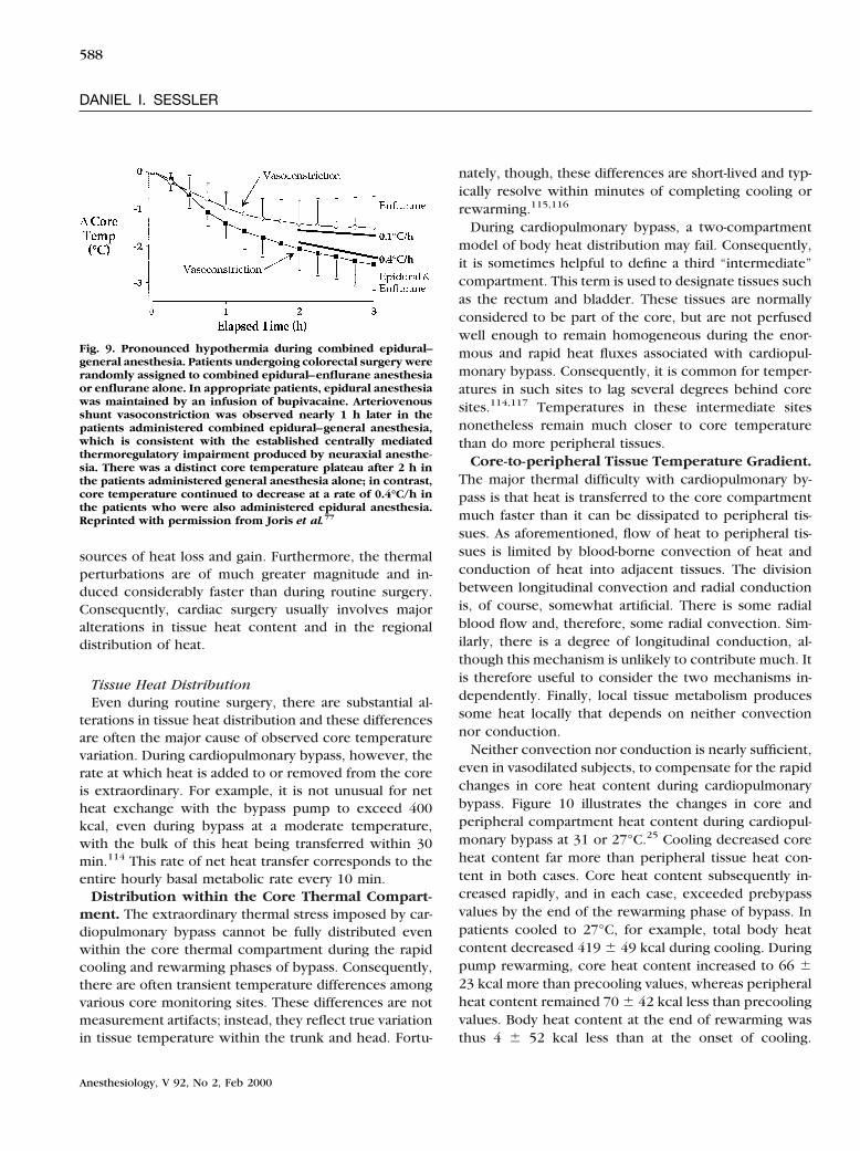
Three additional factors contribute to hypothermiaduring combined regional–general anesthesia. The firstis that neuraxial anesthesia per se reduces the vasocon-striction threshold. General anesthesia does also, but theepidural effect is superimposed on the general anes-thetic effect. As a result, vasoconstriction occurs laterand at a lower core temperature in patients administeredboth epidural and general anesthesia.77 The second fac-tor is that general anesthesia inhibits the shivering thatmight otherwise increase heat production duringneuraxial anesthesia. The third and most important fac-tor is that peripheral nerve block prevents vasoconstric-tion in the legs.96,108,109 As a result, vasoconstriction—once initiated centrally—is relatively ineffective, andcore temperature continues to decrease (fig. 9).
Cardiopulmonary Bypass
Hypothermia is frequently used during cardiac surgerybecause it provides substantial protection against cardiacand cerebral ischemia.110–113 For the same reason, it isalso used during certain neurosurgical procedures, espe-cially repair of aneurysms. Procedures performed withcardiopulmonary bypass differ from other types of sur-gery in that heat exchange to the pump dwarfs all other
587
PERIOPERATIVE HEAT BALANCE
Anesthesiology, V 92, No 2, Feb 2000
sources of heat loss and gain. Furthermore, the thermalperturbations are of much greater magnitude and in-duced considerably faster than during routine surgery.Consequently, cardiac surgery usually involves majoralterations in tissue heat content and in the regionaldistribution of heat.
Tissue Heat DistributionEven during routine surgery, there are substantial al-
terations in tissue heat distribution and these differencesare often the major cause of observed core temperaturevariation. During cardiopulmonary bypass, however, therate at which heat is added to or removed from the coreis extraordinary. For example, it is not unusual for netheat exchange with the bypass pump to exceed 400kcal, even during bypass at a moderate temperature,with the bulk of this heat being transferred within 30min.114 This rate of net heat transfer corresponds to theentire hourly basal metabolic rate every 10 min.
Distribution within the Core Thermal Compart-ment. The extraordinary thermal stress imposed by car-diopulmonary bypass cannot be fully distributed evenwithin the core thermal compartment during the rapidcooling and rewarming phases of bypass. Consequently,there are often transient temperature differences amongvarious core monitoring sites. These differences are notmeasurement artifacts; instead, they reflect true variationin tissue temperature within the trunk and head. Fortu-
nately, though, these differences are short-lived and typ-ically resolve within minutes of completing cooling orrewarming.115,116
During cardiopulmonary bypass, a two-compartmentmodel of body heat distribution may fail. Consequently,it is sometimes helpful to define a third “intermediate”compartment. This term is used to designate tissues suchas the rectum and bladder. These tissues are normallyconsidered to be part of the core, but are not perfusedwell enough to remain homogeneous during the enor-mous and rapid heat fluxes associated with cardiopul-monary bypass. Consequently, it is common for temper-atures in such sites to lag several degrees behind coresites.114,117 Temperatures in these intermediate sitesnonetheless remain much closer to core temperaturethan do more peripheral tissues.
Core-to-peripheral Tissue Temperature Gradient.The major thermal difficulty with cardiopulmonary by-pass is that heat is transferred to the core compartmentmuch faster than it can be dissipated to peripheral tis-sues. As aforementioned, flow of heat to peripheral tis-sues is limited by blood-borne convection of heat andconduction of heat into adjacent tissues. The divisionbetween longitudinal convection and radial conductionis, of course, somewhat artificial. There is some radialblood flow and, therefore, some radial convection. Sim-ilarly, there is a degree of longitudinal conduction, al-though this mechanism is unlikely to contribute much. Itis therefore useful to consider the two mechanisms in-dependently. Finally, local tissue metabolism producessome heat locally that depends on neither convectionnor conduction.
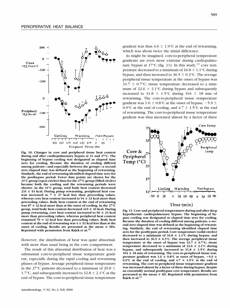
Neither convection nor conduction is nearly sufficient,even in vasodilated subjects, to compensate for the rapidchanges in core heat content during cardiopulmonarybypass. Figure 10 illustrates the changes in core andperipheral compartment heat content during cardiopul-monary bypass at 31 or 27°C.25 Cooling decreased coreheat content far more than peripheral tissue heat con-tent in both cases. Core heat content subsequently in-creased rapidly, and in each case, exceeded prebypassvalues by the end of the rewarming phase of bypass. Inpatients cooled to 27°C, for example, total body heatcontent decreased 419 6 49 kcal during cooling. Duringpump rewarming, core heat content increased to 66 623 kcal more than precooling values, whereas peripheralheat content remained 70 6 42 kcal less than precoolingvalues. Body heat content at the end of rewarming wasthus 4 6 52 kcal less than at the onset of cooling.
Fig. 9. Pronounced hypothermia during combined epidural–general anesthesia. Patients undergoing colorectal surgery wererandomly assigned to combined epidural–enflurane anesthesiaor enflurane alone. In appropriate patients, epidural anesthesiawas maintained by an infusion of bupivacaine. Arteriovenousshunt vasoconstriction was observed nearly 1 h later in thepatients administered combined epidural–general anesthesia,which is consistent with the established centrally mediatedthermoregulatory impairment produced by neuraxial anesthe-sia. There was a distinct core temperature plateau after 2 h inthe patients administered general anesthesia alone; in contrast,core temperature continued to decrease at a rate of 0.4°C/h inthe patients who were also administered epidural anesthesia.Reprinted with permission from Joris et al.77
588
DANIEL I. SESSLER
Anesthesiology, V 92, No 2, Feb 2000
However, the distribution of heat was quite abnormal,with more than usual being in the core compartment.
The result of this abnormal distribution of heat was asubstantial core-to-peripheral tissue temperature gradi-ent, especially during the rapid cooling and rewarmingphases of bypass. Average peripheral tissue temperaturein the 27°C patients decreased to a minimum of 29.8 61.7°C, and subsequently increased to 32.8 6 2.1°C at theend of bypass. The core-to-peripheral tissue temperature
gradient was thus 4.6 6 1.9°C at the end of rewarming,which was about twice the initial difference.
As might be imagined, core-to-peripheral temperaturegradients are even more extreme during cardiopulmo-nary bypass at 17°C (fig. 11). In this study,12 core tem-perature decreased to a minimum of 16.8 6 1.1°C duringbypass, and then increased to 36.5 6 0.3°C. The averageperipheral tissue temperature at the onset of bypass was33.7 6 0.7°C; tissue temperature decreased to a mini-mum of 22.6 6 2.1°C during bypass and subsequentlyincreased to 31.8 6 1.5°C during 104 6 18 min ofrewarming. The core-to-peripheral tissue temperaturegradient was 1.6 6 0.8°C at the onset of bypass, 25.9 60.9°C at the end of cooling, and 4.7 6 1.5°C at the endof rewarming. The core-to-peripheral tissue temperaturegradient was thus increased almost by a factor of three
Fig. 11. Core and peripheral temperatures during and after deephypothermic cardiopulmonary bypass. The beginning of by-pass cooling was designated as elapsed time zero for cooling.Because the duration of cooling differed among patients, a sec-ond zero elapsed time was defined as the beginning of rewarm-ing. Similarly, the end of rewarming identified elapsed timezero for the postbypass period. Core temperature (solid circles)decreased to a minimum of 16.8 6 1.1°C during bypass, andthen increased to 36.5 6 0.3°C. The average peripheral tissuetemperature at the onset of bypass was 33.7 6 0.7°C; tissuetemperature decreased to a minimum of 22.6 6 2.1°C duringbypass, and subsequently increased to 31.8 6 1.5°C during104 6 18 min of rewarming. The core-to-peripheral tissue tem-perature gradient was 1.6 6 0.8°C at onset of bypass, 25.9 60.9°C at the end of cooling, and 4.7 6 1.5°C at the end ofrewarming. The core-to-peripheral tissue temperature gradientwas increased almost by a factor of three during bypass, despitean essentially normal postbypass core temperature. Results arepresented as the mean 6 SD. Reprinted with permission fromRajek et al.12
Fig. 10. Changes in core and peripheral tissue heat contentduring and after cardiopulmonary bypass at 31 and 27°C. Thebeginning of bypass cooling was designated as elapsed timezero for cooling. Because the duration of cooling differedamong patients—and especially between the groups—a secondzero elapsed time was defined as the beginning of rewarming.Similarly, the end of rewarming identified elapsed time zero forthe postbypass period. Fewer data points are shown for the31°C group (open circles) than for the 27°C group (filled circles)because both the cooling and the rewarming periods wereshorter. In the 31°C group, total body heat content decreased231 6 93 kcal. During pump rewarming, peripheral heat con-tent increased to 7 6 27 kcal less than precooling values,whereas core heat content increased to 94 6 33 kcal more thanprecooling values. Body heat content at the end of rewarmingwas 87 6 42 kcal more than at the onset of cooling. In the 27°Cgroup, total body heat content decreased 419 6 49 kcal. Duringpump rewarming, core heat content increased to 66 6 23 kcalmore than precooling values, whereas peripheral heat contentremained 70 6 42 kcal less than precooling values. Body heatcontent at the end of rewarming was 4 6 52 kcal less than at theonset of cooling. Results are presented as the mean 6 SDs.Reprinted with permission from Rajek et al.25
589
PERIOPERATIVE HEAT BALANCE
Anesthesiology, V 92, No 2, Feb 2000
during bypass, despite an essentially normal postbypasscore temperature.
Radial and Longitudinal Tissue Temperature Dis-tributions. Abnormal tissue temperature distributionduring cardiopulmonary bypass is not restricted to thecore-to-peripheral axis. There are also substantial radialand longitudinal inequalities within the peripheral com-partment. This is a natural consequence of the two majorpathways by which heat flows peripherally from thecore: longitudinal convection and radial conduction.Blood-borne convection of heat is faster than radial dif-fusion; however, the distances involved are proportion-ately much greater. That is, the length of the extremitiesfar exceeds their width. Consequently, convection andconduction both remain clinically important impedi-ments to peripheral heat transfer.
The center of the thigh can be considered part of thecore because temperature in this region normally equalscore temperature, even during rapid thermal perturba-tions.12 Calf and foot tissues, however, are normallyconsiderably cooler than the core; furthermore, theirtemperatures change slowly in response to core pertur-bations. The disparity between core and regional tem-perature increases progressively in more distal aspects ofthe extremities.
During one study of bypass at 17°C, for example,temperature in the central axis of the leg at the end ofcooling was 6.0 6 4.3°C greater than core temperatureat the upper calf, 7.1 6 4.2°C greater at the lower calf,and 6.3 6 4.1°C greater in the foot. At the end ofrewarming, the situation was reversed, with the uppercalf being 2.9°C less than the core temperatures. At thattime, temperature at the center of the lower calf ex-ceeded core temperature by 4.9 6 2.0°C, whereas thefoot exceed core temperature by 6.7 6 3.4°C. Thesedata indicate that longitudinal convection of heat alongextremities is insufficient to equilibrate peripheral andcore tissue temperatures during the rapid thermal per-turbations associated with hypothermic bypass.
The second factor limiting transfer of heat from thecore to peripheral tissues is the slow rate at which heatis conducted radially. This is manifested as a radial tissuetemperature gradient. Radial inequalities are far moreimportant in the proximal than in the distal extremitiesbecause the tissue temperature gradients between thecenter axis and skin are greater.
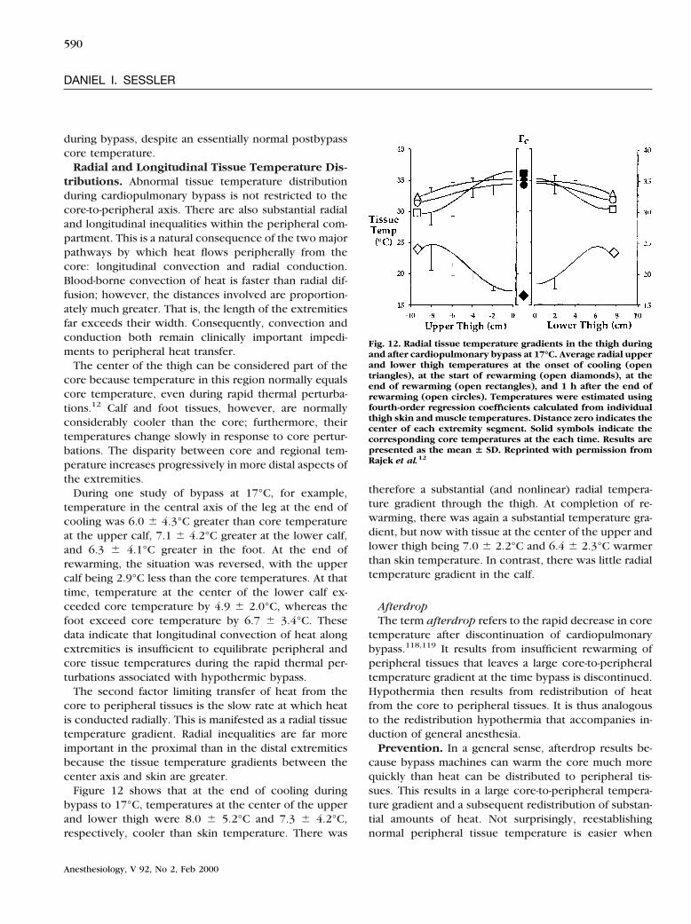
Figure 12 shows that at the end of cooling duringbypass to 17°C, temperatures at the center of the upperand lower thigh were 8.0 6 5.2°C and 7.3 6 4.2°C,respectively, cooler than skin temperature. There was
therefore a substantial (and nonlinear) radial tempera-ture gradient through the thigh. At completion of re-warming, there was again a substantial temperature gra-dient, but now with tissue at the center of the upper andlower thigh being 7.0 6 2.2°C and 6.4 6 2.3°C warmerthan skin temperature. In contrast, there was little radialtemperature gradient in the calf.
AfterdropThe term afterdrop refers to the rapid decrease in core
temperature after discontinuation of cardiopulmonarybypass.118,119 It results from insufficient rewarming ofperipheral tissues that leaves a large core-to-peripheraltemperature gradient at the time bypass is discontinued.Hypothermia then results from redistribution of heatfrom the core to peripheral tissues. It is thus analogousto the redistribution hypothermia that accompanies in-duction of general anesthesia.
Prevention. In a general sense, afterdrop results be-cause bypass machines can warm the core much morequickly than heat can be distributed to peripheral tis-sues. This results in a large core-to-peripheral tempera-ture gradient and a subsequent redistribution of substan-tial amounts of heat. Not surprisingly, reestablishingnormal peripheral tissue temperature is easier when
Fig. 12. Radial tissue temperature gradients in the thigh duringand after cardiopulmonary bypass at 17°C. Average radial upperand lower thigh temperatures at the onset of cooling (opentriangles), at the start of rewarming (open diamonds), at theend of rewarming (open rectangles), and 1 h after the end ofrewarming (open circles). Temperatures were estimated usingfourth-order regression coefficients calculated from individualthigh skin and muscle temperatures. Distance zero indicates thecenter of each extremity segment. Solid symbols indicate thecorresponding core temperatures at the each time. Results arepresented as the mean 6 SD. Reprinted with permission fromRajek et al.12
590
DANIEL I. SESSLER
Anesthesiology, V 92, No 2, Feb 2000
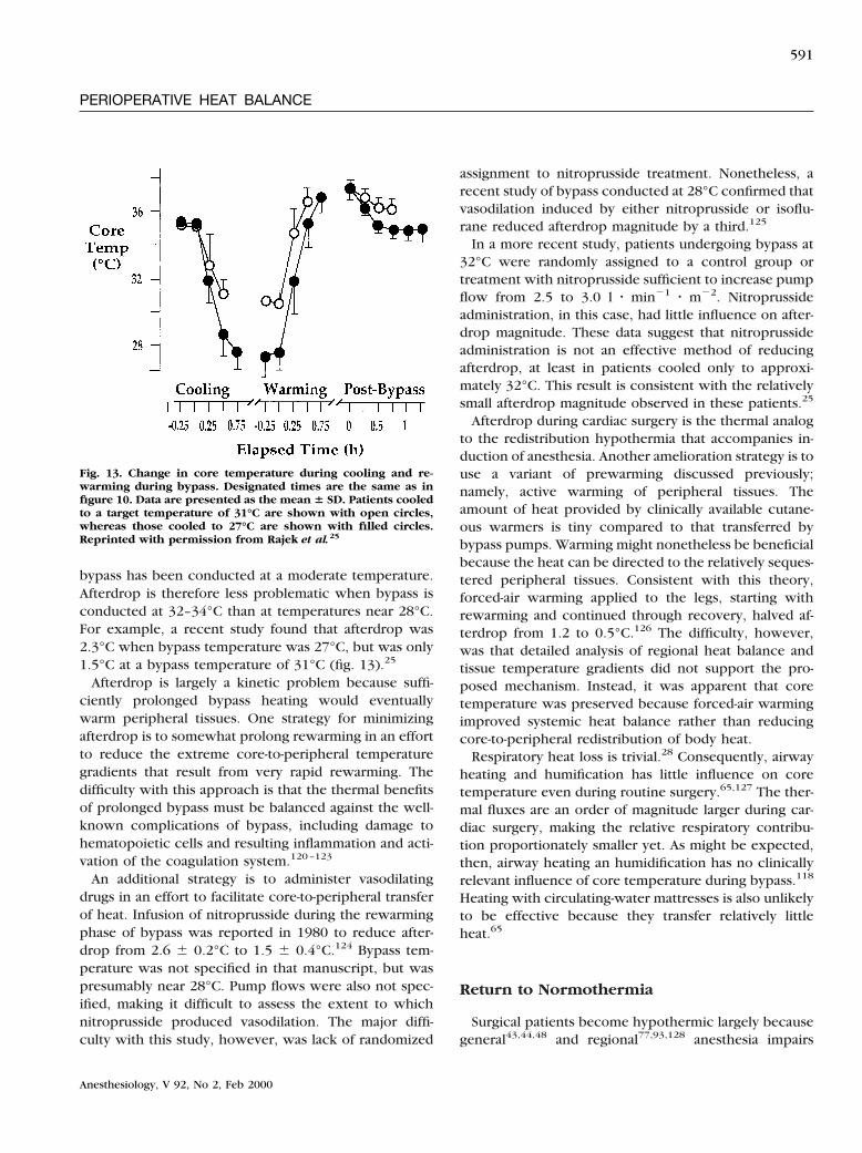
bypass has been conducted at a moderate temperature.Afterdrop is therefore less problematic when bypass isconducted at 32–34°C than at temperatures near 28°C.For example, a recent study found that afterdrop was2.3°C when bypass temperature was 27°C, but was only1.5°C at a bypass temperature of 31°C (fig. 13).25
Afterdrop is largely a kinetic problem because suffi-ciently prolonged bypass heating would eventuallywarm peripheral tissues. One strategy for minimizingafterdrop is to somewhat prolong rewarming in an effortto reduce the extreme core-to-peripheral temperaturegradients that result from very rapid rewarming. Thedifficulty with this approach is that the thermal benefitsof prolonged bypass must be balanced against the well-known complications of bypass, including damage tohematopoietic cells and resulting inflammation and acti-vation of the coagulation system.120–123
An additional strategy is to administer vasodilatingdrugs in an effort to facilitate core-to-peripheral transferof heat. Infusion of nitroprusside during the rewarmingphase of bypass was reported in 1980 to reduce after-drop from 2.6 6 0.2°C to 1.5 6 0.4°C.124 Bypass tem-perature was not specified in that manuscript, but waspresumably near 28°C. Pump flows were also not spec-ified, making it difficult to assess the extent to whichnitroprusside produced vasodilation. The major diffi-culty with this study, however, was lack of randomized
assignment to nitroprusside treatment. Nonetheless, arecent study of bypass conducted at 28°C confirmed thatvasodilation induced by either nitroprusside or isoflu-rane reduced afterdrop magnitude by a third.125
In a more recent study, patients undergoing bypass at32°C were randomly assigned to a control group ortreatment with nitroprusside sufficient to increase pumpflow from 2.5 to 3.0 l z min21 z m22. Nitroprussideadministration, in this case, had little influence on after-drop magnitude. These data suggest that nitroprussideadministration is not an effective method of reducingafterdrop, at least in patients cooled only to approxi-mately 32°C. This result is consistent with the relativelysmall afterdrop magnitude observed in these patients.25
Afterdrop during cardiac surgery is the thermal analogto the redistribution hypothermia that accompanies in-duction of anesthesia. Another amelioration strategy is touse a variant of prewarming discussed previously;namely, active warming of peripheral tissues. Theamount of heat provided by clinically available cutane-ous warmers is tiny compared to that transferred bybypass pumps. Warming might nonetheless be beneficialbecause the heat can be directed to the relatively seques-tered peripheral tissues. Consistent with this theory,forced-air warming applied to the legs, starting withrewarming and continued through recovery, halved af-terdrop from 1.2 to 0.5°C.126 The difficulty, however,was that detailed analysis of regional heat balance andtissue temperature gradients did not support the pro-posed mechanism. Instead, it was apparent that coretemperature was preserved because forced-air warmingimproved systemic heat balance rather than reducingcore-to-peripheral redistribution of body heat.
Respiratory heat loss is trivial.28 Consequently, airwayheating and humification has little influence on coretemperature even during routine surgery.65,127 The ther-mal fluxes are an order of magnitude larger during car-diac surgery, making the relative respiratory contribu-tion proportionately smaller yet. As might be expected,then, airway heating an humidification has no clinicallyrelevant influence of core temperature during bypass.118
Heating with circulating-water mattresses is also unlikelyto be effective because they transfer relatively littleheat.65
Return to Normothermia
Surgical patients become hypothermic largely becausegeneral43,44,48 and regional77,93,128 anesthesia impairs
Fig. 13. Change in core temperature during cooling and re-warming during bypass. Designated times are the same as infigure 10. Data are presented as the mean 6 SD. Patients cooledto a target temperature of 31°C are shown with open circles,whereas those cooled to 27°C are shown with filled circles.Reprinted with permission from Rajek et al.25
591
PERIOPERATIVE HEAT BALANCE
Anesthesiology, V 92, No 2, Feb 2000
thermoregulatory defenses, as do most sedatives.45–47,100
Consequently, most surgical patients become hypother-mic unless they are actively warmed.65,66,129 The amountof heat lost during surgery is substantial. For example,based on the reported specific heat of humans,1 a 3°Creduction in mean body temperature corresponds to adebt of approximately 175 kcal in a 70-kg patient. This isroughly the basal heat production for 3 h.58
Full postoperative recovery of protective thermoregu-latory responses may be limited by residual volatile an-esthetic130 or opioids administered to treat surgicalpain.45,46 Nonetheless, brain anesthetic concentrationsusually decrease rapidly during the initial postoperativeperiod,131 allowing reemergence of thermoregulatoryresponses, including vasoconstriction and shiver-ing.130,132–135 These responses combine to decrease cu-taneous heat loss,33 constrain metabolic heat to the ther-mal core,136 and increase metabolic heat production.132
As a result, core temperatures usually increase towardnormal values after anesthesia is discontinued.
Arteriovenous shunt vasoconstriction is universal dur-ing postanesthetic recovery in hypothermic patients.133
Similarly, shivering is common. Nonetheless, postopera-tive core temperatures in hypothermic patients usuallyincrease relatively slowly, often requiring 2–5 h to returnto normal values.133,137,138 Such slow return to normo-thermia contrasts markedly with the rapid increases ob-served in unanesthetized volunteers,139 suggesting thatprolonged hypothermia does not result simply from themagnitude of the heat debt, but because thermoregula-tory compensations are not maximally activated in sur-gical patients—presumably because of residual volatileanesthetics and opioids administered for treatment ofpain.
Summary
Hypothermia during general anesthesia develops witha characteristic three-phase pattern. The initial rapidreduction in core temperature after induction of anes-thesia results from an internal redistribution of bodyheat. Redistribution results because anesthetics inhibitthe tonic vasoconstriction that normally maintains alarge core-to-peripheral temperature gradient. Core tem-perature then decreases linearly at a rate determined bythe difference between heat loss and production. How-ever, when surgical patients become sufficiently hypo-thermic, they again trigger thermoregulatory vasocon-striction, which restricts core-to-peripheral flow of heat.
Constraint of metabolic heat, in turn, maintains a coretemperature plateau (despite continued systemic heatloss) and eventually reestablishes the normal core-to-peripheral temperature gradient. Together, these mech-anisms indicate that alterations in the distribution ofbody heat contribute more to changes in core tempera-ture than to systemic heat imbalance in most patients.
Just as with general anesthesia, redistribution of bodyheat is the major initial cause of hypothermia in patientsadministered spinal or epidural anesthesia. However,redistribution during neuraxial anesthesia is typically re-stricted to the legs. Consequently, redistribution de-creases core temperature about half as much duringmajor conduction anesthesia. As during general anesthe-sia, core temperature subsequently decreases linearly ata rate determined by the inequality between heat lossand production. The major difference, however, is thatthe linear hypothermia phase is not discontinued byreemergence of thermoregulatory vasoconstriction be-cause constriction in the legs is blocked peripherally. Asa result, in patients undergoing large operations withneuraxial anesthesia, there is the potential of develop-ment of serious hypothermia.
Hypothermic cardiopulmonary bypass is associatedwith enormous changes in body heat content. Further-more, rapid cooling and rewarming produces large core-to-peripheral, longitudinal, and radial tissue temperaturegradients. Inadequate rewarming of peripheral tissuestypically produces a considerable core-to-peripheral gra-dient at the end of bypass. Subsequently, redistributionof heat from the core to the cooler arms and legs pro-duces an afterdrop. Afterdrop magnitude can be reducedby prolonging rewarming, pharmacologic vasodilation,or peripheral warming.
Postoperative return to normothermia occurs whenbrain anesthetic concentration decreases sufficiently toagain trigger normal thermoregulatory defenses. How-ever, residual anesthesia and opioids given for treatmentof postoperative pain decreases the effectiveness ofthese responses. Consequently, return to normothermiaoften needs 2–5 h, depending on the degree of hypo-thermia and the age of the patient.
References
1. Burton AC: Human calorimetry: The average temperature of thetissues of the body. J Nutr 1935; 9:261–80
2. Cooper TE, Trezek GJ: Correlation of thermal properties of somehuman tissue with water content. Aerospace Med 1971; 42:24–7
3. Orkin FK: Physiologic disturbances associated with induced hy-
592
DANIEL I. SESSLER
Anesthesiology, V 92, No 2, Feb 2000

pothermia, Complications in Anesthesiology. Edited by Orkin FK,Cooperman LH. Philadelphia, JB Lippincott Company, 1983, p 626
4. Hynson JM, Sessler DI, Moayeri A, McGuire J: Absence of non-shivering thermogenesis in anesthetized humans. ANESTHESIOLOGY 1993;79:695–703
5. Crosby G, Crane AM, Sokoloff L: A comparison of local rates ofglucose utilization in spinal cord and brain in conscious and nitrousoxide- or pentobarbital-treated rats. ANESTHESIOLOGY 1984; 61:434–8
6. Fagher B, Liedholm H, Monti M, Moritz U: Thermogenesis inhuman skeletal muscle as measured by direct microcalorimetry andmuscle contractile performance during b-adrenoceptor blockade. ClinSci 1986; 70:435–41
7. Fagher B, Monti M, Thulin T: Selective b1-andrenoceptor block-ade and muscle thermogenesis. Acta Med Scand 1988; 223:139–45
8. Adair ER, Berglund LG: On the thermoregulatory consequences ofNMR imaging. Magn Reson Imaging 1986; 4:321–33
9. Matsukawa T, Sessler DI, Sessler AM, Schroeder M, Ozaki M, KurzA, Cheng C: Heat flow and distribution during induction of generalanesthesia. ANESTHESIOLOGY 1995; 82:662–73
10. Bristow GK, Biesbrecht GG, Sessler DI: Leg temperature andheat content in humans during immersion hypothermia and rewarm-ing. Aviat Space Environ Med 1994; 65:220–6
11. Buck SH, Zaritsky AL: Occult core hyperthermia complicatingcardiogenic shock. Pediatrics 1989; 83:782–4
12. Rajek A, Lenhardt R, Sessler DI, Grabenwoger MJK, Mares P,Jantsch U, Gruber E: Tissue heat content and distribution during andafter cardiopulmonary bypass at 17°C. Anesth Analg 1999; 88:1220–5
13. Detry J-MR, Brengelmann GL, Rowell LB, Wyss C: Skin andmuscle components of forearm blood flow in directly heated restingman. J Appl Physiol 1972; 32:506–11
14. Bazett HC, Love L, Newton M, Eisenberg L, Day R, Forster R II:Temperature changes in blood flowing in arteries and veins in man.Blood Heat Exchange 1948; 1:3–19
15. Anderson GS, Martin AD: Calculated thermal conductivities andheat flux in man. Undersea Hyperb Med 1994; 21:431–41
16. LeBlanc J: Subcutaneous fat and skin temperature. Can J Bio-chem Physiol 1954; 32:354–8
17. Tikuisis P, Bell DG, Jacobs I: Shivering onset, metabolic re-sponse, and convective heat transfer during cold air exposure. J ApplPhysiol 1991; 70:1996–2002
18. Pennes HH: Analysis of tissue and arterial blood temperatures inthe resting human forearm. J Appl Physiol 1948; 1:93–122
19. Sessler DI, Sessler AM: Experimental determination of heat flowparameters during induction of anesthesia. ANESTHESIOLOGY 1998; 89:657–65
20. Ducharme MB, Tikuisis P: In vivo thermal conductivity of thehuman forearm tissues. J Appl Physiol 1991; 70:2682–90
21. Wissler EH: A mathematical model of the human thermal sys-tem. Bull Math Biophysics 1964; 26:147–66
22. Wissler EH: Comparison of computed results obtained from twomathematical models—A simple 14-node model and a complex 250-node model. J Physiologie 1971; 63:455–8
23. Ducharme MB, Tikuisis P: Role of blood as heat source or sink inhuman limbs during local cooling and heating. J Appl Physiol 1994;76:2084–94
24. Ducharme MB, VanHelder WP, Radomski MW: Tissue tempera-ture profile in the human forearm during thermal stress at thermalstability. J Appl Physiol 1991; 71:1973–8
25. Rajek A, Lenhardt R, Sessler DI, Kurz A, Laufer G, Christensen R,
Matsukawa T, Hiesmayer M: Tissue heat content and distribution dur-ing and after cardiopulmonary bypass at 31°C and 27°C. ANESTHESIOL-OGY 1998; 88:1511–8
26. Matsukawa T, Sessler DI, Christensen R, Ozaki M, Schroeder M:Heat flow and distribution during epidural anesthesia. ANESTHESIOLOGY
1995; 83:961–727. Bickler P, Sessler DI: Efficiency of airway heat and moisture
exchangers in anesthetized humans. Anesth Analg 1990; 71:415–828. Walker JEC, Wells RE Jr, Merrill EW: Heat and water exchange in
the respiratory tract. Am J Med 1961; 30:259–6729. Hey EN, Katz G: Evaporative water loss in the new-born baby.
J Physiol 1969; 200:605–1930. Adams WC, Mack GW, Langhans GW, Nadel ER: Effects of varied
air velocity on sweating and evaporative rates during exercise. J ApplPhysiol 1992; 73:2668–74
31. Buono MJ, Sjoholm NT: Effect of physical training on peripheralsweat production. J Appl Physiol 1988; 65:811–4
32. Froese G, Burton AC: Heat losses from the human head. J ApplPhysiol 1957; 10:235–41
33. Sessler DI, Moayeri A, Støen R, Glosten B, Hynson J, McGuire J:Thermoregulatory vasoconstriction decreases cutaneous heat loss. AN-ESTHESIOLOGY 1990; 73:656–60
34. Simbruner G, Weninge RM, Popow C, Herholdt WJ: Regionalheat loss in newborn infants: I. Heat loss in healthy newborns atvarious environmental temperatures. S Afr Med J 1985; 68:940–4
35. Vaughan MS, Vaughan RW, Cork RC: Postoperative hypothermiain adults: Relationship of age, anesthesia, and shivering to rewarming.Anesth Analg 1981; 60:746–51
36. Morris RH, Wilkey BR: The effects of ambient temperature onpatient temperature during surgery not involving body cavities. ANES-THESIOLOGY 1970; 32:102–7
37. Morris RH: Influence of ambient temperature on patient tem-perature during intraabdominal surgery. Ann Surg 1971; 173:230–3
38. Morris RH: Operating room temperature and the anesthetized,paralyzed patient. Surgery 1971; 102:95–7
39. Hales JRS, Fawcett AA, Bennett JW, Needham AD: Thermalcontrol of blood flow through capillaries and arteriovenous anastomo-ses in skin of sheep. Pflugers Arch 1978; 378:55–63
40. Hales JRS: Skin arteriovenous anastomoses, their control androle in thermoregulation, Cardiovascular Shunts: Phylogenetic, Onto-genetic and Clinical Aspects. Edited by Johansen K, Burggren W.Copenhagen, Munksgaard, 1985, pp 433–51
41. Rubinstein EH, Sessler DI: Skin-surface temperature gradientscorrelate with fingertip blood flow in humans. ANESTHESIOLOGY 1990;73:541–5
42. Coffman JD, Cohen AS: Total and capillary fingertip blood flowin Raynaud’s phenomenon. N Engl J Med 1971; 285:259–63
43. Xiong J, Kurz A, Sessler DI, Plattner O, Christensen R, DechertM, Ikeda T: Isoflurane produces marked and non-linear decreases in thevasoconstriction and shivering thresholds. ANESTHESIOLOGY 1996; 85:240–5
44. Matsukawa T, Kurz A, Sessler DI, Bjorksten AR, Merrifield B,Cheng C: Propofol linearly reduces the vasoconstriction and shiveringthresholds. ANESTHESIOLOGY 1995; 82:1169–80
45. Kurz A, Ikeda T, Sessler DI, Larson M, Bjorksten AR, Dechert M,Christensen R: Meperidine decreases the shivering threshold twice asmuch as the vasoconstriction threshold. ANESTHESIOLOGY 1997;86:1046–54
46. Kurz A, Go JC, Sessler DI, Kaer K, Larson M, Bjorksten AR:
593
PERIOPERATIVE HEAT BALANCE
Anesthesiology, V 92, No 2, Feb 2000
Alfentanil slightly increases the sweating threshold and markedly re-duces the vasoconstriction and shivering thresholds. ANESTHESIOLOGY
1995; 83:293–947. Talke P, Tayefeh F, Sessler DI, Jeffrey R, Noursalehi M, Richard-
son C: Dexmedetomidine does not alter the sweating threshold, butcomparably and linearly reduces the vasoconstriction and shiveringthresholds. ANESTHESIOLOGY 1997; 87:835–41
48. Annadata RS, Sessler DI, Tayefeh F, Kurz A, Dechert M: Desflu-rane slightly increases the sweating threshold, but produces marked,non-linear decreases in the vasoconstriction and shivering thresholds.ANESTHESIOLOGY 1995; 83:1205–11
49. Weiskopf RB: Cardiovascular effects of desflurane in experimen-tal animals and volunteers. Anaesthesia 1995; 50:S14–27
50. Hall GM: Body temperature and anaesthesia. Br J Anaesth 1978;50:39–44
51. Rowell LB: Active neurogenic vasodilation in man, Vasodilata-tion. Edited by Vanhoutte P, Leusen I. New York, Raven Press, 1981,pp 1–17
52. Rowell LB, Brengelmann GL, Murray JA: Cardiovascular re-sponses to sustained high skin temperature in resting man. J ApplPhysiol 1969; 27:673–80
53. Sessler DI, Schroeder M, Merrifield B, Matsukawa T, Cheng C:Optimal duration and temperature of pre-warming. ANESTHESIOLOGY
1995; 82:674–8154. Hynson JM, Sessler DI, Moayeri A, McGuire J, Schroeder M: The
effects of pre-induction warming on temperature and blood pressureduring propofol/nitrous oxide anesthesia. ANESTHESIOLOGY 1993; 79:219–28
55. Just B, Trevien V, Delva E, Lienhart A: Prevention of intraoper-ative hypothermia by preoperative skin-surface warming. ANESTHESIOL-OGY 1993; 79:214–8
56. Kurz A, Sessler DI, Narzt E, Lenhart R: Morphometric influenceson intraoperative core temperature changes. Anesth Analg 1995; 80:562–7
57. Roe CF: Effect of bowel exposure on body temperature duringsurgical operations. Am J Surg 1971; 122:13–5
58. Stevens WC, Cromwell TH, Halsey MJ, Eger EI II, ShakespeareTF, Bahlman SH: The cardiovascular effects of a new inhalation anes-thetic, Forane, in human volunteers at constant arterial carbon dioxidetension. ANESTHESIOLOGY 1971; 35:8–16
59. Sellden E, Branstrom R, Brundin T: Augmented thermic effect ofamino acids under general anaesthesia occurs predominantly in extra-splanchnic tissues. Clin Sci 1996; 91:431–9
60. Sellden E, Brundin T, Wahren J: Augmented thermic effect ofamino acids under general anesthesia: A mechanism useful for preven-tion of anaesthesia-induced hypothermia. Clin Sci 1994; 86:611–8
61. Acheson KJ, Zahorska-Markiewicz B, Pittet P, Anantharaman K,Jequier E: Caffeine and coffee: Their influence on metabolic rate andsubstrate utilization in normal weight and obese individuals. Am J ClinNutri 1980; 33:989–97
62. Vallerand AL, Jacobs I, Kavanagh MF: Mechanism of enhancedcold tolerance by an ephedrine-caffeine mixture in humans. J ApplPhysiol 1989; 67:438–44
63. Vallerand AL: Effects of ephedrine/xanthines on thermogenesisand cold tolerance. Int J Obes Relat Metab Disord 1993; 17:553–6
64. English MJM, Farmer C, Scott WAC: Heat loss in exposed volun-teers. J Trauma 1990; 30:422–5
65. Hynson J, Sessler DI: Intraoperative warming therapies: A com-parison of three devices. J Clin Anesth 1992; 4:194–9
66. Kurz A, Kurz M, Poeschl G, Faryniak B, Redl G, Hackl W:Forced-air warming maintains intraoperative normothermia better thancirculating-water mattresses. Anesth Analg 1993; 77:89–95
67. Camus Y, Delva E, Just B, Lienhart A: Leg warming minimizescore hypothermia during abdominal surgery. Anesth Analg 1993; 77:995–9
68. Ip Yam PC, Carli F: Maintenance of body temperature in elderlypatients who have joint replacement surgery. Anesthesia 1990; 45:563–5
69. Hardy JD, Milhorat AT, DuBois EF: Basal metabolism and heatloss of young women at temperatures from 22 degrees C to 35 degreesC. J Nutr 1941; 21:383–403
70. Hendrickx HHL, Trahey GE, Argentieri MP: Paradoxical inhibi-tion of decreases in body temperature by use of heated and humidifiedgases (letter). Anesth Analg 1982; 61:393–4
71. Sessler DI, Sessler AM, Hudson S, Moayeri A: Heat loss duringsurgical skin preparation. ANESTHESIOLOGY 1993; 78:1055–64
72. Lee J-H, Mintz PD: A method for estimating the delivery temper-ature of intravenous fluids. Anesth Analg 1994; 79:155–9
73. Monga M, Comeaux B, Roberts JA: Effect of irrigating fluid onperioperative temperature regulation during transurethral prostatec-tomy. Eur Urol 1996; 29:26–8
74. Sessler DI, McGuire J, Sessler AM: Perioperative thermal insula-tion. ANESTHESIOLOGY 1991; 74:875–9
75. Sessler DI, Schroeder M: Heat loss in humans covered withcotton hospital blankets. Anesth Analg 1993; 77:73–7
76. Sessler DI, Hynson J, McGuire J, Moayeri A, Heier T: Thermo-regulatory vasoconstriction during isoflurane anesthesia minimally de-creases heat loss. ANESTHESIOLOGY 1992; 76:670–5
77. Joris H, Ozaki M, Sessler DI, Hardy AF, Lamay M, McGuire J,Blanchard D, Schroeder M, Moayeri A: Epidural anesthesia impairs bothcentral and peripheral thermoregulatory control during general anes-thesia. ANESTHESIOLOGY 1994; 80:268–77
78. Bissonnette B, Sessler DI: Thermoregulatory thresholds for va-soconstriction in pediatric patients anesthetized with halothane orhalothane and caudal bupivacaine. ANESTHESIOLOGY 1992; 76:387–92
79. Bloch EC, Ginsberg B, Binner RA, Sessler DI: Limb tourniquetsand central temperature in anesthetized children. Anesth Analg 1992;74:486–9
80. Kurz A, Sessler DI, Christensen R, Dechert M: Heat balance anddistribution during the core-temperature plateau in anesthetized hu-mans. ANESTHESIOLOGY 1995; 83:491–9
81. Mittleman KD, Mekjavic IB: Effect of occluded venous return oncore temperature during cold water immersion. J Appl Physiol 1988;65:2709–13
82. Bloch EC: Hyperthermia resulting from tourniquet application inchildren. Ann Roy Coll Surg 1986; 68:193–4
83. dos Reis A Jr: Tourniquet use and intraoperative hypothermia(letter). Anesth Analg 1989; 69:549–50
84. Sanders BJ, D’Alessio JG, Jernigan JR: Intraoperative hypother-mia associated with lower extremity tourniquet deflation. J Clin Anesth1996; 8:504–7
85. Lund CC, Browder NC: The estimation of areas of burns. SurgGynecol Obstet 1944; 79:352–8
86. Rudelstorfer R, Simbruner G, Nanz S: Scalp heat flux in postma-ture and in growth-retarded fetuses. Arch Gynecol Obstet 1991; 249:19–25
87. Rudelstorfer R, Simbruner G, Sharma V, Janisch H: Scalp heat
594
DANIEL I. SESSLER
Anesthesiology, V 92, No 2, Feb 2000

flux and its relationship to scalp blood pH of the fetus. Am J ObstetGynecol 1987; 157:372–7
88. Rudelstorfer R, Simbruner G, Bernaschek G, Rogan AM, Szalay S,Janisch H: Heat flux from the fetal scalp during labor and fetal out-come. Arch Gynecol 1983; 233:85–91
89. Simbruner G, Weninger W, Popow C, Herholdt WJ: Regionalheat loss in newborn infants: II. Heat loss in newborns with variousdiseases—A method of assessing local metabolism and perfusion. S AfrMed J 1985; 68:945–8
90. Anttonen H, Puhakka K, Niskanen J, Ryhanen P: Cutaneous heatloss in children during anaesthesia. Br J Anaesth 1995; 74:306–10
91. Bissonnette B, Sessler DI: The thermoregulatory threshold ininfants and children anesthetized with isoflurane and caudal bupiva-caine. ANESTHESIOLOGY 1990; 73:1114–8
92. Emerick TH, Ozaki M, Sessler DI, Walters K, Schroeder M:Epidural anesthesia increases apparent leg temperature and decreasesthe shivering threshold. ANESTHESIOLOGY 1994; 81:289–98
93. Kurz A, Sessler DI, Schroeder M, Kurz M: Thermoregulatoryresponse thresholds during spinal anesthesia. Anesth Analg 1993; 77:721–6
94. Leslie K, Sessler DI: Reduction in the shivering threshold isproportional to spinal block height. ANESTHESIOLOGY 1996; 84:1327–31
95. Arndt JO, Hock A, Stanton-Hicks M, Stuhmeier K-D: Periduralanesthesia and the distribution of blood in supine humans. ANESTHESI-OLOGY 1985; 63:616–23
96. Bonica JJ, Berges PU, Morikawa K-I: Circulatory effects of peri-dural block: I. Effects of level of analgesia and dose of lidocaine.ANESTHESIOLOGY 1970; 33:619–26
97. Hopf HB, Weibbach B, Peters J: High thoracic segmental epi-dural anesthesia diminishes sympathetic outflow to the legs, despiterestriction of sensory blockade to the upper thorax. ANESTHESIOLOGY
1990; 73:882–998. Glosten B, Hynson J, Sessler DI, McGuire J: Preanesthetic skin-
surface warming reduces redistribution hypothermia caused by epi-dural block. Anesth Analg 1993; 77:488–93
99. Vassilieff N, Rosencher N, Sessler DI, Conseiller C: The shiveringthreshold during spinal anesthesia is reduced in the elderly. ANESTHESI-OLOGY 1995; 83:1162–6
100. Delaunay L, Bonnet F, Liu N, Beydon L, Catoire P, Sessler DI:Clonidine comparably decreases the thermoregulatory thresholds forvasoconstriction and shivering in humans. ANESTHESIOLOGY 1993; 79:470–4
101. Joris J, Banache M, Bonnet F, Sessler DI, Lamy M: Clonidine andketanserin both are effective treatments for postanesthetic shivering.ANESTHESIOLOGY 1993; 79:532–9
102. Kim J-S, Ikeda T, Sessler D, Turakhia M, Jeffrey R: Epiduralanesthesia reduces the gain and maximum intensity of shivering. AN-ESTHESIOLOGY 1998; 88:851–7
103. Frank SM, Beattie C, Christopherson R, Norris EJ, Rock P,Parker S, Kimball AW: Epidural versus general anesthesia, ambientoperating room temperature, and patient age as predictors of inadver-tent hypothermia. ANESTHESIOLOGY 1992; 77:252–7
104. Frank SM, Shi RY, Raja SN, Fleisher LA, Beattie C: Core hypo-thermia and skin-surface temperature gradients: Epidural vs. generalanesthesia and the effects of age. ANESTHESIOLOGY 1994; 80:502–8
105. Jenkins J, Fox J, Sharwood-Smith G: Changes in body heatduring transvesical prostatectomy: A comparison of general and epi-dural anesthesia. Anaesthesia 1983; 38:748–53
106. Sessler DI, Ponte J: Shivering during epidural anesthesia. ANES-THESIOLOGY 1990; 72:816–21
107. Frank SM, Nguyen JM, Garcia C, Barnes RA: Temperature mon-itoring practices during regional anesthesia. Anesth Analg 1999; 88:373–7
108. Valley MA, Bourke DL, Hamill MP, Srinivasa NR: Time course ofsympathetic blockade during epidural anesthesia: Laser doppler flow-metry studies of regional skin perfusion. Anesth Analg 1993;76:289–94
109. Modig J, Malmberg P, Karlstrom G: Effect of epidural versusgeneral anesthesia on calf blood flow. Acta Anaesthesiol Scand 1980;24:305–9
110. Kader A, Frazzini VI, Baker CJ, Saloman RA, Trifiletti RR: Effectof mild hypothermia on nitric oxide synthesis during focal cerebralischemia. Neurosurgery 1994; 35:272–7
111. Minamisawa H, Smith M-L, Siesjo BK: The effect of mild hyper-thermia and hypothermia on brain damage following 5, 10, and 15minutes of forebrain ischemia. Ann Neurol 1990; 28:26–33
112. Natale JE, D’Alecy LG: Protection from cerebral ischemia bybrain cooling without reduced lactate accumulation in dogs. Stroke1989; 20:770–7
113. Neubauer RA, James P: Cerebral oxygenation and the recover-able brain. Neurol Res 1998; 20:S33–6
114. Davis FM, Parimelazhagan KN, Harris EA: Thermal balanceduring cardiopulmonary bypass with moderate hypothermia in man.Br J Anaesth 1977; 49:1127–32
115. Cork RC, Vaughan RW, Humphrey LS: Precision and accuracyof intraoperative temperature monitoring. Anesth Analg 1983; 62:211–4
116. Webb GE: Comparison of esophageal and tympanic tempera-ture monitoring during cardiopulmonary bypass. Analg Anesth 1973;52:729–33
117. Horrow JC, Rosenberg H: Does urinary catheter temperaturereflect core temperature during cardiac surgery? ANESTHESIOLOGY 1988;69:986–9
118. Ralley FE, Ramsay JG, Wynands JE, Townsend GE, Whalley DG,DelliColli P: Effect of heated humidified gases on temperature dropafter cardiopulmonary bypass. Anesth Analg 1984; 63:1106–10
119. Pujol A, Fusciardi J, Ingrand P, Baudouin D, Le Guen AF, MenuP: Afterdrop after hypothermic cardiopulmonary bypass: The value oftympanic membrane temperature monitoring. J Cardiothorac VascAnesth 1996; 10:336–41
120. Berendes E, Loick HM, Van Aken H, Erren M, Deng M, MollhoffT: Duration of cardiopulmonary bypass and its relationship to endo-toxemia, systemic inflammation and acute phase reasponse (abstract).Anesth Analg 1998; 86:S53
121. Hill J, Alonso A, Thiele GM, Robbins RA: Glucocorticoids bluntneutrophil CD11b surface glycoprotein upregulation during cardiopul-monary bypass in humans. Anesth Analg 1994; 79:23–7
122. Hill GE, Alonso A, Spurzem JR, Stammers AH, Robbins RA:Aprotinin and methylprednisolone equally blunt cardiopulmonary by-pass-induced inflammation in humans. J Thorac Cardiovasc Surg 1995;110:1658–62
123. Hill GE, Snider S, Galbraith TA, Forst S, Robbins RA: Glucocor-ticoid reduction of bronchial epithelial inflammation during cardiopul-monary bypass. Am J Resp Crit Care Med 1995; 152:1791–5
124. Noback CR, Tinker JH: Hypothermia after cardiopulmonarybypass in man: Amelioration by nitroprusside-induced vasodilationduring rewarming. ANESTHESIOLOGY 1980; 53:277–80
595
PERIOPERATIVE HEAT BALANCE
Anesthesiology, V 92, No 2, Feb 2000

125. Tugrul M, Pembeci K, Camci E, Ozkan T, Telci L: Comparisonof the effects of sodium nitroprusside and isoflurane during rewarmingon cardiopulmonary bypass. J Cardiothorac Vasc Anesth 1997; 11:712–7
126. Rajek A, Lenhardt R, Sessler DI, Watzinger-Jantsch U, BrunnerG, Muhm M, Grabenwoger M, Kurz A: Forced-air warming decreasesafterdrop following cardiopulmonary bypass at 31°C (abstract). ANES-THESIOLOGY 1998; 89:A308
127. Deriaz H, Fiez N, Lienhart A: Influence d’un filtre hygrophobeou d’un humidificateur-rechauffeur sur l’hypothermie peroperatoire.Ann Fr Anesth Reanim 1992; 11:145–9
128. Ozaki M, Kurz A, Sessler DI, Lenhardt R, Schroeder M, MoayeriA, Noyes KM, Rotheneder E: Thermoregulatory thresholds during spi-nal and epidural anesthesia. ANESTHESIOLOGY 1994; 81:282–8
129. Borms SF, Engelen SLE, Himpe DGA, Suy MR, Theunissen WJH:Bair Hugger forced-air warming maintains normothermia more effec-tively than Thermo-Lite insulation. J Clin Anesth 1994; 6:303–7
130. Sessler DI, Rubinstein EH, Moayeri A: Physiological responsesto mild perianesthetic hypothermia in humans. ANESTHESIOLOGY 1991;75:594–610
131. Mills P, Sessler DI, Moseley M, Chew W, Pereira B, James TL,Litt L: An in vivo 19F nuclear magnetic resonance study of isofluraneelimination from the rabbit brain. ANESTHESIOLOGY 1987; 67:169–73
132. Just B, Delva E, Camus Y, Lienhart A: Oxygen uptake duringrecovery following naloxone. ANESTHESIOLOGY 1992; 76:60–4
133. Kurz A, Sessler DI, Narzt E, Bakar A, Lenhardt R, Huemer G:Postoperative hemodynamic and thermoregulatory consequences ofintraoperative core hypothermia. J Clin Anesth 1995; 7:359–66
134. Frank SM, Fleisher LA, Olson KF, Gorman RB, Higgins MS,Breslow MJ, Sitzmann JV, Beattie C: Multivariate determinants of earlypostoperative oxygen consumption in elderly patients. ANESTHESIOLOGY
1995; 83:241–9135. Frank SM, Higgins MS, Breslow MJ, Fleisher LA, Gorman RB,
Sitzmann JV, Raff H, Beattie C: The catecholamine, cortisol, and hemo-dynamic responses to mild perioperative hypothermia. ANESTHESIOLOGY
1995; 82:83–93136. Belani K, Sessler DI, Sessler AM, Schroeder M, McGuire J,
Washington D, Moayeri A: Leg heat content continues to decreaseduring the core temperature plateau in humans. ANESTHESIOLOGY 1993;78:856–63
137. Ramachandra V, Moore C, Kaur N, Carli F: Effect of halothane,enflurane and isoflurane on body temperature during and after surgery.Br J Anaesth 1989; 62:409–14
138. Kurz A, Sessler DI, Lenhardt RA, Study of wound infections andtemperature group: Perioperative normothermia to reduce the inci-dence of surgical-wound infection and shorten hospitalization. N EnglJ Med 1996; 334:1209–15
139. Giesbrecht GG, Sessler DI, Mekjavic IB, Schroeder M, BristowGW: Treatment of immersion hypothermia by direct body-to-bodycontact. J Appl Physiol 1994; 76:2373–9
596
DANIEL I. SESSLER
Anesthesiology, V 92, No 2, Feb 2000