penanganan fraktur (hoppenfeld).doc
DESCRIPTION
zieTRANSCRIPT


1PENANGANAN FRAKTUR KLAVIKULA
0 – 1 Week 2 Weeks 4 – 6 Weeks 6 – 8 Weeks 6 – 12 WeeksBone Healing
Stability None None to minimal With bridging callus, the fracture is usually stable; confirm w/ physical examination
With bridging callus, the fracture is usually stable; confirm w/ physical examination
Stable
Stage Inflammatory phase Beginning of reparative phase Reparative phase Reparative phase Remodeling phaseX-Ray Callus (-) None to early callus; fracture
line is visibleBridging callus is visible. Fracture line is less distinct
Bridging callus is more apparent. Fracture line is less distinct
Bridging callus is very visible. Fracture line becomes even less distinct
Prescription
Precautions Shoulder is held in add & int rotation. Elbow is maintained at 90º of flexion
Shoulder is held in add & int rotation. Elbow is held at 90º of flexion
Limit abduction None. Avoid contact sports None
ROM No ROM to the shoulder Gentle pendulum ex to the shoulder in the sling as pain permits
At the end of 6 weeks, gentle active ROM to the shoulder is allowed. Abd is limited to 80º.
Active to active-assistive ROM in all planes
Active, active-assistive ROM shoulder
Muscle Strength
No strengthening ex to the shoulder
No strengthening ex to the shoulder. Start gentle isometric ex to the deltoid
Pendulum ex are prescribed to the shouler w/ gravity elimination. Start isometric ex to the rotator cuff & deltoids
Resistive ex to the shoulder girdle muscles
Isometric & isotonic ex are prescribed to the shoulder girdle muscles. Resistive ex are prescribed
Functional Act.
The uninvolved extremity is used in self-care & personal hygiene
The uninvolved extremity is used in self-care & personal hygiene
The patient uses the affected extremity for some self-care & personal hygiene
The patient uses the involved extremity for self-care, personal hygiene, stabilization & light activity
The involved extremity is used in self-care & functional activities
Weight Bearing
None None None Gradual WB is allowed FWB
1 Penanganan Fraktur (dikutip dari Treatment & Rehabilitation of Fractures; Hoppenfeld) by Fanny Christina; Printed by Indriana

2PENANGANAN FRAKTUR HUMERUS PROKSIMAL
0 – 1 Week 2 - 4 Weeks 4 – 6 Weeks 6 – 8 Weeks 8 – 12 WeeksBone Healing
Stability None None to minimal With bridging callus, the fracture is usually stable; confirm w/ physical examination
With bridging callus, the fracture is usually stable; confirm w/ physical examination
Stable
Stage Inflammatory phase Beginning of reparative phase Reparative phase Reparative phase Remodeling phaseX-Ray Callus (-). The fracture line
is visibleNo callus; fracture line is still visible
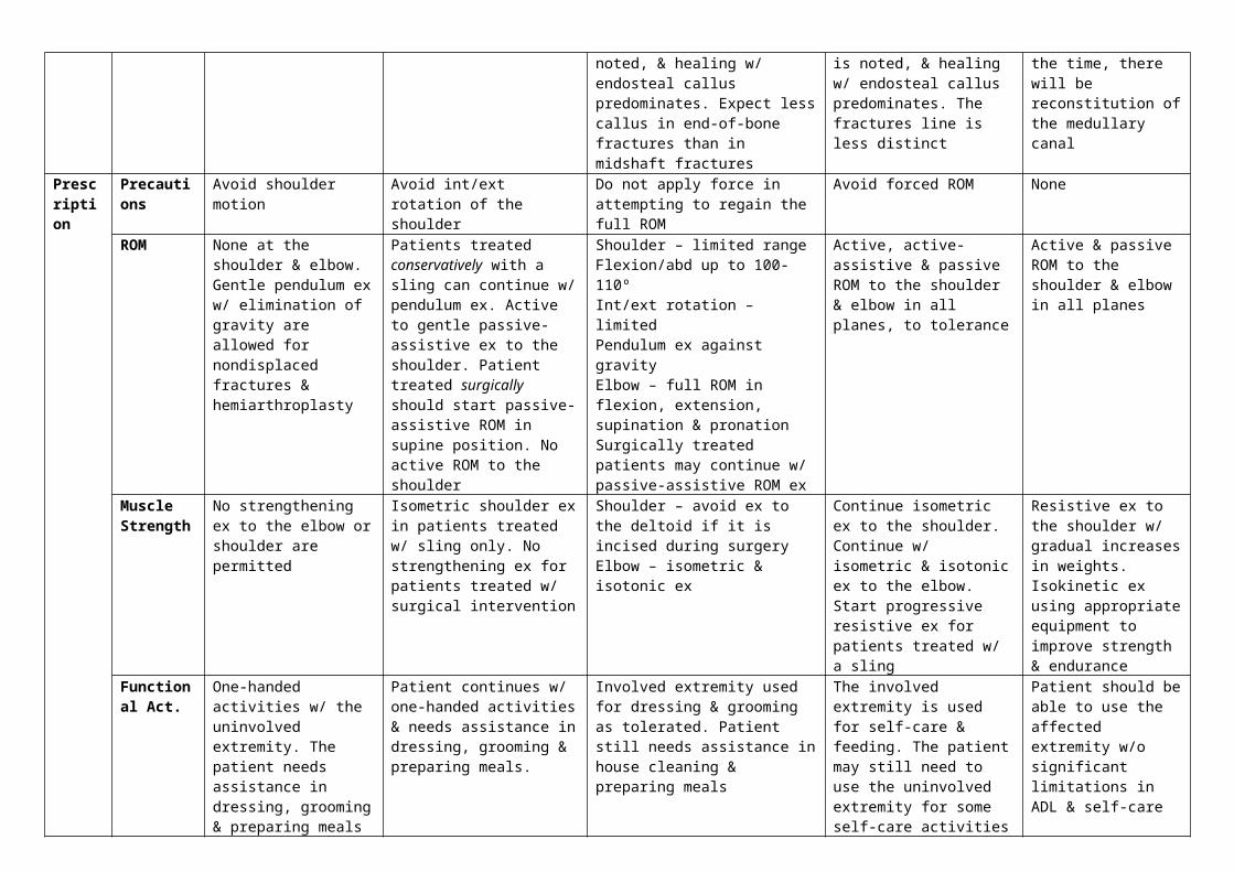
Bridging callus is visible. With increased rigidity of the fixation, less bridging callus is noted, & healing w/ endosteal callus predominates. Expect less callus in end-of-bone fractures than in midshaft fractures
Bridging callus is visible. With increased rigidity, less bridging callus is noted, & healing w/ endosteal callus predominates. The fractures line is less distinct
Abundant callus; fracture line begins to disappear. With the time, there will be reconstitution of the medullary canal
Prescription
Precautions Avoid shoulder motion Avoid int/ext rotation of the shoulder
Do not apply force in attempting to regain the full ROM
Avoid forced ROM None
ROM None at the shoulder & elbow. Gentle pendulum ex w/ elimination of gravity are allowed for nondisplaced fractures & hemiarthroplasty
Patients treated conservatively with a sling can continue w/ pendulum ex. Active to gentle passive-assistive ex to the shoulder. Patient treated surgically should start passive-assistive ROM in supine position. No active ROM to the shoulder
Shoulder – limited rangeFlexion/abd up to 100-110ºInt/ext rotation – limitedPendulum ex against gravityElbow – full ROM in flexion, extension, supination & pronationSurgically treated patients may continue w/ passive-assistive ROM ex
Active, active-assistive & passive ROM to the shoulder & elbow in all planes, to tolerance
Active & passive ROM to the shoulder & elbow in all planes
Muscle Strength
No strengthening ex to the elbow or shoulder are permitted
Isometric shoulder ex in patients treated w/ sling only. No strengthening ex for patients treated w/ surgical intervention
Shoulder – avoid ex to the deltoid if it is incised during surgeryElbow – isometric & isotonic ex
Continue isometric ex to the shoulder.Continue w/ isometric & isotonic ex to the elbow.Start progressive resistive ex for patients treated w/ a sling
Resistive ex to the shoulder w/ gradual increases in weights. Isokinetic ex using appropriate equipment to improve strength & endurance
Functional Act.
One-handed activities w/ the uninvolved extremity. The patient needs assistance in dressing, grooming & preparing meals
Patient continues w/ one-handed activities & needs assistance in dressing, grooming & preparing meals.
Involved extremity used for dressing & grooming as tolerated. Patient still needs assistance in house cleaning & preparing meals
The involved extremity is used for self-care & feeding. The patient may still need to use the uninvolved extremity for some self-care activities
Patient should be able to use the affected extremity w/o significant limitations in ADL & self-care
Weight Bearing
None on affected extremity
None on affected extremity None o affected extremity WB as tolerated FWB
2 Penanganan Fraktur (dikutip dari Treatment & Rehabilitation of Fractures; Hoppenfeld) by Fanny Christina; Printed by Indriana
3PENANGANAN FRAKTUR DIAPHYSIS ATAU MIDSHAFT HUMERUS
0 – 1 Week 2 Weeks 4 – 6 Weeks 8 – 12 WeeksBone Healing
Stability None None to minimal Bridging callus & moderate stability Stable callusStage Inflammatory phase Beginning of reparative phase Reparative phase Remodeling phaseX-Ray Callus (-) None to very early callus Bridging callus is visible Abundant callus, fracture line
begins disappear, reconstitution of medullary canal. Non union is clearly evident
Prescription Precautions No Lifting w/ the affected extremity No Lifting w/ the affected extremity No heavy lifting w/ the affected extremity
No contact sports
ROM Brace / Splint : No ROM to the shoulder & elbowORIF / external fixator : gentle active & active-assistive ROM to the shoulder & elbow if fixation is stable. Pendulum ex. w/ gravity (-) to the shoulder
Active & active-assistive ROM to the shoulder & elbow. W/ splint or brace, no abd shoulder > 60º
Active & active-assistive ROM to the shoulder & elbow
Active, active-assistive & passive ROM tp the shoulder & elbow
Muscle Strength
No strengthening exc. to the elbow or shoulder
Gentle pendulum exercise to the shoulder. No strengthening exercise to shoulder & elbow
Isometric & isotonic exc. To the forearm muscles.After 6 weeks, isometric exc. To biceps & triceps
Progressive resistive exc. to the shoulder & elbow
Functional Activities
Uninvolved extremity may be used for self-care & ADL
ADL w/ uninvolved extremity.In ORIF & external fixation, involved extremity used for feeding, light grooming, writing
Involved extremity may be used for basic self-care & personal hygiene
Involved extremity may be used in ADL. Light lifting is allowed w/ the affected extremity
Weight Bearing
NWB on affected extremity NWB on affected extremity. Limited WB w/ rodding
Early WB is allowed w/ int. fixation FWB is allowed
3 Penanganan Fraktur (dikutip dari Treatment & Rehabilitation of Fractures; Hoppenfeld) by Fanny Christina; Printed by Indriana

4PENANGANAN FRAKTUR HUMERUS DISTAL
0 – 1 Week 2 Weeks 4 – 6 Weeks 8 – 12 WeeksBone Healing
Stability No bony stability. Some stability may be afforded ba an intact periosteum & ligaments
None to minimal Once calus is observed bridging the fracture site, the fracture is usually stable. This should be confirmed by physical examination. The strength of this callus is significantly lower than of normal bone, especially w/ torsional load
Stable
Stage Inflammatory phase Beginning of reparative phase Reparative phase Remodeling phaseX-Ray Callus (-) None to early callus Bridging callus is visible. W/
increased rigidity, less bridging callus is noted & healing w/ endosteal callus predominates
Callus is present but less than in midshaft. The fracture line begins to disappear. Reconstitution of medullary canal occurs w/ time.
Prescription Precautions No int or ext rotation of the shoulder.No passive ROM to the elbow
No int or ext rotation of the shoulder.No passive ROM to the elbow
Avoid rotational stresses across the elbow
Avoid heavy lifting or pushing
ROM Gentle active elbow flexion & extension allowed for stable fractures treated w/ ORIF.No ROM to the elbow if treated by other methods
Gentle active flexion & extension exc. to the elbow for fractures only when treated w/ ORIF.Gentle assistive supervised active flexion & extension for nondisplaced stable fractures
Active / active-assistive flexion & extension to the elbow
Active & passive ROM to the elbow
Muscle Strength
No strengthening exc. to the elbow No strengthening exc. to the elbow No strengthening exc. to the elbow
Progressive resistive exc. to the elbow musculature
Functional Activities
The uninvolved extremity is used for self-care & ADL
The uninvolved extremity is used for self-care & ADL
The uninvolved extremity is used for self-care & ADL
The involved extremity used for self-care & personal hygiene
Weight Bearing
NWB on affected extremity NWB on affected extremity NWB on affected extremity FWB by 12 weeks
4 Penanganan Fraktur (dikutip dari Treatment & Rehabilitation of Fractures; Hoppenfeld) by Fanny Christina; Printed by Indriana

5PENANGANAN FRAKTUR OLEKRANON
0 – 1 Week 2 Weeks 4 – 6 Weeks 6 – 8 Weeks 8 – 12 WeeksBone Healing
Stability None None to minimal W/ bridging callus, the fracture line is usually stable
Stable Stable
Stage Inflammatory phase Beginning of reparative phase Reparative phase Reparative phase Remodeling phaseX-Ray Callus (-) None to early callus. Fracture
line is visibleBridging callus is visible. Fracture line is less distinct. Endosteal callus formation will predominate
Bridging callus is more apparent, especially w/ less-rigid fixation. Fracture line is less distinct. There is less callus formation if the fracture site is at the end of the ulna than in a midshaft fracture..
More callus is seen 7 fracture line becomes even less distinct
Prescription Precautions Avoid premature elbow motion
Cast or splint : no extension to the elbow < 90º
Active to active-assitive ROM to the elbow & wrist
None None
ROM No ROM to the elbow or wrist in a cast or splint. Gentle active elbow flexion & active ROM to the wrist if treated surgically
No ROM to the elbow or wrist in a cast or splint. Active elbow flexion & active ROM to the wrist if treated surgically
Encourage active ROM to the elbow in flexion & extension
Full active to active-assitive ROM in all planes to the elbow & wrist
Full active & active-assisted ROM in all planes to the elbow & wrist
Muscle Strength
No strengthening exc. to the elbow.Three or 4 days after fracture, isometric exc. to the wrist within the cast
No strengthening exc. to the elbow in extension.Isometric exc. to the elbow in flexion in a cast.Isometric exc. to the wrist
Isometric exc. to the elbow & wrist in flexion & extension
Resistive exc. to the elbow & wrist
Resistive exc. to the elbow & wrist
Functional Activities
One-handed activities. The patient uses the uninvolved extremity for personal hygiene & self-care
The patient uses the uninvolved extremity for personal hygiene & self-care
The patient uses the affected extremity for stability & light self-care
The patient uses the affected extremity for personal hygiene & self-care
The patient uses the affected extremity for personal hygiene & self-care
Weight Bearing
None None NWB Gradual WB is allowed FWB is allowed
5 Penanganan Fraktur (dikutip dari Treatment & Rehabilitation of Fractures; Hoppenfeld) by Fanny Christina; Printed by Indriana

6PENANGANAN FRAKTUR RADIAL HEAD
0 – 1 Week 2 Weeks 4 – 6 Weeks 8 – 12 WeeksBone Healing
Stability None None to minimal W/ bridging callus, the fracture line is usually stable; confirm w/ physical examination
Stable
Stage Inflammatory phase Beginning of reparative phase Reparative phase Remodeling phaseX-Ray Callus (-) Callus (-) Bridging callus is visible. W/
increased rigidity, less bridging callus is noted & healing w/ endosteal callus predominates. The amount of callus formation is less at the ends of the long bones, compared to midshaft fractures
Visible bridging callus in nonoperative patients. There is less callus with int fixation
Prescription Precautions No passive ROM to the elbow No passive ROM to the elbow Avoid valgus stresses to the elbow to avoid stress on the radial head
No pushing or lifting heavy objects
ROM Gentle, active ROM to the elbow in flexion & pronation
Active ROM to the elbow Active, active-assistive & passive ROM to the elbow for nonoperative cases. Active & active-assistive ROM for patients w/ int. fixation
Active & passive ROM to the elbow
Muscle Strength
No strengthening exc. to the elbow.
No strengthening exc. to the elbow. Start isometric exc. to the deltoid, biceps & triceps
Isometric exc. to the biceps, triceps & deltoid
Progressive resistive exc. are given to the elbow flexor, extensors, supinators & pronators
Functional Activities
The uninvolved extremity is used for ADL
The uninvolved extremity is used for self-care
The uninvolved extremity is used in self-care. The involved extremity is used to assist in gentle activities
The affected extremity is used in self-care
Weight Bearing
None None PWB for patients w/ nonoperative fixation. NWB for patients w/ int fixation
WB allowed for self-care & light-duty activities
6 Penanganan Fraktur (dikutip dari Treatment & Rehabilitation of Fractures; Hoppenfeld) by Fanny Christina; Printed by Indriana

7PENANGANAN FRAKTUR FOREARM
0 – 1 Week 2 Weeks 4 – 6 Weeks 8 – 12 WeeksBone Healing
Stability None None to minimal Once callus is observed bridging the fracture site, the fracture is usually stable. This should be confirmed w/ physical examination. The strength of this callus is significantly lower than that of normal bone.
Stable
Stage Inflammatory phase Beginning of reparative phase Reparative phase Woven bone is replaced by lamellar bone. The process of remodeling takes months to years. Patients whose treatment is w/ rigid fixation have direct bridging osteomes.
X-Ray Callus (-) None to early callus Bridging callus is visible in patient w/ a cast. Patient who have had anatomic rigid int fixation show little or no callus, because primary bone healing predominates. The fracture line becomes less visible.
Abundant callus is present if cast treatment was used. The fracture line begins disappear & reconstitution of the medullary canal occurs w/ time. Patient who have had anatomic rigid int fixation show little or no callus; rather, the fracture line disappear as primary bone healing progresses. The amount of callus is inversely proportional to the stability.
Prescription Precautions No passive ROM No passive ROM No passive ROM to the forearm No heavy lifting or sports activitiesROM If there is adequate fixation &
the forearm is not in a cast, gentle active ROM exc. are prescribed to the elbow & wrist, including supination & pronation exc.
Gentle active ROM to the elbow & wrist if there is adequate fixation & the forearm is not in a cast
Active to active-assistive ROM to the elbow & wrist, including supination & pronation if the patient is out of cast.
Full active & passive ROM to the elbow & wrist. Stress supination & pronation of the forearm
Muscle Strength
Isometric exc. to the deltoid, biceps & triceps if the fracture is rigidly fixed. No strengthening exc. to the forearm if treated w/ cast only
No strengthening exc. to the forearm if treated w/ cast only. Isometric exc. to the deltoid, biceps & triceps w/ rigid fixation
If fixation is adequate at end of 6 weeks, start gentle isokinetic exc. to the forearm muscles w/ < 5 lb of resistance
Progressive resistive exc. are prescribed for the forearm muscles. Use free weights of 5 lb & more
Functional Activities
The uninvolved extremity is used for self-care
The uninvolved extremity is used for self-care
The involved extremity is used for light self-care activities.
The affected extremity is used for self-care
Weight Bearing
NWB on the affected extremity NWB on the affected extremity NWB on the affected extremity FWB as tolerated
7 Penanganan Fraktur (dikutip dari Treatment & Rehabilitation of Fractures; Hoppenfeld) by Fanny Christina; Printed by Indriana

8PENANGANAN FRAKTUR COLLES
0 – 1 Week 2 Weeks 4 – 6 Weeks 6 – 8 Weeks 8 – 12 WeeksBone Healing
Stability None None to minimal W/ bridging callus, the fracture is usually stable; confirm w/ physical examination
W/ bridging callus, the fracture is usually stable; confirm w/ physical examination
Satble
Stage Inflammatory phase Beginning of reparative phase
Reparative phase Reparative phase Remodeling phase
X-Ray Callus (-); fracture line is visible
None to early callus; fracture line is visible
Bridging callus is visible. W/ increased rigidity, less bridging callus is noted, & healing w/ endosteal callus predominates. The fracture line is less distinct.
Bridging callus is visible. W/ increased rigidity, less bridging callus is noted, & healing w/ endosteal callus predominates. The fracture line is less distinct.
Callus is seen. The fracture line begins to disappear; w/ time, the contour of the bone is being restored. Metaphyseal areas do not produce as much callus as diaphyseal regions
Prescription Precautions No supination & pronation No ROM to wrist
No supination & pronation if treated w/ cast & ORIFNo passive ROM
No passive ROM to the forearm None, unless pseudoarthrosis or nonunion is suspected
None
ROM Full active ROM of digits of MCP joint.Full opposition of thumb
Full ROM of MCP & IP joint.Attempt gentle active ROM of wrist if treated by ORIF & fixation is rigid.
Full active ROM of wrist, MCP & IP joints.Supination & pronation encouraged. Active ulnar & radial deviation.
Full ROM of all joints of upper extremity.Stress supination & ulnar deviation.Active assistive to passive ROM attempted or initiated.
Full ROM, active & passive in all planes to the wrist & digits. Stress supination & ulnar deviation
Muscle Strength
Attempt isometric exc. to the intrinsic muscles of the hand
Isometric exc. given to the intrinsic muscles of the hand & wrist flexor & extensor.
Gentle resistive exc. given to the digits of the hand.Improve power grip Isometric exc. to wrist flexors, extensors & radial and ulnar deviators. Gentle resistive exc. given to the wrist if treated by ORIF
Gentle resistive exc. to the digits & wrist.Improve power grip
Progressive resistive exc. to the wrist & digits & to all the groups of muscles
Functional Activities
Use the uninvolved extremity for self-care & ADL
Uninvolved extremity is used for self-care & ADL
The involved extremity may be used as a stabilizer in two-handed activities. The patient may attempt self-care w/ involved extremity.
The affected extremity is used for self-care & ADL
The patient may use the involved extremity in self-care & ADL
Weight Bearing
NWB on the affected extremity
NWB on the affected extremity
Avoid WB until the end of 6 weeks
WB as tolerated, because the fracture is stable
FWB as tolerated on the involved extremity
8 Penanganan Fraktur (dikutip dari Treatment & Rehabilitation of Fractures; Hoppenfeld) by Fanny Christina; Printed by Indriana

9PENANGANAN FRAKTUR SCAPHOID (NAVICULAR)
0 – 1 Week 2 Weeks 4 – 6 Weeks 8 – 12 Weeks 12 – 16 WeeksBone Healing
Stability No bony stability, although ligamentous stability may be present
None to minimal Bridging callus indicates stability
Stable Stable
Stage Inflammatory phase Beginning of reparative phase
Reparative phase Remodeling phase Remodeling phase
X-Ray Callus (-); fracture line is visible
Callus (-). Resorption at fracture site may be seen
Callus is not seen because there is no periosteum. This is a membranous bone. Trabecular bone may be visible
Fracture line begins to disappear w/ reconstitution of trabecular bone pattern
Fracture line begins to disappear. There is reconstitution of the trabecular bone pattern
Prescription Precautions Avoid supination & pronation of the elbow
Avoid supination & pronation at the elbow
Avoid passive ROM to the thumb & wrist
Avoid heavy lifting None if fracture is healed
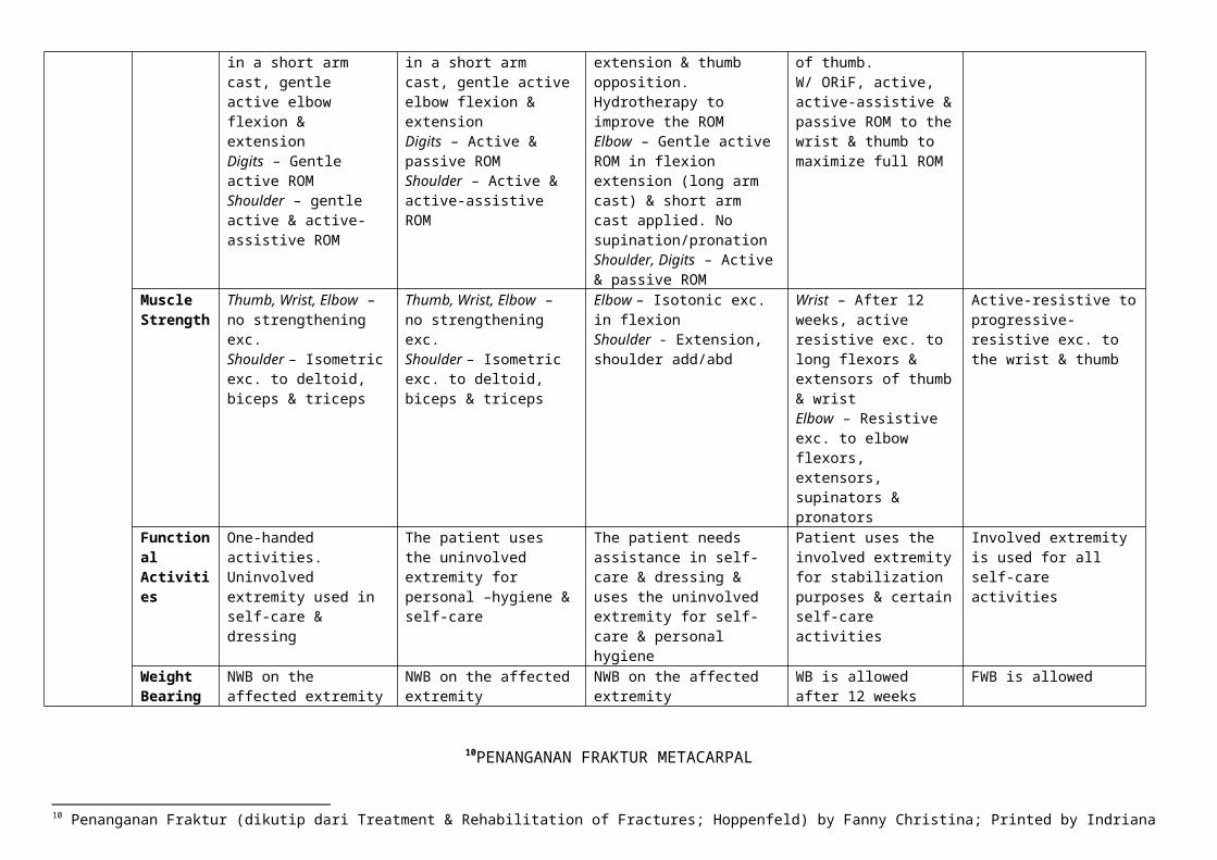
ROM Thumb, Wrist – none (immobilized)Elbow – none if immobilized in a long arm cast. If in a short arm cast, gentle active elbow flexion & extensionDigits – Gentle active ROMShoulder – gentle active & active-assistive ROM
Thumb, Wrist – none (immobilized)Elbow – none if immobilized in a long arm cast. If in a short arm cast, gentle active elbow flexion & extensionDigits – Active & passive ROMShoulder – Active & active-assistive ROM
Thumb – If short arm cast is removed (ORIF), gentle active ROM to the wrist & thumb in flexion, extension & thumb opposition. Hydrotherapy to improve the ROMElbow – Gentle active ROM in flexion extension (long arm cast) & short arm cast applied. No supination/pronationShoulder, Digits – Active & passive ROM
Cast is removed after 12 weeks. Gentle active ROM to wrist & digits & MCP & IP joints of thumb.W/ ORiF, active, active-assistive & passive ROM to the wrist & thumb to maximize full ROM
Active-resistive, passive ROM of wrist & thumb.
Muscle Strength
Thumb, Wrist, Elbow – no strengthening exc.Shoulder – Isometric exc. to deltoid, biceps & triceps
Thumb, Wrist, Elbow – no strengthening exc.Shoulder – Isometric exc. to deltoid, biceps & triceps
Elbow – Isotonic exc. in flexionShoulder - Extension, shoulder add/abd
Wrist – After 12 weeks, active resistive exc. to long flexors & extensors of thumb & wristElbow – Resistive exc. to elbow flexors, extensors, supinators & pronators
Active-resistive to progressive-resistive exc. to the wrist & thumb
Functional Activities
One-handed activities. Uninvolved extremity used in self-care & dressing
The patient uses the uninvolved extremity for personal –hygiene & self-care
The patient needs assistance in self-care & dressing & uses the uninvolved extremity for self-care & personal hygiene
Patient uses the involved extremity for stabilization purposes & certain self-care activities
Involved extremity is used for all self-care activities
Weight Bearing
NWB on the affected extremity
NWB on the affected extremity
NWB on the affected extremity WB is allowed after 12 weeks
FWB is allowed
9 Penanganan Fraktur (dikutip dari Treatment & Rehabilitation of Fractures; Hoppenfeld) by Fanny Christina; Printed by Indriana

10PENANGANAN FRAKTUR METACARPAL
0 – 1 Week 2 Weeks 4 – 6 Weeks 6 – 8 Weeks 8 – 12 WeeksBone Healing
Stability None None to minimal W/ bridging callus, the fracture is usually stable; confirm w/ physical examination
W/ bridging callus, the fracture is usually stable; confirm w/ physical examination
Stable
Stage Inflammatory phase Beginning of reparative phase
Reparative phase Reparative phase Remodeling phase
X-Ray Callus (-) Callus (-) Bridging callus is visible. W/ increased rigidity, less bridging callus is noted & healing w/ endosteal callus predominates. Fracture line is less distinct
Bridging callus is visible. W/ increased rigidity, less bridging callus is noted & healing w/ endosteal callus predominates. Fracture line is less distinct
Abundant callus is seen & the fracture line begins to disappear; w/ time, there will be reconstitution of the medullary canal. Metaphyseal areas do not produce as much callus as diaphyseal regions
Prescription Precautions No passive ROM No passive ROM to the affected digit
No passive ROM to the affected digit
None None
ROM Active ROM to non-splinted digits
1. If rigid fixation is achieved, active ROM to the affected digit
2. Active, active-assistive & passive ROM to non-splinted digits
1. Full active ROM to all digits & wrist
2. Active pronation & supination of wrist & ulnar & radial deviation of the wrist
Active, active-assistive & passive ROM to all digits
Full active & passive ROM to all digits
Muscle Strength
Isometric exc. prescribed within the cast of the non-splinted fingers
Isometric exc. to the intrinsic muscles of non-splinted digits
1. Gentle ball-squeezing & Silly Putty exc.
2.Gentle add & abd resistive exc. of the digits
Active-resistive exc. to all digits & wrist
Progressive resistive exc. to the all digits w/ increasing weights
Functional Activities
Uninvolved extremity used in self-care & personal hygiene
Uninvolved extremity used in self-care & personal hygiene
Bimanual activities are encouraged at 6 weeks
The patient uses affected extremity for self-care & personal hygiene
The affected extremity used for self-care
Weight Bearing
None None None FWB as tolerated FWB
10 Penanganan Fraktur (dikutip dari Treatment & Rehabilitation of Fractures; Hoppenfeld) by Fanny Christina; Printed by Indriana

11PENANGANAN FRAKTUR PHALANG
0 – 1 Week 2 Weeks 4 – 6 Weeks 6 – 8 Weeks 8 – 12 WeeksBone Healing
Stability None None to minimal W/ bridging callus, the fracture is usually stable; confirm w/ physical examination
W/ bridging callus, the fracture is usually stable. However, the strength of this callus, especially w/ torsional load, is significantly lower than that of normal lamellar bone. Confirm w/ physical examination
Stable
Stage Inflammatory phase Beginning of reparative phase
Reparative phase Reparative phase Remodeling phase
X-Ray Callus (-); fracture line is visible
None to early callus; fracture line is visible
Bridging callus is visible. W/ increased rigidity, less bridging callus is noted & healing w/ endosteal callus predominates. Fracture line is less distinct
Bridging callus is visible. W/ increased rigidity, less bridging callus is noted & healing w/ endosteal callus predominates. Fracture line is less distinct
Abundant callus is seen & the fracture line begins to disappear; there is reconstitution of the medullary canal. Metaphyseal areas do not produce as much callus as diaphyseal regions
Prescription Precautions No ROM to the digit if the fracture is unstable
No ROM to the splinted joint
No passive ROM to the affected joint
Night splint is used if necessary
None
ROM Active ROM to the unaffected digits & to the fractured digit if the fracture is stable
Active ROM to all non-splinted joints & digits
Full active & active-assistive ROM to all digits
Active, active-assistive & passive ROM to all digits
Full active & passive ROM to all digits & wrist.
Muscle Strength
Isometric exc. to the intrinsic muscles of the non-splinted fingers
Isometric strengthening exc. to the intrinsic muscles
Isometric & isotonic exc. to the flexors, extensors, abd & add of the digit
Gentle resistive exc. to all digits
Progressive resistive exc. to the digits & wrist
Functional Activities
The uninvolved extremity used for self-care & personal hygiene
The uninvolved extremity used for self-care
Bimanual activities using the involved extremity are encouraged for self-care
The involved extremity is used for self-care
The involved extremity is used in all activities to tolerance
Weight Bearing
None None WB as tolerated by the patient
FWB FWB
11 Penanganan Fraktur (dikutip dari Treatment & Rehabilitation of Fractures; Hoppenfeld) by Fanny Christina; Printed by Indriana

12PENANGANAN FRAKTUR COLLUM / NECK FEMUR
12 Penanganan Fraktur (dikutip dari Treatment & Rehabilitation of Fractures; Hoppenfeld) by Fanny Christina; Printed by Indriana

0 – 1 Week 2 Weeks 4 – 6 Weeks 8 – 12 Weeks 12 – 16 WeeksBone Healing
Stability No stability is present from bone healing.Impacted femoral neck fracture : partial bony stabilityTreated w/ screw, except severe osteopenia : immediate mechanical stabilityTreated w/ hemiarthroplasty : full mechanical stability
Only minimal stability.Impacted femoral neck fracture : partial bony stabilityTreated w/ screws, except severe osteopenia : immediate mechanical stabilityTreated w/ hemiarthroplasty : full mechanical stability
Moderate stability from bone healing is present as endosteal callus bridges the fracture; correlate w/ physical examination.Mechanical stability from hardware or endoprosthesis is unchanged
Moderate stability from bone healing is present as endosteal callus bridges the fracture; correlate w/ physical examination.Mechanical stability from hardware or endoprosthesis is unchanged
Significant stability is now present from bone healing as endosteal callus bridges the fracture; correlate w/ physical examination.Mechanical stability from hardware or endoprosthesis is unchanged
Stage Inflammatory phase Beginning of reparative phase Reparative phase Late reparative, early remodeling phase
Remodelling phase
X-Ray Callus (-), fracture line is clearly visible. No periosteum, all healing is endosteal
No callus is visible (healing is endosteal/intenal)Fracture line is visible
No external callus is visible because healing is endosteal (internal) & composed of cartilage & fibrous tissue; this gradually becomes visible as it undergoes endochondral ossification
No external callus is visible because healing is endosteal (internal) & composed of cartilage & fibrous tissue; this gradually becomes visible as it undergoes endochondral ossification
No external callus is visible because healing is endosteal (internal) & composed of cartilage & fibrous tissue; this gradually becomes visible as it undergoes endochondral ossification. Fracture line is obliterated
Prescription
Precau- tions
Avoid passive ROM.Patient treated w/ endoprotheses avoid int. rotation & add past midline
Avoid passive ROM on fractures that have been reduced.Treated w/ endoprotheses : avoid int. rotation & add past midline
No passive ROM on fractures that have been reduced.Treated w/ hemiarthroplasty : avoid int rotation & add past midline
Avoid excessive add & int rotation if use endoprosthesis
Avoid excessive add if use endoprosthesis
ROM Active ROM hip & knee Active, active-assistive ROM to hip & knee
Active, active-assistive ROM to hip & knee
Active, active-assitive & passive ROM to hip & knee
Full active & passive ROM to hip & knee
Muscle Strength
Isometric gluteal & quadriceps exc.Isotonic exc. to ankle
Isometric gluteal & quadriceps exc.
Isometric & isotonic exc. to hip & knee
Isotonic & isokinetic exc. to hip & knee. Progressive resistive exc. instituted
Isokinetic & isotonic exc. & progressive resistive exc.
Functional Act.
Stand-pivot transfers & ambulation w/ assistive devices; raised toilet seat & chair
Stand-pivot transfers & ambulation w/ assistive devices
Stand-pivot transfers & ambulation w/ assistive devices
WB transfers & ambulation w/ assistive devices
Independent in transfers & ambulation w/o assistive devices
Weight Bearing
Stable impacted fracture or endoprotheses : WB as toleratedUnstable fracture that require reduction : NWB
Stable impacted fracture or endoprotheses : WB as toleratedUnstable fracture that require reduction : NWB
Stable impacted fracture or endoprotheses : WB as toleratedUnstable fracture that require reduction : NWB
FWB to WB as tolerated FWB
13PENANGANAN FRAKTUR INTERTROCHANTER FEMUR
13 Penanganan Fraktur (dikutip dari Treatment & Rehabilitation of Fractures; Hoppenfeld) by Fanny Christina; Printed by Indriana
0 – 1 Week 2 Weeks 4 – 6 Weeks 8 – 12 WeeksBone Healing
Stability None None to minimal With a bridging callus, the fracture is usually stable; confirm w/ physical examination
Stable
Stage Inflammatory phase Beginning of reparative phase Reparative phase Early remodeling phaseX-Ray Callus (-), fracture line is visible. None to very early callus; fracture
line is visible. Bone in the metaphyseal region has very thin periosteum & does not form an abundant external callus
Bridging callus is beginning to be visible. Endosteal callus may predominate in the metaphyseal region & the fracture line should become less visible
Abundant callus has formed & fracture line begins to disappear. The medullary canal & metaphyseal region begin to be reconstituted.
Prescription
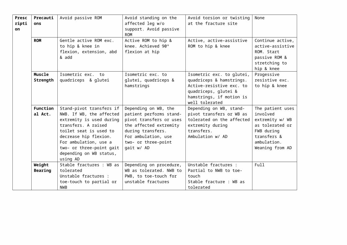
Precautions Avoid passive ROM Avoid standing on the affected leg w/o support. Avoid passive ROM
Avoid torsion or twisting at the fracture site
None
ROM Gentle active ROM exc. to hip & knee in flexion, extension, abd & add
Active ROM to hip & knee. Achieved 90º flexion at hip
Active, active-assistive ROM to hip & knee
Continue active, active-assistive ROM. Start passive ROM & stretching to hip & knee
Muscle Strength
Isometric exc. to quadriceps & glutei
Isometric exc. to glutei, quadriceps & hamstrings
Isometric exc. to glutei, quadriceps & hamstrings. Active-resistive exc. to quadriceps, glutei & hamstrings, if motion is well tolerated
Progessive resistive exc. to hip & knee
Functional Act.
Stand-pivot transfers if NWB. If WB, the affected extremity is used during transfers. A raised toilet seat is used to decrease hip flexion.For ambulation, use a two- or three-point gait depending on WB status, using AD
Depending on WB, the patient performs stand-pivot transfers or uses the affected extremity during transfers. For ambulation, use two- or three-point gait w/ AD
Depending on WB, stand-pivot transfers or WB as tolerated on the affected extremity during transfers. Ambulation w/ AD
The patient uses involved extremity w/ WB as tolerated or FWB during transfers & ambulation. Weaning from AD
Weight Bearing
Stable fractures : WB as toleratedUnstable fractures : toe-touch to partial or NWB
Depending on procedure, WB as tolerated. NWB to PWB, to toe-touch for unstable fractures
Unstable fractures : Partial to NWB to toe-touchStable fracture : WB as tolerated
Full
14PENANGANAN FRAKTUR SUBTROCHANTER FEMUR
14 Penanganan Fraktur (dikutip dari Treatment & Rehabilitation of Fractures; Hoppenfeld) by Fanny Christina; Printed by Indriana

0 – 1 Week 2 Weeks 4 – 6 Weeks 8 – 12 Weeks 12 – 16 WeeksBone Healing
Stability None None to minimal Callus is beginning to bridge fracture fragments in the femoral region (thick periosteum) & endosteal healing is bridging the metaphyseal region (thin periosteum but rich intramedullary blood supply). Unless bone loss or severe comminution is present, the fracture is usually stable; confirm w/ physical examination
Stable Stable
Stage Inflammatory phase Beginning of reparative phase Reparative phase Early remodeling phase Remodeling phaseX-Ray Callus (-), fracture line is
clearly visible. None to very early callus in the region below the lesser trochanter. Callus (-) in the intertrochanteric region where periosteum is thin & healing is predominately endosteal. Fracture line is visible
Bridging callus is beginning to be visible. W/ increased rigidity of fixation, less bridging callus is noted & healing w/ endosteal callus predominates. Fracture line is less visible in both the shaft & metaphyseal regions
Abundant callus in fracture w/ intact periosteum. Fracture line begins disappear
Abundant callus is present & fracture line begins to disappear
Prescription
Precau-tions
No add & abd to hip. No isometric exc. to quads & hamstrings
Avoid torsional forces on fracture. Avoid excessive abd or add
Avoid torsional forces on fracture site. None None
ROM Active ROM to hip & knee in flexion & extension
Active, active-assistive to gentle passive ROM to hip in flexion & extension
Active, active-assistive, passive ROM to hip in flexion & extension. Active ROM to hip in abd & add
Full ROM in all planes to hip & knee
Full ROM in all planes to hip & knee
Muscle Strength
Isometric exc. to glutei Isometric exc. to glutei, quadriceps & hamstrings
Isometric exc. to glutei, quadriceps & hamstrings.
Gradual resistive exc. to hip & knee
Prgressive resistive exc. to hip & knee
Functional Act.
WB as tolerated or toe-touch WB during transfers w/ AD & 3-point gait w/ AD
Toe-touch WB or WB as tolerated during transfers & 3-point gait; WB as tolerated or toe-touch WB w/ AD
Toe-touch WB or WB as tolerated during transfers & ambulation w/ AD
WB as tolerated or FWB during transfers & ambulation w/ AD
FWB during transfer & ambulation
Weight Bearing
Stable fractures treated w/ intramedullary nails: WB as tolerated on affected extremityUnstable fractures or those treated by ORIF : toe-touch WB
Stable fractures treated w/ intramedullary nails: WB as tolerated on affected extremityUnstable fractures or those treated by ORIF : toe-touch WB
Stable fractures treated w/ intramedullary nails: WB as tolerated on affected extremityUnstable fractures or those treated by ORIF : toe-touch WB
Almost all fractures have sufficient bone healing & callus to be FWB as tolerated. Limited WB should be necessary only for fractures w/ no callus present that are being considered for bone grafting
Almost all fractures have sufficient bone healing & callus to be FWB as tolerated. Limited WB should be necessary only for fractures w/ no callus present that are being considered for bone grafting
15PENANGANAN FRAKTUR SHAFT FEMUR
15 Penanganan Fraktur (dikutip dari Treatment & Rehabilitation of Fractures; Hoppenfeld) by Fanny Christina; Printed by Indriana

0 – 1 Week 2 - 4 Weeks 4 – 6 Weeks 8 – 12 Weeks 12 – 16 WeeksBone Healing
Stability None None to minimal With bridging callus, the fracture is usually stable; confirm w/ PE
Stable Stable
Stage Inflammatory phase Beginning of reparative phase Reparative phase Early remodeling phase Remodeling phaseX-Ray Callus (-), fracture line is clearly
visible. None to very early callus; fracture line is visible
Bridging callus is beginning to be visible. W/ increased rigidity of fixation, less bridging callus will be noted, & healing w/ endosteal callus will predominate. The amount of callus formation is greater for diaphyseal than metaphyseal fractures. Fracture line is less visible
Abundant callus in fractures not rigidly fixed by plates. Fracture line begins to disappear; with time, there will be reconstitution of the medullary canal, except w/ an intramedullary nail
Abundant callus in fractures not rigidly fixed by plates. Fracture line begins to disappear; w/ time, there will be reconstitution of the medullary canal, except w/ an intramedullary nail
Prescription
Precautions No passive ROM to hip & kneeNo rotation on planted foot
Avoid rotation on the affected extremity w/ the foot planted
Avoid rotation on the affected extremity w/ foot planted
Avoid torsion loading of the femur
None
ROM Active ROM to hip & knee Active, active-assistive ROM to hip & knee, passive ROM closer to 4 weeks
Active/passive ROM to hip & knee Active/passive ROM to hip & knee
Active/passive ROM to hip & knee
Muscle Strength
Isometric exc. to quads & glutei
Isometric ex. to quads & glutei; straight leg raising
Resistive isotonic exc. & isometric exc. to quads, hamstrings & glutei
Progessive resistive exc. to quads, hamstrings & glutei
Progressive resistive exc. to quads, hamstrings & glutei. Isokinetic exc. to quadriceps & hamstrings
Functional Act.
Ambulatory stand-pivot transfers & ambulation w/ crutches
Ambulatory stand-pivot transfers w/ crutches & ambulation w/ crutches
Stand/pivot transfers & ambulation w/ crutches
Regular transfers. May need crutches for ambulation
Regular transfers. May need crutches for ambulation
Weight Bearing
Unstable fractures or those treated by plating or external fixator : toe-touch or NWBStable fracture : progress to FWB as tolerated
Unstable fractures or those treated by plating or external fixator : toe-touch or NWBStable fracture : WB as tolerated
Unstable fractures & those treated w/ plating or external fixator : PWBStable fracture : FWB
Stable fracture : FWB or WB as toleratedUnstable fracture : PWB
FWB
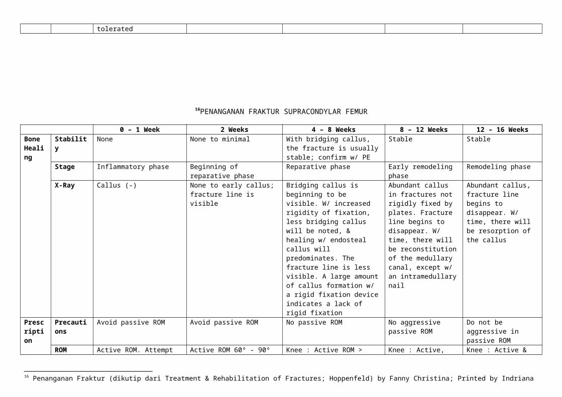
16PENANGANAN FRAKTUR SUPRACONDYLAR FEMUR
16 Penanganan Fraktur (dikutip dari Treatment & Rehabilitation of Fractures; Hoppenfeld) by Fanny Christina; Printed by Indriana

0 – 1 Week 2 Weeks 4 – 8 Weeks 8 – 12 Weeks 12 – 16 WeeksBone Healing
Stability None None to minimal With bridging callus, the fracture is usually stable; confirm w/ PE
Stable Stable
Stage Inflammatory phase Beginning of reparative phase Reparative phase Early remodeling phase Remodeling phaseX-Ray Callus (-) None to early callus; fracture
line is visibleBridging callus is beginning to be visible. W/ increased rigidity of fixation, less bridging callus will be noted, & healing w/ endosteal callus will predominates. The fracture line is less visible. A large amount of callus formation w/ a rigid fixation device indicates a lack of rigid fixation
Abundant callus in fractures not rigidly fixed by plates. Fracture line begins to disappear. W/ time, there will be reconstitution of the medullary canal, except w/ an intramedullary nail
Abundant callus, fracture line begins to disappear. W/ time, there will be resorption of the callus
Prescription
Precautions Avoid passive ROM Avoid passive ROM No passive ROM No aggressive passive ROM
Do not be aggressive in passive ROM
ROM Active ROM. Attempt full extension & 60º - 90º of flexion to the knee. Avoid passive ROM
Active ROM 60º - 90º in flexion & full extension to the knee.
Knee : Active ROM > 90º; active, active-assistive ROM in flexion & extension, if the fracture is stable
Knee : Active, active-assistive ROM; gentle passive ROM
Knee : Active & passive ROM; emphasize terminal extension to reduce extension lag
Muscle Strength
No strengthening exc. prescribed to the knee
Isometric exc. to quadriceps in supine position & knee in full extension
Knee : Isometric exc. to quadriceps & hamstrings
Knee : Isometric & isotonic exc. to quadriceps & hamstrings
Knee : Isometric, isotonic & isokinetic exc. to quadriceps & hamstrings. Gentle progressive resistive exc. Muscle strength 4+ or 5
Functional Act.
NWB stand/pivot transfers & NWB ambulation
NWB ambulation & stand/pivot transfers
NWB ambulation & stand/pivot transfers
NWB ambulation & stand/pivot transfers
PWB w/ crutches, progressing to FWB during ambulation & transfers
Weight Bearing
None None None None Toe-touch to PWB progressing to FWB
17PENANGANAN FRAKTUR PATELLA
17 Penanganan Fraktur (dikutip dari Treatment & Rehabilitation of Fractures; Hoppenfeld) by Fanny Christina; Printed by Indriana

0 – 1 Week 2 Weeks 4 – 6 Weeks 8 – 12 WeeksBone Healing
Stability None None to minimal None to minimal Stable
Stage Inflammatory phase Beginning of reparative phase Reparative phase Remodeling phaseX-Ray Fracture line is visible; no callus
formationCallus (-); fracture line is visible No callus; fracture line is less
visible. Sesamoid bones produce minimal callus
Small amount of callus noted. Fracture line begins to disappear w/ time. Amount of callus formed is small, because this is a sesamoid bone
Prescription
Precautions Avoid passive ROM Avoid passive ROM Maintain knee immobilizer if tenderness is present
ROM Knee : None if in a cast.If open reduction & stable internal fixation is achieved, active ROM of the knee in a sitting position w/o WB
Knee : NoneIf treated w/ open reduction & stable internal fixation, active knee flexion w/ no WB
Knee : Active ROM in flexion/extension
Knee : Active & passive ROM. Patient may have extension lag secondary to quad weakness & immobilization
Muscle Strength
No strengthening exc. prescribed to the knee
Knee : None Knee : Isometric exc. to quadriceps & hamstrings.At 6 weeks, isotonic exc. to quadriceps w/ active knee extension: 45º to 0º & then from 90º to 0º where 0º is full extension
Knee : Progressive resistive exc. to quadriceps & hamstrings w/ weights; isokinetic exc. using Cybex machine; pylometric closed chain exc.
Functional Act.
FWB during transfers & ambulation using AD
FWB during ambulation & transfers
FWB during ambulation & transfers. Remove immobilizer for level ground walking if fracture is stable
FWB during ambulation 7 transfers w/o AD
18PENANGANAN FRAKTUR TIBIAL PLATEAU
0 – 1 Week 2 Weeks 4 – 6 Weeks 8 – 12 Weeks 12 – 16 Weeks
18 Penanganan Fraktur (dikutip dari Treatment & Rehabilitation of Fractures; Hoppenfeld) by Fanny Christina; Printed by Indriana

Bone Healing
Stability None None to minimal W/ bridging callus, the fracture is usually stable; confirm w/ PE
Stable Stable
Stage Inflammatory phase Beginning of reparative phase
Reparative phase Early Remodeling phase Remodeling phase
X-Ray No callus None to early callus; fracture line is visible
Bridging callus is beginning to be visible. W/ increased rigidity of fixation, less bridging callus is noted & healing w/ endosteal callus predominates. The fracture line is less visible
Abundant callus in fracture not rigidly fixed by plates. Fracture line begins to disappear, w/ time the medullary canal will be reconstituted
Fracture line has disappeared
Prescription
Precautions No varus or valgus stress on knee; no passive ROM
No varus or valgus stress on knee; no passive ROM
No varus or valgus stress on knee; no passive ROM
No varus or valgus stress None
ROM Active & active-assistive flexion/extension: 40º to 60º of flexion allowed initially, increasingly to 90º of flexion after 1 week
Active & active-assistive flexion/extension up to 90º
Active & active-assistive ROM to the knee
Active, active-assistive & passive ROM to the knee
Full active & passive ROM to the knee
Muscle Strength
No strengthening exc. to knee Isometric exc. to the quadriceps
No strengthening exc. to the knee Gentle resistive exc. to the quadriceps & hamstrings
Progressive resistive exc. to the knee
Functional Act.
NWB stand/pivot transfers & ambulation w/ crutches
NWB stand/pivot transfers & ambulation w/ crutches
NWB transfers & ambulation w/ crutches
WB transfers & ambulation at the end of 12 weeks
FWB transfers & ambulation
Weight Bearing
NWB on the affected extremity NWB on affected extremity NWB on affected extremity Partial to FWB at the end of 12 weeks
FWB
19PENANGANAN FRAKTUR SHAFT TIBIA
0 – 1 Week 2 Weeks 4 – 6 Weeks 8 – 12 WeeksBone Stability None None to minimal W/ advancing callus, the fracture Fractures having minimal to no
19 Penanganan Fraktur (dikutip dari Treatment & Rehabilitation of Fractures; Hoppenfeld) by Fanny Christina; Printed by Indriana

Healing becomes stable for axial loading but must still be protected from torsional loading
comminution are increasingly stable to completely stable. Fractures that have significant bone loss or have required bone grafting for bone loss have limited stability until the bone graft begins consolidate & the callus is visible
Stage Inflammatory phase Beginning of reparative phase Reparative phase Early remodeling phaseX-Ray Callus (-) No callus; fracture line is visible Early callus may be visible in the
posterolateral aspect of the tibia where blood supply is best. If the fracture is rigidly fixed, little callus is seen
Bony consolidation is progressing, & the callus should be visible at the posterolateral surface of the tibia in extending around to the other surfaces. The fracture line should become cloudy & begin to disappear. If bone grafting was required, consolidation of this bone graft should begin to be seen
Prescription
Precautions Avoid rotary motion w/ the foot on the floor
Avoid rotary movements w/ the foot planted
Avoid rotation of the extremity on a fixed foot
ROM Active ROM ankle & knee if not in a cast
Active ROM ankle & knee if not in a cast
Active ROM to ankle & knee if not in a cast
Active, active-assistive & passive ROM to knee & ankle
Muscle Strength
Isometric ex to quadriceps, tibialis anterior & gastroc-soleus
Isometric exc. to quadriceps, tibialis anterior & gastroc-soleus
Isometric & isotonic exc. to knee & ankle
Gentle progressive resistive exc. prescribed to quadriceps, dorsiflexors & plantar flexors.
Functional Act.
Unstable fractures : stand-pivot transfers & NWB ambulation w/ AD Stable fracture : WB as tolerated to PWB transfers w/ AD
Unstable fractures : stand/pivot transfers & NWB ambulation w/ AD Stable fracture : WB as tolerated or PWB w/ AD, depending on the method of treatment
Unstable fractures : stand/pivot transfers & NWB ambulation w/ AD Stable fracture : WB as tolerated or PWB, to FWB transfers & ambulation w/ AD, depending on the method of treatment
If fracture site is still tender, patient may still need AD for transfers & ambulation
Weight Bearing
Stable fracture patterns (restoration of cortical contact, no comminution, no segmental bone loss) : WB as toleratedUnstable fracture (minimal cortical contact, comminution, segmental bone loss) : NWB to toe-touch
Stable fracture patterns (restoration of cortical contact, no comminution, no segmental bone loss) : WB as toleratedUnstable fracture (minimal cortical contact, comminution, bone loss) : NWB to toe-touch
Stable fracture patterns (restoration of cortical contact, no comminution, no segmental bone loss) : WB as toleratedUnstable fracture (minimal cortical contact, comminution, bone loss) : NWB to toe-touch
As tolerated
20PENANGANAN FRAKTUR TIBIAL PLAFOND
0 – 1 Week 2 Weeks 4 – 6 Weeks 6 – 8 Weeks 8 – 12 WeeksBone Stability None None to minimal Usually stable. Fractures should W/ bridging callus, the Stable. Bridging callus is being
20 Penanganan Fraktur (dikutip dari Treatment & Rehabilitation of Fractures; Hoppenfeld) by Fanny Christina; Printed by Indriana
Healing be showing bridging callus & are stable. However, the strength of this callus, especially w/ torsional load, is significantly less than that of normal bone. Confirm this w/ PE & x-rays
fracture is usually stable. However, the strength of this callus, especially w/ torsional load, is significantly less than that of normal lamellar bone. Confirm w/ PE
reorganized as lamellar bone. There is increased rigidity. Ligamentous healing across the ankle joint is well established
Stage Inflammatory phase Beginning of reparative phase
Reparative phase Reparative phase Reparative phase / early remodeling phase
X-Ray Callus (-). Fracture lines are visible.
None to very early callus Bridging callus is visible as a small amount of fluffy material on the periosteal surface of cortical bone. Fractures rigidly fixed w/ screws & plates : callus may not be visible, because there is primary bone healing.Fractures treated in a cast, expect more callus formation. There is a consolidation of the fracture & filling in of lucent lines
Bridging callus is visible & indicates increasing rigidity. W/rigid fixation, less callus is seen & fracture lines are less distinct. Less bridging callus is noted & healing w/ endosteal bone predominates
Bridging callus is visible across the fracture. W/ fracture consolidation, fracture lines are less visible. Healing w/ endosteal callus predominates. There is evidence of incorporation of bone graft.
Prescription
Precau-tions
Ankle & leg are immobilized in either a cast, splint, fixation or traction
Patients in a long cast or external fixator do not have stable fractures
Unstable fractures or those w/ limited fixation are still in a cast
Patients undergoing conservative treatment may not yet have stable fractures
Avoid heavy pounding activities
ROM Rigidly fixed fractures : active ROM at MTP & knee joints; gentle active ROM to the ankle while in a compressive dressing.Nonrigidly fixed fractures : ROM at the MTP joints.
Rigidly fixed fractures : active ROM at MTP & knee joints; active ROM to the ankle out of splint or bivalve cast.Nonrigidly fixed fractures : active ROM at the MTP joints.
Rigidly fixed fractures : active ROM to ankle, MTP joints & kneeNonrigidly fixed fractures : active ROM to the MTP joints, ankle & knee as immobilization devices allow
Rigidly fixed fractures : begin active ROM in all planes of the ankle & subtalar joint. Nonrigidly fixed fractures : range the ankle & knee as the immobilization device allows. Continue active ROM to MTP joints
Rigidly fixed fractures : begin more aggressive resistive exc. in all planes of the ankle & subtalar joint. Nonrigidly fixed fractures : begin active & active-assistive as well as passive ROM of the ankle & subtalar joints. Patients in a cast may actively range the MTP joints & perform isometric exc. of the ankle & subtalar joints within their cast.
Muscle Strength
No strengthening exc. to the ankle or foot. Quadriceps isometric exc. as tolerated
Rigidly fixed fractures : isometric exc. to dorsiflexors & plantarflexors of the ankle & toes; no resistive exc.; isometric quadriceps exc.Nonrigidly fixed fractures :
Rigidly fixed fractures : isometric exc. to dorsiflexors & plantarflexors of the ankle. No resistive exc. to long flexors & extensors of the toes. Quadriceps strengthening
Rigidly fixed fractures : continue isometric exc. to dorsiflexors & plantarflexors of the ankle; no resistive exc. to long flexors & extensors of the toes; continue quadriceps
Rigidly fixed fractures : begin more aggressive resistive exc. to dorsiflexors & plantarflexors, as well as the invertors & evertors. Nonrigidly fixed fractures :

no strengthening or resistive exc.
continuesNonrigidly fixed fractures : gentle isometric exc. to dorsiflexors & plantarflexors within a cast. No resistive exc. to the long flexors & extensors of the toes. Quadriceps strengthening continues.
isotonic strengthening Nonrigidly fixed fractures : continue gentle isometric exc. to dorsiflexors & plantarflexors within a cast; no resistive exc. to the long flexors & extensors of the toes. Quadriceps strengthening continues.
begin gentle patient controlled resistive exc.
Functional Act.
NWB stand/pivot transfers & ambulation w/ AD
NWB stand/pivot transfers; ambulation w/ AD
NWB stand/pivot transfers & ambulation w/ AD
Rigidly fixed fractures : begin PWB w/ 3-point stance. For fractures w/ evidence of healing, ambulation w/ AD
Rigidly fixed fractures : progress from partial to FWB as tolerated for transfers & ambulation using AD as necessary. Non rigidly fixed fractures : begin PWB using AD
Weight Bearing
None None None None for fractures that have not shown evidence of healing. PWB for fractures that are nontender to palpation & appear stable on radiograph
Toe-touch to FWB
21PENANGANAN FRAKTUR ANKLE
0 – 1 Week 2 Weeks 4 – 6 Weeks 6 – 8 Weeks 8 – 12 Weeks
21 Penanganan Fraktur (dikutip dari Treatment & Rehabilitation of Fractures; Hoppenfeld) by Fanny Christina; Printed by Indriana

Bone Healing
Stability None None to minimal Acute fractures should be showing bridging callus & are stable. However, the strength of this callus, especially w/ torsional load, is significantly less than that of normal bone.
W/ bridging callus, the fracture is usually stable. However, the strength of this callus, especially w/ torsional load, is significantly less than that of normal bone. Confirm w/ PE
Stable, except for the most comminuted fractures
Stage Inflammatory phase Beginning of reparative phase
Reparative phase Reparative phase Remodeling phase
X-Ray Callus (-) No changes noted. Fracture lines are visible; no callus present
Bridging callus is visible as a small amount of fluffy material on the periosteal surface of cortical bone. Fractures rigidly fixed w/ screws & plates : callus may not be visible, because there is a consolidation of the fracture & filling in of lucent lines. Amount of callus deposition is less than that at a midshaft fracture
Bridging callus is visible & indicates increased rigidity. W/rigid fixation, less callus is seen & fracture lines are less distinct. Healing w/ endosteal bone predominates
Rigidly fixed bones should show a disappearance of the fracture line. Fractures treated in a cast show a small amount of fluffy callus at the medial malleolus & along the shaft of the distal fibula.
Prescription
Precau-tions
Patients treated in long leg cast or external fixation do not have stable fractures
Keep unstable fractures or those w/ limited fixation in a cast or cam walker. Stable fractures are out of a cast.
Keep unstable fractures or those w/ limited fixation in a cast or cam walker. Stable fractures are out of a cast.
Essentially none
ROM Rigidly fixed fractures : active ROM at MTP & knee joints. No ankle ROM.Nonrigidly fixed fractures : ROM at the MTP joints. No ROM at ankle or knee
Rigidly fixed fractures : active ROM at MTP & knee joints. No ankle ROM.Nonrigidly fixed fractures : active ROM at the MTP joints. No ROM at ankle or knee
Rigidly fixed fractures : active ROM to ankle, MTP joints & kneeNonrigidly fixed fractures : active ROM to the MTP joints. Range the ankle & knee as immobilization devices allow
Rigidly fixed fractures : active, active-assistive & passive ROM in all planes of the ankle & subtalar joint. Nonrigidly fixed fractures : begin active & active-assistive ROM to the ankle & subtalar joint. Patients still in a cast may actively range the MTP joints & try to actively range the ankle in their casts
Rigidly fixed fractures : active, active-assistive & passive ROM in all planes of the ankle & subtalar joint. Nonrigidly fixed fractures : begin active & active-assistive ROM to the ankle & subtalar joint. Patients still in a cast may actively range the MTP joints & try to actively range the ankle in their casts
Muscle Strength
No strengthening exc. to ankle or foot. Quadriceps isometric exc. as tolerated
Rigidly fixed fractures : isometric exc. to dorsiflexors & plantarflexors of toes & ankle. No resistive exc.Nonrigidly fixed fractures : no strengthening exc.
Rigidly fixed fractures : isometric & isotonic exc. to dorsiflexors & plantarflexors of the ankle, evertors & invertors of the ankle & foot. No resistive exc. prescribed. Quadriceps strengthening continued.Nonrigidly fixed fractures :
For rigidly & nonrigidly fixed fractures, begin resistive exc. to dorsiflexors & plantarflexors as well as invertors & evertors of the ankle.
Rigidly fixed fractures : begin progressive resistive exc. to dorsiflexors & plantarflexors, as well as the invertors & evertors. Nonrigidly fixed fractures : continue gentle resistive exc.

gentle isometric exc. to dorsiflexors & plantarflexors within a cast. No resistive exc. prescribed. Quadriceps strengthening continued.
Functional Act.
NWB stand/pivot transfers & ambulation w/ AD
NWB stand/pivot transfers; ambulation w/ AD
NWB stand/pivot transfers & ambulation w/ AD for fractures w/ little evidence of healing. Toe-touch to PWB w/ AD for fractures showing evidence of healing.
Rigidly fixed fractures : PWB to FWB w/ AD for fractures showing evidence of healing. Use AD as necessary.Nonrigidly fixed fractures : toe-touch to PWB using AD for transfers & ambulation
Rigidly fixed fractures : PWB to FWB as tolerated for transfers & ambulation, using AD as necessary.Nonrigidly fixed fractures : begin PWB. AD required for transfers & ambulation
Weight Bearing
None, except WB as tolerated for nondisplaced distal fibula fractures
None, except for stable fractures of the distal fibula. Toe-touch WB for rigidly fixed fractures
None for fractures showing little evidence of healing. PWB for fractures that are nontender to palpation & appear stable on radiography. WB as tolerated for nondisplaced distal fibula fractures.
PWB to FWB PWB to FWB
22PENANGANAN FRAKTUR TALAR
22 Penanganan Fraktur (dikutip dari Treatment & Rehabilitation of Fractures; Hoppenfeld) by Fanny Christina; Printed by Indriana
0 – 1 Week 2 Weeks 4 – 6 Weeks 6 – 8 Weeks 8 – 12 WeeksBone Healing
Stability None None to minimal Some stability at fracture site. There is some callus formation, but the strength of this callus, especially w/ torsional load, is significantly lower than that of normal bone. The foot requires further protection to avoid refractures. Confirm w/ PE & radiography.
Increasing stability. There is callus formation, but the strength of this callus, especially w/ torsional load, is significantly lower than that of normal lamellar bone. The foot requires further protection to avoid refracture. Confirm w/ PE & radiography
Fractures treated w/ internal fixation are stable. Talar neck fractures that are not rigidly fixed may not be stable
Stage Inflammatory phase Beginning of reparative phase
Reparative phase Reparative phase Reparative / early remodeling phase
X-Ray Callus (-); visible fracture lines.
No changes noted. Fracture lines are visible; no callus formation
The tarsal bone, which mainly cancellous in composition, w/ minimal periosteum, begin to show consolidation of the fracture & filling in of lucent lines. W/ increased rigidity, lucency disappears & healing w/ endosteal callus predominates because there is little periosteum
The fracture lines is less distinct. In the tarsal bones, which are mainly cancellous, no appreciable amount of callus is visible because the periosteum is thin.
Tarsal bones show that fracture lines are disappearing. This is more obvious w/ fracture that have had internal fixation. The amount of callus formation is significantly less than in midshaft long bone fractures because the periosteum is quite thin in this region
Prescription
Precau-tions
Fixation is not rigid unless the patient has had ORIF. Avoid passive ROM
Fixation is not rigid unless the patient has had ORIF. Avoid passive ROM
No passive ROM Nonrigidly fixed fractures may need to limit the amount of WB & the performance of resistive exc.
ROM Active ROM of the toes & MTP joints as well as the knee. Before casting, do not move the ankle & subtalar joint unless rigidly fixed.
Rigidly fixed fractures of the talus may begin active ankle & subtalar ROM. Continue MTP joints exc. Patients who have not had internal fixation may range the MTP joints only
Rigidly fixed fractures : begin active, active-assistive ROM in dorsiflexion & plantarflexion as well as inversion & eversion at the ankle & subtalar joint, out of the cast. Nonrigidly fixed fractures : actively range the MTP joints as well as ankle & subtalar joints within or w/o a cast.
Rigidly fixed fractures : active, active-assistive & passive ROM at the ankle & subtalar joints. Nonrigidly fixed fractures : allow active ROM at the MTP joints & isometric exc. of the ankle & subtalar joints out of the casts
Muscle Strength
No strengthening exc. to ankle & foot.
Rigidly fixed fractures may begin isometric exc. in dorsiflexion & plantarflexion as well as inversion & eversion out of the bivalve cast or cam walker
Rigidly fixed fractures : begin isometric exc. out of the cast.Nonrigidly fixed fractures : continue isometric exc. at the ankle & subtalar joint in the cast. Continue quadriceps strengthening
Rigidly fixed fractures : begin gentle resistive exc. to dorsiflexors & plantarflexors, invertors & evertors & flexor & extensor of the toes. Nonrigidly fixed fractures : no resistive exc.

Functional Act.
NWB stand/pivot transfers & ambulation w/ AD
Toe-touch WB transfers w/ AD for rigidly fixed talar fractures
Rigidly fixed fractures : PWB for transfers & ambulation w/ AD.Nonrigidly fixed fractures : continue NWB transfers & mobilization
Rigidly fixed fractures : progress to FWB as tolerated for transfers & ambulation, using AD as necessary.Nonrigidly fixed fractures : NWB or PWB. They require the use of AD for transfers & ambulation
Weight Bearing
None Talar fractures that have been rigidly fixed may begin toe-touch WB
Rigidly fixed fractures : begin PWB as tolerated in a castNonrigidly fixed fractures : must remain NWB
Rigidly fixed fractures : PWB to FWB Nonrigidly fixed fractures : NWB to PWB
23PENANGANAN FRAKTUR CALCANEAL
0 – 1 Week 2 Weeks 4 – 6 Weeks 6 – 8 Weeks 8 – 12 Weeks
23 Penanganan Fraktur (dikutip dari Treatment & Rehabilitation of Fractures; Hoppenfeld) by Fanny Christina; Printed by Indriana

Bone Healing
Stability None None to minimal Some stability at fracture site. There is some callus formation, but the strength of this callus, especially w/ torsional load, is significantly lower than that of normal bone. The foot requires further protection to avoid refractures. Confirm w/ PE & radiography.
Increasing stability. There is callus formation, but the strength of this callus, especially w/ torsional load, is significantly lower than that of normal lamellar bone. The foot requires further protection to avoid refracture. Confirm w/ PE & radiography
Fractures treated w/ internal fixation are stable.
Stage Inflammatory phase Beginning of reparative phase
Reparative phase Reparative phase Remodeling phase
X-Ray Callus (-); visible fracture lines.
No changes noted. Fracture lines are visible; no callus formation
The tarsal bone, which mainly cancellous in composition, w/ minimal periosteum, begin to show consolidation of the fracture & filling in of lucent lines. W/ increased rigidity, lucency disappears & healing w/ endosteal callus predominates because there is little periosteum
The fracture lines is less distinct. In the tarsal bones, which are mainly cancellous, no appreciable amount of callus is visible because the periosteum is thin.
Tarsal bones show that fracture lines are disappearing. This is more obvious w/ fracture that have had internal fixation. The amount of callus formation is significantly less than in midshaft long bone fractures because the periosteum is quite thin in this region
Prescription
Precau-tions
Fixation is not rigid unless the patient has had ORIF. Avoid passive ROM
Fixation is not rigid unless the patient has had ORIF. Avoid passive ROM
All calcaneus fractures are still in NWB short leg cast
No passive ROM Nonrigidly fixed fractures may need to limit the amount of WB & the ability to perform resistive exc.
ROM Active ROM of the toes & MTP joints & knee. Before casting, do not move the ankle & subtalar joint unless rigidly fixed.
Rigidly & nonrigidly fixed fractures may range the MTP joints only.
Rigidly fixed fractures : still casted. Continue active ROM to the MTP joints as well as isometric exc. of the ankle, plantarflexion & dorsiflexion, inversion & eversion in the cast.Nonrigidly fixed fractures : continue active ROM at MTP joints only. The patient is still in a cast.
Rigidly fixed fractures : begin active ROM in dorsiflexion & plantarflexion as well as inversion & eversion to the ankle & subtalar joint, out of the cast. Nonrigidly fixed fractures : actively range the MTP joints as well as ankle & subtalar joints in or out of a cast.
Rigidly fixed fractures : active & active-assistive as well as passive ROM at the ankle & subtalar joints. Nonrigidly fixed fractures : actively range the MTP joints & perform isometric exc. of the ankle & subtalar joints within their casts
Muscle Strength
No strengthening exc. to ankle & foot.
Rigidly fixed calcaneal fractures may begin isometric exc. in dorsiflexion & plantarflexion as well as inversion & eversion in the cast only
Rigidly fixed fractures : begin isometric exc. to the dorsiflexors & plantarflexion of the ankle & the invertors & evertors in the cast.Nonrigidly fixed fractures : o strengthening exc.
Rigidly fixed fractures : begin isometric exc. out of the cast.Nonrigidly fixed fractures : continue isometric exc. at the ankle & subtalar joint in the cast. Continue quadriceps strengthening
Rigidly fixed fractures : begin gentle resistive exc. to the dorsiflexors & plantarflexors, invertors & evertors & flexor & extensor of the toes. Nonrigidly fixed fractures : no resistive exc.
Function NWB stand/pivot transfers NWB stand/pivot transfers Rigidly fixed fractures of the Rigidly fixed fractures : PWB Rigidly fixed fractures :

al Act. & ambulation w/ AD for calcaneus fractures calcaneus & talus may continue PWB stand/pivot transfers & a 3-point gait
for transfers & ambulation w/ AD.Nonrigidly fixed fractures : continue NWB transfers
progress to FWB as tolerated for transfers & ambulation, using AD as necessary.Nonrigidly fixed fractures : NWB or PWB & require the use of AD for transfers & ambulation
Weight Bearing
None Calcaneus fractures are NWB Rigidly fixed fractures : continue toe-touch to PWB.Nonrigidly fixed fractures : NWB in a short leg cast.
Rigidly fixed fractures : begin PWB as tolerated in a castNonrigidly fixed fractures : must remain NWB
Rigidly fixed fractures : PWB to FWB Nonrigidly fixed fractures : NWB to PWB
24PENANGANAN FRAKTUR MIDFOOT
0 – 1 Week 2 Weeks 4 – 6 Weeks 6 – 8 Weeks 8 – 12 WeeksBone Stability None, except stress None to minimal Usually stable. Acute fractures W/ bridging callus, the Stable.
24 Penanganan Fraktur (dikutip dari Treatment & Rehabilitation of Fractures; Hoppenfeld) by Fanny Christina; Printed by Indriana

Healing fracture of the navicular should show bridging callus. Confirm w/ PE & radiography/ W/ ligamentous injuries that occurs in Lisfranc fracture/dislocations & tarsal bone avulsions, the reconstruction may not yet be stable secondary to the slower healing of ligaments.
fracture is usually stable. Confirm w/ PE
Stage Inflammatory phase Beginning of reparative phase
Reparative phase Reparative phase Remodeling phase
X-Ray Callus (-) No changes to early callus noted in the periosteal aspects of the bone.
Bridging callus is visible as a fluffy material on the periosteal surface of cortical bone. The tarsal bones, which are mainly cancellous in composition, begin to show consolidation & filling in of lucent fracture lines. W/ increased rigidity, less bridging callus & lucency are noticed, & healing w/ endosteal callus predominates. In stress fractures & nonunions of the tarsal navicular, a fibrous nonunion w/ a smooth fracture edge may be observed
Bridging callus is visible in cortical bone, indicating increased rigidity. Healing w/ endosteal bone oredominates. In the region of the tarsal bone, which are mainly cancellous, an appreciable amount of callus is not seen because the cortex is quite thin, but the fracture line is less distinct
Callus is seen in all fractures in cortical regions of bone. Tarsal bones show fracture lines beginning to disappear. Trabeculae reform & strengthen over time
Prescription
Precau-tions
Fixation is not rigid unless the patient has had ORIF. No ROM to the midfoot.
Fixation is rigid & stable only for treated w/ ORIF.
The fracture/dislocation is not fully stable unless the rigid fixation device is in place. However, the fractures is still not fully healed & cannot bear weight.
Avoid passive ROM to the midfoot. Stability of fracture/dislocations not full unless rigid fixation devices in place.
A rigid shoe or cam walker can be used as necessary
ROM Active ROM to the toes & MTP joints.
Active ROM to the toes & MTP joints
Active ROM to toes & MTP joints. If out of cast, gentle active ROM to the ankle & subtalar joint.
Gentle active to active-assistive to gentle passive ROM as tolerated to the ankle & subtalar joint if not in a cast
Active, active-assistive & passive ROM to the ankle & subtalar joints
Muscle Strength
No strengthening exc. to ankle & foot.
No resistive exc. to the long flexor & extensors of the toes & MTP joints. Isometric exc. to the dorsiflexors & plantarflexors & invertors & evertors of the ankle are performed in the cast.
Isometric exc. to the dorsiflexors & plantarflexors of the ankle. No resistive exc. to the long flexors or extensors of the toes.
Isometric exc. & isotonic exc. to the ankle & subtalar joint if not in a cast
Gentle resistive exc. to the dorsiflexors & plantarflexors, evertors, invertors, long flexors & extensors of the toes

Functional Act.
NWB stand/pivot transfers & ambulation w/ AD.PWB transfers & ambulation w/ AD for some fractures of the navicular & cuboid
NWB stand/pivot transfers & ambulation w/ AD, depending on type of fracture.PWB to WB as tolerated w/ AD for stable fractures of the navicular & cuboid.
PWB or NWB stand/pivot transfers & ambulation w/ AD, depending on type of fracture
PWB is permitted during transfers except in fractures treated w/ ORIF
Partially to FWB transfers & ambulation w/ AD or independently, as healing dictates
Weight Bearing
PWB for cortical avulsion & tuberosity fractures of navicular, as well as avulsion or nondisplaced fractures of cuboid. Remainder are NWB.
None except for stable fractures of the tarsal navicular & cuboid.
None for patients w/ ORIF, or multiple cuneiform fractures & displaced stress fractures of the tarsal navicular. PWB as tolerated for all other fractures, including percutaneous pinning after hardware removal.
Depending on tenderness at fracture site & callus formation, WB is partial or full, w/ the exception of any fracture w/ ORIF
PWB to FWB
25PENANGANAN FRAKTUR FOREFOOT
0 – 1 Week 2 Weeks 4 – 6 Weeks 6 – 8 Weeks 8 – 12 WeeksBone Healing
Stability None None to minimal Acute fracture should be showing bridging callus & the
W/ bridging callus, the fracture is usually stable.
Stable.
25 Penanganan Fraktur (dikutip dari Treatment & Rehabilitation of Fractures; Hoppenfeld) by Fanny Christina; Printed by Indriana

fracture is usually stable. This is confirmed by PE & radiography. However, the strength of this callus, especially w/ torsional load, is significantly lower than that of normal bone.
Confirm w/ PE
Stage Inflammatory phase Beginning of reparative phase
Reparative phase Reparative phase Remodeling phase
X-Ray Callus (-) No changes to early callus noted in the periosteal aspects of the bone.
Bridging callus is visible as a fluffy material on the periosteal surface of the bone. W/ increased rigidity, less bridging callus is noted, & healing w/ endosteal callus predominates. For stress fractures & nonunions of the sesamoids & 5th metatarsal, a fibrous nonunion w/ smooth fracture edges may be observed
Bridging callus is visible w/ increased rigidity. Less bridging callus is noted & healing w/ endosteal callus predominates. Fracture line is less distinct. Sesamoid fractures do not show callus but the fracture line is less distinct.
Abundant callus is seen in all fractures w/ the exception of the sesamoids. The fracture line begin to disappear. W/ time, there is reconstitution of the medullary canal. Apophyseal areas do not produce as much callus as diaphyseal regions.
Prescription
Precau-tions
No passive ROM No passive ROM No passive ROM No repetitive impact exc.
ROM For stable phalangeal fractures, active ROM to MTP joints.For fractures of the sesamoids, 1st phalanx & 1st metatarsal, no ROM
Stable phalangeal fractures : active ROM to the MTP jointsFractures of 1st metatarsal & Jones fracture : no ROMSesamoids & 1st phalanx : immobilized, no ROMFractures of the 2nd – 5th metatarsal : active ROM to the MTP & IP joints
Stable phalangeal fractures : full active ROM to the metatarsal jointsMetatarsal fractures out of cast: active ROM to metatarsal joints. Active to active-assistive ROM to the ankle.Fractures of the 1st & 5th metatarsal (Jones fracture), sesamoids & 1st phalanx : immobilized, no ROM
Active & active-assistive to gentle passive ROM to all phalangeal, metatarsal & ankle joints.
Active, active-assistive & passive ROM to the MTP, IP & ankle joints
Muscle Strength
No strengthening exc. Stable phalangeal fractures : no strengthening exc. to the long flexors & extensors of the toes.Metatarsal fractures : no exc. however, isometric strengthening exc. to all the ankle musculature
Stable phalangeal fractures : isotonic exc. to the long flexors & extensors of the toes.Metatarsal fractures : isometric & isotonic strengthening exc. to the ankle plantarflexors, dorsiflexors, evertors & invertors.
Isometric & isotonic exc. w/ resistance to ankle dorsiflexors, plantarflexors, evertors & invertors. Isometric & isotonic strengthening exc. to longflexors & extensors of the toes
Progressive resistive exc. to the longflexors, extensors of the toes, dorsiflexors, plantarflexors, evertors & invertors of the ankle
Functional Act.
NWB stand/pivot transfers & ambulation w/ AD for fractures of sesamoi, 1st
NWB stand/pivot transfers & ambulation w/ AD for fractures of the 1st phalanx,
WB transfers & ambulation w/ AD as needed. PWB to NWB transfers & ambulation for 1st
Stable fractures : FWB transfers & ambulationFractures of sesamoids, 1st &
FWB transfers & ambulation

phalanx & 1st & 5th metatarsals.NWB as tolerated, transfers & ambulation for stable fractures of metatarsals, lesser phalanges & lesser metatarsal
sesamoids, 1st & 5th metatarsals.WB as tolerated transfers 7 ambulation for single lesser phalangeal fractures
phalanx, 1st & 5th metatarsals & sesamoids.
5th metatarsal & 1st phalanx : PWB to FWB transfers & ambulation
Weight Bearing
WB to tolerance for stable fractures of phalanges & lesser metatarsals.NWB for fractures of the sesamoid, 1st phalanx & 1st & 5th metatarsals.
Lesser phalangeal & stable metatarsal fractures : WB as tolerated.Sesamoid, 1st & 5th metatarsal fractures : NWB
Stable fractures, lesser phalangeal fractures & metatarsal fractures : WB as toleratedFractures of the 1st phalanx, 1st & 5th metatarsal (Jones fracture) & sesamoids : NWB to PWB
FWB for phalangeal & metatarsal fractures. PWB to FWB for fractures of sesamoids, 1st & 5th metatarsal & 1st phalanx
FWB

26PENANGANAN FRAKTUR C1 (Jefferson Fracture)
0 – 1 Week 2 – 4 Weeks 4 – 8 Weeks 8 – 12 Weeks 12 – 16 WeeksBone Healing
Stability Unstable. The degree of instability is dependent
Unstable. Stability continues to be a function of intact
Early healing at the fracture site & early graft consolidation give
Bone stability achieved but ligamentous instability may
Stable.
26 Penanganan Fraktur (dikutip dari Treatment & Rehabilitation of Fractures; Hoppenfeld) by Fanny Christina; Printed by Indriana

upon intact bony & ligamentous structures, internal fixation & external immobilization
bony & ligamentous elements, internal fixation & external immobilization
added stability. persist
Stage of bone healing
Inflammatory phase Reparative phase Reparative phase Remodeling phase Remodeling phase
Stage of arthrodesis
Fibrovascular stroma arises Trabeculation of fusion mass is occurring. Remodeling is an ongoing process
Remodeling phase
X-Ray Fracture lines & bone graft, if used, are visible
Fracture lines & graft remain visible. Early callus formation occurs but is usually not seen
Fracture lines are less obvious; bone graft is consolidating. Callus is observed & it may be minimal in cervical spine fractures because of the small size of the bones.
Fracture lines begin to disappear. Trabeculation of bone graft is at varying stages.
Healed fractures; maturation of bone graft
Prescription
Precau-tions
Cervical spine is immobilized. Avoid overhead ROM of upper extremities.
Maintain cervical spine immobilization
Maintain immobilization Be aware of ligamentous instability
No contact sports
ROM No ROM is allowed to the cervical spine. Gentle active ROM to upper & lower extremities
No ROM to the cervical spine. Active ROM to the upper & lower extremities
Avoid ROM to the cervical spine. Active ROM to the upper & lower extremities.
Gentle active ROM to the cervical spine if the fracture has healed at 10 to 12 weeks.Gentle passive ROM may begin if the fracture has healed at 12 weeks.
Active, gentle passive ROM to the cervical spine
Muscle Strength
No strengthening exc. allowed to the cervical spine. Isometric exc. to the abdominal, gluteal & quadriceps muscles. If the cervical spine is immobilized, gentle strengthening exc. to both upper extremities.
No strengthening exc. to the cervical spine. Isometric exc. to the abdominal, gluteal & quadriceps.
No strengthening exc. to the cervical spine. Isometric exc. to the abdominal, gluteal & quadriceps.
Isometric strengthening exc. to the cervical spine as tolerated.
Isometric strengthening exc. to the cervical spine muscles
Functional Act.
Bed mobility : log-rolling w/ assistance.Transfers & ambulation : w/ AD & w/ assistance.
Bed mobility : log-rolling w/ assistance.Transfers & ambulation : w/ AD & w/ assistance.
Bed mobility : log-rolling Transfers & ambulation : w/ AD as needed.
Independent in bed mobility, transfers & ambulation
Independent in transfers & ambulation
Weight Bearing
WB w/ AD. WB as tolerated w/ AD FWB FWB FWB

27PENANGANAN CERVICAL SPINE COMPRESSION & BURST FRACTURES
0 – 1 Week 2 – 4 Weeks 4 – 8 Weeks 8 – 12 Weeks 12 – 16 WeeksBone Healing
Stability Dependent upon intact bony & ligamentous elements, internal fixation
Stability continues to be a function of intact bony & ligamentous elements,
Early healing at the fracture site & early graft consolidation give added stability.
Bone stability achieved but ligamentous instability may persist
Stable.
27 Penanganan Fraktur (dikutip dari Treatment & Rehabilitation of Fractures; Hoppenfeld) by Fanny Christina; Printed by Indriana

& external immobilization internal fixation & external immobilization
Stage of bone healing
Inflammatory phase Reparative phase Reparative phase Remodeling phase Remodeling phase
Stage of arthrodesis
Bone graft is at a similar phase
Fibrovascular stroma arises Trabeculation of fusion mass is occurring. Remodeling is an ongoing process
Remodeling phase
X-Ray Fracture lines & bone graft are visible
Fracture lines & graft remain visible. Early callus may be seen
Fracture lines become obscured; bone graft is consolidating.
Fracture lines begin to disappear. Trabeculation of bone graft is at varying stages.
Healed fractures; maturation of bone graft
Prescription
Precau-tions
Cervical spine is immobilized. Avoid overhead ROM of upper extremities.
Cervical spine immobilized No passive ROM. Maintain immobilization in patients w/ unstable injuries
Be aware of ligamentous instability
No contact sports
ROM No ROM is allowed to the cervical spine. Gentle active ROM to upper & lower extremities
No ROM is allowed to the cervical spine. Active ROM to the upper & lower extremities
Avoid ROM to the cervical spine. Gentle active ROM to the cervical spine, if the fracture has healed at 10 to 12 weeks.Passive ROM is allowed at 12 weeks if the fracture has healed.
Active, gentle passive ROM to the cervical spine
Muscle Strength
No strengthening exc. allowed to the cervical spine. Isometric exc. to the abdominal, gluteal & quadriceps muscles. If the cervical spine is immobilized, gentle strengthening exc. to both upper extremities.
No strengthening exc. to the cervical spine. Isometric exc. to the abdominal, gluteal & quadriceps. Light isotonic exc. to the upper extremities
No strengthening exc. to the cervical spine. Isometric exc. to the abdominal, gluteal & quadriceps.
Isometric strengthening exc. to the cervical spine as tolerated.
Isometric strengthening exc. to the cervical spine muscles
Functional Act.
Bed mobility : log-rolling w/ assistance.Transfers & ambulation : w/ AD & w/ assistance.
Bed mobility : log-rolling w/ assistance.Transfers & ambulation : w/ AD
Bed mobility : log-rolling Transfers & ambulation : w/ AD as needed.
Independent in bed mobility, transfers & ambulation
Independent in transfers & ambulation
Weight WB w/ AD. WB w/ AD FWB FWB FWB28PENANGANAN CERVICAL SPINE UNILATERAL & BILATERAL FACET DISLOCATION
0 – 1 Week 2 – 4 Weeks 4 – 8 Weeks 8 – 12 Weeks 12 – 16 WeeksBone Healing
Stability Complex, depending on intact bony & ligamentous elements, internal fixation
Stability continues to be a function of intact bony & ligamentous elements,
Early healing at the fracture site & early graft consolidation give added stability.
Bone stability achieved but ligamentous instability may persist
Stable.
28 Penanganan Fraktur (dikutip dari Treatment & Rehabilitation of Fractures; Hoppenfeld) by Fanny Christina; Printed by Indriana

& external immobilization internal fixation & external immobilization
Stage of bone healing
Inflammatory phase Reparative phase Reparative phase Remodeling phase Remodeling phase
Stage of arthrodesis
Bone graft is at a similar phase
Fibrovascular stroma arises Trabeculation of fusion mass is occurring. Remodeling is an ongoing process
Remodeling phase
X-Ray If an associated fracture was present, a fracture line is visible & if a fusion was performed, the bone graft is visible. Facets should appear reduced & spinous processes aligned
Fracture lines & graft remain visible. Early callus may be seen
Fracture lines become obscured; bone graft is consolidating.
Fracture lines begin to disappear. Trabeculation of bone graft is at varying stages.
Any fractures that were present should be healed. There is maturation of bone graft in surgically treated patients. Ligamentous instability may still be present as evidenced by motion on dynamic active flexion/extension radiographs
Prescription
Precau-tions
Cervical spine is immobilized.
Maintain cervical spine immobilization
Maintain cervical spine immobilization
Be aware of persistent ligamentous instability
No contact sports
ROM No ROM is allowed to the cervical spine. Gentle active ROM to upper & lower extremities
No ROM to the cervical spine. Active ROM to the upper & lower extremities
No ROM to the cervical spine. Active ROM to the upper & lower extremities
Gentle active & passive ROM to the cervical spine if the fracture has healed.
Active, gentle, passive ROM to the cervical spine
Muscle Strength
No strengthening exc. allowed to the cervical spine. Isometric exc. to the abdominal, gluteal & quadriceps in neurologically intact patients. If the cervical spine is immobilized, gentle strengthening exc. to both upper extremities in intact patients. Passive ROM in neurologically impaired patients to prevent contractures
No strengthening exc. to the cervical spine. Isometric exc. to the abdominal, gluteal & quadriceps in neurologically intact patients. If the cervical spine is immobilized, gentle strengthening exc. to both upper extremities in intact patients. Passive ROM in neurologically impaired patients to prevent contractures
No strengthening exc. to the cervical spine. Isometric exc. to the abdominal, gluteal & quadriceps in neurologically intact patients. If the cervical spine is immobilized, gentle strengthening exc. to both upper & lower extremities in intact patients. Passive ROM in neurologically impaired patients to prevent contractures
Isometric strengthening exc. to the cervical spine as tolerated.
Isometric strengthening exc. to the cervical spine muscles
Functional Act.
Bed mobility : log-rolling w/ assistance.Transfers & ambulation : w/ AD & w/ assistance.
Bed mobility : log-rolling w/ assistance.Transfers & ambulation : w/ AD & w/ assistance.
Bed mobility : log-rolling w/ assistance.Transfers & ambulation : w/ AD & w/ assistance.
Independent in bed mobility, transfers & ambulation in neurologically intact patients
Neurologically intact patients are independent in transfers & ambulation
Weight Bearing
FWB w/ AD in neurologically intact
FWB w/ AD as needed FWB w/ AD as needed FWB in neurologically intact patients
FWB for neurologically intact patients

patients.
29PENANGANAN THORACOLUMBAR SPINE FRACTURES
0 – 1 Week 2 Weeks 4 – 8 Weeks 8 – 12 Weeks 12 – 16 WeeksBone Healing
Stability Complex, depending on intact bony & ligamentous elements, internal fixation & external immobilization
Stability continues to be a function of intact bony & ligamentous elements, internal fixation & external immobilization
Early healing at the fracture site & early graft consolidation provides some stability.
Bone stability is established but ligamentous instability may persist
Stable.
Stage of Inflammatory phase Early reparative phase Reparative phase Remodeling phase Remodeling phase
29 Penanganan Fraktur (dikutip dari Treatment & Rehabilitation of Fractures; Hoppenfeld) by Fanny Christina; Printed by Indriana

bone healingStage of arthrodesis
Bone graft is at a similar phase
Fibrovascular stroma arises Early trabeculation of the fusion mass seen at 12 week. Remodeling is an ongoing process
Remodeling phase
X-Ray Fracture line is visible & not incorporated
Fracture lines & bone graft remain visible. Early callus may be seen. The amount of callus formation is minimal compared to the long bones
Fracture lines become obscured; bone graft is consolidating.
Fracture lines begin to disappear. Trabeculation of bone graft is at varying stages.
Healed fractures; maturation of fusion mass. Bone fragments in spinal canal associated w/ a burst fracture may show signs of resorption
Prescription
Precau-tions
Avoid flexion, sit-ups & spinal rotation
Avoid spinal flexion, torsion & sit-ups
No passive ROM to the thoracolumbar spine. Avoid rotator & flexion movements to the thoracolumbar spine.
No passive ROM to the thoracolumbar spine.
Avoid extreme ROM
ROM Active ROM to the upper & lower extremities. No ROM of the thoracolumbar spine allowed.
No ROM to the thoracolumbar spine. Active ROM to the upper & lower extremities
At the end of 6 weeks, active extension is allowed to the thoracolumbar spine for stable compression fractures
Active flexion, extension, lateral bending & rotary movement allowed to the thoracolumbar spine
Active, active-assistive, gentle passive ROM to the thoracolumbar spine
Muscle Strength
Abdominal isometrics & gluteal & quadriceps sets. No strengthening exc. to the spinal muscles
Abdominal isometrics exc. Isotonic exc. w/ light weights to the upper & lower extremities. No strengthening exc. to the spinal muscles
No strengthening exc. to paraspinal muscles. Isotonic exc. w/ weights to the upper & lower extremities.
Trunk strengthening & paraspinal strengthening exc. once the fusion is solid or the fracture is healed.
Progressive resistive exc. to the paraspinal muscles
Functional Act.
Bed mobility : log-rolling. Avoid lying proneTransfers & ambulation : to a chair using AD.
Bed mobility : log-rolling. Avoid lying proneTransfers & ambulation : w/ AD.
Bed mobility : log-rolling encouraged.Transfers & ambulation : w/ AD.
Bed mobility : patients can be prone by 12 weeks postoperatively.Transfers & ambulation : independent
Independent transfers & ambulation
Weight Bearing
WB as tolerated w/ AD WB w/ AD WB w/ AD FWB FWB
PE : Physical Examination
W/ : with
W/O : without
ORIF : Open reduction and Internal Fixation
WB : Weight Bearing

NWB : Non Weight Bearing
PWB : Partial Weight Bearing
FWB : Full Weight Bearing
AD : Assistive Devices
MCP : Metacarpophalangeal
MTP : Metatarsophalangeal
Inflammatory phase : The fracture hematoma is colonized by inflammatory cells, & debridement of the fracture begins.
Beginning of reparative phase : Osteoprogenitor cell differentiate into osteoblasts, which lay down woven bone.
Reparative phase : There is further organization of the callus, and formation of lamellar bone begins. Once callus is observed bridging the fracture site, the fracture is usually stable. However, the strength of the callus, especially with torsional load, is significantly lower than that of normal bone. Further protection of bone (if not further immobilization) is required to avoid refracture.
Remodeling phase : There is further organization of the callus, & formation of lamellar bone continues. Woven bone is replaced w/ lamellar bone. The process of remodeling takes month to years for completion (years for radial head).