pelvic fracture guideline - agency for clinical innovation · with a pelvic fracture, nsw institute...
TRANSCRIPT

ADULT TRAUMA CL IN ICAL PRACT ICE GU IDEL INES
:: Management of haemodynamicallyunstable patients with a Pelvic Fracture
PE
LVIC
FR
AC
TU
RE
GU
IDE
LIN
E
PelvicFracture_FullRCvR.qxd 3/2/07 12:09 PM Page 1

Suggested citation:
Dr Martin Heetveld 2007, The Management of Haemodynamically Unstable Patients
with a Pelvic Fracture, NSW Institute of Trauma and Injury Management.
Author
Dr Martin Heetveld, Trauma Fellow, Trauma Service, Liverpool Hospital
Editorial team
NSW ITIM Clinical Guidlines Committee
Dr Ian Harris, Director of Orthopaedics, Liverpool Hospital
Ms Kathleen Peters (RN), Case Manager, Trauma Service, Liverpool Hospital
Dr Glen Schlaphoff, Director of Radiology, Liverpool Hospital
Assoc. Prof. Michael Sugrue, Trauma Director, Trauma Service, Liverpool Hospital
Ms Joan Lynch (RN), Project Manager, Trauma Service, Liverpool Hospital
Mr Glenn Sisson (RN), Trauma Clinical Education Manager, NSW ITIM
This work is copyright. It may be reproduced in whole or in part for study
training purposes subject to the inclusion of an acknowledgement of the source.
It may not be reproduced for commercial usage or sale. Reproduction for purposes
other than those indicated above requires written permission from the NSW Insititute
of Trauma and Injury Management.
© NSW Institute of Trauma and Injury Management
SHPN (TI) 070026
ISBN 978-1-74187-143-5
For further copies contact:
NSW Institute of Trauma and Injury Management
PO Box 6314, North Ryde, NSW 2113
Ph: (02) 9887 5726
or can be downloaded from the NSW ITIM website
http://www.itim.nsw.gov.au
or the
NSW Health website http://www.health.nsw.gov.au
January 2007
PelvicFracture_FullRCvR.qxd 3/2/07 12:09 PM Page 2

The Management of Haemodynamically Unstable Patients with a Pelvic Fracture :: NSW ITIM PAGE i
PE
LVIC
FR
AC
TU
RE
GU
IDE
LIN
E
Important notice!'The Management of Haemodynamically Unstable Patients with a Pelvic Fracture’ clinicalpractice guidelines are aimed at assisting clinicians in informed medical decision-making.They are not intended to replace decision-making. The authors appreciate theheterogeneity of the patient population and the signs and symptoms they may present with and the need to often modify management in light of a patient's co-morbidities.
The guidelines are intended to provide a general guide to the management of specifiedinjuries. The guidelines are not a definitive statement on the correct procedures, rather theyconstitute a general guide to be followed subject to the clinicians judgement in each case.
The information provided is based on the best available information at the time of writing, which is December 2003. These guidelines will therefore be updated every five years and consider new evidence as it becomes available.
These guidelines are intended for use in adults only.
All guidelines regarding pre-hospital care should be read and considered in conjunction with NSW Ambulance Service protocols.
Pelvic_FullRep.qxd 6/8/07 1:30 PM Page i

::
Pelvic_FullRep.qxd 6/8/07 1:30 PM Page ii
The Management of Haemodynamically Unstable Patients with a Pelvic Fracture :: NSW ITIM PAGE iii
PE
LVIC
FR
AC
TU
RE
GU
IDE
LIN
E
ContentsAlgorithm 1 :: Management of the HaemodynamicallyUnstable Patient with a Pelvic Fracture with Angiography Services available .............1
Algorithm 2 :: Management of the HaemodynamicallyUnstable Patient with a Pelvic Fracture without Angiography Services available ........2
Summary of guidelines.....................................3
1 Introduction................................................9
2 Methods...................................................12
3 How to determine the source of bleeding in haemodynamically unstable pelvic fracture patients? ...........14
4 How to control pelvic with or withoutassociated intraabdominal bleeding?......16
5 What is the optimal angiography and embolisation technique? ..................22
6 How to optimally mechanically stabilise the pelvis? .................................22
Evidence table................................................28
Appendices
APPENDIX A ::
Pelvic sheeting.........................................................36
References .....................................................37
List of tables
Table 1. Levels of evidence ......................................12
List of figures
Figure 1. The Young-Burgess classification...............10
Figure 2. The proximity of the arteries in
relation to the pelvis .................................15
Figure 3. Type of pelvic fractures that
benefit from stabilisation ..........................25
Pelvic_FullRep.qxd 6/8/07 1:30 PM Page iii

::
Pelvic_FullRep.qxd 6/8/07 1:30 PM Page iv

The Management of Haemodynamically Unstable Patients with a Pelvic Fracture :: NSW ITIM PAGE 1
PE
LVIC
FR
AC
TU
RE
GU
IDE
LIN
E
ABDOMEN POSITIVE
Immediate interventional angiography
Admit to ICU for stabilisationAdmit to ICU for stabilisation
Immediate interventional angiography
OT for fixation of pelvis
ABDOMEN NEGATIVE
Stabilise pelvis in OT using Single Pin V Shaped technique
ABDOMEN POSITIVE
Immediate laparotomy
ABDOMEN NEGATIVE
Immediate interventional angiography
Primary Survey (ABCDE)
1 Stop external blood loss
1 Assess long bones
1 Treat haemo / pneumothorax
1 Chest and pelvic x-ray
1 Assess abdomen with DPA* and / or FAST** if available
Pelvic fracture identified, haemodynamically unstable
Stabilise pelvis with non-invasive device*** in ED
Fluid resus using small aliquots of fluid with early use of blood to maintain systolic BP 80-90 mmHg. Use caution in the elderly. Contraindicated in the unconscious patient without a palpable blood pressure. Maintain the systolic blood pressure >90mmHg for those with a traumatic brain injury. Treat any other serious injury identified in Primary Survey.
Remains haemodynamically unstable or large pelvic haematoma?
Management of the Haemodynamically Unstable Patient with a Pelvic Fracture with Angiography Services available
NO YES
Immediate Laparotomy
Remains haemodynamically unstable?
Repeat FAST**
* Diagnostic Peritoneal Aspiration (DPA) 10mls of frank blood = Positive DPA
** Focused Abdominal Sonography in Trauma (FAST). Free fluid = Positive FAST.
***Non-invasive pelvic stabilisation with sheet or binder.
NS
W In
stitu
te o
f Tra
uma
and
Inju
ry M
anag
emen
t ©
Jan
uary
200
7 S
HP
N: (
TI) 0
7003
5
Algorithm 1 :: Management of the Haemodynamically Unstable Patientwith a Pelvic Fracture with Angiography Services available
Pelvic_FullRep.qxd 6/8/07 1:30 PM Page 1

PAGE 2 The Management of Haemodynamically Unstable Patients with a Pelvic Fracture :: NSW ITIM
PE
LVIC
FR
AC
TU
RE
GU
IDE
LIN
EAlgorithm 2 :: Management of the Haemodynamically Unstable Patient with
a Pelvic Fracture without Angiography Services available
SBP <70 mmHg despite fluid resus
Immediate laparotomy for surgical control of arteries and pelvic packing with large sponges
Keep patient warm. Await Retrieval Service for transfer to definitive care and interventional angiography.
Primary Survey (ABCDE)
* Diagnostic Peritoneal Aspiration (DPA) 10mls of frank blood = Positive DPA
** Focused Abdominal Sonography in Trauma (FAST). Free fluid = Positive FAST.
***Non-invasive pelvic stabilisation with sheet or binder.
SBP >80 mmHg with fluid resus
Reassess patient
Keep patient warm. Await Retrieval Service for transfer to definitive care and interventional angiography.
Continue fluid resus (maintain SBP 80-90 mmHg)
ABDOMEN NEGATIVE OR UNKNOWN
Make early call to arrange time critical inter hospital transfer Aeromedical and Medical Retrieval Services (AMRS fomerly MRU) 1800 650 004
Stabilise pelvis with non-invasive device*** in ED
1 Stop external blood loss 1 Assess long bones1 Treat haemo / pneumothorax1 Chest and pelvic x-ray 1 Assess abdomen with DPA* and / or FAST** if available
Pelvic fracture identified, haemodynamically unstable
Ensure Retrieval Service is aware
Management of the Haemodynamically Unstable Patient with a Pelvic Fracture without Angiography Services available
Immediate transfer to OT for combined laparotomy and invasive external fixation of pelvis
Ensure Retrieval Service is aware
ABDOMEN POSITIVE
Keep patient warm. Await Retrieval Service for transfer to definitive care and interventional angiography.
Fluid resus using small aliquots of fluid with early use of blood to maintain systolic BP 80-90 mmHg. Use caution in the elderly. Contraindicated in the unconscious patient without a palpable blood pressure. Maintain the systolic blood pressure >90mmHg for those with a traumatic brain injury. Treat any other serious injury identified in Primary Survey.
NS
W In
stitu
te o
f Tra
uma
and
Inju
ry M
anag
emen
t ©
Jan
uary
200
7 S
HP
N: (
TI) 0
7003
6
Pelvic_FullRep.qxd 6/8/07 1:30 PM Page 2

GUIDELINE LEVEL OF EVIDENCE
How to determine the source of bleeding in haemodynamically unstable pelvic fracture patients?
When the haemodynamically unstable patient enters the resuscitation room, III-3a primary survey with full exposure takes place. Carefully inspect for external bleeding sources and examine the long bones. A supine chest x-ray and pelvic X-ray must be obtained within ten minutes of arrival. The CXR will identify a large haemothorax. If the pelvic x-ray shows a pelvic fracture, the remaining two sites of significant bleeding are the abdomen and the pelvic retroperitoneum.
The probability of associated intraabdominal bleeding with a major pelvic III-3fracture is 32%. The options for assessing intraabdominal bleeding are: Diagnostic Peritoneal Aspiration (DPA) and / or Focused Abdominal Sonography in Trauma (FAST). Both should be completed within 30 minutes of the patient's arrival.
DPA is a reliable diagnostic test for determining frank blood in the abdominal III-3cavity. The aspiration is positive in the presence of >10 ml of frank blood.
FAST is a good alternative diagnostic modality for evaluating free fluid in the III-2presence of a pelvic fracture. Most frequently FAST is positive in the right upper quadrant. The suprapubic view is unreliable in pelvic fracture patients. If practical, FAST should be repeated to increase sensitivity.
When external sources, long bones, intrathoracic injury and intra-abdominal III-3injury do not account for the hypotension in a haemodynamically unstable patient with a major pelvic fracture, pelvic arterial bleeding must be evaluated. The patient should go for immediate angiography.
In the presence of a pelvic fracture and haemodynamic instability, pelvic arterial IVbleeding must be evaluated even if other sources of haemorrhage have been identified.
In the rural or urban environment where DPA or FAST is not available, Consensusidentification of the source of bleeding is reliant on the method of exclusion. Upon exclusion of the other four sources of bleeding and in light of continuing haemodynamic instability it must be assumed that the patient has intraabdominal bleeding until proven otherwise.
The Management of Haemodynamically Unstable Patients with a Pelvic Fracture :: NSW ITIM PAGE 3
PE
LVIC
FR
AC
TU
RE
GU
IDE
LIN
E
Summary of guidelines
Pelvic_FullRep.qxd 6/8/07 1:30 PM Page 3

If intra-abdominal bleeding has been excluded by DPA and / or FAST, immediate III-3angiography and embolisation is warranted. Transfer to the angiography suite should take place within 45 minutes of the patient's arrival. Close monitoring, ongoing resuscitation and re-assessment of an intra-abdominal bleeding source utilising FAST and / or clinical parameters are mandatory in the angiography suite.
Haemodynamically unstable patients with pelvic fractures should be managed III-1with early non-invasive external stabilisation to aid in controlling small venous and cancellous bone bleeding. In the case of a vertical shear type pelvic fracture, supplementary femoral pin traction is necessary on the affected side. External stabilisation does not control arterial bleeding.
If laparotomy is warranted, a non-invasive or invasive external stabilisation IVdevice should be placed concomitantly with laparotomy in the operating theatre. If angiography is warranted, a non-invasive external stabilisation device should be placed prior to transfer in the resuscitation room.
If intra-abdominal bleeding has been determined by DPA and / or FAST, III-3immediate laparotomy is warranted. Concomitant intestinal perforation shouldbe dealt with in a damage control fashion if necessary.
If at laparotomy, performed for intraabdominal haemorrhage, there is a freely IVbleeding ruptured pelvic haematoma, packing and immediate transfer to angiography is preferable. Where angiography is not available or the patient is too unstable (blood pressure unable to be restored above 70 mmHg), the haematoma should be evacuated, the internal iliac arteries bilaterally ligated and the pelvis packed.
In the situation of exsanguination at presentation, with little or no response IVto fluid resuscitation, immediate operation is the only life saving option: Laparotomy with surgical ligation of bleeding arteries, pelvic packing with large sponges and stabilisation of the pelvis.
Internal fixation of major pelvic fractures is contra-indicated in haemodynamically IVunstable patients.
In the situation where angiography services are not available, management Consensusis determined by the degree of haemodynamic instability. In the presence of reasonable haemodynamic stability (SBP>80mmhg), the pelvis should be non-invasively externally stabilised and Retrieval Services should be contacted as a matter of urgency. If the systolic blood pressure drops below 80-90mmHg, small aliquots of fluid (100-200mls) may administered. If the blood pressure becomes increasingly unstable (SBP<80mmHg) and cannot be maintainedwith small boluses of fluid, immediate laparotomy with surgical ligation of bleeding arteries, pelvic packing with large sponges and invasive external stabilisation of the pelvis is the only life saving option available. An unstable patient should not be transferred where surgical services are available until surgical haemostasis has been achieved.
GUIDELINE LEVEL OF EVIDENCE
How to control pelvic bleeding with or without associated intraabdominal bleeding?
:: SUMMARY OF GUIDELINES
PAGE 4 The Management of Haemodynamically Unstable Patients with a Pelvic Fracture :: NSW ITIM
PE
LVIC
FR
AC
TU
RE
SU
MM
AR
Y
Pelvic_FullRep.qxd 6/8/07 1:30 PM Page 4

The Management of Haemodynamically Unstable Patients with a Pelvic Fracture :: NSW ITIM PAGE 5
PE
LVIC
FR
AC
TU
RE
GU
IDE
LIN
E
:: SUMMARY OF GUIDELINES
If a major pelvic fracture is seen on the initial pelvic x-ray in the IVhaemodynamically unstable patient, the interventional radiologist and nursing staff should be notified immediately. It is critically important that the general surgeon coordinates care between emergency clinicians, interventional radiologist, orthopaedic surgeon, intensive care specialist, nursing and blood bank.
Angiography should be performed as soon as the abdomen has been cleared IVas a source of bleeding or within 90 minutes of presentation. A femoral approachis preferred, but a left brachial puncture may be needed in patients in whichfemoral approach fails.
Using 4 or 5 Fr catheters, angiography begins with a midstream abdominal III-3flush to reassess any abdominal bleeding. A pelvis flush is then performed at aortic bifurcation level. An arterial haemorrhage requiring embolisation should present itself at the pelvis midstream flush.
Any sites of contrast extravasation, false aneurysms and occlusion of the IVmainstem internal iliac artery warrant embolisation. Vasospasm due to major haemorrhage may obscure bleeding distal to the spasm.
Non-selective embolisation of main arteries, such as the internal iliac arteries IVor their first divisional branches should be performed with steel coils. Steel coils are also preferred for selective embolisation of bleeding branches. In patients with multiple distal, small branch bleeding sites a shower of gelfoam slurry can be utilised.
Selective embolisation of smaller branches should be used judiciously. IVIf respiratory or hemodynamic instability increases during angiography, non-selective bilateral embolisation of the internal iliac arteries with steel coils should be performed. Complete occlusion of the internal iliac artery is an acceptable alternative to exsanguination.
Once a vessel is embolised for distal branch extravasation, the potential IVcollateral vessels of both internal iliac arteries should be evaluated to identify additional supply to the injured vascular bed. Completion angiography is necessary to document cessation of bleeding.
GUIDELINE LEVEL OF EVIDENCE
What is the optimal angiography and embolisation technique?
Pelvic_FullRep.qxd 6/8/07 1:30 PM Page 5
PAGE 6 The Management of Haemodynamically Unstable Patients with a Pelvic Fracture :: NSW ITIM
PE
LVIC
FR
AC
TU
RE
GU
IDE
LIN
E:: SUMMARY OF GUIDELINES
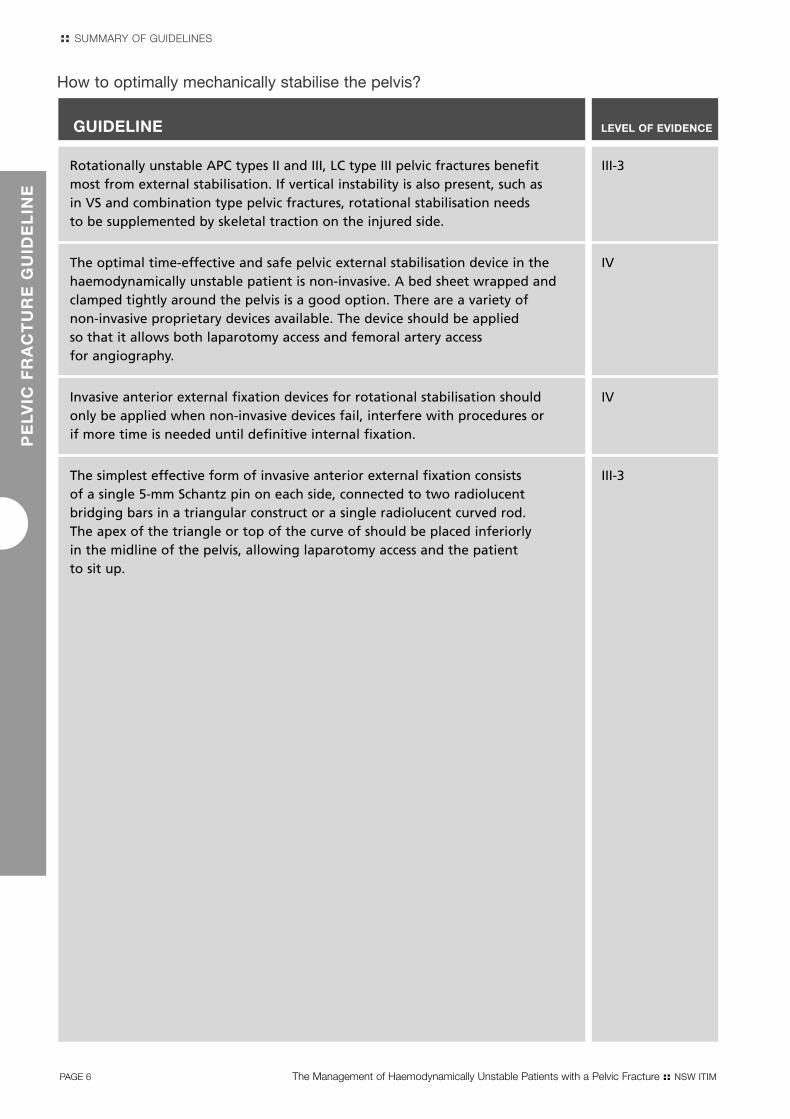
Rotationally unstable APC types II and III, LC type III pelvic fractures benefit III-3most from external stabilisation. If vertical instability is also present, such as in VS and combination type pelvic fractures, rotational stabilisation needs to be supplemented by skeletal traction on the injured side.
The optimal time-effective and safe pelvic external stabilisation device in the IVhaemodynamically unstable patient is non-invasive. A bed sheet wrapped and clamped tightly around the pelvis is a good option. There are a variety ofnon-invasive proprietary devices available. The device should be applied so that it allows both laparotomy access and femoral artery access for angiography.
Invasive anterior external fixation devices for rotational stabilisation should IVonly be applied when non-invasive devices fail, interfere with procedures or if more time is needed until definitive internal fixation.
The simplest effective form of invasive anterior external fixation consists III-3of a single 5-mm Schantz pin on each side, connected to two radiolucent bridging bars in a triangular construct or a single radiolucent curved rod. The apex of the triangle or top of the curve of should be placed inferiorlyin the midline of the pelvis, allowing laparotomy access and the patientto sit up.
GUIDELINE LEVEL OF EVIDENCE
How to optimally mechanically stabilise the pelvis?
Pelvic_FullRep.qxd 6/8/07 1:30 PM Page 6

Pelvic_FullRep.qxd 6/8/07 1:30 PM Page 7

::
Pelvic_FullRep.qxd 6/8/07 1:30 PM Page 8

Mortality rates from pelvic fractures remain thegreatest of any skeletal injury, with retroperitonealhaemorrhage representing the major potentiallyreversible issue. For clearer understanding, mortalityreporting can divided into three categories: 1. Overall mortality of pelvic fractures with andwithout haemodynamic instability, 2. Closed pelvicfractures with haemodynamic instability and 3. Openpelvic fractures.
PE
LVIC
FR
AC
TU
RE
GU
IDE
LIN
E
1 IntroductionHaemorrhage is the major contribution to mortalityin 42% of pelvic trauma patients. Of these, 62% is due to pelvic haemorrhage and 38% is due toassociated external, intrathoracic and intraabdominalhaemorrhage sources.5-7;14;24;25 Non-haemorrhagecauses of mortality from associated injuries arerelated to head injury (32%), followed by sepsisleading to multi-organ failure (14%), and cardio-respiratory failure (8%).2;5;6;14;25;26
Pelvic fracture classification andcorrelation with haemorrhageThe two pelvic fracture classification systems mostfrequently used are the Young-Burgess system shownbelow, which is based on the direction of injuringforce and the Tile classification system, which basedon rotational and vertical stability of the pelvis.27
The Young-Burgess system was utilised in six studies,which compared the potential association betweenpelvic fracture classification based on the initial APradiograph and pelvic haemorrhage. A significantcorrelation was reported between higher bloodproduct requirement and the more severe type ofpelvic fracture: LC II and III, APC II and III, VS andcombined mechanism.6;7;17;19;23;28
Using the Tile system16 and a new classificationsystem developed by the authors, based on differentlocations of pelvic ring fractures,14 these two studiesreported no significant correlation between fracturepattern and blood requirement. A reason for thesedifferent findings is the confounding factor ofhaemorrhage from associated external, intrathoracicor intraperitoneal sources. Up to 50% of pelvicfracture patients may have associated bleedingsources.5;6;15;20;24;28-30 Another limitation of thepredictive value of classification systems based on the initial AP radiograph to predict haemorrhage is that the initial AP radiograph may not clearlydemonstrate major posterior element disruption.14;31
In two studies comparing plain pelvic radiographswith CT, 9% and 22% of fractures were not detectedon the plain radiographs.32;33 Fracture classificationsystems are also not foolproof in determiningwhether the bleeding site is anterior or posterior.34
In summary, it is clear that fracture types causingmajor ligament disruption of the sacro-iliac, sacro-spinous and sacro-tuberous ligaments carry anincreased probability of pelvic arterial bleeding.34
Category 1Overall pelvic fracture mortality is 16% (range 5-30%).1-11
Category 2Closed pelvic fracture with haemodynamic
instability mortality is 27% (range 10-42%).12-19
Category 3Open pelvic fracture is a lethal injury with
a reported 55% mortality (range 50-60%).2;4
The pelvis possesses exceptional inherent strengthand the major external force that is required tofracture a pelvis is also transferred to head, chest,abdomen, the genitourinary system and other skeletalstructures. In the pelvis the close proximity of majorarteries, veins and highly vascularised cancellous boneincrease the risk of severe haemorrhage.20 Varyingdegrees of venous and cancellous bone bleeding are present in all types of pelvic fractures.
Physical assessment is highly sensitive in determiningthe presence of fracture of the pelvis in a consciousand orientated patient. Findings warranting a plainpelvic X-ray are hip pain, groin pain, low back pain,tenderness to palpation over the pelvic girdle,obvious deformities and ecchymosis or abrasionsaround the pelvic area.21 To detect an unstablefracture pattern of the pelvic bones at physicalexamination springing of the pelvis is advocated, but unless one is experienced in pelvic fracturesurgery, the sensitivity of pelvic springing is only 59% and is painful in the awake patient.22
The most common mechanisms of pelvic fractures are motor vehicle accidents (57%), pedestrians hit by motor vehicles (18%), motorcycle accidents (9%),falls (9%) and crush injuries (4%).23
The Management of Haemodynamically Unstable Patients with a Pelvic Fracture :: NSW ITIM PAGE 9
Pelvic_FullRep.qxd 6/8/07 1:30 PM Page 9

PAGE 10 The Management of Haemodynamically Unstable Patients with a Pelvic Fracture :: NSW ITIM
ADULT TRAUMA CL IN ICAL PRACT ICE GU IDEL INESP
ELV
IC F
RA
CT
UR
E G
UID
EL
INE
Figure 1. The Young-Burgess classification, based on direction of force, showing lateral compression (LC), anterior-posterior compression (APC) and vertical shear (VS) pelvic injuries. Subtypes I, II and III of each classification represent increasing degree of severity.35 The injury classification key is shown below: 27
LC
I
II
III
I
II
III
APC
VS
Pelvic_FullRep.qxd 6/8/07 1:30 PM Page 10

1.1 DefinitionsHaemodynamic instability is defined as a class III or IV hemorrhage leading to a drop in systolic bloodpressure below 100 mmHg at the scene and / orinitial fluid resuscitation requirement of more than 2000 mL.36
Pelvic fractures are classified according to the Young-Burgess system (Figure 1, P.10) as a disruptionof the pelvic ring from a lateral compression (LC) type I,II and III injury, an anterior-posterior compression (APC)type I, II and III injury, a vertical shear (VS) injury or acombination mechanism injury. The more severe types(LC II and III, APC II and III, VS and combined types)often involve disruption of the major pelvic ligaments.27
When following EMST/ATLS principles and facing a circulation problem in combination with a pelvicfracture on the initial pelvic radiograph, clinicalpractice guidelines are needed on how to determinesources of bleeding, how to control bleeding andwhich techniques are most rapid and effective. To implement these guidelines, a significant realitycomponent is needed to accommodate for the limitsof the institution and availability of personnel.34
A major challenge for clinicians is the relativeinfrequent presentation of haemodynamicallyunstable pelvic trauma patients.37 Furthermore, the potential for associated injuries is a confounding management problem.
The Management of Haemodynamically Unstable Patients with a Pelvic Fracture :: NSW ITIM PAGE 11
PE
LVIC
FR
AC
TU
RE
GU
IDE
LIN
E
:: INTRODUCTION
Incidence of pelvic fractures at the MajorTrauma Service (Liverpool Health Service) and a Rural hospital (Bowral hospital) withinSouth Western Sydney Area Health Service
Between 1999 and 2001, 187 patients presented to Liverpool Health Service with pelvic fractures, representing1.2% of all fractures seen in this period. The overall mortalityrate was 11.2%.
Sixty-seven (36%) of the 187 patients were deemedhaemodynamically unstable. Fifteen (22%) of these 67haemodynamically unstable patients died compared to 6 (5%) deaths out of 120 haemodynamically stable patients. (p = 0.0003, chi-squared = 13.05). The odds ratio of death in haemodynamically unstable patients was 5.4 compared with death in haemodynamically stable patients. The primarycause of death in seven of the 15 (46%) haemodynamicallyunstable patients was caused by pelvic haemorrhage. All 15haemodynamically unstable patients that died had associatedinjuries including 7 (46%) head injuries, 5 (33%) chest injuries, 2 (13%) abdominal injuries and 1 (7%) extremity injury.
In the same period, 18 patients presented to Bowral hospitalwith a fracture of the pelvis. None were haemodynamicallyunstable and the Injury Severity Score (ISS) varied between 4 and 13. Bowral is the only rural hospital within SouthWestern Sydney Area Health Service.
Pelvic_FullRep.qxd 6/8/07 1:30 PM Page 11

2.3 Assessment of the evidence
2.3.1 Level of EvidenceThe articles were classified according to their general purpose and study type. From this each article was allocateda Level of Evidence:
Table 1. Levels of evidence
2.1 Scope of the guidelinesThis guideline is intended for use by all clinicians whoare involved in the initial care of patients presentingwith haemodynamic instability associated with pelvicfractures; ambulance officers, emergency physicians,emergency nurses, surgeons, radiology staff andoperating theatre staff.
This guideline has been developed to assist cliniciansin providing a more selective, evidence-basedapproach to the management of these patients.
These guidelines however, are not prescriptive norare they rigid procedural paths. It is recognised thatthe guidelines may not suit all patients in all clinicalsituations. The guidelines rely on individual cliniciansto decipher the needs of individuals. They aim toprovide information on what decisions can be made,rather than dictate what decisions should be made.
PAGE 12 The Management of Haemodynamically Unstable Patients with a Pelvic Fracture :: NSW ITIM
2 Methods
PE
LVIC
FR
AC
TU
RE
GU
IDE
LIN
E
1 How to determine the source of bleeding in haemodynamically unstable pelvic fracture patients?
2 How to control pelvic retroperitoneal bleeding with or without intra-abdominal bleeding?
3 What is the optimal angiography and embolisation technique?
4 What is the optimal way of physically stabilising the pelvis?
Level I Evidence obtained from a systematic review of all relevant randomised control trials
Level II Evidence obtained from at least one properly-designed randomised control trial.
Level III-1 Evidence obtained from well-designed pseudo-randomised controlled trials
(alternate allocation or some other method).
Level III-2 Evidence obtained from comparative studies (including systematic reviews of such studies) with concurrent controls
and allocation not randomised, cohort studies, case-control studies, or interrupted time series with a control group.
Level III-3 Evidence obtained from comparative studies with historical control, two or more
single arm studies or interrupted time series without a parallel control group.
Level IV Evidence obtained from a case-series, either post-test or pre-test / post-test
2.2 Aims and objectives of the guideline
AimTo facilitate the management of haemodynamicallyunstable pelvic fracture patients, clinical practiceguidelines were developed to answer the four keyclinical questions shown below.
Key clinical questionsThe following points represent the common key situations that arise during the treatment of haemodynamically unstable patients with pelvic fractures:
Pelvic_FullRep.qxd 6/8/07 1:30 PM Page 12
The Management of Haemodynamically Unstable Patients with a Pelvic Fracture :: NSW ITIM PAGE 13
PE
LVIC
FR
AC
TU
RE
GU
IDE
LIN
E
:: METHODS
2.4 Consultation processesA multidisciplinary committee was established consisting of clinicians from Emergency Department,Orthopaedics, Interventional Radiology, and TraumaSpecialists.
In the absence of suitable evidence the multidisciplinaryworking party worked to develop consensus guidelines.
Pelvic_FullRep.qxd 6/8/07 1:30 PM Page 13

GUIDELINE LEVEL OF EVIDENCE
When the haemodynamically unstable patient enters the resuscitation room, III-3a primary survey with full exposure takes place. Carefully inspect for external bleeding sources and examine the long bones. A supine chest x-ray and pelvic X-ray must be obtained within ten minutes of arrival. The CXR will identify a large haemothorax. If the pelvic x-ray shows a pelvic fracture, the remaining two sites of significant bleeding are the abdomen and the pelvic retroperitoneum.
The probability of associated intraabdominal bleeding with a major pelvic III-3fracture is 32%. The options for assessing intraabdominal bleeding are: Diagnostic Peritoneal Aspiration (DPA) and / or Focused Abdominal Sonography in Trauma (FAST). Both should be completed within 30 minutes of the patient's arrival.
DPA is a reliable diagnostic test for determining frank blood in the abdominal III-3cavity. The aspiration is positive in the presence of >10 ml of frank blood.
FAST is a good alternative diagnostic modality for evaluating free fluid in the III-2presence of a pelvic fracture. Most frequently FAST is positive in the right upper quadrant. The suprapubic view is unreliable in pelvic fracture patients. If practical, FAST should be repeated to increase sensitivity.
When external sources, long bones, intrathoracic injury and intra-abdominal III-3injury do not account for the hypotension in a haemodynamically unstable patient with a major pelvic fracture, pelvic arterial bleeding must be evaluated. The patient should go for immediate angiography.
In the presence of a pelvic fracture and haemodynamic instability, pelvic arterial IVbleeding must be evaluated even if other sources of haemorrhage have been identified.
In the rural or urban environment where DPA or FAST is not available, Consensusidentification of the source of bleeding is reliant on the method of exclusion. Upon exclusion of the other four sources of bleeding and in light of continuing haemodynamic instability it must be assumed that the patient has intraabdominal bleeding until proven otherwise.
PAGE 14 The Management of Haemodynamically Unstable Patients with a Pelvic Fracture :: NSW ITIM
3 How to determine the source of bleeding in haemodynamicallyunstable pelvic fracture patients?
PE
LVIC
FR
AC
TU
RE
GU
IDE
LIN
E
Pelvic_FullRep.qxd 6/8/07 1:30 PM Page 14

Probabilities
In a haemodynamically unstable trauma patient witha pelvic fracture there are five potential sites of majorblood loss:
1 Externally
2 Long bones
3 Chest
4 Abdomen
5 Retroperitoneum38
Determining the site of bleeding is paramount.External blood loss requires careful visual inspection.Bleeding from long bone fractures is present inapproximately 40% of unstable trauma patients andis usually evident from swelling due to haematomaformation. This is usually a contribution, not a majorongoing cause of blood loss.12;28;30
Intrathoracic haemorrhage is to be expected in 4-29% of cases15;28-30 and can be evaluated on a chest X-ray, which should be performed within10 minutes of the patients arrival.39 There are minorlimitations to first mobile supine chest X-ray. A smallhaemothorax can be initially missed in 5% ofsurviving patients and in up to 18% in non-survivingpatients.40 However, a large haemothoraxcontributing to haemodynamic instability should not be missed.
The next part of the decision tree is crucial, trying todecide whether the blood loss is in the abdomen or inthe pelvic retroperitoneum or in both. At this pointthe AP pelvic radiograph should be reviewed. If apelvic fracture with possible disruption of the pelvicligaments causing an unstable fracture pattern (APCtype II and III, LC type II and III, VS or a combinationtype injury) is seen or suspected, the probability ofpelvic arterial bleeding is 52%.6;17;19;20;41-43 In the first prospective study on the safety and efficacy ofpelvic angiography Velmahos et al have identifiedthree independent risk factors of pelvic arterialbleeding: age >55 years (Odds Ratio: 8.1), absence of long bone fractures (OR: 3.2) and need foremergent angiography (OR: 2.9). If all three riskfactors were present, the probability of pelvic arterialbleeding was 94%, if all three were absent theprobability was 18%.44
Reported bleeding sites are anteriorly the internalpudendal (27%) and the obturator (16%) arteries.Posteriorly the superior gluteal (25%) and the lateralsacral (23%) arteries are most frequently involved.Other sources include the iliolumbar artery and theinferior gluteal artery (9%). Therefore 43% of arterial bleeding sources are anterior and 57% are posterior.31 Varying degrees of venous andcancellous bone bleeding within the retroperitoneumare present in all haemodynamically unstable pelvicfracture patients.
The Management of Haemodynamically Unstable Patients with a Pelvic Fracture :: NSW ITIM PAGE 15
PE
LVIC
FR
AC
TU
RE
GU
IDE
LIN
E
:: HOW TO DETERMINE THE SOURCE OF BLEEDING IN HAEMODYNAMICALLY UNSTABLE PELVIC FRACTURE PATIENTS?
Figure 2. The proximity of the arteries in relation to the pelvis: IL, iliolumbar artery; SG, superiorgluteal artery; LS, lateral sacral artery; IP, internal pudendal artery; O, obturator artery
IL
LSSG
IP
O
Pelvic_FullRep.qxd 6/8/07 1:30 PM Page 15

The overall probability of a pelvic fracture withhaemodynamic instability associated withintraperitoneal bleeding source is32%.2;6;12;15;19;20;28;30 Seventy-eight per centof intraperitoneal injuries result in haemorrhageincluding; the spleen (22%), the liver (20%), thebladder (15%), the bowel mesentery (10%) anddiaphragmatic lesions (4%). Renal haemorrhageis found in 7% of cases.5;12;19;28;30;45 The remaining22% of intraperitoneal injuries are not associatedwith bleeding.
InvestigationsDiagnostic Peritoneal Aspiration (DPA) and FocusedAbdominal Sonography in Trauma (FAST) are thepreferred diagnostic means to determine if there isintra-abdominal bleeding.5;14;15;43
DPA takes less time than Diagnostic Peritoneal Lavage (DPL); only aspiration of intra-peritoneal blood is determined. More than 10 cc of gross bloodis considered positive. When performing DPA inpatients with a suspected pelvic haematoma, an open umbilical46;47 or supra-umbilical technique,5
if there is a large retroperitoneal haematoma, with a large bore catheter (20F) is advised.46 In the absenceof >10 cc of gross blood on direct aspiration througha 20 French catheter, it must be assumed there is nomajor intra-abdominal bleeding going on at that time.If DPA is negative, Diagnostic Peritoneal Lavage (DPL)effluent should be sent for routine analysis forintestinal perforation, but this has little impact duringthe phase of haemodynamic instability.
FAST is generally 95% accurate in identifying freefluid in the abdominal cavity.48-51 The few studiesreporting the utilisation of FAST in pelvic traumapatients report accuracy between 84% and 97%.49;52;53 However, in the presence of a pelvic fracture,FAST should be interpreted cautiously as up to 19% false negative rates for free fluid have beenreported.54 An explanation for this may be that the FAST is performed so soon after the injury thathaemorrhage was not yet significant enough to bedetected.55 Repeating the FAST is a good option as it decreases false negative results and increasessensitivity to around 95%.55;56 Therefore, if FASTis not able to be repeated, those with pelvic ringfractures and those that remain haemodynamicallyunstable in the presence of an initial negative FASTresult require continued assessment of clinicalcondition to exclude intra-abdominal bleeding.51
In general, regardless of type of injury, intraperitoneal fluid is found most frequently inMorrison's pouch – the right upper quadrant.48;51
If a pelvic fracture with retroperitoneal haematoma is present, the haematoma may compress the bladder that is needed as an acoustic window for the detection of free fluid. Therefore thesuprapubic FAST view can be difficult to interpret.
Assessment and decision-makingIn the presence of a positive DPA and/or free fluidin the abdomen using FAST, in the face of continuinghaemodynamic instability immediate laparotomy is warranted.5;16;17;34;57
If absence of intra-abdominal bleeding has beendetermined using FAST and/or DPA, the pelvicretroperitoneum is the last possible source ofbleeding. Angiography is recommended as the best next step for locating a retroperitonealsource.5;11;13;14;16;17;44;58-62 The haemodynamicallyunstable patient should not go to CT scanning. If there is a fracture pattern suggestive of a majorpelvic ligament disruption, ie LC type III, APC types II and III, VS or a combination type fracture, the probability of a pelvic arterial bleeding source is increased.6;17;19;20;23
The initial AP pelvic radiograph is the only guideto determine the probability of pelvic bleeding.Disruptions involving only the pubic rami do notvertically or rotationally unstabilise the pelvic ring, but when recognising a fracture of the pubic bone,posterior disruption and probability of arterialbleeding must always be suspected.14;31 One mustalso bear in mind that bilateral inferior/superior pubicramus fractures (butterfly type fracture from APcompression mechanism), acetabular fractures andeven simple ramus fractures in the elderly can lead to arterial bleeding causing hypotension.41;44
The abdomen may be the most difficult to assess in the rural and urban environment if DPA or FAST is not available. Determining the source of bleeding is then reliant on method of exclusion. The clinicianshould examine the other 4 sources for blood loss,upon exclusion of these and in light of continuinghaemodynamic instability it must be assumed thatthe patient has intraabdominal bleeding until proven otherwise.
PAGE 16 The Management of Haemodynamically Unstable Patients with a Pelvic Fracture :: NSW ITIM
ADULT TRAUMA CL IN ICAL PRACT ICE GU IDEL INESP
ELV
IC F
RA
CT
UR
E G
UID
EL
INE
Pelvic_FullRep.qxd 6/8/07 1:30 PM Page 16

TimeframesFor every three minutes of haemodynamic instability elapsed without haemorrhage control in the Emergency Department, there is a 1% increasein mortality.63 Therefore decision making within pre-determined timeframes is crucial. Thehaemodynamically unstable pelvic fracture patient should leave the resuscitation room within 45 minutes heading for either angiography or laparotomy. Assessment of external bleedingsources and long bone fractures should take placewithin the first 5 minutes. The chest X-ray and pelvicX-ray should be performed within 10 minutes of thepatients arrival. Assessment of the abdomen with DPAand/or FAST should be completed within 30 minutes.
The Management of Haemodynamically Unstable Patients with a Pelvic Fracture :: NSW ITIM PAGE 17
PE
LVIC
FR
AC
TU
RE
GU
IDE
LIN
E
:: HOW TO DETERMINE THE SOURCE OF BLEEDING IN HAEMODYNAMICALLY UNSTABLE PELVIC FRACTURE PATIENTS?
Pelvic_FullRep.qxd 6/8/07 1:30 PM Page 17

If intra-abdominal bleeding has been excluded by DPA and / or FAST, immediate III-3angiography and embolisation is warranted. Transfer to the angiography suite should take place within 45 minutes of the patient's arrival. Close monitoring, ongoing resuscitation and re-assessment of an intra-abdominal bleeding source utilising FAST and / or clinical parameters are mandatory in the angiography suite.
Haemodynamically unstable patients with pelvic fractures should be managed III-1with early non-invasive external stabilisation to aid in controlling small venous and cancellous bone bleeding. In the case of a vertical shear type pelvic fracture, supplementary femoral pin traction is necessary on the affected side. External stabilisation does not control arterial bleeding.
If laparotomy is warranted, a non-invasive or invasive external stabilisation IVdevice should be placed concomitantly with laparotomy in the operating theatre. If angiography is warranted, a non-invasive external stabilisation device should be placed prior to transfer in the resuscitation room.
If intra-abdominal bleeding has been determined by DPA and / or FAST, III-3immediate laparotomy is warranted. Concomitant intestinal perforation shouldbe dealt with in a damage control fashion if necessary.
If at laparotomy, performed for intraabdominal haemorrhage, there is a freely IVbleeding ruptured pelvic haematoma, packing and immediate transfer to angiography is preferable. Where angiography is not available or the patient is too unstable (blood pressure unable to be restored above 70 mmHg), the haematoma should be evacuated, the internal iliac arteries bilaterally ligated and the pelvis packed.
In the situation of exsanguination at presentation, with little or no response IVto fluid resuscitation, immediate operation is the only life saving option: Laparotomy with surgical ligation of bleeding arteries, pelvic packing with large sponges and stabilisation of the pelvis.
Internal fixation of major pelvic fractures is contra-indicated in haemodynamically IVunstable patients.
In the situation where angiography services are not available, management Consensusis determined by the degree of haemodynamic instability. In the presence of reasonable haemodynamic stability (SBP>80mmhg), the pelvis should be non-invasively externally stabilised and Retrieval Services should be contacted as a matter of urgency. If the systolic blood pressure drops below 80-90mmHg, small aliquots of fluid (100-200mls) may administered. If the blood pressure becomes increasingly unstable (SBP<80mmHg) and cannot be maintainedwith small boluses of fluid, immediate laparotomy with surgical ligation of bleeding arteries, pelvic packing with large sponges and invasive external stabilisation of the pelvis is the only life saving option available. An unstable patient should not be transferred where surgical services are available until surgical haemostasis has been achieved.
GUIDELINE LEVEL OF EVIDENCE
PAGE 18 The Management of Haemodynamically Unstable Patients with a Pelvic Fracture :: NSW ITIM
4 How to control pelvic bleedingwith or without associatedintraabdominal bleeding?
PE
LVIC
FR
AC
TU
RE
GU
IDE
LIN
E
Pelvic_FullRep.qxd 6/8/07 1:30 PM Page 18

SummaryNumerous techniques to control pelvic haemorrhagebleeding have been described: angiography andembolisation, external invasive fixation of the pelvis,laparotomy with ligation of the internal iliac arteriesand pelvic packing with large sponges, internalfixation of the pelvis and non-invasive externalstabilisation.
When the abdomen has been cleared as a bleeding source, haemodynamically unstable patients with pelvic fractures should undergoimmediate angiography and embolisation to controlarterial pelvic bleeding and immediate externalstabilisation to control small venous and cancellousbone bleeding.
Laparotomy and external stabilisation have priorityover angiography if there is an intraabdominalbleeding source. The only two indications remainingfor open surgical arterial ligation and pelvic packing are:
1 Ruptured pelvic haematoma at laparotomy when transfer to angiography is not possible or angiography facilities are not available
2 The exsanguinating patient unresponsiveto resuscitation.
Internal fixation in the haemodynamically unstablepatient is contra-indicated.
Pelvic haemorrhage withoutintraabdominal bleeding
Angiography and embolisationOnce an intraabdominal bleeding source has been excluded or treated, angiography is both diagnostic and therapeutic for pelvichaemorrhage.11;13;41;60;64 Margolies was oneof the first to report the use of angiography andembolisation as an effective means of controllingpelvic haemorrhage in 1972.58
In management strategies in the late seventies andeighties, angiography was indicated for those patientswho, after initial fluid resuscitation and orthopaedicpelvic fixation, still had ongoing bleeding from asuspected retroperitoneal source.12;14;31;45;65;66
Angiography was used selectively or as a last resortand reported pelvic arterial bleeding rates were lessthan 15%.15;59;67;68
In more recent and prospective series, whenangiography is used liberally and early in thetreatment algorithm, pelvic arterial bleeding ratesof 57-64% have been reported.19;41;42;44 Embolisationsuccess rates for these patients approach 90%.13;41;44
This success of early interventional angiography has shifted the indications for its use and it is nowrecommended at an early stage in the managementof the haemodynamically unstable patient after other sources of haemorrhage have been ruled out.5;6;11;16;17;20;44;67;69
The various forms of arterial injury includetransection, laceration, intimal tears, thrombosis,vasospasm, false aneurysm, arteriovenous fistulae and vessel tortuosity. Arteries may be injured by bluntforce, stretching force and sharp force from bonyedges or tense fascia, such as the piriformis muscle fascia. Transection, laceration, vasospasm,arteriovenous fistulae, false aneurysms and vesseltortuosity are important to recognise and treat in the haemodynamically unstable patient.31;58 Injury to the obturator and internal pudendal arteries arefrequently seen in association with fractures of thepubic rami.31;58 The superior gluteal artery (25%) and the lateral sacral artery (23%) may be traumatized with disruption/fractures near the sacro-iliac joint.31
Arterial embolisation is effective by stopping thearterial bleeding and allowing the tamponade effectof the haematoma to control venous and cancellousbone bleeding.
Complications of embolisation have been reported in limited case series and include necrosis of the distal colon and ureter, bladder necrosis and perinealwound sepsis.60 In larger and prospective patientseries, even using bilateral internal iliac arteryembolisation, the use of unselective and selectiveembolisation has been reported to be safe, with minimal morbidity.13;41;42;44
Physicians should abandon the reluctant attitude totransport haemodynamically unstable patients to theangiography suite. Left in the Emergency Departmentor Intensive Care unit, these patients may die fromexsanguination.34 Rather, the angiography suiteshould be prepared as a mini-ICU with closemonitoring, ongoing resuscitation and re-assessmentof other bleeding sources by dedicated clinicians.44
FAST should be repeated at least once duringangiography to monitor an intraabdominal bleeding source.
The Management of Haemodynamically Unstable Patients with a Pelvic Fracture :: NSW ITIM PAGE 19
PE
LVIC
FR
AC
TU
RE
GU
IDE
LIN
E
:: HOW TO CONTROL PELVIC WITH OR WITHOUT ASSOCIATED INTRAABDOMINAL BLEEDING?
Pelvic_FullRep.qxd 6/8/07 1:30 PM Page 19

Pelvic stabilisationOsseous stabilisation of the pelvis has a role in themanagement of haemodynamically unstable pelvicfractures. In rotationally unstable, but vertically stablefractures (LC type III, APC types II and III), a form ofexternal stabilisation is recommended, achieving 64-83% stabilisation rates. Biomechanically, rotationalstabilisation is not effective if the hemipelvis is alsovertically displaced on the initial AP pelvic X-ray (VS type, combination type), achieving only 27%stabilisation rates without supplementary skeletaltraction or subsequent internal fixation.70;71
In promoting control of haemorrhage in patients with major pelvic fractures, the role of externalstabilisation has not been fully elucidated. Manyauthors have advocated early invasive external fixation devices.8;14;19;25;34;59;70;72 However, there is only one prospective trial with randomisationpromoting immediate invasive external fixation. In the group allocated to immediate invasive external fixation, blood transfusion, post-operativepain and late deformities were significantly less.73
One theory is that reducing the pelvis back to itsnormal configuration with external fixation reducespelvic volume, and therefore limits the amount ofblood loss to the retroperitoneal pelvic haematoma.74
This theory supported smaller pelvic volumepromoting tamponade of pelvic bleeding sources.66
However, other studies showed there was little effect of external fixation on the diameter of thepelvic ring and the pressure-volume characteristics ofthe pelvic retroperitoneum. In all pelvic fractures withhaemodynamic instability there is a variable amountof retroperitoneal pelvic bleeding from the exposedcancellous bone surfaces and the disrupted pelvicvenous plexus.
Current opinion favours a concept that re-apposingthese bony pelvic surfaces back into reasonablealignment allows the haemostatic pathways to controlvenous bleeding from small veins and exposedcancellous bone.75-77 Low-pressure venous bleedingmay be tamponaded by external stabilisation giventhat enough fluid volume (clot) is present in the pelvic retroperitoneum.76 Maintaining them in stable, non-moving apposition could also prevent repeatedtrauma to already clotted vessels, thereby alsolessening consumption of clotting factors.76
It seems likely that aspects of these theories arecorrect and supports the rationale behind earlyexternal stabilisation to achieve a better degree ofvenous haemorrhage control and bony alignment.75
Another advantage of early external stabilisation isthat it relieves pain from shearing bony surfaces andallows patients to sit up in the Intensive Care Unit.This contributes to preventing ventilatory adverseeffects and allows better nursing care.69;71 In addition,better mobility, less leg-shortening and less obstetriccomplications in the longer term are described afterearly external fixation.69;71;72
If an intra-abdominal bleeding source hasbeen excluded and angiography is warranted, non-invasive external stabilisation (see Section 6 –How to optimally mechanically stabilise the pelvis,p.25) should be applied in the emergencydepartment. If laparotomy for intra-abdominalbleeding is warranted, immediate transfer to theoperating theatre takes place and non-invasive orinvasive radiolucent external stabilisation (see Section 6 – How to optimally mechanically stabilisethe pelvis, p.25) should be applied concomitantly with laparotomy. Through a tension band effect on the iliac wings, the anterior abdominal wallcontributes to limiting the degree of anterior pubic diastasis, which will increase if the pelvis is not stabilised prior to the midline incision.70;74;75;77;78
This theory supports external stabilisation of the pelvisbefore or concomitantly with laparotomy.75
Laparotomy to control pelvic haemorrhageWith the availability of therapeutic angiography,laparotomy to control pelvic retroperitonealhaemorrhage in pelvic fracture patients is no longerindicated. Laparotomy and pelvic retroperitonealexploration may not only fail to arrest haemorrhagebut may also put the patient at risk. By opening the abdomen, the surgeon can directly contribute to aggravation of pelvic haemorrhage due todecompression of the tamponade effect of theabdominal wall.44;57;58;62 In this scenario laparotomymay be non-therapeutic.
In most circumstances, venous pelvic haemorrhage is contained within the pelvis by the pelvic bones, the fibromuscular pelvic floor and the parietalperitoneum superiorly. If the parietal peritoneum is breached by the expanding retroperitonealhaematoma or iatrogenically during laparotomy,exsanguination may occur. Before therapeuticangiography there was frustration reported withthe inadequacies of direct operative exposure andligation of the internal iliac artery or branches of itdeep in the pelvis.2;57;68;79 This is primarily becauseanastomotic pathways remain between the peripheralinternal iliac branches and the inferior mesentericartery, inferior epigastric arteries, circumflex andperforating branches of the deep femoral arteries and lumbar arteries.20;64
PAGE 20 The Management of Haemodynamically Unstable Patients with a Pelvic Fracture :: NSW ITIM
ADULT TRAUMA CL IN ICAL PRACT ICE GU IDEL INESP
ELV
IC F
RA
CT
UR
E G
UID
EL
INE
Pelvic_FullRep.qxd 6/8/07 1:30 PM Page 20

Pelvic haemorrhage withintraabdominal haemorrhage
Laparotomy for intra-abdominal haemorrhageA positive DPA and/or FAST in the haemodynamicallyunstable patient with a pelvic fracture warrantsimmediate laparotomy.5,75 Concomitant small bowel or colon injury should be dealt with in a damage control fashion if the patient's physiology necessitates this.
During laparotomy, in the situation where a ruptured pelvic haematoma is freely bleeding into the abdomen, there are two possible scenarios. If the patient's physiologic status is poor, bloodpressure is unable to be supported above 70 mmHgand/or interventional radiology is not available withinone hour, surgical ligation of arterial bleeding andpacking of the pelvis remains the only life-savingoption.18 If physiologic status and blood pressure isstable and interventional radiology is available withinone hour, the haematoma can be packed and theabdomen closed in a temporary fashion, followed by immediate angiography and either non-selectivebilateral embolisation of the internal iliac arteries44
or selective embolisation.
In the situation of controlled intra-abdominalhaemorrhage and a contained moderate to largepelvic haematoma is seen during laparotomy, thepelvis should be packed with large sponges to add totamponade and the abdomen closed in a temporaryfashion, followed by immediate angiography andembolisation.44
The exsanguinating patientIn the desperate setting of an exsanguinating patientin the resuscitation room with little or no response to resuscitative measures and blood pressure unableto be restored to above 70 mmHg, immediateoperation is the only life-saving option. If the patientcan be brought to the operating room and assumingassociated external and thoracic injuries arecontrolled, the last option at laparotomy to controlpelvic arterial bleeding in the face of exsanguination is bilateral internal iliac artery ligation. To control smallarterial and venous bleeding, packing the pelvis with
large abdominal sponges is effective.18;69 In Ertel'sseries of twenty consecutive patients, five patientsunderwent emergency room laparotomy with pelvicpacking and nine patients underwent immediateoperating room laparotomy and pelvic packing. In three patients an additional thoracotomy wasperformed due to lack of vital signs, of which onlyone patient survived. In two patients the aorta wascross-clamped and both of these patients died.18
A posterior external fixation C-clamp was applied inall patients. Utilising this approach 64% of patientsundergoing emergency operating procedures survivedand 36% developed an abdominal compartmentsyndrome, which was successfully relieved bydecompressive laparotomy. One case of ischaemicileum and subsequent multi-organ failure was seen.18
Other techniques
Internal FixationInternal Fixation provides a better biomechanicalstabilisation of the pelvis, but is contra-indicated inthe haemodynamically unstable patient, as it is time-consuming and exposing the fracture surfaces cancause decompression of the pelvic haematoma anduncontrollable haemorrhage.71;80
The Military Anti-Shock Trousers (MAST) Historically, the Military Anti-Shock Trousers (MAST)has been advocated as an external splinting device forpelvic fractures and promoting tamponade of smallvessel bleeding.5;6;15;69 In the late 1980's there wasincreasing evidence that early external fixation was as good as or better than the MAST for haemorrhagecontrol.5;14 The MAST has also been criticized for itslimiting access to the abdomen, groin and perineum.Other described risks are untreatable hypovolaemia at removal and compartment syndrome when applied for too long.28;71 In a prospective study, the complications associated with the MAST, causingcompression of the lower extremities and abdomenoutweighed its benefits.81 For these reasons the useof the MAST has been limited. If any, its role lies inthe pre-hospital setting when ambulance personnelmay use it as a valuable temporising manoeuvreduring transport, especially in areas with longertransportation times or in rural to urban transfers.6;14;28;69;71
The Management of Haemodynamically Unstable Patients with a Pelvic Fracture :: NSW ITIM PAGE 21
PE
LVIC
FR
AC
TU
RE
GU
IDE
LIN
E
:: HOW TO CONTROL PELVIC WITH OR WITHOUT ASSOCIATED INTRAABDOMINAL BLEEDING?
Pelvic_FullRep.qxd 6/8/07 1:30 PM Page 21

PAGE 22 The Management of Haemodynamically Unstable Patients with a Pelvic Fracture :: NSW ITIM
5 What is the optimal angiographyand embolisation technique?
If a major pelvic fracture is seen on the initial pelvic x-ray in the IVhaemodynamically unstable patient, the interventional radiologist and nursing staff should be notified immediately. It is critically important that the general surgeon coordinates care between emergency clinicians, interventional radiologist, orthopaedic surgeon, intensive care specialist, nursing and blood bank.
Angiography should be performed as soon as the abdomen has been cleared IVas a source of bleeding or within 90 minutes of presentation. A femoral approachis preferred, but a left brachial puncture may be needed in patients in whichfemoral approach fails.
Using 4 or 5 Fr catheters, angiography begins with a midstream abdominal III-3flush to reassess any abdominal bleeding. A pelvis flush is then performed at aortic bifurcation level. An arterial haemorrhage requiring embolisation should present itself at the pelvis midstream flush.
Any sites of contrast extravasation, false aneurysms and occlusion of the IVmainstem internal iliac artery warrant embolisation. Vasospasm due to major haemorrhage may obscure bleeding distal to the spasm.
Non-selective embolisation of main arteries, such as the internal iliac arteries IVor their first divisional branches should be performed with steel coils. Steel coils are also preferred for selective embolisation of bleeding branches. In patients with multiple distal, small branch bleeding sites a shower of gelfoam slurry can be utilised.
Selective embolisation of smaller branches should be used judiciously. IVIf respiratory or hemodynamic instability increases during angiography, non-selective bilateral embolisation of the internal iliac arteries with steel coils should be performed. Complete occlusion of the internal iliac artery is an acceptable alternative to exsanguination.
Once a vessel is embolised for distal branch extravasation, the potential IVcollateral vessels of both internal iliac arteries should be evaluated to identify additional supply to the injured vascular bed. Completion angiography is necessary to document cessation of bleeding.
GUIDELINE LEVEL OF EVIDENCE
PE
LVIC
FR
AC
TU
RE
GU
IDE
LIN
E
Pelvic_FullRep.qxd 6/8/07 1:30 PM Page 22

Considerations and timingEarly anticipation of the need for embolisation and alerting the interventional radiologist and nursing staff in advance is essential. Coordinationbetween Emergency department, general surgeon,interventional radiologist, orthopaedic surgeon,intensive care specialist, nursing and blood bank are critical in the management of haemodynamicallyunstable patients going to the angiography suite.41
When anticipating angiography, a non-invasive pelvic stabilising device (sheet, proprietary device)should not obscure access to the femoral arteries. If applying an invasive external fixation device beforeangiography, the orthopaedic surgeon should usecarbonated (radiolucent) connecting rods to facilitate angiography or subsequent CT.
Agolini reported a mortality of 17% if embolisationwas performed within three hours of presentation.This increased to 75% mortality after three hours.Patients who had delayed arrival to the angiographysuite because of prolonged resuscitation, non-therapeutic laparotomy or unnecessary diagnostictests had a higher mortality rate.41 Just as laparotomy controls intraabdominal haemorrhage,angiography with embolisation controls arterial pelvic haemorrhage. Angiography should therefore be performed as soon as possible. The preferredtimeframe is within 90 minutes of arrival or as soonafter the abdomen has been cleared as a source of haemorrhage.
Catheter insertionA femoral puncture approach is preferred with the Seldinger technique. Arterial duplex or fluoroscopically guided puncture may be needed if massive haematoma prevents palpation of thefemoral pulse. Fluoroscopic guidance utilises themiddle third of the acetabular roof as a landmark for the Seldinger needle.13 Bilateral punctures may be required. A sheath with a reflux preventingseal is recommended to protect access duringcatheter exchanges.82
A left brachial approach with a Headhunter-1 catheter50 is an alternative in patients when the femoral approach fails. The 100-110-cm-longHeadhunter (selective cerebral) catheter is longenough to reach the internal iliac artery and becauseit's gently curved tip is designed to enter the carotidsthrough brachial approach, it is also effective whencannulating the internal iliac artery from above.82
Acquisitions and findings The procedure begins with an abdominal flush to re-assess any abdominal bleeding focus. A pelvic flush is then performed using 4 or 5 fr catheters at aortic bifurcation levels (40ml contrast at 15ml/s) with extended filming. As a general rule an arterialhaemorrhage requiring embolisation should presentitself at the midstream flush. However, selectiveinternal and external iliac artery runs usingappropriate oblique projections are recommendedand extravasation of contrast on these views alsowarrant embolisation. A 4 or 5 Fr Cobra or Simmons1 or 2 catheters are used for these selective runs.82
The lumbar arteries should be surveyed, as these can be injured in pelvic fracture patients.13;82
In descending order of frequency, the internal iliacbranches that are most commonly injured are the:
:: internal pudendal (27%)
:: superior gluteal (25%)
:: ateral sacral (23%)
:: obturator (15%)
:: inferior gluteal (6%)
:: iliolumbar (3%) arteries.31;83
The manifestations of arterial injury may consist of any arterial abnormality:
:: Extravasation is the clearest and most obviousfinding in transected or lacerated arteries or in arteriovenous fistulae. The typical, increasinglydense extravascular collection of contrast materialmay be transitory if it dissipates into a largehematoma. Transient areas of increased opacityseen at nonselective imaging require furtherevaluation.
:: Extravasation from injured smaller vessels (1-2 mm in diameter) is often self-limited innormothermic patients with normal coagulationparameters; in trauma patients with hypothermiaand coagulopathy, this may not occur. A corollaryis that the angiographic abnormality is not alwaysproportional to the actual amount of blood lossfrom the injury.
:: False aneurysms, or well-circumscribed collectionsof contrast material that extend beyond theconfines of the arterial wall, may appear stable.However, after an acute injury, these collectionsare not confined by chronic fibrotic tissue and are best characterized as pulsatile hematomas that require treatment.
The Management of Haemodynamically Unstable Patients with a Pelvic Fracture :: NSW ITIM PAGE 23
PE
LVIC
FR
AC
TU
RE
GU
IDE
LIN
E
:: WHAT IS THE OPTIMAL ANGIOGRAPHY AND EMBOLISATION TECHNIQUE?
Pelvic_FullRep.qxd 6/8/07 1:30 PM Page 23

:: Occlusions and vessel tortuosity may be stableinjuries, but bleeding may be intermittent and may depend on the changing parameters of coagulation, blood pressure, and motion of fracture fragments. Larger branch occlusionswarrant embolisation.83;84
:: Vasospasm is a frequent finding inhaemodynamically unstable patients. Profusevasoconstriction from major haemorrhage mayobscure bleeding sites distal to the spasm.44
Embolisation and materialsMain arteries, such as the internal iliac arteries or their first divisional branches should be embolisedby steel coils. Steel coils are preferred over gelfoampledgets in these large-bore arteries. Selectiveembolisation of bleeding branches is preferred byplacing steel coils proximal to the extravasation site. In patients with multiple distal, small branch bleedingsites, scatter embolisation with a gelfoam suspension(small gelfoam particles mixed with contrast material)is utilised. The bleeding sites are showered with thesemultiple gelfoam particles and provide temporaryocclusion without significant ischaemia until thegelfoam dissolves.82 Fine particles, utilised in cancertherapy, should not be introduced because of risk of pelvic ischaemia.13;41;60
Selective embolisation should be used judiciously in patients who are unstable. If ventilation becomesdifficult or hemodynamic instability increases duringthe procedure, rapid non-selective embolisation of anentire anterior or posterior division or even bilateralembolisation of the internal iliac arteries (97% successrate) is preferable to an elegant but long
selective embolisation. Complete occlusion of the internal iliac artery is an acceptable alternative to exsanguination.44 After this temporising intervention, leaving the groin sheath in case of repeat angiography, the patient can be transferred for operative management if necessary or to the intensive care unit.
Afterward, planned repeat angiography can beperformed for selective embolisation of lumbar, lateral sacral, iliolumbar, or replaced obturator arteries that may require additional time for selective catheterisation.
Haemorrhage is identified and treated, after whichthe diagnostic evaluation is resumed. Once a vessel is embolised for distal branch extravasation, the localpotential collateral vessels are evaluated to identifyadditional supply to the injured vascular bed. Multiplecommunicating channels between the two internaliliac arteries exist. In particular, evaluation of theipsilateral and contralateral potential collateral supplyfollows embolisation of internal iliac anterior divisionvisceral branches or lateral sacral branches.82-84
Similarly, after treatment of extravasation in pubic ring fractures, the common femoral artery isevaluated for injury to a replaced obturator or external pudendal artery.
Completion angiography is necessary to documentcessation of bleeding and to screen for previouslyunsuspected sites of extravasation or collateralsupply.83;84 The catheter sheath can be left in, so angiography can be repeated if necessary.
PAGE 24 The Management of Haemodynamically Unstable Patients with a Pelvic Fracture :: NSW ITIM
ADULT TRAUMA CL IN ICAL PRACT ICE GU IDEL INESP
ELV
IC F
RA
CT
UR
E G
UID
EL
INE
Pelvic_FullRep.qxd 6/8/07 1:30 PM Page 24

Lateral Compression IIIAnterior-Posterior
Compression IIAnterior-Posterior Compression III
The Management of Haemodynamically Unstable Patients with a Pelvic Fracture :: NSW ITIM PAGE 25
PE
LVIC
FR
AC
TU
RE
GU
IDE
LIN
E
6 How to optimally mechanicallystabilise the pelvis?
Rotationally unstable APC types II and III, LC type III pelvic fractures benefit III-3most from external stabilisation. If vertical instability is also present, such as in VS and combination type pelvic fractures, rotational stabilisation needs to be supplemented by skeletal traction on the injured side.
The optimal time-effective and safe pelvic external stabilisation device in the IVhaemodynamically unstable patient is non-invasive. A bed sheet wrapped and clamped tightly around the pelvis is a good option. There are a variety ofnon-invasive proprietary devices available. The device should be applied
so that it allows both laparotomy access and femoral artery access for angiography.
Invasive anterior external fixation devices for rotational stabilisation should IVonly be applied when non-invasive devices fail, interfere with procedures or if more time is needed until definitive internal fixation.
The simplest effective form of invasive anterior external fixation consists III-3of a single 5-mm Schantz pin on each side, connected to two radiolucent bridging bars in a triangular construct or a single radiolucent curved rod. The apex of the triangle or top of the curve of should be placed inferiorlyin the midline of the pelvis, allowing laparotomy access and the patientto sit up.
GUIDELINE LEVEL OF EVIDENCE
Types of pelvic fractures that benefit from stabilisationThe best results of pelvic stabilisation are achieved in rotationally unstable pelvic fractures: LC type III and APCtypes II and III. In these types, stabilisation can be achieved in 64-83% of cases.70;71 If vertical instability is alsopresent, such as in vertical shear (VS) type injury or a combination type injury, stabilisation can be achieved in only 27% of cases and additional skeletal traction is needed in the other 73%.70;71;85
Figure 3. Types of pelvic fractures that benefit from stabilisation
Pelvic_FullRep.qxd 6/8/07 1:30 PM Page 25
BiomechanicsInternal fixation has been proven to give the greatestbiomechanical stability, but it is impractical foremergency use.71;86 To obtain rotational stability,multiple types of anterior and posterior externalfixation devices are in use in bridging the gap todefinitive internal fixation. When comparing thesedifferent kinds of anterior and posterior invasiveexternal fixation devices, all performed similarlybased on their ability to prevent rotational pelvicexpansion and application times.85-87
Approach in the emergency settingIn the situation of haemodynamic instability,stabilisation of the pelvis in APC type II and III, LC type III, VS and combinations of these fractures,must be performed in the shortest possible time. Two kinds of pelvic stabilisation can be considered:non-invasive techniques and invasive fixation.
Non-invasive techniques have advantages: they aresafe, time-effective, do not require the presence of an orthopaedic surgeon in the resuscitation room and do not interfere with further resuscitationefforts.88 Simple, popular options include the use of a bed sheet wrapped and clamped tightly aroundthe pelvis or a vacuum beanbag to provide emergentpelvic stability. (36;78;88) There are also a number of more expensive proprietary devices specificallydesigned and marketed for such use. These non-invasive options should be considered astemporising measures bridging the gap from injury to more definitive stabilisation.75
Pelvic stability promotes venous bleeding tamponadeand protects the soft tissues from further injury.70;87
The application of an invasive anterior externalfixation frame is recommended in cases of ongoingvenous bleeding.70;78 These fixators, althougheffective in preventing pelvic rotational expansion,have problems. Certain fracture patterns do not allow placement of the pins into the iliac crest. Pin site infections compromise subsequent openreduction and internal fixation.78;89 Large, bulkyframes with multiple pins into the iliac crest are to be considered obsolete in patients, which have beenhaemodynamically unstable. They prevent proper re-laparotomy access, interfere with angiography and prevent chair mobility by limiting hip flexion.78
The pelvic antishock clamp or C-clamp effectivelylimits expansion of the posterior elements if placedproperly.90 However, clamp application is unfamiliar to most orthopaedic surgeons to apply, particularly in patients with morbid obesity or severe pelvicdeformity. Without fluoroscopy, pins might be placedthrough the sciatic notch, with the potential forcomplications. In addition, transforaminal sacralfractures make application dangerous, as overcompression can damage nerve roots in these cases.78
If an invasive anterior external fixation device isapplied, the simplest effective form consists of asingle 5-mm Schantz pin on each side either into theiliac crest or the supraacetabular region anteriorly,connected to two carbonated, radiolucent bridgingbars in a triangular construct.91 The apex of thetriangle should be placed inferiorly in the midline of the pelvis and over the groin area rather than overthe lower abdomen to allow access into the abdomenshould laparotomy be necessary.70;91 Alternatively a single curved radiolucent rod may be used.
PAGE 26 The Management of Haemodynamically Unstable Patients with a Pelvic Fracture :: NSW ITIM
ADULT TRAUMA CL IN ICAL PRACT ICE GU IDEL INESP
ELV
IC F
RA
CT
UR
E G
UID
EL
INE
Pelvic_FullRep.qxd 6/8/07 1:30 PM Page 26

Pelvic_FullRep.qxd 6/8/07 1:30 PM Page 27

PAGE 28 The Management of Haemodynamically Unstable Patients with a Pelvic Fracture :: NSW ITIM
Author Level of
& year evidence Finding
Evidence table
Agolini199741
IV A review of 35 unstable pelvic fracture patients treated with early angiography. Fifteen requiredembolisation. Angiographic yield was 64% and success rate of embolisation was 100%. Morbidityand mortality increased if angiography was performed later than three hours after presentation toendotracheal tube in success rate, difficulty of insertion, and time to position correctly in thispatient population. The laryngeal mask however does not reliably protect against aspiration and is therefore only recommended when more conventional methods of airway management fail.Further studies in the trauma scenario are indicated.
Ali200237
III-2 Cognitive and clinical performance was analysed among physicians who treated more than 50 and less than 50 trauma patients per year. Immediate and progressive cognitive skill attrition wasworse in the low volume group. Global skills and adherence to ATLS principles were similar in both groups.
Ballard199956
III-2 A prospective protocolled study assessing the accuracy of Focused Abdominal Sonography in Trauma (FAST) in 74 pelvic trauma patients. True negative exams: 71%, true positive exams:5%. There were no false positive exams, but 17.5% false negative exams. Of the false negativeultrasounds, most occurred in patients with pelvic fractures. Sensitivity was therefore 23.5% with a specificity of 100% and an accuracy of 81.4%.
Bassam199811
III-2 Results of protocolled management of 15 patients with pelvic fracture after clearing the abdomenof hemorrhage. External fixation was performed for anterior pelvic fractures and angiography wasperformed for posterior fractures. Bleeding was primarily controlled in all patients undergoing initialangiography, but in only 50% of the patients undergoing initial external fixation. The other 50%required further angiography. Blood transfusion requirements were increased in the externalfixation group due to delay in hemorrhage control.
Bode199355
III-2 Retrospective review of 353 nontrivial blunt abdominal trauma patients undergoing abdominalultrasound. Sensitivity was 92.8% and specificity was 100%. Accuracy was 99.4%; positivepredictive value was 100%. Findings at ultrasound were compared with findings at laparotomy,other diagnostic studies and clinically at follow-up.
Broos199271
III-3 Prospective review of 35 patients with complex pelvic ring fractures treated with anterior external fixation. 18 patients had rotationally unstable and 17 patients had rotationally andvertically unstable pelvic fractures. Six months to three years post-injury rotationally and vertically(Young Burgess VS type, Tile C type) unstable fractures had significantly more pain, diminishedfunctional capacity and permanent deformity compared to only rotationally unstable fractures. The external fixator provides a good initial splint, but cannot maintain stability in both rotationaland vertical unstable fractures.
Cryer19886
III-3 A review of 243 consecutive pelvic fracture patients. Based on the Pennel and Sutherlandclassification, fracture patterns were correlated with blood transfusion requirement. With 90%confidence 50 to 69% of patients with unstable fractures will require more than 4 units of blood,30 to 49% will require more than 10 units, 36 to 55% will have intra-abdominal associated injuryand 6 to 18% will have pelvic arterial bleeding. In contrast 75 to 85% of patients with stablefracture types will require less than 4 units. Classification of pelvic fractures based on the initial APradiograph can predict the group of patients at risk for severe hemorrhage.
Dalal198923
III-3 Review of 343 patients subdivided into APC, LC, VS and combined mechanism injury. As LC increased from grade 1 to 3 there was increased incidence of pelvic bleeding andretroperitoneal haematoma. As APC increased from 1 to 3 there was increased incidence of injury to spleen, liver, bowel and pelvic bleeding. Grade of APC correlated most with 24-hourvolume requirement. Mechanical force type and severity of pelvic fracture were considered keys to the expected organ injury pattern.
ADULT TRAUMA CL IN ICAL PRACT ICE GU IDEL INESP
ELV
IC F
RA
CT
UR
E G
UID
EL
INE
Pelvic_FullRep.qxd 6/8/07 1:30 PM Page 28

The Management of Haemodynamically Unstable Patients with a Pelvic Fracture :: NSW ITIM PAGE 29
:: EVIDENCE TABLE
Author Level of
& year evidence Finding
Eastridge199710
IV
Retrospect
ive review of 111 pedestrians with major pelvic fractures. Mean ISS was 29.2. Associated injurieswere 27% neurologic, 26% thoracic, 14% abdominal. There were 71% lateral compression, 21%anterior-posterior compression, 5% vertical shear and 3% combined mechanism type fractures.As severity of pelvic increased according to the Young Burgess classification, Glasgow ComaScale decreased and fluid resuscitation volume, associated injuries and mortality increased.Severity of injury was correlated with the degree of destructive energy on the body as a whole, as manifested by the number and severity of associated injuries and mortality.
Eastridge200219
III-3 Retrospective review of 86 haemodynamically unstable pelvic fracture patients requiring ongoingresuscitation. All underwent laparotomy and/or angiography. In the stable fracture pattern group85% of 40 patients had intraabdominal haemorrhage contributing to hypotension and 25% died.In the 46 unstable fracture pattern patients (Lateral Compression type II, III, Anterior-PosteriorCompression type II, III, Vertical shear or Combined mechanism), 59% had arterial retroperitonealbleeding and 52% died. Conclusion was that in unstable fracture patterns, even in the presence ofhaemoperitoneum, consideration should be given to angiography before laparotomy.
Ertel200118
IV Prospective study of 20 consecutive multiply injured patients (mean ISS 41.2 ± 15.3) withhaemodynamically unstable pelvic fractures. A posterior C-clamp was placed for primarystabilisation within 57 ± 30 minutes after arrival. With massive haemorrhage 14 patients underwentlaparotomy with pelvic packing. Three required additional thoracotomy and aortic clamping. Fourdied of exsanguination after a mean of 5.4 hours. One died from sepsis after 23 days. Laparotomywith pelvic packing after initial C-clamp application ameliorates mortality in torrential haemorrhage.
Evers19895
III-2 A DPL driven protocol was used in 83 pelvic fracture patients. DPL was highly accurate of intra-abdominal hemorrhage when grossly positive. Microscopic positive DPL did not reflect intra-abdominal hemorrhage contributing to haemodynamic instability. Grossly positive DPL warrantsimmediate laparotomy. External fixation should be performed during laparotomy. If haemodynamicinstability persists or a large pelvic haematoma is identified at laparotomy, angiography should beorganized. When the supraumbilical is grossly negative, the pelvis should be stabilised followed by pelvic angiography.
Fleming197379
IV Case series of 11 patients with pelvic crush injuries from the early 1970s. Five patients wereexplored in the pelvis control bleeding. Four of the 6 non-explored patients died as compared totwo of the five explored patients. The 80% decrease in blood product requirement of the second48 hours in hospital indicated favorable results of retroperitoneal surgical hemorrhage control.
Broos199271
III-3 Prospective review of 35 patients with complex pelvic ring fractures treated with anterior external fixation. Eighteen patients had rotationally unstable and 17 patients had rotationally andvertically unstable pelvic fractures. Six months to three years post-injury rotationally and vertically(Young Burgess VS type, Tile C type) unstable fractures had significantly more pain, diminishedfunctional capacity and permanent deformity compared to only rotationally unstable fractures. The external fixator provides a good initial splint, but cannot maintain stability in both rotationaland vertical unstable fractures.
Cryer19886
III-3 A review of 243 consecutive pelvic fracture patients. Based on the Pennel and Sutherlandclassification, fracture patterns were correlated with blood transfusion requirement. With 90%confidence 50 to 69% of patients with unstable fractures will require more than four units of blood,30 to 49% will require more than 10 units, 36 to 55% will have intra-abdominal associated injuryand 6 to 18% will have pelvic arterial bleeding. In contrast 75 to 85% of patients with stablefracture types will require less than 4 units. Classification of pelvic fractures based on the initial AP radiograph can predict the group of patients at risk for severe hemorrhage.
PE
LVIC
FR
AC
TU
RE
GU
IDE
LIN
E
Pelvic_FullRep.qxd 6/8/07 1:30 PM Page 29
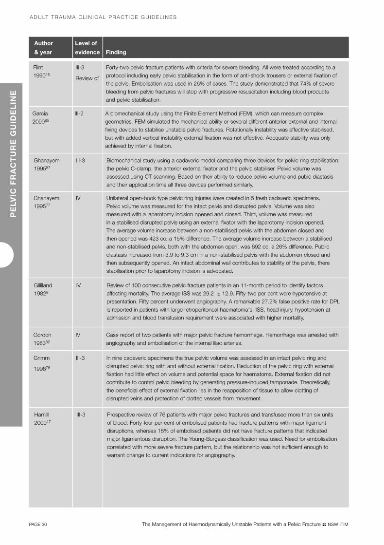
Author Level of
& year evidence Finding
Flint199015
III-3
Review of
Forty-two pelvic fracture patients with criteria for severe bleeding. All were treated according to a protocol including early pelvic stabilisation in the form of anti-shock trousers or external fixation ofthe pelvis. Embolisation was used in 26% of cases. The study demonstrated that 74% of severebleeding from pelvic fractures will stop with progressive resuscitation including blood products and pelvic stabilisation.
Garcia200085
III-2 A biomechanical study using the Finite Element Method (FEM), which can measure complexgeometries. FEM simulated the mechanical ability or several different anterior external and internalfixing devices to stabilise unstable pelvic fractures. Rotationally instability was effective stabilised,but with added vertical instability external fixation was not effective. Adequate stability was onlyachieved by internal fixation.
Ghanayem199587
III-3 Biomechanical study using a cadaveric model comparing three devices for pelvic ring stabilisation:the pelvic C-clamp, the anterior external fixator and the pelvic stabiliser. Pelvic volume wasassessed using CT scanning. Based on their ability to reduce pelvic volume and pubic diastasisand their application time all three devices performed similarly.
Ghanayem199577
IV Unilateral open-book type pelvic ring injuries were created in 5 fresh cadaveric specimens. Pelvic volume was measured for the intact pelvis and disrupted pelvis. Volume was also measured with a laparotomy incision opened and closed. Third, volume was measured in a stabilised disrupted pelvis using an external fixator with the laparotomy incision opened. The average volume increase between a non-stabilised pelvis with the abdomen closed and then opened was 423 cc, a 15% difference. The average volume increase between a stabilisedand non-stabilised pelvis, both with the abdomen open, was 692 cc, a 26% difference. Publicdiastasis increased from 3.9 to 9.3 cm in a non-stabilised pelvis with the abdomen closed andthen subsequently opened. An intact abdominal wall contributes to stability of the pelvis, therestabilisation prior to laparotomy incision is advocated.
Gilliland19828
IV Review of 100 consecutive pelvic fracture patients in an 11-month period to identify factorsaffecting mortality. The average ISS was 29.2 ± 12.9. Fifty-two per cent were hypotensive atpresentation. Fifty percent underwent angiography. A remarkable 27.2% false positive rate for DPLis reported in patients with large retroperitoneal haematoma's. ISS, head injury, hypotension atadmission and blood transfusion requirement were associated with higher mortality.
Gordon198362
IV Case report of two patients with major pelvic fracture hemorrhage. Hemorrhage was arrested withangiography and embolisation of the internal iliac arteries.
Grimm
199876
III-3 In nine cadaveric specimens the true pelvic volume was assessed in an intact pelvic ring anddisrupted pelvic ring with and without external fixation. Reduction of the pelvic ring with externalfixation had little effect on volume and potential space for haematoma. External fixation did notcontribute to control pelvic bleeding by generating pressure-induced tamponade. Theoretically, the beneficial effect of external fixation lies in the reapposition of tissue to allow clotting ofdisrupted veins and protection of clotted vessels from movement.
ADULT TRAUMA CL IN ICAL PRACT ICE GU IDEL INESP
ELV
IC F
RA
CT
UR
E G
UID
EL
INE
Hamill200017
III-3 Prospective review of 76 patients with major pelvic fractures and transfused more than six units of blood. Forty-four per cent of embolised patients had fracture patterns with major ligamentdisruptions, whereas 18% of embolised patients did not have fracture patterns that indicatedmajor ligamentous disruption. The Young-Burgess classification was used. Need for embolisationcorrelated with more severe fracture pattern, but the relationship was not sufficient enough towarrant change to current indications for angiography.
PAGE 30 The Management of Haemodynamically Unstable Patients with a Pelvic Fracture :: NSW ITIM
Pelvic_FullRep.qxd 6/8/07 1:30 PM Page 30

The Management of Haemodynamically Unstable Patients with a Pelvic Fracture :: NSW ITIM PAGE 31
PE
LVIC
FR
AC
TU
RE
GU
IDE
LIN
E
:: EVIDENCE TABLE
Author Level of
& year evidence Finding
Hawkins197057
III-3 A review from the early 1970s of 192 patients with pelvic fractures but focused on 35 patientswho underwent exploratory laparotomy. Twenty laparotomies were preceded by DPL. Thirty hadsurgically controllable intra-peritoneal injuries. Non-therapeutic laparotomy rate was 15%. Mortalitywas 20%. Six patients who died required a mean transfusion requirement of 22.4 units of blood.No correlation between pelvic fracture pattern and the need for laparotomy. It is not clear,however, which classification was used. The authors report a dismal salvage rate of trying tosurgically control retroperitoneal hemorrhage. However, if indications of intra-abdominal injuriesrequiring surgery are also present, the surgeon should still perform laparotomy.
Hehir199040
III-2 Prospective review of 100 patients presenting with a chest injury and an AIS above 3. Reportedinterpretations of the first CXR were compared with chest injuries finally diagnosed. In 19 out of 77surviving patients and 8 out of 17 non-surviving patients significant abnormalities were notdiagnosed on the initial CXR.
Holting199267
IV Analysis of angiographic management of 20 patients with pelvic fractures and arterial bleeding.Angiography was introduced in the management when control of haemodynamic instability hadfailed. Aggressive volume resuscitation with or without laparotomy usually preceded angiography.Mean time to angiography was seven hours in survivors (n=9) and 21 hours for non-survivors (n=11).
Huang199552
IV Review of 43 patients, in which 18 DPLs and 25 FAST were performed separately. Sensitivity,specificity and accuracy were 100%, 40% and 66% for DPL and 95%, 50% and 84% for FAST.
Kam198131
IV A description of angiographic findings in 63 pelvic fracture patients. The internal pudendal artery(27%) was the most frequent bleeding site identified, followed by the superior gluteal artery (25%).No correlation of angiographic findings to the mechanism of injury or fracture pattern was found,but this was demonstrated only by plain films of two cases.
Klein199228
III-3 A review of 429 blunt pelvic trauma patients. The Kane modification of the Key and Conwell pelvicfracture classification was used. Posterior pelvic ring disruptions required significantly more bloodtransfusions in the first 24 hours, was associated with arterial injury requiring intervention, whichwas angiography and embolisation if no intra-abdominal injury requiring surgery was present. Alsosignificantly increased mortality and associated injuries were seen in posterior ring disruptions.
Latenser199172
III-3 Comparative study with historical control of 37 patients divided into two groups. In group 1external fixation was not routinely used between 1981 and 1984. In group 2 external fixation wasperformed unless contra-indicated between 1985 and 1988. Groups matched well for age, sex,ISS and mechanism of injury. In group 1 60% were disabled for at least six months compared to17% in group 2. Survival in group 1 was 83%, in group 2 100%. Hospital stay was decreased by38% in group 2. These were significant differences.
Looser197630
III-3 Retrospective review of 100 consecutive patient with violent pelvic trauma between 1970 and1971. There were 70 associated injuries in group 1 and 125 associated injuries in group 2. Group 1 (57 patients) had purely anterior fractures and group 2 (43 patients) had purely posterior fractures. In group 1 30% had retroperitoneal bleeding compared to 83% in group 2.
Lu200039
IV One hundred trauma team activations in a level 1 trauma centre were analysed. The radiographerwas present in 69% of cases on arrival of the patient. Median time to achieve shooting of the CXRwas five minutes, with a range of two to 22 minutes. Together with other parameters this analysisprovided a template to improve performance in early trauma management.
Mattox198981
II Prospective -randomized study of 911 hypotensive patients randomly assigned to MAST and no-MAST groups. Groups were matched for age, sex, ISS and etiology of injury. MAST did not improve survival (31% mortality in MAST group and 25% in No-MAST group), increased pre-hospital time and most adversely affected outcome in cardiac and thoracic vascular injury.
Pelvic_FullRep.qxd 6/8/07 1:30 PM Page 31

PAGE 32 The Management of Haemodynamically Unstable Patients with a Pelvic Fracture :: NSW ITIM
ADULT TRAUMA CL IN ICAL PRACT ICE GU IDEL INESP
ELV
IC F
RA
CT
UR
E G
UID
EL
INE
Author Level of
& year evidence Finding
Margolies197258
IV Case-series in which angiography with embolisation was used to control pelvic fracturehaemorrhage. The authors attributed higher morbidity and mortality to late embolisation.
McKenney199653
III-2 Prospective study in 200 patients with BAT. Ultrasound was initially performed and subsequentlywithout knowledge of the US results, DPL or CT was performed. US sensitivity was 83%,specificity 100% and accuracy was 975 at predicting intra-abdominal injuries. Six injuries were missed, but only one was felt to be significant.
McMurtry198012
III-3 A review of 76 patients with combined anterior/posterior fractures. Their classification is not widely accepted. Forty-three had posterior ring disruption and a 100% increase in bloodtransfusion requirement and a 150% increase in mortality compared with historical controls. Open-technique DPL drove the treatment protocol in haemodynamically unstable patients. Positive results warranted laparotomy and negative results warranted angiography. Large arteries were surgically controlled and smaller arteries were embolised.
Moore198147
IV Case series in which the open DPL technique at the umbilical ring is described. Fewer complications were found.
Moreno198614
III-3 The study group consisted of 92 haemodynamically unstable pelvic fracture patients, indicated by requirement of more than 6 units of packed red cells. When applied, anti-shock trouserscontrolled pelvic bleeding in 71% of patients. External fixation was effective for hemorrhage control in 95% of cases. If external fixation and/or laparotomy failed, angiography was utilised.Overall mortality was 26%.
Moss199674
III-3 A cadaveric study using a balloon in the true pelvis to measure volume. Open-book type pelvicring disruptions were simulated by increasing pubic diastasis to specified distances and thenmeasuring true pelvic volume. Varying sacroiliac joint and combined symphysis sacroiliac jointseparations were also simulated. Changes in the true pelvic volume as a result of pelvic ringdisruptions were much smaller than previously reported.
Mucha198459
III-3 Analysis of pelvic fracture management over a five-year period of 533 patients. Overall mortalitywas 6.4%. Based on the Key and Conwell classification 36% of fractures were consideredcomplicated due to fracture pattern and, more often, associated injuries. Mean ISS was 35.8 and mortality of these patients, when haemodynamically unstable was 42%, compared to a 3.4% mortality in stable patients. Additionally there was significantly increased ISS and moresevere pelvic fracture types in unstable patients.
Angiography and embolisation was utilised in only 13 patients, of whom 4 had prior laparotomy,with an 86% success rate. In relation to pelvic fracture deaths the importance of associatedinjuries is stressed. Only in 12% of deaths was the pelvic fracture unquestionably the major cause. In 53% pelvic trauma played a contributing role and in 35% pelvic trauma was considered inconsequential.
Lu200039
IV One hundred trauma team activations in a level 1 trauma centre were analysed. The radiographerwas present in 69% of cases on arrival of the patient. Median time to achieve shooting of the CXRwas five minutes, with a range of two to 22 minutes. Together with other parameters this analysisprovided a template to improve performance in early trauma management.
Namm
198345
IV A review of 102 patients, but is focused on a small series of 11 haemodynamically unstablepatients. This group required increased blood transfusion requirements. Anti-shock trousers,external fixation and angiography and embolisation were very useful.
Nunes200149
III-2 Prospective study of 147 patients after blunt and penetrating trauma who underwent USexamination of the abdomen, followed by CT and/or laparotomy. Sensitivity, specificity andaccuracy for predicting abdominal and pelvic injury were 57%, 99% and 95%. Repeat USexamination decreased false negative rates by 50%. A learning curve was also observed, with 67% of the false negative findings occurring in the first three months of the 19-month study.
Pelvic_FullRep.qxd 6/8/07 1:30 PM Page 32

Author Level of
& year evidence Finding
Palmer199789
III-3 A prospective cohort study to assess the safety and efficacy of pelvic external fixators over a three-year period in 24 patients. All external fixators were applied within 6 hours of arrival. The complication rate was 47%, mostly due to poor pin positioning and pin site infection with skin necrosis. This interferes with subsequent management.
Panetta198513
III-2 Report of a protocolled treatment for pelvic fracture patients with blood transfusion requirements.The protocol involved angiographic embolisation to control pelvic bleeding once the abdomen was controlled by laparotomy, if necessary. Embolisation success rate was 87%. Three of the 11 deaths in the series did not respond to embolisation.
Perez199860
IV A review over a 10-year period in which 721 pelvic fracture patients were seen. Only eightunderwent pelvic angiography. Bleeding pelvic vessels were identified in four patients (50% yield)and success rate of embolisation was 50%. Emphasis is placed on the ischaemic complicationsseen after embolisation and the safety of the technique is questioned.
Poole19917
III-3 A multivariate multiple regression analysis correlating outcome with ISS in patients with pelvicfractures. Two-hundred and thirty-six patients were reviewed. Mortality was 7.6%. Hemorrhageaccounted for only 1 death. Analysis showed degree of pelvic fracture severity correlated with ISS,but death could not be predicted. Therefore associated injuries predicted outcome (mortality)following pelvic fractures.
Poole199420
IV Analysis of 90 unstable pelvic fracture patients of whom 66 underwent external fixation.Subsequently 10 of these 66 patients had internal fixation after external fixation had led tohaemodynamic stability. Eighteen patients underwent pelvic angiography, revealing pelvic arterial bleeding in 33% of cases. Mortality was 16% in the angiography group.
Richardson19823
IV Utilizing a graded approach to management of hemorrhage in 37 open pelvic fracture patients a94.5% survival rate was achieved. The approach included wound packing, direct suturing, anti-shock trousers and hemipelvectomy (in two cases) for external control. For intrapelvic hemorrhagecontrol anti-shock trousers, pelvic packing and angiographic embolisation was used.
Riemer199325
III-2 Comparison of mortality in a group of 605 pelvic fracture patients from 1981 to 1988. After 1983 early external fixation as a resuscitative measure was introduced as part of anorganized multidisciplinary protocol. From 1981 to 1988 mean ISS of pelvic fracture patients and blunt abdominal trauma patients did not change. As controls patients with blunt trauma, but without pelvic trauma were studied. As further controls head injured patients with pelvicfractures were compared with head injured patients without pelvic fractures. Mortality fell from26% to 6% after 1983 in pelvic fracture patients. Mortality in head injured patients with pelvicfractures fell from 43% to 7 % after 1983. The organized protocol including external fixationreduced mortality.
The Management of Haemodynamically Unstable Patients with a Pelvic Fracture :: NSW ITIM PAGE 33
PE
LVIC
FR
AC
TU
RE
GU
IDE
LIN
E
:: EVIDENCE TABLE
Rothen-berger19782d
IV Review of 22 open pelvic fracture and 582 closed pelvic fracture patients. Mortality of openfractures was 50%, whereas in closed pelvic fractures mortality was 10.5%. The open pelvicfracture was the cause of death in 73% of patients, in closed pelvic fractures this was 30%.
Rothen-Berger197824d
IV A review of 604 patients, but is focused on the 72 who died. Of these 72 deaths, 71 hadassociated injuries, but in 36.1% death was entirely attributed to the pelvic fracture. In these pelvic fracture related deaths 69% died of hemorrhagic shock. Of the major pelvic vesseldisruptions described, it is not clear which part is arterial and which part is venous.
Pelvic_FullRep.qxd 6/8/07 1:30 PM Page 33

PAGE 34 The Management of Haemodynamically Unstable Patients with a Pelvic Fracture :: NSW ITIM
ADULT TRAUMA CL IN ICAL PRACT ICE GU IDEL INES
Author Level of
& year evidence Finding
Velmahos200043
III-3 Retrospective review of utilisation of angiography and embolisation in 137 consecutive patientswith blunt and penetrating intra- and retroperitoneal trauma. There were 71 blunt pelvic traumacases. Most frequently the internal iliac was the embolised vessel (84%), followed by the superiorgluteal artery (6%), internal pudendal artery (4%), lateral sacral artery (3%) and the obturator andiliolumbar arteries (1.5% each). Twenty-eight bilateral internal iliac embolisations took place withoutadverse sequelae.
Velmahos200244
III-2 Prospective safety and efficacy study of 100 patients with abdominal visceral organ injuries ormajor pelvic fractures sent for emergent or pre-emptive angiography and embolisation. Theefficacy of AE was based on its ability to control bleeding radiographically and clinically; the safetyof AE was determined by the complications related to transport, vascular access, catheterinsertion, contrast administration, and tissue necrosis following interruption of blood supply toorgans.
Of 100 consecutive patients evaluated by angiography for bleeding from major pelvic fractures (65)or solid visceral organ injuries (35), 80 were embolised. AE was effective and safe in 95% and94% respectively of 80 patients who were embolised. Three independent factors were predictiveof bleeding identified on angiography: age older than 55 years, absence of long-bone fractures,and “emergent” angiography. The presence of all three independent predictors was associatedwith a 95% probability of bleeding.
Vrahas199886
III-3 Cadaveric biomechanical study in which three methods of internal fixation and external fixationwere analyzed and compared. Open internal antero-posterior fixation provided the greatest pelvicstability expansion overall, but external fixation proved the most practical method in theresuscitation situation.
Waikukul199973
III-1 Prospective comparison with randomisation of 112 patients with unstable pelvic fractures. Group1 (n=70) was submitted to conventional management without early external fixation. Group 2(n=42) received early orthopaedic consultation and early external fixation. Baseline parameterswere similar in both groups. Blood transfusion, post-operative pain and skeletal deformity wereless in group 2.
PE
LVIC
FR
AC
TU
RE
GU
IDE
LIN
E
Pelvic_FullRep.qxd 6/8/07 1:30 PM Page 34

Pelvic_FullRep.qxd 6/8/07 1:30 PM Page 35
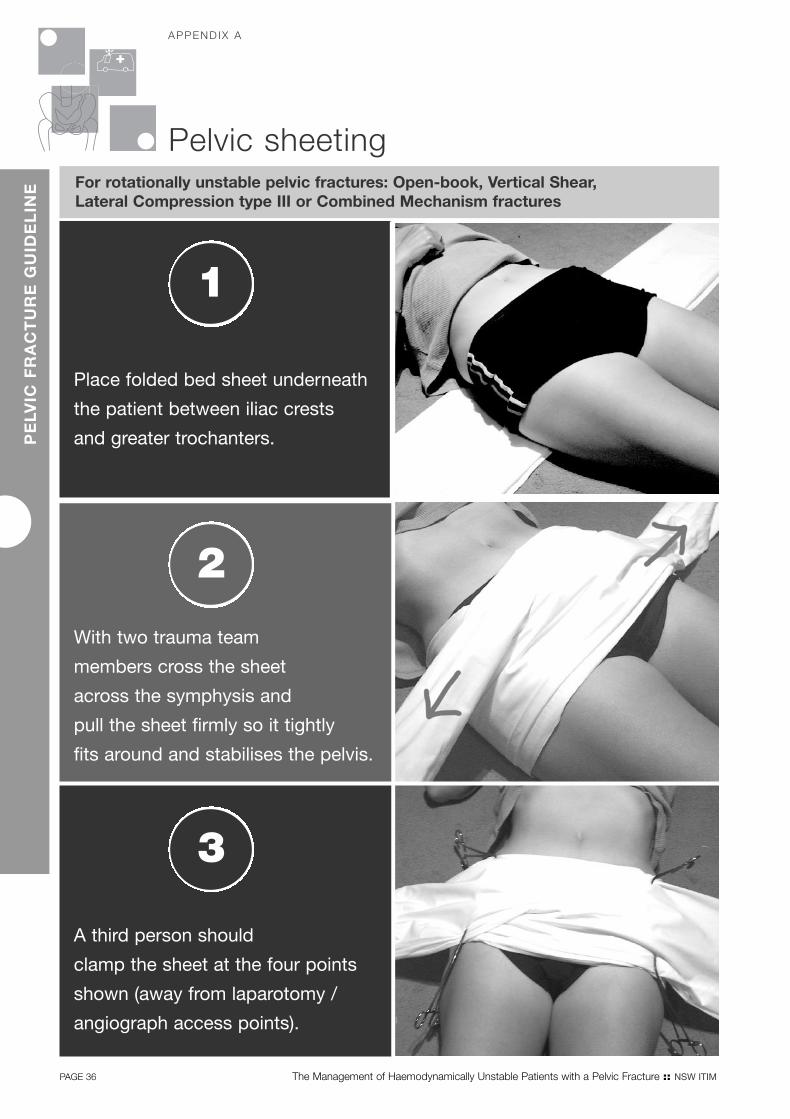
Place folded bed sheet underneath
the patient between iliac crests
and greater trochanters.
For rotationally unstable pelvic fractures: Open-book, Vertical Shear, Lateral Compression type III or Combined Mechanism fractures
1
With two trauma team
members cross the sheet
across the symphysis and
pull the sheet firmly so it tightly
fits around and stabilises the pelvis.
2
A third person should
clamp the sheet at the four points
shown (away from laparotomy /
angiograph access points).
3
PAGE 36 The Management of Haemodynamically Unstable Patients with a Pelvic Fracture :: NSW ITIM
Pelvic sheeting
APPEND IX AP
ELV
IC F
RA
CT
UR
E G
UID
EL
INE
Pelvic_FullRep.qxd 6/8/07 1:30 PM Page 36

The Management of Haemodynamically Unstable Patients with a Pelvic Fracture :: NSW ITIM PAGE 37
PE
LVIC
FR
AC
TU
RE
GU
IDE
LIN
E
References1 Trunkey DD, Chapman MW, Lim RC Jr, Dunphy JE
1974, Management of pelvic fractures in blunttrauma injury, Journal of Trauma-Injury Infection &
Critical Care 14(11):912-23.
2 Rothenberger D, Velasco R, Strate R, Fischer RP,Perry JF Jr 1978, Open pelvic fracture: a lethalinjury, Journal of Trauma-Injury Infection & Critical
Care 18(3):184-7.
3 Richardson JD, Harty J, Amin M, Flint LM 1982,Open pelvic fractures, Journal of Trauma-Injury
Infection & Critical Care 22(7):533-8.
4 Naam NH, Brown WH, Hurd R, Burdge RE,Kaminski DL 1983, Major pelvic fractures, Archives of Surgery 118(5):610-6.
5 Evers B, Cryer HM, Miller F 1989, Pelvic Fracture Haemorrhage, Archives of Surgery
1989;124:422-4.
6 Cryer HM, Miller FB, Evers BM, Rouben LR,Seligson DL 1988, Pelvic fracture classification:correlation with hemorrhage, Journal of Trauma-
Injury Infection & Critical Care 28(7):973-80.
7 Poole GV, Ward EF, Muakkassa FF, Hsu HS,Griswold JA, Rhodes RS 1991, Pelvic fracture frommajor blunt trauma. Outcome is determined byassociated injuries, Annals of Surgery 213(6):532-8;discussion 538-9.
8 Gilliland MD, Ward RE, Barton RM, Miller PW,Duke JH 1982, Factors affecting mortality in pelvicfractures, Journal of Trauma-Injury Infection & Critical
Care 22(8):691-3.
9 Tile M 1988, Pelvic ring fractures: Should they befixed? Journal of Bone & Joint Surgery – British
Volume Vol 70(1); 34-37.
10 Eastridge BJ, Burgess AR 1997, Pedestrian pelvicfractures: 5-year experience of a major urbantrauma center, Journal of Trauma-Injury Infection
& Critical Care 42(4):695-700, 1997 Apr.
11 Bassam D, Cephas GA, Ferguson KA, Beard LN,Young JS 1998, A protocol for the initialmanagement of unstable pelvic fractures, American Surgeon 64(9):862-7.
12 McMurtry R, Walton D, Dickinson D, Kellam J, TileM 1980, Pelvic disruption in the polytraumatizedpatient: a management protocol, Clinical
Orthopaedics & Related Research (151):22-30.
13 Panetta T, Scalfani S, Goldstein AS, Phillips TF,Shaftan GW 1985, Percutaneous transcatherterembolisation for massive bleeding from pelvicfractures, Journal of Trauma-Injury Infection &
Critical Care 1985;25:1021-9.
14 Moreno C, Moore EE, Rosenberger A, ClevelandHC 1986, Hemorrhage associated with majorpelvic fracture: a multispecialty challenge, Journal
of Trauma-Injury Infection & Critical Care 26(11):987-94, 1986 Nov.
15 Flint L, Babikian G, Anders M, Rodriguez J,Steinberg S 1990, Definitive control of mortalityfrom severe pelvic fracture, Annals of Surgery
211(6):703-6; discussion 706-7.
16 Gruen GS, Leit ME, Gruen RJ, Peitzman AB 1994, The acute management of hemodynamicallyunstable multiple trauma patients with pelvic ringfractures, Journal of Trauma-Injury Infection & Critical
Care 36(5):706-11; discussion 711-3.
17 Hamill J, Holden A, Paice R, Civil I 2000, Pelvic fracture pattern predicts pelvic arterialhaemorrhage, Australian & New Zealand Journal of
Surgery 70(5):338-43.
18 Ertel W, Keel M, Eid K, Platz A, Trentz O 2001,Control of severe hemorrhage using C-clamp andpelvic packing in multiply injured patients withpelvic ring disruption [see comment], Journal of
Orthopaedic Trauma 15(7):468-74.
19 Eastridge BJ, Starr A, Minei JP, O'Keefe GE, Scalea TM 2002, The importance of fracturepattern in guiding therapeutic decision-making in patients with hemorrhagic shock and pelvic ring disruptions, Journal of Trauma-Injury Infection &
Critical Care 53(3):446-50; discussion 450-1.
20 Poole GV, Ward EF 1994, Causes of mortalityin patients with pelvic fractures, Orthopedics
17(8):691-6.
Pelvic_FullRep.qxd 6/8/07 1:30 PM Page 37

PAGE 38 The Management of Haemodynamically Unstable Patients with a Pelvic Fracture :: NSW ITIM
ADULT TRAUMA CL IN ICAL PRACT ICE GU IDEL INESP
ELV
IC F
RA
CT
UR
E G
UID
EL
INE
21 Duane TM, Tan BB, Golay D, Cole FJ Jr, Weireter LJ Jr, Britt LD 2002, Blunt trauma and the role ofroutine pelvic radiographs: a prospective analysis,Journal of Trauma-Injury Infection & Critical Care
53(3):463-8.
22 Grant P 1990, The diagnosis of pelvic fractures by springing, Archives of Emergency Medicine
1990;7(3):178-82.
23 Dalal SA, Burgess AR, Siegel JH, Young JW,Brumback RJ, Poka A, et al 1989, Pelvic fracturein multiple trauma: classification by mechanism is key to pattern of organ injury, resuscitativerequirements, and outcome, Journal of Trauma-
Injury Infection & Critical Care 29(7):981-1000;discussion 1000-2, 1989 Jul.
24 Rothenberger DA, Fischer RP, Strate RG, Velasco R,Perry JF Jr 1978, The mortality associated withpelvic fractures, Surgery 84(3):356-61.
25 Riemer BL, Butterfield SL, Diamond DL, Young JC,Raves JJ, Cottington E et al 1993, Acute mortalityassociated with injuries to the pelvic ring: the roleof early patient mobilization and external fixation,Journal of Trauma-Injury Infection & Critical Care
35(5):671-5; discussion 676-7.
26 Poole GV, Ward EF, Griswold JA, Muakkassa FF,Hsu HS 1992, Complications of pelvic fracturesfrom blunt trauma, American Surgeon 58(4):225-31.
27 Burgess AR, Tile M 1991, Fractures of the pelvis.In: Rockwood C, Green D, editors, Fractures in
Adults, Philadelphia: JB Lippincott; p.1399-442.
28 Klein SR, Saroyan RM, Baumgartner F, Bongard FS1992, Management strategy of vascular injuriesassociated with pelvic fractures, Journal of
Cardiovascular Surgery 33(3):349-57.
29 McMurtry RY 1989, Pelvic fractures anddislocations. (i) Initial assessment andmanagement, Current Orthopaedics
Vol 3(4); 233-227, 1989
30 Looser KG, Crombie HD Jr 1976, Pelvic fractures:an anatomic guide to severity of injury, Review of 100 cases, American Journal of Surgery
132(5):638-42.
31 Kam J, Jackson H, Ben-Menachem Y 1981,Vascular injuries in blunt pelvic trauma, Radiologic
Clinics of North America Vol 19(1); 171-186).
32 Resnik CS, Stackhouse DJ, Shanmuganathan K,Young JW 1992, Diagnosis of pelvic fractures inpatients with acute pelvic trauma: efficacy of plainradiographs, AJR American Journal of
Roentgenology 158(1):109-12.
33 Robertson D, Sutherland C, Chan B, Hodge J,Scott W, Fishman E 1995, Depiction of pelvicfractures using 3D volumetric hologrpahy:comparison of plain X-ray and CT, J Comput Assist Tomogr 1995;19(6):967-74.
34 Ben-Menachem Y, Coldwell DM, Young JW,Burgess AR 1991, Hemorrhage associated withpelvic fractures: causes, diagnosis, and emergentmanagement, [Review] [50 refs], AJR American
Journal of Roentgenology 157(5):1005-14.
35 Kellam JF, Trafton PG. Fractures of the Pelvic Ring.In: Browner BD, Jupiter J, Levine A, Trafton PG,editors 1992, Skeletal Trauma, Volume 1 ed.Philadelphia: WB Saunders; 1992. p. 849-97.
36 American College of Surgeons Committee onTrauma 1997, Advanced Trauma Life Support for
Doctors (6th Edition).
37 Ali J, Howard M, Williams J 2002, Is attrition of advanced trauma life support acquired skillsaffected by trauma patient volume? American
Journal of Surgery 183(2):142-5.
38 American College of Surgeons 1997, Advanced
Trauma life support for Doctors Instruction Course
Manual Book 1, First Impression: USA 1997.
39 Lu W, Kolkman K, Seger M, Sugrue M 2000, An evaluation of trauma team response in a major trauma hospital in 100 patients withpredominantly minor injuries, ANZ Journal
of Surgery 70(5):329-32.
40 Hehir MD, Hollands MJ, Deane SA 1990, The accuracy of the first chest X-ray in the trauma patient, Australian & New Zealand Journal of
Surgery 60(7):529-32.
41 Agolini SF, Shah K, Jaffe J, Newcomb J, Rhodes M,Reed JF, III 1997, Arterial embolisation is a rapidand effective technique for controlling pelvicfracture hemorrhage [see comment], Journal of
Trauma-Injury Infection & Critical Care 43(3):395-9.
42 Velmahos GC, Chahwan S, Hanks SE, Murray JA,Berne TV, Asensio J, et al 2000, Angiographicembolisation of bilateral internal iliac arteries tocontrol life-threatening hemorrhage after blunttrauma to the pelvis, American Surgeon
66(9):858-62.
Pelvic_FullRep.qxd 6/8/07 1:30 PM Page 38

The Management of Haemodynamically Unstable Patients with a Pelvic Fracture :: NSW ITIM PAGE 39
PE
LVIC
FR
AC
TU
RE
GU
IDE
LIN
E
:: REFERENCES
43 Velmahos GC, Chahwan S, Falabella A, Hanks SE,Demetriades D 2000, Angiographic embolisationfor intraperitoneal and retroperitoneal injuries,World Journal of Surgery 24(5):539-45.
44 Velmahos GC, Toutouzas KG, Vassiliu P, SarkisyanG, Chan LS, Hanks SH, et al 2002, A prospectivestudy on the safety and efficacy of angiographicembolisation for pelvic and visceral injuries, Journal
of Trauma-Injury Infection & Critical Care 53(2):303-8; discussion 308.
45 Namm NHBW, Hurd R, Burdge R, Kaminski D1983, Major pelvic fractures, Archives of Surgery
118:610-6.
46 Sugrue M, Seger M, Gunning K, Sloane D, Deane S 1995, A modified combination techniquefor performing diagnostic peritoneal lavage, ANZ
Journal of Surgery 1995;65:604-6.
47 Moore JB, Moore EE, Markovchick VJ, Rosen P1981, Diagnostic peritoneal lavage for abdominaltrauma: Superiority of the open technique at theinfraumbilical ring, Journal of Trauma-Injury Infection
& Critical Care Vol 21(7); 570-572.
48 McKenney M, Lentz K, Nunez D, Sosa JL, SleemanD, Axelrad A et al 1994. Can ultrasound replacediagnostic peritoneal lavage in the assessment ofblunt trauma? [see comment], Journal of Trauma-
Injury Infection & Critical Care 37(3):439-41.
49 Nunes LW, Simmons S, Hallowell MJ, Kinback R,Trooskin S, Kozar R 2001, Diagnostic performanceof trauma US in identifying abdominal or pelvicfree fluid and serious abdominal or pelvic injury,Academic Radiology 8(2):128-36.
50 Rozycki GS, Newman PG 1999, Surgeon-performed ultrasound for the assessment ofabdominal injuries, Advances in Surgery 33:243-59.
51 Rozycki GS, Ochsner MG, Feliciano DV, Thomas B,Boulanger BR, Davis FE et al 1998, Early detectionof hemoperitoneum by ultrasound examination ofthe right upper quadrant: a multicenter study,Journal of Trauma-Injury Infection & Critical Care
45(5):878-83.
52 Huang J, Fang J, Chen R 1995, Diagnosis ofintraperitoneal injuries in patients with pelvicfractures, Changgeng Yi Xue Za Zhi
1995;18(2):154-9.
53 McKenney KL, McKenney MG, Nunez DB,McDowell L, Martin L 1996, Interpreting thetrauma ultrasound: Observations in 62 positivecases, Emergency Radiology Vol 3(3); 113-117.
54 Ballard RB, Rozycki GS, Newman PG, Cubillos JE1999, Salomone JP, Ingram WL et al, An algorithmto reduce the incidence of false-negative FASTexaminations in patients at high risk for occultinjury, Focused Assessment for the SonographicExamination of the Trauma patient, Journal of the
American College of Surgeons 189(2):145-50;discussion 150-1.
55 Bode PJ, Niezen RA, Van Vugt AB, Schipper J1993, Abdominal ultrasound as a reliable indicatorfor conclusive laparotomy in blunt abdominaltrauma [see comment], Journal of Trauma-Injury
Infection & Critical Care 34(1):27-31, 1993 Jan.
56 Ballard RB, Rozycki GS, Newman PG, Cubillos JE,Salomone JP, Ingram WL et al 1999, An algorithmto reduce the incidence of false-negative FASTexaminations in patients at high risk for occultinjury, Journal of the American College of Surgeons
Vol 189(2); 145-151.
57 Hawkins L, Pomerantz M, Eisman B 1970,Laparotomy at time of pelvic fracture, Journal
of Trauma-Injury Infection & Critical Care
1970;10:619-23.
58 Margolies MN, Ring E, Waltman AC, Kerr W, Baum F 1972, Arteriography in the managementof haemorrhage from pelvic fractures, New England
Journal of Medicine 287:317-21.
59 Mucha P, Jr., Farnell MB 1984, Analysis of pelvicfracture management, Journal of Trauma-Injury
Infection & Critical Care 24(5):379-86.
60 Perez J, Hughes P, Bowers S 1998, Angiographicembolisation in pelvic fracture, Injury 29:187-91.
61 Stephen DJ, Kreder HJ, Day AC, McKee MD,Schemitsch EH, ElMaraghy A, et al 1999, Earlydetection of arterial bleeding in acute pelvictrauma, Journal of Trauma-Injury Infection & Critical
Care 47(4):638-42.
62 Gordon RL, Fast A, Aner H 1983, Control of massive retroperitoneal bleeding associated with pelvic fractures by angiographic embolisation,Israel Journal of Medical Sciences Vol 19(2);185-188.
63 Clarke JR, Trooskin SZ, Doshi PJ, Greenwald L,Mode CJ 2002, Time to laparotomy for intra-abdominal bleeding from trauma does affectsurvival for delays up to 90 minutes, Journal of
Trauma-Injury Infection & Critical Care 52(3):420-5.
64 Van UH, Perlberger RR, Muller H 1978, Selectivearterial embolisation for control of traumatic pelvichemorrhage, Surgery 83(2):133-7.
Pelvic_FullRep.qxd 6/8/07 1:30 PM Page 39

PAGE 40 The Management of Haemodynamically Unstable Patients with a Pelvic Fracture :: NSW ITIM
ADULT TRAUMA CL IN ICAL PRACT ICE GU IDEL INESP
ELV
IC F
RA
CT
UR
E G
UID
EL
INE
65 Slatis P, Huittinen VM 1972, Double verticalfractures of the pelvis, Acta Chirurgica Scandinavia
1972;138:799-802.
66 Huittinen V, Slatis P. Post mortem angiography anddissection of the hypogastric artery in pelvicfractures, Surgery 1973;73:454-62.
67 Holting T, Buhr HJ, Richter GM, Roeren T, Friedl W, Herfarth C 1992, Diagnosis andtreatment of retroperitoneal hematoma in multiple trauma patients, Archives of Orthopaedic
& Trauma Surgery 111(6):323-6.
68 Flint LM, Brown A, Richardson JD, Polk HC Jr1979, Definitive control of bleeding from severepelvic fractures, Annals of Surgery 89:709-16.
69 Mucha JP, Welch TJ 1988, Hemorrahge in majorpelvic fractures, Surgical Clinics of North America
Vol 68(4); 757-773.
70 Kellam JF 1989, The role of external fixation inpelvic ring disruptions, Clinical Orthopaedics &
Related Research 1989;241:66-82.
71 Broos P, Vanderschot P, Craninx L, Rommens P1992, The operative treatment of unstable pelvicring fractures, International Surgery 77(4):303-8,1992 Oct;-Dec.
72 Latenser BAGL, Tarver A 1991, Improved outcome with early fixation of skeletally unstable pelvic fractures, Journal of Trauma-
Injury Infection & Critical Care 31:28-31.
73 Waikakul S, Harnroonroj T, Vanadurongwan V1999, Immediate stabilisation of unstable pelvicfractures vs delayed stabilisation, Journal of the
Medical Association of Thailand 82(7):637-42.
74 Moss MC, Bircher MD 1996, Volume changeswithin the true pelvis during disruption of thepelvic ring – Where does the haemorrhage go?Injury Vol 27(SUPPL 1).
75 DiGiacomo JC, Bonadies J, Cole F, Diebel L, HoffW, Holevar M et al 2001, Practice ManagementGuidelines for Haemorrhage in Pelvic Fracture,EAST.
76 Grimm M, Vrahas M, Thomas K 1998, Pressure-volume characteristics of the intact and disruptedpelvic retroperitoneum, Journal of Trauma-Injury
Infection & Critical Care 44:454-9.
77 Ghanayem AJ, Wilber JH, Lieberman JM, MottaAO 1995, The effect of laparotomy and externalfixator stabilisation on pelvic volume in an unstablepelvic injury, Journal of Trauma-Injury Infection &
Critical Care 38(3):396-400; discussion 400-1.
78 Routt ML, Jr., Simonian PT, Ballmer F 1995, A rational approach to pelvic trauma, Resuscitation and early definitive stabilisation,Clinical Orthopaedics & Related Research
(318):61-74.
79 Fleming W, Bowen J 1973, Control of hemorrhagein pelvic crush injuries, Journal of Trauma-Injury
Infection & Critical Care 1973;13:567-70.
80 Matta JM 1996, Indications for anterior fixation ofpelvic fractures, Clinical Orthopaedics & Related
Research (329):88-96.
81 Mattox KL, Bickell WH, Pepe PE, Mangelsdorff AD,Prospective randomized evaluation of antishockMAST in post-traumatic hypotension, Journal of
Trauma-Injury Infection & Critical Care 26(9):779-86,1986 Sep.
82 Ben-Menachem Y, Handel SF, Ray RD, Childs III TL1985, Embolisation procedures in trauma; The pelvis. Seminars in Interventional Radiology
Vol 2(2);158-181.
83 Kaufman J, Waltman A 1997, Angiogrpahicmanagement of haemorrhage in pelvic fracture,Abrams Angiography- Interventional Radiology,
Volume III. 4th ed. Little, Brown and Co p.869-83.
84 Hoffer EK 1999, Interventional angiography for treatment of hemorrhage in pelvic trauma,Emergency Radiology Vol 6(4); 216-226, 1999
85 Garcia J, Doblare M, Seral B, Seral F, Palanca D2000, Three dimensional finite element analysis of several internal and external pelvis fixations,Journal of Biomechanical Engineering 122:516-22.
86 Vrahas M, Wilson SC, Cummings P, Paul E 1998,Comparison of fixation methods for preventingpelvic ring expansion, Orthopedics 21:285-9.
87 Ghanayem AJ, Stover MD, Goldstein JA, Bellon E,Wilber JH 1995, Emergent treatment of pelvicfractures, Comparison of methods for stabilisationClinical Orthopaedics & Related Research
(318):75-80.
88 Simpson T, Kreig J, Heuer F, Bottlang M 2002,Stabilisation of pelvic ring disruptions with acircumferential sheet, Journal of Trauma-Injury
Infection & Critical Care 52:158-61.
89 Palmer S, Fairbank A, Bircher M 1997, Surgicalcomplications and implications of external fixationfor pelvic fractures, Injury 28:649-53.
90 Ganz R 1991, The antishock pelvic clamp, Clinical
Orthopaedics & Related Research 267:71-8.
Pelvic_FullRep.qxd 6/8/07 1:30 PM Page 40

The Management of Haemodynamically Unstable Patients with a Pelvic Fracture :: NSW ITIM PAGE 41
PE
LVIC
FR
AC
TU
RE
GU
IDE
LIN
E
:: REFERENCES
91 Simonian PT, Routt ML Jr, Harrington RM, TencerAF 1995, Anterior versus posterior provisionalfixation in the unstable pelvis. A biomechanicalcomparison, Clinical Orthopaedics & Related
Research (310):245-51.
92 Rozycki GS, Ballard RB, Feliciano DV, Schmidt JA, Pennington SD 1998, Surgeon-performed ultrasound for the assessment oftruncal injuries: lessons learned from 1540patients, Annals of Surgery 228(4):557-67.
Pelvic_FullRep.qxd 6/8/07 1:30 PM Page 41

::
SHPN (TI) 070026
PelvicFracture_FullRCvR.qxd 3/2/07 12:09 PM Page 4