pegylated aza-bodipy derivatives as nir probes for ... supporting information pegylated aza-bodipy...
TRANSCRIPT

S1
Supporting Information
PEGylated Aza-BODIPY Derivatives as NIR Probes for Cellular Imaging
Daniel Collado,a,b Yolanda Vida,a,b Francisco Najeraa,b and Ezequiel Perez-Inestrosa*a,b
a Department of Organic Chemistry, University of Malaga, Campus Teatinos, E-29071 Málaga (Spain) b Andalusian Centre for Nanomedicine and Biotechnology-BIONAND, Parque Tecnológico de Andalucía, E-29590
Malaga, (Spain).
Table of Contents
General experimental and Computational methods ...................................................... S-2 Experimental procedures .............................................................................................. S-3 Additional figures ........................................................................................................ S-6 Additional tables ......................................................................................................... S-12 1H and 13C NMR spectra ............................................................................................. S-13 References ................................................................................................................... S-20
Electronic Supplementary Material (ESI) for RSC AdvancesThis journal is © The Royal Society of Chemistry 2013

S2
General Experimental Instruments. All photophysical measurements performed in solutions were carried out using dilute solutions with absorbance around 0.1 at the maximum absorption wavelength in 1 cm path length quartz cells and in air-equilibrated solutions at room temperature. The UV–Vis absorption spectra were recorded with a HP 8452A Diode Array Spectrophotometer spectrophotometer. The fluorescence spectra were recorded with a JASCO FP-750 Spectrofluorometer. Fluorescence quantum yields were determined using Rhodamine B (ΦF = 0.53 in EtOH) as a reference and corrected for the corresponding refractive index.1 Proton and carbon nuclear magnetic resonance spectra (1H and 13C NMR) of the newly synthesized compounds were recorded on a Bruker Advance III 400 MHz Ascend instrument and were referenced to the signal of the residual solvent. (1H NMR, CHCl3 at 7.26 ppm, CD3OD at 3.34 ppm; 13C NMR, CDCl3 at 77.0 ppm, CD3OD at 49.9 ppm).2 Data for 1H NMR are reported as follows: chemical shift (ppm), integration, multiplicity (s = singlet; d = doublet; t = triplet; q = quartet; m = multiplet) and coupling constants, J, in (Hz). The HRMS data were recorded on a Bruker MicroTOF mass spectrometer. Materials. All reagents (including the starting compound 5a) used in the synthetic procedures were commercially available and were purchased in the highest purity available (≥ 98%). Aldehydes 4a3-4b4
, acetophenones 5b5-5c6, aza-BODIPY 97 and compound 88 were synthesized according to
previously reported procedures. Unless otherwise noted, all reagents and chemicals were used without further purification. Chromatography on silica gel was performed using silica gel 60 (230-400 mesh). Thin layer chromatography (TLC) was performed on silica gel 60 plates. Visualization of spots on TLC plates was accomplished with UV light (254 nm). Solvents for synthesis and NMR were used without further purification. The overall pH of the PBS buffer was 7.4. Cell Culture and aza-BODIPY incubations. 8-well chambered coverglass slides were seeded with the HeLa human tumor cell line. Cells were cultured at 37ºC with 5% CO2 in RPMI (1640 medium supplemented with L-Glutamine and Fetal Bovine Serum until they reached 50-70% confluence, 1-2 days). HeLa cell-containing wells were incubated for 1 h with 200µl of fresh, pre-heated RPMI 1640 culture medium containing different concentrations of aza-BODIPY plus a negative control. After incubation, dye-containing media was removed from each well, washed two times with preheated PBS to remove unbound aza-BODIPY, and then fixed using 4% paraformaldehyde for 10 min at room temperature. Finally cells were washed with PBS three times and kept at 4ºC until analysis. Confocal Microscopy. The fluorescence properties of aza-BODIPY 2 in HeLa cells were analyzed using an inverted confocal microscope. 3D image series containing 7 images were captured at ~1µm intervals. Aza-BODIPY fluorescence was visualized using excitation at 633 nm and detection of emissions between 643nm–758nm. Non-confocal transmitted light images were obtained simultaneously. All samples were analyzed using identical imaging conditions. Emission spectra for cell and background Regions of Interest (ROIs) were calculated using the manufacturer´s software. The 633 nm emission spectrum was measured using a dynamic 20 nm wide emission detection window moving in 20 steps between 650 nm and 795 nm. Computational Methods. All calculations were achieved using the Gaussian 09 package.9 The ground-state (S0) geometrical parameters have been determined with the density functional level of theory, employing the PBE0 functional10,11 and the 6-311G(2d,p) basis set, and within C2 point group, as recommended.12 The nature of the minima was confirmed by the absence of a negative frequency in the vibrational analysis. We have replaced the PEG chains by methoxy groups, as it is expected that the length of these chains has no substantial influence on the optical properties. Restricted formalism was applied for the singlet electronic states and unrestricted formalism for the triplets states. Absorption
Electronic Supplementary Material (ESI) for RSC AdvancesThis journal is © The Royal Society of Chemistry 2013

S3
spectra were computed as vertical electronic excitations from the minima of the ground-state structures by using time-dependent density functional response theory13 employing the PBE0 functional, and with the 6-311+G(2d,p) basis set.12,14 The solvent effect has been accounted for with the polarizable continuum model (PCM),15 and water as solvent.
Experimental procedures
O H
OR1 O
OR2
+
O
OR2
OR1
O
OR2
OR1
NO2
N
N
N
R2O OR2
R1O OR1
BF F
KOH
EtOH / ∆MeNO2
KOH / EtOH / ∆
4a R1 = [(CH2)2O]3(CH2)2OH4b R1 = CH2CCH5a R2 = OCH35b R2 = [(CH2)2O]3(CH2)2OH5c R2 = CH2CCH
4a-b 5a-c
6a R1 = [(CH2)2O]3(CH2)2OH; R2 = CH36b R1 = R2 = [(CH2)2O]3(CH2)2OH6c R1 = R2 = CH2CCH
7a R1 = [(CH2)2O]3(CH2)2OH; R2 = CH37b R1 = R2 = [(CH2)2O]3(CH2)2OH7c R1 = R2 = CH2CCH
1 R1 = [(CH2)2O]3(CH2)2OH; R2 = CH32 R1 = R2 = [(CH2)2O]3(CH2)2OH9 R1 = R2 = CH2CCH
1) AcONH4 / n-BuOH / 120ºC2) NEt3 / BF3 / CH2Cl2
O
O
O
N3OH
H2O / MeOH / THF (1:1:1)CuSO4 / ascorbic acid / TBTA
3
8
Scheme S1. General route to generate aza-BODIPYs 1, 2 and 3. General procedure for the synthesis of chalcones 6a, 6b and 6c7 A solution of aromatic ketone (10 mmol) in ethanol (10 mL) was added gradually to an aqueous solution of 10% KOH (30 mL) at 0ºC. After stirring for 15 min, aromatic aldehyde (10 mmol) was added and stirred at 0ºC for 15 min. The mixture was then allowed to attain room temperature and stirred for 4h. The solvent was removed in vacuo, the residue diluted with CH2Cl2 (100 mL), washed with water (2 x 15 mL) and dried with anhydrous MgSO4. The organic layer was concentrated, and the residue was purified by chromatography on a silica gel column (AcOEt/MeOH 20/1) to give the title product. (E)-3-(4-(2-(2-(2-(2-hydroxyethoxy)ethoxy)ethoxy)ethoxy)phenyl)-1-(4-ethoxyphenyl)prop-2-en-1-one), (6a). Colorless solid, (232 mg, 60%); δH(400 MHz, CDCl3) 8.00 (2H, d, J = 9.2 Hz), 7.74 (1H, d, J = 15.4 Hz), 7.56 (2H, d, J = 9.2 Hz), 7.40 (1H, d, J = 15.4 Hz), 6.98-6.91 (4H, m), 4.17 (2H, t, J = 4.3 Hz), 3.87-3.84 (4H, m), 3.70-3.56 (14H, m); δC(100 MHz, CDCl3) 188.3, 162.4, 160.6, 144.0, 131.6, 130.1, 129.3, 127.4, 119.5, 114.4, 114.2, 72.4, 70.7, 70.5, 70.4, 70.1, 69.5, 69.4, 67.5, 67.4, 61.5, 55.6; HRMS (ESI) m/z calcd for C24H30O7+H+, 431.2070 [M+H+]; found, 431.2074.
Electronic Supplementary Material (ESI) for RSC AdvancesThis journal is © The Royal Society of Chemistry 2013

S4
(E)-1,3-bis(4-(2-(2-(2-(2-hydroxyethoxy)ethoxy)ethoxy)ethoxy)phenyl)prop-2-en-1-one, (6b). Colorless oil, (408 mg, 72%); δH(400 MHz, CDCl3) 8.00 (2H, d, J = 8.6 Hz), 7.75 (1H, d, J = 16.0 Hz), 7.57 (2H, d, J = 8.6 Hz), 7.40 (1H, d, J = 16.0 Hz), 7.00-6.90 (4H, m), 4.23-4.14 (6H, m), 3.89-3.83 (6H, m), 3.71-3.57 (22H, m); δC(100 MHz, CDCl3) 188.7, 162.3, 160.5, 143.7, 131.3, 130.6, 130.0, 127.9, 119.5, 114.9, 114.3, 72.5, 70.7, 70.5, 70.4, 70.1, 69.5, 69.4, 67.5, 67.4, 61.6; HRMS (ESI) m/z calcd for C31H44O11+H+, 593.2962 [M+H+]; found 593.2965. General procedure for the synthesis of nitro derivatives 7a, 7b and 7c7 A solution of chalcone 6a-c (5 mmol) in EtOH (50 mL) was treated with KOH (25 mmol) and nitromethane (25 mmol) and heated under reflux for 24 h. After cooling to room temperature, the solvent was removed in vacuo and the oily residue obtained was dissolved in CH2Cl2 (75 mL) and washed with water (2 x 50 mL). The combined organic layers were washed with brine, dried over anhydrous MgSO4, and concentrated to give the target compound, which could be used directly in the next step without further purification. 3-(4-(2-(2-(2-(2-hydroxyethoxy)ethoxy)ethoxy)ethoxy)phenyl)-1-(4-methoxyphenyl)-4-nitrobutan-1-one, (7a). Brown oil, (240 mg, 90%); δH(400 MHz, CDCl3) 7.87 (2H, d, J = 9.1 Hz), 7.16 (2H, d, J = 8.5 Hz), 6.92-6.82 (4H, m), 4.83-4.53 (2H, m), 4.16-4.05 (4H, m), 3.85-3.78 (5H, m), 3.69-3.57 (14H, m); δC(100 MHz, CDCl3) 195.4, 163.7, 158.1, 131.3, 130.3, 129.4, 128.4, 114.9, 113.6, 79.8, 72.6, 70.8, 70.6, 70.5, 70.4, 70.1, 70.0, 69.7, 69.5, 67.2, 66.5, 61.5, 55.4, 41.1, 38.7; HRMS (ESI) m/z calcd for C25H33NO9+H+, 492.2234 [M+H+]; found, 492.2238. 1,3-bis(4-(2-(2-(2-(2-hydroxyethoxy)ethoxy)ethoxy)ethoxy)phenyl)-4-nitrobutan-1-one, (7b). Brown oil, (148 mg, 72%); δH(400 MHz, CDCl3) 7.86 (2H, d, J = 8.5 Hz), 7.16 (2H, d, J = 8.5 Hz), 6.92 (2H, d, J = 9.1 Hz), 6.84 (2H, d, J = 9.1 Hz) 4.83-4.53 (2H, m), 4.17-4.08 (6H, m), 3.88-3.79 (5H, m), 3.75-3.56 (26H, m); δC(100 MHz, CDCl3) 188.7, 162.3, 160.5, 160.2, 143.7, 131.3, 130.6, 130.0, 127.8, 119.5, 114.9, 114.3, 72.5, 70.7, 70.5, 70.4, 70.1, 69.5, 69.4, 67.4, 67.3, 61.5, 41.3, 39.1; HRMS (ESI) m/z calcd for C32H47NO13+H+, 654.3126 [M+H+]; found 654.3130. Synthesis of aza-BODIPY 1-2 Ammonium acetate (10 mmol) was added to a stirred solution of 7a-c (0.5 mmol) in n-BuOH (20 mL). The mixture was heated at 120 ºC for 24 h. The solvent was removed in vacuo, the residue diluted with CH2Cl2 (100 mL), washed with water (2 x 15 mL) and dried with anhydrous MgSO4. The organic layer was concentrated in vacuo. The resulting solid was subsequently dissolved in dry CH2Cl2 (10 mL), cooled to 0ºC, and treated with triethylamine (5 mL) followed by slow addition of BF3·OEt2 (5 mL). The mixture was stirred for 24 h. The reaction was quenched with crushed ice and extracted with CH2Cl2 (2 x 30 mL). The organic layer was dried over anhydrous MgSO4 and evaporated to dryness. Purification by column chromatography on silica gel (AcOEt/MeOH 8/2) gave the title compound. Aza-BODIPY 1. Purple solid (56 mg, 12%). δH(400 MHz, CDCl3) 8.13 (4H, d, J = 8.5 Hz), 7.72-7.64 (6H, m), 7.06-6.98 (8H, m), 4.20 (4H, t, J = 4.3 Hz), 3.90-3.86 (10H, m), 3.74-3.57 (26H, m); δC(100 MHz, CDCl3) 160.4, 159.4, 156.8, 149.4, 132.4, 131.7, 128.3, 128.2, 115.2, 115.1, 113.9, 72.5, 70.8, 70.6, 70.5, 69.6, 70.2, 67.4, 61.7, 55.3; HRMS (MALDI) m/z calcd for C50H58BF2N3O12+H+, 942.4160 [M+H+]; found, 942.4166. Aza-BODIPY 2. Purple solid (43 mg, 15%). δH(400 MHz, CDCl3) 8.10 (4H, d, J = 8.5 Hz), 7.71-7.63 (6H, m), 7.04-6.99 (8H, m), 4.20-4.17 (8H, m), 3.87-3.85 (8H, m), 3.74-3.57 (52H, m); δC(100 MHz, CDCl3) δ 159.5, 142.7, 136.2, 131.4, 130.7, 128.3, 115.1, 114.7, 72.5, 70.8, 70.6, 70.5, 70.3, 69.7, 67.6, 61.7; HRMS (MALDI) m/z calcd for C64H86BF2N3O20+H+, 1266.5944 [M+H+]; found 1266.5950.
Electronic Supplementary Material (ESI) for RSC AdvancesThis journal is © The Royal Society of Chemistry 2013

S5
Synthesis of aza-BODPIPY 3 Aza-BODIPY 9 (116 mg, 0.29 mmol) was suspended in a 2:1:1 mixture of MeOH:H2O:THF (6 mL), to which was added 8 (325 mg, 0.61 mmol), CuSO4·5H2O (184 mg, 2.90 mmol), and TBTA (3.1 mg, 0.006 mmol). The reaction mixture was allowed to stir vigorously at room temperature for 24 h after which it was diluted with CH2Cl2, and then filtered through a bed of Celite to remove the excess CuSO4. The filtrate was dried over anhydrous MgSO4, and concentrated in vacuo to yield the crude material. The crude material was purified by reversed-phase column chromatography using MeOH for the elution to yield aza-BODIPY 3 as a blue solid. Blue solid (34 mg, 78%); δH(400 MHz, CD3OD) 7.94 (4H, d, J = 8.5 Hz), 7.41-7.30 (10H, m), 7.11 (4H, d, J = 8.5 Hz), 6.97 (4H, d, J = 8.8 Hz), 5.29 (8H, s), 3.92-3.90 (8H, m), 3.74-3.50 (60H, m); δC(100 MHz, CD3OD) δ 177.7, 160.8, 145.3, 135.4, 132.3, 130.2, 128.7, 128.2, 127.7, 125.5, 125.1, 124.1, 114.8, 114.5, 114.4, 72.3, 70.3, 70.2, 70.1, 70.0, 69.9, 69.8, 69.7, 68.9, 60.8, 50.4; HRMS (MALDI) m/z calcd for C76H98BF2N15O20+H+, 1590.7252 [M+H+]; found 1590.7260.
Electronic Supplementary Material (ESI) for RSC AdvancesThis journal is © The Royal Society of Chemistry 2013
S6
Additional figures
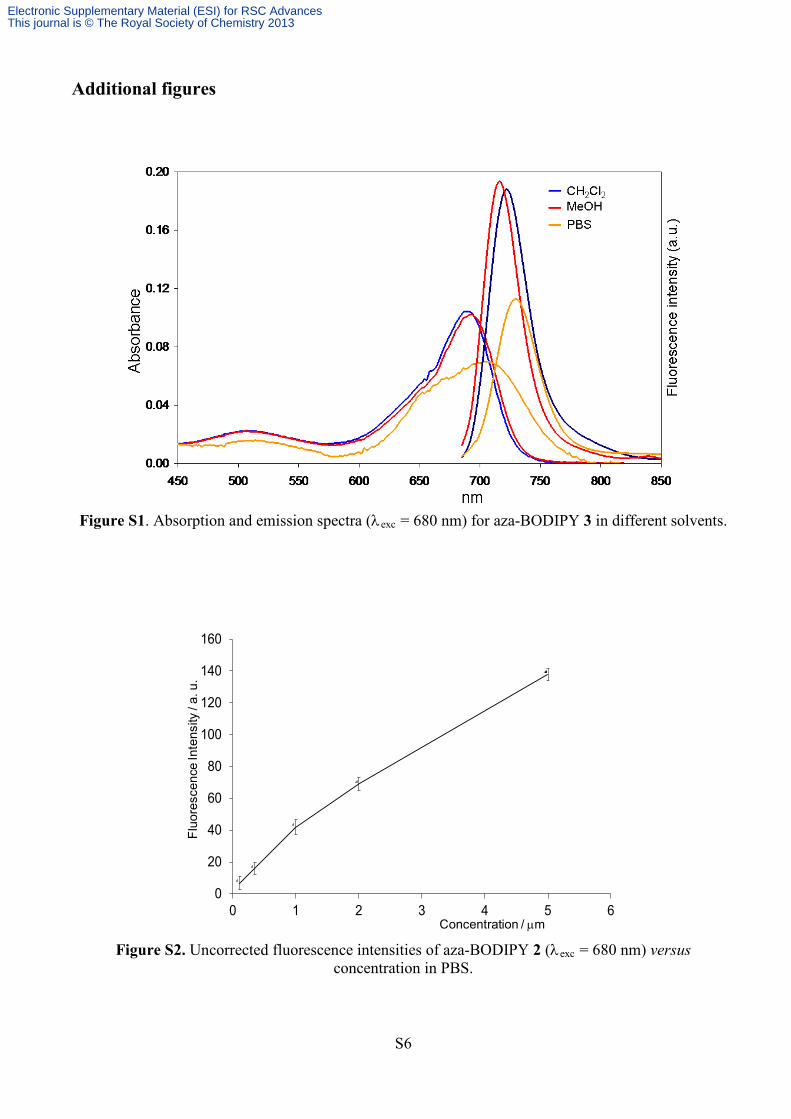
Figure S1. Absorption and emission spectra (λexc = 680 nm) for aza-BODIPY 3 in different solvents.
Figure S2. Uncorrected fluorescence intensities of aza-BODIPY 2 (λexc = 680 nm) versus
concentration in PBS.
Fluo
resc
ence
Inte
nsity
/ a.
u.
Concentration / µm
0
20
40
60
80
100
120
140
160
0 1 2 3 4 5 6
Electronic Supplementary Material (ESI) for RSC AdvancesThis journal is © The Royal Society of Chemistry 2013

S7
Figure S3. Transient absorption spectra of aza-BODIPY 2 following 355 nm laser pulse excitation.
S0 geometry S1 geometry
Figure S4. Ground and excited state geometries for the model aza-BODIPY obtained at the PCM-(TD)-PBE0/6-311G(2d,p) level.
-0.15
-0.1
-0.05
0
0.05
0.1
385 485 585 685
Series1Series2Series3Series4
∆O
D
Wavelength / nm
0.04 µs0.09 µs0.21 µs2.1 µs
Electronic Supplementary Material (ESI) for RSC AdvancesThis journal is © The Royal Society of Chemistry 2013

S8
HOMO LUMO
HOMO-1 HOMO-2
Figure S5. Computed molecular occupied and virtual orbitals for the model aza-BODIPY obtained at the PCM(H2O)-TD-PBE0/6-311G+(2d,p) level using an isosurface value of 0.02.
Figure S6. Plot of change in fluorescence emission of solution of aza-BODIPY 2 in PBS at 720 nm
versus irradiation time (absorbance 0.1, λexc = 680 nm, 8 W, 20 nm/5 nm excitation/emission slit widths).
0
5
10
15
20
25
0 1000 2000 3000 4000
Time / s
Rel
ativ
eflu
ores
cenc
ein
tens
ity/ a
.u.
Electronic Supplementary Material (ESI) for RSC AdvancesThis journal is © The Royal Society of Chemistry 2013

S9
Figure S7. Lower magnification confocal fluorescence images of HeLa cells with
aza-BODIPY 2 (6 µM) following 1 h incubation, washing, and fixation (laser excitation λ = 633 nm).
Figure S8. Confocal fluorescence images of HeLa cells with aza-BODIPY 2 following 1 h incubation
at different concentration, washing, and fixation.
0 M 1,2x10⁻⁴ M 6x10⁻⁵ M 1.2x10⁻⁵ M 6x10⁻⁶ M 1.2x10⁻⁶ M
Electronic Supplementary Material (ESI) for RSC AdvancesThis journal is © The Royal Society of Chemistry 2013

S10
Figure S9. Confocal fluorescence images of HeLa cells with aza-BODIPY 2 at different incubation
times at 6x10⁻⁵M, washing, and fixation.
-ve
2´
10´
20´
30´
40´
50´
60´
Electronic Supplementary Material (ESI) for RSC AdvancesThis journal is © The Royal Society of Chemistry 2013

S11
Figure S10. Average of fluorescence emission intensity obtained from confocal microscopy images
following different aza-BODIPY 2 incubation times (6x10⁻⁵M).
Figure S11. Localized emission spectrum of cytoplasmic HeLa cell fluorescence treated with aza-
BODIPY 2 (ROI1) compared to fluorescence emission in PBS solution of compound 2 and background fluorescence (ROI2).
0
5
10
15
20
25
30
35
0 2 10 20 30 40Incubation time / min
Ave
rage
fluor
esce
nce
inte
nsity
/ a.u
.
negative
Electronic Supplementary Material (ESI) for RSC AdvancesThis journal is © The Royal Society of Chemistry 2013

S12
Table S1. Total energies and singlet-triplet energetic gaps (∆E) for the model aza-BODIPY at PCM(H2O)-TD-UPBE0/6-311+G(2d,p) level.
Electronic state Total energy (hartrees) ∆E (eV) 1A -2077.9907 - 3A -2077.9634 0.74
Geometry: the computed structural parameters in water solution for both ground (S0) and excited state (S1) geometries of the model aza-BODIPY are represented in Table S2. The bond lengths and plane angles slightly change in the excited state S1. The main modifications involve the dihedral angles (φ1, φ2, φ3, φ4) describing the spatial orientation of the phenyl rings relative to the aza-BODIPY core. In the state S1 these angles are closed to 180º and the molecule is then more planar.
1
2
3
8
45
6
7C
NB
N
CN
OCH3H3CO
OCH3H3CO
F F
φ1 φ2
φ3 φ4
Table S2. Selected geometric parameters for PCM(H2O)-(TD)-PBE0 optimized geometric structures of ground (S0) and excited (S1) states of the model aza-BODIPY. S0 S1 bond lengths [Å] B-F 1.392 1.400 B-N 1.554 1.546 N4a-C7a 1.388 1.370 C7a-N8 1.313 1.328 C-CPhenyl 1.455 1.445 CPhenyl-O 1.346 1.344 valence angles [º] B-N4a-C7a 121.9 121.6 F-B-F 110.3 109.6 C7a-N8-C8a 120.4 118.9 torsion angles [º] φ1 149.8 156.7 φ2 149.8 156.7 φ3 156.0 163.1 φ4 156.0 163.1 Table S3. Selected excitation energies (∆E), oscillator strengths (f) and transition coefficients computed, at PCM(H2O)-TD-PBE0/6-311+G(2d,p) level, for the model aza-BODIPY. All electronic states belong to 1A.
Excited state Transition character Weight ∆E (eV) f 1 HOMO→LUMO 0.707 1.91 0.858 2 HOMO-1→LUMO 0.698 2.37 0.015 3 HOMO-2→LUMO 0.702 2.44 0.605
Exp. 2 (in H2O) 1.76
Electronic Supplementary Material (ESI) for RSC AdvancesThis journal is © The Royal Society of Chemistry 2013

S13
1H and 13C NMR spectra
Figure S1. 1H (top) and 13C NMR (bottom) spectra of 6a in CDCl3.
O
O
OCH3
OO
OOH
6a
Electronic Supplementary Material (ESI) for RSC AdvancesThis journal is © The Royal Society of Chemistry 2013

S14
Figure S2. 1H (top) and 13C NMR (bottom) spectra of 6b in CDCl3.
O
O
O
OO
OOH
6b
OO
OOH
Electronic Supplementary Material (ESI) for RSC AdvancesThis journal is © The Royal Society of Chemistry 2013

S15
Figure S3. 1H (top) and 13C NMR (bottom) spectra of 7a in CDCl3.
O
O
OCH3
OO
OOH
7aNO2
Electronic Supplementary Material (ESI) for RSC AdvancesThis journal is © The Royal Society of Chemistry 2013

S16
Figure S4. 1H (top) and 13C NMR (bottom) spectra of 7b in CDCl3.
O
O
O
OO
OOH
7b
OO
OOH
NO2
Electronic Supplementary Material (ESI) for RSC AdvancesThis journal is © The Royal Society of Chemistry 2013

S17
Figure S5. 1H (top) and 13C NMR (bottom) spectra of 1 in CDCl3.
N
N
N
O
H3CO OCH3
BF F
α
γ
1
O
OO
O
OH
OO
O
HO
Electronic Supplementary Material (ESI) for RSC AdvancesThis journal is © The Royal Society of Chemistry 2013

S18
Figure S6. 1H (top) and 13C NMR (bottom) spectra of 2 in CDCl3.
N
N
N
O O
BF F
2OO
OO
O
OH
OO
O
HO
OO
O
OH
O O
O
HO
Electronic Supplementary Material (ESI) for RSC AdvancesThis journal is © The Royal Society of Chemistry 2013

S19
Figure S7. 1H (top) and 13C NMR (bottom) spectra of 3 in CD3OD.
3
N
N
N
O O
O O
BF F
NNNN
NN
NN
N
O
O
OOH
O
O
OHO
O
O
O
HO
NN
NO
O
O
OH
Electronic Supplementary Material (ESI) for RSC AdvancesThis journal is © The Royal Society of Chemistry 2013

S20
References
1 D. F. Eaton, Pure Appl. Chem., 1988, 60, 1107–1114. 2 G. R. Fulmer, A. J. M. Miller, N. H. Sherden, H. E. Gottlieb, A. Nudelman, B. M. Stoltz, J. E.
Bercaw and K. I. Goldberg, Organometallics, 2010, 29, 2176–2179. 3 W. Wang and A. D. Q. Li, Bioconjugate Chem., 2007, 18, 1036–1052. 4 J.-B. Giguère, D. Thibeault, F. Cronier, J.-S. Marois, M. Auger and J.-F. Morin, Tetrahedron
Lett., 2009, 50, 5497–5500. 5 M. Ono, R. Watanabe, H. Kawashima, Y. Cheng, H. Kimura, H. Watanabe, M. Haratake, H.
Saji and M. Nakayama, J. Med. Chem., 2009, 52, 6394–6401. 6 K. Kumar, P. Singh, L. Kremer, Y. Guérardel, C. Biot and V. Kumar, Dalton Trans., 2012, 41,
5778–5781. 7 M. Yuan, X. Yin, H. Zheng, C. Ouyang, Z. Zuo, H. Liu and Y. Li, Chem. Asian J., 2009, 4,
707–713. 8 D. J. Leaver, R. M. Dawson, J. M. White, A. Polyzos and A. B. Hughes, Org. Biomol. Chem.,
2011, 9, 8465–8474. 9 M. J. Frisch, G. W. Trucks, H. B. Schlegel, G. E. Scuseria, M. A. Robb, J. R. Cheeseman, G.
Scalmani, V. Barone, B. Mennucci, G. A. Petersson, H. Nakatsuji, M. Caricato, X. Li, H. P. Hratchian, A. F. Izmaylov, J. Bloino, G. Zheng, J. L. Sonnenberg, M. Hada, M. Ehara, K. Toyota, R. Fukuda, J. Hasegawa, M. Ishida, T. Nakajima, Y. Honda, O. Kitao, H. Nakai, T. Vreven, Jr. J. A. Montgomery, J. E. Peralta, F. Ogliaro, M. Bearpark, J. J. Heyd, E. Brothers, K. N. Kudin, V. N. Staroverov, R. Kobayashi, J. Normand, K. Raghavachari, A. Rendell, J. C. Burant, S. S. Iyengar, J. Tomasi, M. Cossi, N. Rega, J. M. Millam, M. Klene, J. E. Knox, J. B. Cross, V. Bakken, C. Adamo, J. Jaramillo, R. Gomperts, R. E. Stratmann, O. Yazyev, A. J. Austin, R. Cammi, C. Pomelli, J. M. Ochterski, R. L. Martin, K. Morokuma, V. G. Zakrzewski, G. A. Voth, P. Salvador, J. J. Dannenberg, S. Dapprich, A. D. Daniels, O. Farkas, J. B. Foresman, J. V. Ortiz, J. Cioslowski and D. J. Fox, Gaussian 09, revision C.01; Gaussian Inc.: Wallingford CT, 2009.
10 M. Ernzerhof and G. E. Scuseria, J. Chem. Phys., 1999, 110, 5029−5036. 11 C. Adamo and V. Barone, J. Chem. Phys., 1999, 110, 6158−6170. 12 B. Le Guennic, O. Maury and D. Jacquemin, Phys. Chem. Chem. Phys., 2012, 14, 157−164. 13 M. E. Casida, in Time-Dependent Density-Functional Response Theory for Molecules, ed. D. P.
Chong, World Scientific, Singapore, 1995, vol. 1, pp. 155−192. 14 D. Jacquemin, E. A. Perpète, I. Ciofini and C. Adamo, Acc. Chem. Res., 2009, 42, 326−334. 15 J. Tomasi, B. Mennucci and R. Cammi, Chem. Rev., 2005, 105, 2999−3093.
Electronic Supplementary Material (ESI) for RSC AdvancesThis journal is © The Royal Society of Chemistry 2013