peds unit 2 study guide
TRANSCRIPT

Pediatrics – Unit 2 Study Guide
• Respiratory infection
• Described according to anatomic area of involvement
• Upper respiratory tract consists of oronasopharynx, pharynx, larynx, and upper part of trachea
• Lower respiratory tract consists of lower trachea, mainstem bronchi, segmental bronchi, subsegmental bronchioles, terminal bronchioles, and alveoli
• Infections of epiglottis and larynx categorized as croup syndromes
• Often spread from one structure to another due to contiguous nature of mucus membrane lining entire tract
• Account for majority of acute illnesses in children • Infants are nose breathers
• < 6 years old most common breathing pattern = diaphragmatic
• Most infections caused by viruses respiratory syncytial virus (RSV), adenoviruses, parainfluenza viruses, pneumonia
• RSV can cause severe respiratory compromise in infants
• ≤ 5 years old = very susceptible to upper respiratory infections (URIs)
• ≥ 6 years old = bacterial infections
• Etiology influenced by age of child, season, living conditions, and preexisting medical problems
Age Size Resistance Seasonal Variations
• < 3 months = infection rate vs. older children due to protective function of maternal antibodies
• 3 – 6 months = infection rate (maternal antibodies disappear and infant develops own antibodies)
• Viral infection rate remains during toddler and preschool years
• Smaller child = smaller airway
• Diameter of airways is smaller in young children = risk for considerable narrowing from edematous mucus membranes and production of secretions
• Distance between structures within respiratory tract is shorter in young child organisms may move rapidly down respiratory tract = extensive involvement
• Relatively short and open eustachian tube in infants and young children allows pathogens easy access to middle ear
• Deficiencies of immune system place child at risk for infection
• resistance due to malnutrition, anemia, fatigue, and chilling of body
• Allergies, preterm birth, broncho-pulmonary dysplasia (BPD), asthma, history of RSV infection, cardiac anomalies, cystic fibrosis (CF)
• Day care attendance
• Smoke in home
• Most common season for URI = late fall
• Most common respiratory pathogens appear during winter and spring months
• Mycoplasmal infections occur more often in autumn and early winter
• Infection-related asthma occurs more frequently during cold weather
• RSV typically occurs during winter and spring
• Manifestations infants and young children, especially between 6 months – 3 years, react more severely to acute respiratory tract infection vs. older children
Respiratory Sounds Sore Throat Anorexia
• Sounds associated with respiratory disease hoarseness, grunting, stridor, wheezing, cough
• Common feature = cough may be evident only during acute phase and can persist several months after disease
• Auscultation sounds wheezing, crackles, absence of breath sounds
• Frequent complaint of older children
• Young children are unable to describe symptoms may not complain even when highly inflamed
• Child will often refuse to take oral fluids or solids
• Common with most childhood illnesses
• Frequently 1st
evidence of illness
• Persists to a greater or lesser degree throughout febrile stage of illness
• Often extends into convalescence

Vomiting Diarrhea Abdominal Pain
• Typical with small children during illness clue to onset of infection (may precede other signs)
• Usually short-lived but may persist during illness
• Frequent cause of dehydration if fluid intake is impaired
• Usually mild, transient diarrhea but may become severe
• Often accompanies viral respiratory infections
• Frequent cause of dehydration
• Common complaint sometimes indistinguishable from pain of appendicitis
• May be caused by mesenteric lymphadenitis
• Can be caused by muscle spasms from vomiting
Nasal Meningismus Fever
• Small nasal passages of infants easily blocked by mucosal swelling and exudation
• Blockage can interfere with respiration and feeding in infants and may contribute to development of otitis media and sinusitis
• Frequent occurrence of nasal discharge
• Discharge may be thin and watery (rhinorrhea) or thick and purulent depends on type or stage of infection
• Nasal discharge is associated with itching and may irritate upper lip and skin surrounding nose
• Meningeal signs without infection of meninges
• Occurs with abrupt onset of fever
• Signs and symptoms = headache, pain + stiffness in back and neck, Kernig and Brudzinski signs
• Kernig sign flexing patient’s hip 90
o then
extending patient’s knee causes pain
• Brudzinski sign flexing patient’s neck causes flexion of patient’s hips and knees
• Subsides as temperature
• May be absent in newborn infants
• Greatest at ages 6 months – 3 years
• Temperature may reach 103° – 105° F even with mild infections
• Often appears as 1st
sign of infection
• May be listless and irritable or activity
• Tendency to develop high temperatures with infection in certain families
• May precipitate febrile seizures uncommon after 3 – 4 years old
• Nursing interventions
• Monitor for signs of respiratory distress = tachypnea, stridor, coughing, wheezing, retractions, grunting, nasal flaring, accessory muscle usage
• Assess O2 levels pulse oximetry, color (cyanotic, pallor)
• Assess child’s LOC, response to stimuli, and hydration status (insatiable loss from breathing)
Respirations Other Observations
• Rate rapid (tachypnea), normal, or slow for particular child
• Depth normal depth, too shallow (hypopnea), too deep (hyperpnea)
• Ease effortless, labored (dyspnea), orthopnea (difficult breathing except in upright position), associated with intercostal or substernal retractions, flaring nares, head bobbing, grunting, or wheezing
• Labored breathing continuous, intermittent, becoming steadily worse, sudden onset, at rest or on exertion, associated with wheezing or grunting, associated with pain
• Rhythm variation in rate and depth of respirations
• Evidence of infection look for temperature, enlarged cervical lymph nodes, inflamed mucus membranes, purulent discharges from nose, ears, or lungs (sputum)
• Cough under what circumstances cough is heard (e.g., night only, on arising), nature of cough (paroxysmal with or without wheeze, “croupy” or “brassy”), frequency , associated with swallowing or other activity, character of cough (moist and dry), productivity
• Wheeze expiratory or inspiratory, high-pitched or musical, prolonged, slowly progressive or sudden, associated with labored breathing
• Cyanosis distribution (peripheral, perioral, facial, trunk, and face), degree, duration, associated with activity
• Abdominal pain seen with preschooler and school-age children, referred pain from chest
• Chest pain seen with older children, localized or generalized, referred to base of neck or abdomen, dull or sharp, deep or superficial, associated with rapid, shallow respirations or grunting
• Sputum volume, color, viscosity, and odor
• Bad breath may be associated with some lung infections

• Ease respiratory efforts warm or cool mist is a common therapeutic measure for symptomatic relief of respiratory discomfort
• Moisture soothes inflamed membranes and is beneficial with hoarseness or laryngeal involvement
• Cool vaporizers in home is better than warm vaporizers due to risk of burns from steam
• Can also run hot water shower to produce warm mist
• Promote rest acute febrile illness = place on bed rest
• Promote comfort saline nose drops, nasal irrigation, vasoconstrictive nose drops or sprays
• DO NOT administer nose drops or sprays for > 3 days can cause rebound nasal constriction
• Prevent spread of infection use a tissue or hand to cover nose and mouth when coughing or sneezeing, dispose of tissues properly, frequent handwashing, do NOT share cups/washcloths/towels
• temperature acetaminophen (or ibuprofen if child > 6 months) and administer cool liquids
• Promote hydration dehydration is a potential complication when children have respiratory tract infections and are febrile or anorexic, especially when vomiting or diarrhea is present
• Infants are especially prone to fluid and electrolyte with respiratory illness
• Rapid respiratory rate precludes adequate fluid intake
• Fever total body fluid turnover in infants
• Nasal secretions can block narrow nasal passages when infant reclines to feed stops compensatory mouth breathing effort = intake of fluids
• AVOID high-calorie liquids (soda, fruit juice), fluids with caffeine (tea, coffee)
• Offer oral rehydration solutions Infalyte or Pedialyte (for infants) or Gatorade (for older children)
• Breastfeeding infants should continue to be breastfed because human milk confers some degree of protection from infection
• Fluids should NOT be forced
• Assess child's level of hydration by observing frequency of voiding
• Provide nutrition loss of appetite is characteristic of children with acute infections
• Nasopharyngitis upper respiratory tract infection equivalent of “common cold”
• Most common respiratory illness
• Commonly caused by virus rhinovirus, RSV, adenovirus, influenza virus, and parainfluenza virus
• Symptoms are more severe in infants and children vs. adults
• Fever, irritability, restlessness, sneezing, coughing, nasal congestion, anorexia, general malaise
• Treatment managed at home
• Antipyretics for mild fever and discomfort NO aspirin!
• Rest recommended until child is free of fever for at least 1 day
• Decongestants may be used for children > 5 years of age to shrink swollen nasal passages
• Affect ALL vascular beds give with caution to children with diabetes
• DO NOT give oral decongestants to infants < 6 months
• Used for MAXIMUM of 3 days
• Cough suppressants may be prescribed for a dry, hacking cough in older children
• Antihistamines ineffective in treatment of nasopharyngitis
• Prevent spread of infection frequent handwashing, avoid contact with affected persons
• Hydration essential to maintain adequate fluid intake
• Early evidence of respiratory complications
• Earache
• Fever > 38.3° C (101° F)
• irritability, crying
• Respirations > 50 – 60 breaths/min, wheezing
• Persistent cough for ≥ 2 days
• Restlessness and poor sleep patterns

• Tonsillitis inflammation of palantine tonsils (masses of lymphoid tissue located in pharyngeal cavity)
• Tonsils filter and protect respiratory and alimentary tracts from invasion by pathogenic organisms and play a role in antibody formation
• Children generally have much larger tonsils than adolescents or adults
• Proximity to nares and eustachian tubes causes difficulties in instances of inflammation
• Etiology tonsillitis often occurs with pharyngitis
• Can be caused by virus or bacteria
• Manifestations caused by inflammation
• Tonsils and pharynx may be inflamed and covered with exudates
• Palatine tonsils enlarge from edema = obstructs passage = difficulty swallowing and breathing
• Adenoids enlarge = space behind posterior nares becomes blocked = difficult or impossible for air to pass from nose to throat = child must breathe through mouth
• Throat culture should be performed to rule out GABHS (Group A Beta Hemolytic Streptococcus)
• GABHS infection of upper airway (aka strep throat) = risk for rheumatic fever (lead to heart and joing complications) and acute glomerulonephritis (kidney infection)
• Nodes are often tender (due to pharyngitis) pain can be relatively mild to severe enough to make swallowing difficult
• Thick, nasal voice + pain
• Treatment tonsillitis is usually self-limiting, treat symptoms
• Administration of antibiotics, if bacterial in origin children should NOT return to school until they have been taking antibiotics for a full 24 hours
• Oral erythromycin is indicated for children allergic to penicillin
• Azithromycin, clarithromycin, oral cephalosporins, amoxicillin, and amoxicillin with clavulanic acid
• Advise parents that entire dose of antibiotics MUST be completed
• Discard toothbrush and replace it with a new one after taking antibiotics for 24 hours
• Prevent sharing of drinking or eating items to prevent spread of infection
• Warm saline gargles offer relief of throat discomfort
• Tonsillectomy = surgical removal of palatine tonsils
• Indicated for recurrent tonsillitis (2 – 3 occurances in a year), peritonsillar abcess, airway obstruction, sleep apnea, or recurrent ear infections
• Adenoidectomy = surgical removal of adenoids (aka pharengeal tonsils)
• Recommended for hypertrophied adenoids that obstruct nasal breathing, recurrent adenoiditis and sinusitis, otitis media (OM) with effusion, sleep apnea, and recurrent rhinorrhea
• Nursing interventions provide comfort and activites or interventions that precipitate bleeding
• Soft to liquid diet is preferred
• Cool-mist vaporizer keeps mucus membranes moist during periods of mouth breathing
• Opioids may be needed to pain for child to drink
• Preoperative interventions
• Teach what to expect after surgery, preoperative requirements, lab work
• Check for any loose teeth poses aspiration risk if knocked loose during surgery
• Postoperative interventions
• Place child on abdomen or side until fully awake facilitates drainage of secretions
• Careful suctioning only when needed AVOID trauma to oropharynx
• AVOID gargling, coughing, clearing throat, or blowing nose aggravates operative site
• Assure parents that dark brown (old) blood is usually present in emesis, in nose, and between teeth
• Anticipate low grade fever, slight ear pain, and mouth odor for 1st
few days (due to healing process)

• Throat will be sore after surgery provide analgesics at regular intervals (tendency is to undermedicate kids), ice collar, anesthetic pops
• Food and fluids are restricted until children are fully alert and there are no signs of hemorrhage
• fluids = healing (keep child hydrated!)
• Start with liquids the move to soft solids NO citrus, spices, hot food, irritating foods
• AVOID any red or brown colored fluids (need to be able to distinguish fresh or old blood)
• Milk, ice cream, and pudding are usually NOT offered coat mouth and throat with mucus and may cause child to clear throat = can initiate bleeding
• Monitor vital signs look for signs of bleeding and shock
• Observe throat directly for evidence of bleeding first 24 hours after surgery, then again 7 – 10 days after surgery (when scab falls off)
• Most obvious early sign of bleeding = child's continuous swallowing of trickling blood
• Signs of hemorrhage tachycardia, pallor, frequent clearing of throat, vomiting of bright red blood, restlessness, blood pressure (late sign of shock)
• Airway obstruction can occur due to edema or accumulated secretions
• Look for signs of respiratory distress stridor, drooling, restlessness, agitation, respiratory rate, and progressive cyanosis
• Otitis Media inflammation of middle ear
• Incidence is highest in winter months many cases of bacterial OM are preceded by a viral respiratory infection (RSV or influenza)
• Etiology one of most prevalent diseases of early childhood
• Most episodes occur between 6 months – 2 years old
• incidence with age, except for a small at age 5 – 6 years when children enter school
• Occurs infrequently in children > 7 years old
• risk for boys, children in large households, presence of smoke in home, siblings with history of OM
• Can be caused by bacteria or by blocked eustachian tubes from edema, URIs, allergic rhinitis, hypertrophic adenoids
• Relationship with infant feeding methods
• Breast milk = incidence vs. formula-fed infants
• Semivertical feeding position = incidence
• incidence of OM if bottle left in bed with infant
• Pathophysiology primarily a result of bacteria and/or malfunctioning eustachian tubes which causes obstruction
• Diagnosis is made via visual inspection of tympanic membrane
• Serous or purulent discolored fluid line (effusion)
• Membrane is bulging or full, opacified, reddened, immobile and bony landmarks are not visible
Acute Otitis Media Chronic Otitis Media
Definition • Sudden, rapid onset
• Lasts ≤ 3 weeks
• Lasts > 3 weeks
Causative Agent • Strep, RSV, influenza frequently occurs after URIs
• Eustachian tube dysfunction
• Frequently an extension of an acute episode of OM
Manifestations • Irritability, otalgia (earache), high fever
• Pulling at ear, drainage
• Purulent discharge (otorrhea) may or may not be present
• Poor feeding
• Hearing loss (most frequent)
• Difficulty communicating
• Feeling of fullness in ear
• Tinnitus, vertigo

• Treatment most are viral in nature, therefore there is movement away from giving antibiotics
• Broad spectrum antibiotics given amoxicillin, Septra, erythromycins take all doses!
• Should wait up to 72 hours for spontaneous resolution before taking antibiotics
• ALL cases of AOM in infants < 6 months old should be treated with antibiotics due to infant's immature immune system
• NOT recommended for children < 2 years old who have persistent acute symptoms of fever and severe ear pain
• Analgesic-antipyretic (acetaminophen or ibuprofen if > 6 months old), warm compress to ear, lying on affected side to promote drainage
• Antihistamines and decongestants are NOT recommended
• Myringotomy surgical incision of eardrum used to provide continuous drainage of infected middle ear fluid
• Can be done with or without tympanostomy tube placement and/or adenoidectomy
• NOT recommended for children < 3 years old unless there is pathology
• NOT used for initial management of OM!
• Can cause child to hear much better, but sounds can be very loud talk in muted voice so child is not scared
• Child will have low-grade fever (< 100.4o F) for 24 – 48 hours postoperatively
• Advise parents of grommet appearance (usually a tiny, white, plastic spool-shaped tube) so they can recognize it if it falls out normal, requires no immediate intervention (but should still advise HCP)
• Croup syndrome croup = general term used for symptom complex that can affect larynx, trachea, bronchi, and epiglottis
• Characterized by
• Hoarseness
• Resonant cough described as “barking” or “brassy” (croupy)
• Varying degrees of inspiratory stridor
• Varying degrees of respiratory distress resulting from swelling or obstruction in larynx
• Acute infections of larynx in infants and small children = risk for significant narrowing with inflammation
• Laryngotracheobronchitis and epiglottitis DO NOT occur together
Epiglottitis Laryngotracheobronchitis
Definition • Serious obstructive inflammation of epiglottis that covers airway
• Medical emergency requires IMMEDIATE attention!
• Obstruction is supraglottic
• Most common croup syndrome
• Usually preceded by a URI gradually descends to adjacent structures
• Typically occurs and/or worsens during night
• Subglottic obstruction of laryngitis

Epiglottitis Laryngotracheobronchitis
Age Group • 2 – 8 yr • < 2 yr (boys > girls)
Etiology • Usually H. influenzae
• Bacterial or viral
• Parainfluenza virus, RSV, Influenza A and Bi
• Mycoplasma pneumonia (microbacteria)
Onset • Abrupt
• Can rapidly progress to severe respiratory distress
• Gradual onset
• Slowly progressive
Signs & Symptoms
• NO spontaneous cough + NO hoarseness
• High fever (> 102o F) + toxic appearance
• Restlessness + irritability
• Thick, muffled voice + frog-like croaking sound on inspiration (stridor when supine)
• Drooling + red and inflamed throat + distinctive large, cherry-red, edematous epiglottis + pain on swallowing (dysphagia)
• Tripod position child insists of sitting upright and leans forward with chin thrust out (to expand neck), mouth open, tongue protruding
• Barky, brassy cough (seal-like) + hoarseness
• Low-grade fever (< 100.5o F) + does NOT look toxic
• Restlessness + irritability
• Inspiratory stridor problem getting air IN vs. wheezing, which is problem getting air OUT
• Suprasternal retractions due to narrowing of airway from inflamed mucosa (dyspnea)
• Stage 1 (hoarseness, croupy cough, some agitation) can be treated at home need to treat in hospital if advances to stage 2 – 4
Complications • Respiratory obstruction can appear suddenly
• Hypoxia, hypercapnia, acidosis followed by muscle tone
• level of consciousness
• Sudden death
• Respiratory distress nasal flaring, intercostal retractions, tachypnea, and continuous stridor (stage 2)
• Hypoxia, anoxia, hypercapnia, acidosis (stage 3)
• Cyanosis, respiratory failure (stage 4)
Treatment • Keep child quiet, calm, and comfortable avoid invasive procedures
• Keep child NPO
• DO NOT inspect throat or stick anything into mouth if child is suspected to have epiglottisis! can cause complete airway obstruction (have intubation tray at bedside)
• DO NOT take child to x-ray
• Fluids + humidified O2 (cool mist)
• Monitor airway pulse ox, respiratory status
• Antibiotic therapy (if bacterial) 7 – 10 days
• High dose corticosteroids used to edema PO or nebulized (racemic) epinephrine
• Keep child calm
• Elevate head of bed ( breathing ability), maintain airway, and provide adequate respiratory exchange
• Continuous, vigilant observation + accurate assessment of respiratory status
• High humidity + cool mist
• Nebulized (racemic) epinephrine upper airway bronchodilator used for severe disease, stridor at rest, retractions, or difficulty breathing
• Corticosteroids dexamethasone
• Fluids cool, clear liquids
• Educate parents on how to make child more comfortable
• Bronchiolitis most common acute viral infection that mainly affects bronchiolar level (lower airways)
• Etiology most cases caused by respiratory syncytial virus (RSV) also caused by adenoviruses and parainfluenza
• Primarily occurs in late fall through spring
• Spread by hand – nose contact
• Mostly occurs in children < 2 years old rare in children > 2 years old
• Peak age = 6 months
• By age 3 years most children have been infected at least once with RSV severe RSV infections in 1
st year of life = risk for development of asthma up to age 13
• Pathophysiology bronchiolar mucosa swells = lumina filled with mucus and exudate
• Obstruction in small air passages = hyperinflation, obstructive emphysema, and patchy areas of atelectasis
• Dilation of bronchial passages on inspiration allows for adequate intake of air (can breathe in)
• Narrowing of passages on expiration prevents air from leaving lungs air is trapped distal to obstruction = causes progressive overinflation (emphysema)

• Manifestations usually begins with a URI
• Infants can have URI symptoms, slight lethargy, poor feeding, or irritability apena may be 1st
recognized indicator of RSV infection in very young infants
Initial Symptoms Progression of Illness Severe Illness
• Rhinorrhea (thick clear to white discharge)
• Pharyngitis
• Productive cough + sneezing
• Wheezing (work of breathing)
• Intermittent low-grade fever (< 100.5o F)
• Possible otitis media and conjunctivitis can cause fever > 100.5
o F
• coughing and wheezing
• Tachypnea, dyspnea
• Retractions
• Crackles
• Cyanosis
• Tachypnea > 70 breaths/min
• Listlessness
• Apneic spells
• Poor air exchange
• breath sounds
• Diagnosis tests done on nasal or nasopharyngeal secretions
• ELISA techniques used for RSV antigen detection
• Treatment manged at home with cool humidified O2, adequate fluid intake, airway maintenance via saline nose drops + nasal suction, rest, and medications
• Hospitalization usually recommended for children:
• ≤ 6 months old due to being obligate nose breathers (can cause apnea)
• With respiratory distress
• Who cannot maintain adequate hydration
• Have underlying lung or heart disease
• Whose home environment cannot adequately manage disease (exhausted parent)
• Medical therapy for bronchiolitis is primarily supportive aimed at airway hyperresonance and inflammation and adequate fluid intake
• Bronchodilators may provide short-term benefits
• Corticosteroids and antihistamines NOT recommended for routine use
• Antibiotics are NOT part of treatment of RSV unless there is a coexisting bacterial infection (otitis media, conjunctivitis) that causes temperature > 100.5
o F
• If respirations > 60 breaths/min child is kept NPO due to risk for aspiration
• Ribavirin only antiviral agent approved for hospitalized children
• Drug is aerosolized and delivered via a small-particle aerosol generator
• Administered by mist tent for 18 hours/day for 7 – 10 days
• Teratogenic drug no pregnant women can be around it
• Prevention of RSV infection
• RSV is very contagious contact and standard precautions are used
• Frequent handwashing
• NO touching nasal mucosa or conjunctiva
• Use gloves and gowns when entering patient's room
• Make patient assignments so that nurses assigned to children with RSV are NOT caring for other patients who are considered high risk
• Children suspected RSV infection may be assigned separate rooms or grouped with other RSV-infected children
• Prophylactic medications only given to children who are high risk (cardiac, premies, immunocompromised)
• RSV-IG IV med given monthly for 8 months
• Synagis IM med given monthly for 8 months

• Asthma chronic inflammatory disorder of airways
• Most common chronic disease in children
• Etiology allergy and viral respiratory infections (RSV in infants) influence persistence and severity of disease
• Boys are affected more frequently than girls until adolescence, when trend reverses
• 1st
asthma attack frequently occurs < 5 years old
• Pathophysiology asthma results from complex interactions mediated by mast cells, eosinophils, histamine, and T lymphocytes present in airways
• Exaggerated inflammatory response to stimuli (hyper-responsiveness of airway)
• Allergens and environmental factors
• Physical, chemical, pharmacological agents
• Infection, URIs
• Stress psychsocial factors
• Exercise
• Hereditary runs in families, but NOT considered a genetic disease
• Hyper-responsiveness of airways = bronchospasm shortening and narrowing of airway
• Spasm of smooth muscle of bronchi and bronchioles = ↓ caliber of bronchioles
• Inflammation = airway edema + secretion of mucus
• Air and mucus becomes trapped in distended aveoli
• Hyperinflation of alveoli = ↓ effect of cough
• ↑ respiratory effort during expiratory phase of respiration (wheeze)
• Trapped O2 takes up lung space actelectasis causes no sound
• Person fights to inspire sufficient air due to trapped air
• Causes fatigue, ↓ respiratory effectiveness, and ↑ O2 consumption
• ↓ alveolar ventilation = ↑ CO2 retention, hypoxemia, respiratory acidosis, and eventually respiratory failure
• Chronic inflammation may cause permanent damage to airway structures airway remodeling
• Classified into 4 categories based on symptom indicators of disease severity
• Provide a stepwise approach to pharmacologic management, environmental control, and educational interventions needed for each category
Intermittent Mild Persistent Moderate Persistent Severe Persistent
Frequency of Day Symptoms
• < 2 days/week • > 2 days/week
• NOT daily
• Daily • Continual symptoms despite taking meds
Frequency of Night Symptoms
• < 2 times/month • 3 – 4 times/month • 2 – 3 times/week
• NOT nightly
• Frequent
• Nightly
Interference with Normal Activity
• None • Minor limitation • Some limitation • Extremely limited (due to ↓ pulmonary function)
Use of Meds for Symptom Control
• < 2 days/week • > 2 days/week
• NOT daily
• Daily • Multiple times/day

• Manifestations main symptoms are dyspnea, wheezing, and coughing
Cough Respiratory-Related Signs Chest With Repeated Episodes
• Initially dry, hacking, nonproductive
• Paroxysmal attack comes on suddenly
• Irritative
• Becomes rattling
• Can eventually produce frothy, clear, gelatinous sputum
• Shortness of breath
• expiratory phase (takes longer to breath out) with audible wheeze
• Malar flush, red ears, with lips deep dark red color (due to hypoxia)
• Possible progression to cyanosis of nail beds or circumoral cyanosis
• Restlessness, apprehension
• sweating as attack progresses
• May sit in tripod position upright with shoulders in a hunched-over position, hands on bed or chair, and arms braced
• Speak in short, panting, broken phrases
• Hyperresonance on percussion (hollow drum)
• Coarse, loud breath sounds
• Bilateral wheezes throughout lung fields
• Prolonged expiration
• Crackles (with URI pneumonia)
• Retractions
• Barrel chest AP diameter ≥ transverse diameter
• Elevated shoulders
• Use of accessory muscles of respiration (causes stomache to hurt)
• Facial appearance: flattened malar bones, circles beneath eyes, narrow nose, big upper teeth
• Retractions
Mild Distress Moderate Distress Severe Distress
• Isolated intercostal
• Anything that starts with an “S”
• Subcostal, suprasternal, and supraclavicular
• Retractions + use of accessory muscles
• Diagnosis determined primarily on basis of clinical manifestations, history, physical examination, and some labs
• Chronic cough in absence of infection or diffuse wheezing during expiratory phase of respiration is sufficient to establish a diagnosis
• Chest x-rays show infiltrated and hyperexpansion of airways, pneumonias or respiratory tract infections
• Pulmonary function tests (PFTs) evaluates presence and degree of impact on lung fields
• Peak expiratory flow rate (PEFR) measures maximum flow of air that can be forcefully exhaled in 1 second
• Treatment continuous care approach with regular visits (at least every 1 – 6 months) to HCP
• Early recognition of symptoms is crucial
• Overall goals of asthma management:
• Maintain normal activity levels and pulmonary function
• Prevent chronic symptoms and recurrent exacerbations avoid triggers and allergens + use meds
• Provide optimal drug therapy with minimal or no adverse effect used to ↓ underlying inflammation and ↓ or prevent symptomatic airway narrowing
• Patient and family education + monitoring severity of disease = correct course of therapy
• Explain pathophysiology to parents used of steroid + drink fluids = inflammation and mucus
• Medications used to prevent and control asthma symptoms, ↓ frequency and severity of asthma exacerbations, and reverse airflow obstruction
• Therapy is directed toward long-term suppression of inflammation
• Administered as PO meds or by inhalation with a nebulizer or a metered-dose inhaler (MDI)
• MDI (method of choice for kids) should always be attached to a spacer when:
• An inhaled corticosteroid is given to prevent yeast infections in mouth
• Children have difficulty coordinating or learning proper inhalation technique

• 2 general classes often used in combination
Preventive Medications Rescue Medications
• Long-term control medications
• Used to achieve and maintain control of inflammation
• Inhaled corticosteroids, cromolyn sodium and nedocromil, long-acting β2-agonists, methylxanthines, and leukotriene modifiers
• Quick-relief medications
• Used to treat symptoms and exacerbations
• Short-acting β2-agonists, anticholinergics, and systemic corticosteroids
Drug Indication Nursing Interventions
Short Acting β-Adrenergic Agonists
• Albuterol
• Epinephrine
• levalbuterol (Xopenex)
• Terbutaline
• 1st
line drug used for treatment of acute exacerbations and prevention of exercise induced asthma
• Binds with β2-receptors to relax bronchial smooth muscle, dilate airways, and eliminate bronchospasm
• β1-effects ( heart rate and GI disturbances) have been minimized
• Administered PO or via inhalation (MDI or aerosol) inhaled has more rapid onset of action (within minutes)
• Inhaled β-adrenergic agents should NOT be taken > 3 – 4 times/day for acute symptoms
• Side effects irritability, tremor, nervousness, insomnia, tachycardia (except with Xopenex)
Long Acting β-Adrenergic Agonists
• salmeterol (Serevent)
• Bronchodilator used for long-term prevention of symptoms especially nighttime symptoms and exercise-induced bronchospasm
• Typically added to antiinflammatory therapy
• Used twice daily (q12h)
• NOT used in children < 12 years old
• NOT used for acute symptoms or exacerbations
Corticosteroids
• budesonide
• fluticasone
• Antiinflammatory drug
• 2nd
line drug used to control chronic or acute inflammatory process
• Treats reversible airflow obstruction and bronchial hyper-responsiveness
• Administered IV, PO, or by inhalation takes about 2 – 6 hours for onset
• Given in “bursts” over several days
• Frequently monitor growth of children and adolescents taking corticosteroids to assess systemic effects of these drugs
Nonsteroidal Anti-Inflammatory Drug (NSAID)
• cromolyn sodium (Intal)
• nedocromil sodium (Tilade)
• Inhibits mast cells + inhibits activation and release of mediators from eosinophil and epithelial cells
• Inhibits bronchoconstrictor response to inhaled antigens
• Used as daily maintenance therapy
• Administered via nebulizer or MDI
• Minimal side effects occasional coughing from inhalation of powder
• NOT effective for acute exacerbation long onset of action time
• NOT used in children < 5 years old
CNS Stimulant
• theophylline
• Bronchodilator + CNS stimulant
• Mainly used for adult COPD treatments
• NOT used as mainline management neurotoxic and can cause hypertension
• Used primarily in ER when child is not responding to maximal therapy
• MUST obtain therapeutic levels with this drug has a narrow therapeutic window
Leukotriene Modifiers
• zafirlukast (Accolate)
• montelukast sodium (Singulair)
• Mediate inflammation that cause in airway hyper-responsiveness
• Block inflammation and bronchospasms
• Used for long-term control and prevention of mild persistant asthma
• Administered in combination with β-agonists and steroids
• NOT used to treat acute episodes
• Montelukast for ≥ 12 months old
• Zafirlukast for children ≥ 7 years old
Anticholinergics
• Atropine
• ipratropium (Atrovent)
• Used for relief of acute bronchospasm
• Significantly improves lung function
• Side effects drying of respiratory secretions, blurred vision, and cardiac + CNS stimulation (except for ipratropium)

• Exercise induced bronchospasm (EIB) acute, reversible, usually self-terminating airway obstruction that develops during or after vigorous activity
• Reaches its peak 5 – 10 minutes after stopping activity
• Episode typically ends after another 20 – 30 minutes
• Manifestations cough, shortness of breath, chest pain, tightness, wheezing, endurance problems during exercise
• Does NOT usually occur during activities that require short bursts of energy baseball, sprints, gymnastics
• More common during activities that involve endurance exercise soccer, basketball, distance running
• Swimming is well tolerated by children with EIB breathing air fully saturated with moisture
• Diagnosis requires exercise challenge test in a laboratory setting
• Status asthmaticus continued respiratory distress despite vigorous therapeutic measures
• Medical emergency can result in respiratory failure and death
• Rapid onset + immediate progression
• Often coincides with complicating conditions pneumonia, respiratory virus, exposure to triggers
• Treatment usually requires hospitalization
• Most important treatment goals
• ventilation
• airway resistance
• Relieving bronchspasm
• Correcting dehydration and acidosis
• child and parent anxiety
• Treating any concurrent infection
• Place child in position of comfort = high Fowler’s expands airway
• Humidified O2 administed to maintain O2 saturation > 90%
• Inhaled aerosolized short-acting β2-agonists (albuterol)
• Systemic corticosteroid used to effects of inflammation
• IV flids for hydration and to administer medications
• Can also give IV magnesium sulfate potent muscle relaxant that acts to inflammation and pulmonary function and peak flow rate
• Antibiotics should NOT be used to treat acute asthma attacks EXCEPT with an underlying bacterial infection
• Cystic Fibrosis genetic disease that causes viscosity of exocrine (mucus producing) gland secretions
• Autosomal recessive trait affected child inherits defective gene from both parents
• Progressive and incurable disease
• Pathophysiology affects multiple organ systems
• of sweat electrolytes (Na+ and Cl
-)
• Chloride channel impeded
• Salty taste on skin
• Mucus glands produce a thick mucoprotein that accumulates and causes gland to dilate
• viscosity of secretions = mechanical obstruction in organs
• Obstruction = secretions precipitate or coagulate to form concretions in glands and ducts
• Affects organ defined by mucus production
• Pulmonary = lungs
• Gastrointestinal = liver, pancreas, intestines, saliva, stomach
• Sexual organs
• Develop abnormalities in autonomic nervous system function

• Manifestations
Meconium Ileus Gastrointestinal Tract Pulmonary Function
• Small intestine blocked with meconium = earliest postnatal manifestation in newborn
• Abdominal distention
• Vomiting
• Failure to pass stools
• Rapid development of dehydration
• Large, bulky, loose, frothy, extremely foul-smelling stools
• Abdominal distention
• Weight loss, failure to grow
• Marked tissue wasting, thin extremities
• Sallow skin, anemia
• Deficiency of fat-soluble vitamins (A, D, E, and K)
• Wheezing + dry, nonproductive cough (initially)
• dyspnea, paroxysmal cough
• Obstructive emphysema and patchy areas of atelectasis
• Overinflated, barrel-shaped chest
• Hypoxia, cyanosis, hypercapnia
• Clubbed fingers
• Repeated episodes of bronchitis and bronchopneumonia
• Possible complications as disease progresses
• Pancreas thick secretions block ducts = causes pancreatic fibrosis
• Prevents essential pancreatic enzymes from reaching duodenum = digestion and absorption of nutrients
• Formation of bulky stools that are frothy from undigested fat (steatorrhea) and foul smelling from putrefied protein (azotorrhea)
• incidence of insulin dependent cystic fibrosis–related diabetes (CFRD)
• Liver biliary obstruction and fibrosis
• Rectum prolapse occurs in infancy and childhood
• Related to large, bulky stools, malnutrition, and intra-abdominal pressure secondary to paroxysmal cough
• Intestinal obstruction from inspissated or impacted feces
• Pulmonary destruction of lung tissue and respiratory failure
• Secretions are difficult to expectorate gradually obstruct bronchi and bronchioles
• Causes scattered areas of bronchiectasis, atelectasis, and hyperinflation
• Stagnant mucus offers a favorable environment for bacterial growth = risk for passing infection to other CF children
• Most children show respiratory symptoms before 1 year old
• Reproductive systems inhibited fertility or sterility
• Growth and development restricted as a result of absorption of nutrients, including vitamins and fat
• Diagnosis family history, absence of pancreatic enzymes, chest radiography (shows patchy atelectasis and obstructive emphysema), chronic pulmonary involvement
• Can also be diagnosied via newborn screening, DNA identification of mutant genes, and abnormal nasal potential difference measurement
• Gold standard = positive sweat chloride test
• Chloride concentration > 60 mEq/L is diagnostic of CF (normal = 35 – 40 mEq/L)

• Therapeutic management requires multidisciplinary approach to treatment
• Goals of CF therapeutic management:
• Prevent or pulmonary complications
• Ensure adequate nutrition for growth
• Encourage appropriate physical activity
• Promote a reasonable quality of life for child and family
Manage Pulmonary Problems Manage Gastrointestinal Problems Manage Endocrine Problems
• Prevention and treatment of pulmonary infection remove mucopurulent secretions, ventilation, antibiotics
• Recurrent pulmonary infections = damage to small airways = bronchiectasis
• Daily routine of chest physical therapy (CPT) to maintain pulmonary hygiene
• Medications bronchodilators, expectorants, mucolytics
• Most children will have central venous line
• O2 administration for acute episodes
• CO2 retention in CF patients can lead to bronchial cysts and emphysema if cysts rupture = pneumothroax (tachypnea, tachycardia, dyspnea, pallor, cyanosis)
• Administration of fat-soluble vitamins (A, D, E, and K) + multivitamin + pancreatic enzymes with meals and snacks
• Well-balanced, high-protein, high-caloric diet due to intestinal absorption
• 3 daily meals + 3 snacks recommended to meet energy and growth requirements
• Experience frequent anorexia nighttime gastrostomy feedings or parenteral alimentation to supplement
• salt intake, especially in during activities and in hot weather
• Monitor growth treatments working if child still has stools (but no steatorrhea) and child is growing
• Management of CFRD is critical
• Monitor blood glucose
• Administer oral glucose-lowering agents or insulin injections
• Diet and exercise management
• Ketoacidosis rare with CFRD due to no fat absorption
• Assessment of bone health to detect and prevent osteoporosis or osteopenia
• Nursing interventions most patients with CF require hospitalization only for treatment of pulmonary infection, uncontrolled diabetes, or a coexisting medical problem
• Standard precautions with meticulous handwashing should be implemented to nosocomial spread of organisms to CF patient and between hospitalized CF patients
• Evaluate effectiveness of aerosol therapy, chest percussion therapy, and postural drainage
• CPT should NOT be performed before or immediately after meals
• Provide supplemental O2 therapy and evaluate effectiveness pulse oximetry, observation of respiratory pattern, work of breathing, and lung auscultation
• Educate parents in breathing exercises and techniques for removal of mucus
• Cardiovascular disorders
• Divided into 2 major groups:
Congenital Heart Disease (CHD) Acquired Heart Disorders
• Anatomic abnormalities that result in abnormal cardiac function
• Present at birth
• Clinical consequences = congestive heart failure (CHF) and hypoxemia
• Result from infection, autoimmune responses, environmental factors, and familial tendencies
• Disease processes or abnormalities that occur AFTER birth
• Can be seen in normal heart or in presence of congenital heart defects
• History and physical examination
• Ask details about mother's health history, pregnancy, and birth history + family health history
• Chronic maternal health conditions (diabetes type 1, lupus)
• Exposure to infections (rubella)
• Medication use during pregnancy (seizure meds) that could be teratogenic to fetus
• Maternal alcohol or illicit drug use
• Maternal age > 40 years old
• Low birth weight = risk for CHD
• High birth weight = risk for heart disease
• incidence of congenital cardiac defects if either parent or a sibling has a heart defect
• Congenital heart defects occur with chromosomal defects Down and Turner's syndromes
• Genetic predisposition to cardiomyopathies

• Observations during physical examination
• Nutritional state failure to thrive, poor weight gain, poor feeding
• Skin/Color cyanosis, pallor (due to poor perfusion), sweating (especially unusual in infants)
• Chest deformities enlarged heart can distort chest configuration
• Unusual pulsations visible pulsations of neck veins seen in some patients
• Respiratory excursion tachypnea, dyspnea, expiratory grunt, frequent URIs, shortness of breath
• Clubbing of fingers associated with cyanosis
• Abdomen hepatomegaly or splenomegaly
• Peripheral pulses discrepancies in rate, regularity, and amplitude (strength)
• Heart tachycardia, bradycardia, irregular rhythms, murmurs, and additional heart sounds
• Diagnosis
• Chest x-ray provides information on heart size and pulmonary blood flow patterns
• EKG graphic measure of electrical activity of heart, heart rate, and rhythm
• Echocardiogram produces image of cardiac structures
• Cardiac catheterization radiopaque catheters placed in a peripheral blood vessel and advanced into heart to measure pressures and O2 levels in heart chambers and visualize heart structures and blood flow patterns
• Potential complications = cardiac irregularities, venous spasm, hemorrhage
Preprocedural Care Postprocedural Care
• Accurate height and weight determines correct size and length of catheter
• History of allergic reactions to iodine betadine, shellfish
• Assess for signs and symptoms of infection
• Assess diaper area diaper rash risk for infection if femoral access is required
• Assess, mark, and document pedal pulses (dorasalis pedis, posterior tibial) for later comparison after procedure
• Record baseline O2 levels
• Educate child and family on procedure (based on developmental level)
• Child is kept NPO for 4 – 6 hours if sedated
• Vital signs taken every 15 minutes blood pressure, heart rate (for 1 FULL MINUTE), temperature, respirations, O2 levels (pulse ox)
• Compare pulses distal to catheterization site with baseline values
• Assess temperature and color of affected extremity coolness or blanching may indicate arterial obstruction
• Monitor dressing site for signs of bleeding
• Monitor fluid intake to ensure hydration
• Immobilize and straighten affected extremity
• Child is encouraged to void to clear contrast material from blood
• Pain control acetaminophen or ibuprofen
• Blood flow during fetal life blood carrying O2 from placenta enters fetal system through large umbilical vein
1) Blood travels to inferior vena cava and enters heart through right atrium
2) pressure of blood entering right atrium = blood directed posteriorly in a straight pathway across right atrium and through foramen ovale to left atrium
3) Better-oxygenated blood enters left atrium and ventricle to be pumped through aorta to head and upper extremities
4) Blood from head and upper extremities enters right atrium from superior vena cava is directed downward through tricuspid valve into right ventricle
5) Blood is pumped through pulmonary artery, where major portion is shunted to descending aorta via ductus arteriosus bypasses fetal pulmonary system (only a small amount of blood flows to and from nonfunctioning fetal lungs)

• Changes to blood flow after birth cessation of placental blood flow from clamping of umbilical cord and expansion of lungs at birth causes hemodynamics of fetal vascular system to undergo pronounced and abrupt changes
• With 1st
breath lungs are expanded, and O2 causes pulmonary vasodilation
• Pulmonary pressures start to as systemic pressures start to
• Foramen ovale closes as pressure in left atrium > pressure in right atrium
• Ductus arteriosus starts to close in presence of O2 concentration in blood and other factors
• Blood flows in response to pumping action of heart:
• From area of pressure to area of pressure L side pressure > R side pressure
• Toward path of least resistance pulmonary system resistance < systemic circulation
• pressure gradient = rate of flow
• resistance = rate of flow
• Abnormal connection between heart chambers (septal defect) causes blood to flow from an area of pressure (left side) to one of pressure (right side) = left-to-right shunt
• Anomalies resulting in cyanosis may result from a change in pressure blood is shunted from right to left side of heart (right-to-left shunt)
• Congenital heart disease (CHD) incidence of CHD in children is approximately 5 – 8 per 1000 live births major cause of death (other than prematurity) in 1
st year of life
• Most common heart anomaly = ventricular septal defect (VSD)
• Main consequences of CHD = congestive heart failure (CHF), hypoxia ( O2), polycythemia ( RBCs)
• Categories based on hemodynamic characteristics (blood flow patterns within heart):
Acyanotic Cyanotic
Left-to-right shunt = pulmonary blood flow
Obstruction to blood flow out of ventricles
Pulmonary blood flow
Mixed blood flow
CHF
Atrial septal defectVentrical septal defect
Patent ductus arteriosusAtrioventricular canal
Left side obstruction = CHF
Right side obstruction = Cyanosis
Coarctation of aortaAortic stenosis
Pulmonic stenosis
Tetralogy of FallotTricuspid atresia
Transposition of great arteriesTotal anomalous pulmonary venous return
Truncus arteriosusHypoplastic left heart syndrome

• Defects with pulmonary blood flow blood flows from pressure LEFT side of heart to pressure RIGHT side of heart
• Caused by abnormal connection between septum wall of heart or abnormal connection between great arteries
• blood volume on right side of heart = pulmonary blood flow at expense of systemic blood flow ()
• Patients show signs and symptoms of CHF
• Atrial septal defect (ASD) abnormal opening between atria
• Pathophysiology blood flows from pressure LEFT atrium into pressure RIGHT atrium = flow of O2 blood into right side of heart
• There is right atrial and ventricular enlargement, but cardiac failure is unusual in an uncomplicated ASD
• Manifestations least symptomatic
• Characteristic systolic murmur fixed split second heart sound
• At risk for atrial dysrhythmias due to right atrial enlargement and stretching of conduction fibers to cause electrical system changes
• Complications pulmonary vascular obstructive disease and emboli formation later in life from chronically pulmonary blood flow
• Treatment surgical patch closure or closure with a device during cardiac catheterization
• Ventricular septal defect (VSD) Abnormal opening between right and left ventricles
• Frequently associated with other defects pulmonary stenosis, transposition of great vessels, patent ductus arteriosus (PDA), atrial defects, and coarctation of aorta
• Many VSDs close spontaneously during 1st
year of life
• Pathophysiology blood flows through defect into pulmonary artery (area of least resistance) = blood volume pumped into lungs = pulmonary vascular resistance
• Left-to-right shunting + pulmonary resistance + back up of blood at vena cava = pressure in right ventricle
• Right side works harder to push blood = right ventricle hypertrophy can lead to right sided heart failure
• Manifestations CHF is common
• Characteristic systolic murmur loud, booming sound
• Complications at risk for bacterial endocarditis and pulmonary vascular obstructive disease
• Treatment pulmonary artery banding (palliative), closing of defect with sutures or patch (complete repair), or device closure during cardiac catheterization
• Patent ductus arteriosus (PDA) failure of fetal ductus arteriosus (artery connecting aorta and pulmonary artery) to close within 1
st weeks of life
(most common in premies)
• Pathophysiology systemic pressure > pulmonary pressure = blood shunts from pressure aorta across duct to pressure pulmonary artery = left-to-right shunt
• Additional blood is recirculated through lungs returned to left atrium and left ventricle
• amount of blood flow to pulmonary system = pulmonary resistance + vascular congestion
• workload on left side of heart and right ventricular pressure and hypertrophy
• Manifestions may be asymptomatic or show signs of CHF
• Characteristic machinery-like murmur
• pulse pressure + bounding pulses due to runoff of blood from aorta to pulmonary artery

• Complications at risk for bacterial endocarditis and pulmonary vascular obstructive disease in later life from chronic excessive pulmonary blood flow
• Treatment administration of indomethacin (prostaglandin inhibitor), surgical division or ligation of patent vessel, or placement of coils to occlude PDA via cardiac catherization
• Obstructive defects blood exiting heart meets an area of anatomic narrowing (stenosis) = obstruction to blood flow
• ↑ pressure in ventricle and great artery before obstruction + ↓ pressure in area after obstruction = ↑ pressure load on ventricle + ↓ cardiac output
• Coarctation of aorta localized narrowing near insertion of ductus arteriosus (right after great vessels)
• Pathophysiology effect of a narrowing within aorta = ↑ blood pressure proximal to defect (head and upper extremities) and ↓ blood pressure distal to obstruction (body and lower extremities)
• Manifestations dizziness, headaches, fainting, and epistaxis resulting from hypertension
• High blood pressure + bounding pulses in arms
• Weak or absent femoral pulses
• Lower blood pressure + weak pulses in lower extremities
• Complications at risk for hypertension, ruptured aorta, aortic aneurysm, and stroke
• Treatment surgical repair by resection of coarctated portion with an end-to-end anastomosis of aorta, enlargement of constricted section using a graft, or balloon angioplasty
• Aortic stenosis narrowing or stricture of aortic valve
• Causes resistance to blood flow in LEFT ventricle, ↓ cardiac output, LEFT ventricular hypertrophy, and pulmonary edema
• Left ventricular wall hypertrophy leads to pulmonary venous and pulmonary arterial hypertension
• Obstruction tends to be progressive and can cause sudden episodes of myocardial ischemia
• Pathophysiology stricture in aortic outflow tract = ↑ resistance to ejection of blood from left ventricle
• Extra workload on left ventricle causes hypertrophy
• Left ventricular failure = ↑ left atrial pressure = ↑ pressure in pulmonary veins = pulmonary vascular congestion (pulmonary edema)
• Manifestations faint pulses, ↓ cardiac output, hypotension, tachycardia, poor feeding, exercise intolerance, chest pain, and dizziness
• Complications at risk for bacterial endocarditis, coronary insufficiency, and ventricular dysfunction
• Treatment balloon dilation, aortic valve replacement
• Pulmonic stenosis narrowing at entrance to pulmonary artery
• Pathophysiology resistance to blood flow = RIGHT ventricular hypertrophy + ↓ pulmonary blood flow
• Right ventricular failure = ↑ right atrial pressure = reopening of foramen ovale
• Causes shunting of unoxygenated blood into left atrium + systemic cyanosis
• Manifestations cyanosis, CHF, cardiomegaly
• Loud systolic ejection murmur
• Complications at risk for bacterial endocarditis
• Treatment balloon angioplasty, pulmonary valvotomy

• Defects with ↓ pulmonary blood flow obstruction of pulmonary blood flow + an anatomic defect (ASD or VSD) between right and left sides of heart
• Blood has difficulty exiting right side of heart via pulmonary artery = R side pressure > L side pressure
• O2 saturated blood mixes with desaturated blood
• Allows desaturated blood to shunt right-to-left = desaturation in left side of heart and in systemic circulation = child is hypoxemic and appears cyanotic
• Survival postnatally depends on mixing of lood with heart chambers
• Tetralogy of Fallot combination of 4 cardiac defects
• Pathophysiology alteration in hemodynamics
• Ventricular septal defect is usually large = equal pressures in right and left ventricles
• Shunt direction depends on difference between pulmonary and systemic vascular resistance
• Pulmonary resistance > systemic resistance = right-to-left shunt
• Pulmonary resistance < systemic resistance = left-to-right shunt
• Pulmonic stenosis = ↓ blood flow to lungs = ↓ amount of O2 blood returning to left side of heart
• Back up of blood = RIGHT ventricular hypertrophy
• Overriding aorta aortic valve is off normal position = blood from both ventricles may be distributed systemically
• Manifestations cyanosis (“blue babies”)
• Characteristic systolic murmur moderate in intensity
• Stress can cause acute episodes of cyanosis and hypoxia “blue spells” or “tet spells”
• O2 requirements > blood supply to heart and system
• Obstruction of blood flow related to infundibular spasm
• ↑ right-to-left shunt = ↓ pulmonary flow
• Mostly seen in 1st
year of life relationship to feeding, stooling, crying, stress
• Treatment morphine sulfate (to relieve spasm) + knee-to-chest position (↓ resistance in system to ↑ O2 levels)
• Complications at risk for emboli, seizures, loss of consciousness or sudden death following an anoxic spell
• Treatment palliative shunt, surgical repair and patches of defects
• Congestive heart failure (CHF) inability of heart to pump an adequate amount of blood to systemic circulation at normal filling pressures to meet body's metabolic demands (major consequence of congential heart disease
• Pathophysiology 2 categories: right-sided and left-sided failure typically when one side fails, other fails too
LEFT-Sided Failure RIGHT-Sided Failure
• Left ventricle unable to pump blood effectively into systemic circulation
• Results in pressure in left atrium and pulmonary veins
• Lungs become congested with blood = pulmonary pressures + pulmonary edema
• Right ventricle unable to pump blood effectively into pulmonary artery
• Results in pressure in right atrium and systemic venous circulation = systemic congestion
• Systemic venous hypertension = hepatosplenomegaly + edema
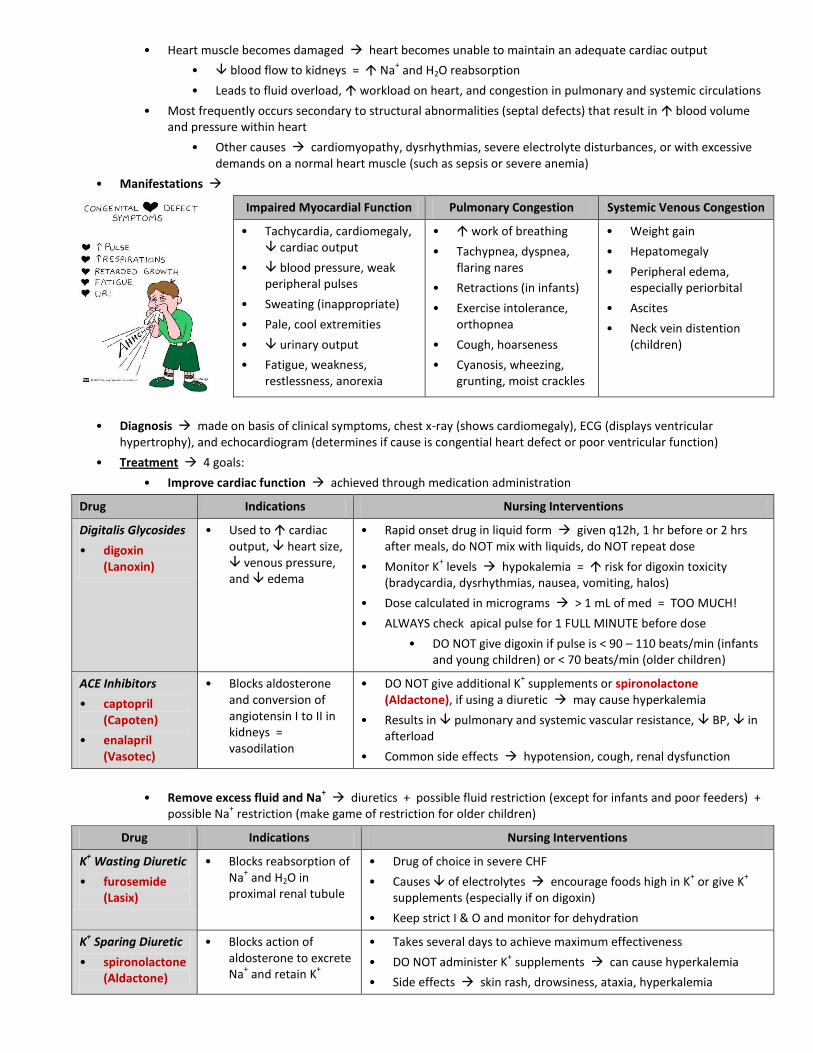
• Heart muscle becomes damaged heart becomes unable to maintain an adequate cardiac output
• blood flow to kidneys = Na+ and H2O reabsorption
• Leads to fluid overload, workload on heart, and congestion in pulmonary and systemic circulations
• Most frequently occurs secondary to structural abnormalities (septal defects) that result in blood volume and pressure within heart
• Other causes cardiomyopathy, dysrhythmias, severe electrolyte disturbances, or with excessive demands on a normal heart muscle (such as sepsis or severe anemia)
• Manifestations
Impaired Myocardial Function Pulmonary Congestion Systemic Venous Congestion
• Tachycardia, cardiomegaly, cardiac output
• blood pressure, weak peripheral pulses
• Sweating (inappropriate)
• Pale, cool extremities
• urinary output
• Fatigue, weakness, restlessness, anorexia
• work of breathing
• Tachypnea, dyspnea, flaring nares
• Retractions (in infants)
• Exercise intolerance, orthopnea
• Cough, hoarseness
• Cyanosis, wheezing, grunting, moist crackles
• Weight gain
• Hepatomegaly
• Peripheral edema, especially periorbital
• Ascites
• Neck vein distention (children)
• Diagnosis made on basis of clinical symptoms, chest x-ray (shows cardiomegaly), ECG (displays ventricular hypertrophy), and echocardiogram (determines if cause is congential heart defect or poor ventricular function)
• Treatment 4 goals:
• Improve cardiac function achieved through medication administration
Drug Indications Nursing Interventions
Digitalis Glycosides
• digoxin (Lanoxin)
• Used to cardiac output, heart size, venous pressure, and edema
• Rapid onset drug in liquid form given q12h, 1 hr before or 2 hrs after meals, do NOT mix with liquids, do NOT repeat dose
• Monitor K+ levels hypokalemia = risk for digoxin toxicity
(bradycardia, dysrhythmias, nausea, vomiting, halos)
• Dose calculated in micrograms > 1 mL of med = TOO MUCH!
• ALWAYS check apical pulse for 1 FULL MINUTE before dose
• DO NOT give digoxin if pulse is < 90 – 110 beats/min (infants and young children) or < 70 beats/min (older children)
ACE Inhibitors
• captopril (Capoten)
• enalapril (Vasotec)
• Blocks aldosterone and conversion of angiotensin I to II in kidneys = vasodilation
• DO NOT give additional K+ supplements or spironolactone
(Aldactone), if using a diuretic may cause hyperkalemia
• Results in pulmonary and systemic vascular resistance, BP, in afterload
• Common side effects hypotension, cough, renal dysfunction
• Remove excess fluid and Na+ diuretics + possible fluid restriction (except for infants and poor feeders) +
possible Na+ restriction (make game of restriction for older children)
Drug Indications Nursing Interventions
K+ Wasting Diuretic
• furosemide (Lasix)
• Blocks reabsorption of Na
+ and H2O in
proximal renal tubule
• Drug of choice in severe CHF
• Causes of electrolytes encourage foods high in K+ or give K
+
supplements (especially if on digoxin)
• Keep strict I & O and monitor for dehydration
K+ Sparing Diuretic
• spironolactone (Aldactone)
• Blocks action of aldosterone to excrete Na
+ and retain K
+
• Takes several days to achieve maximum effectiveness
• DO NOT administer K+ supplements can cause hyperkalemia
• Side effects skin rash, drowsiness, ataxia, hyperkalemia

• cardiac demands minimize metabolic needs of child
• Limit physical activity (bed rest)
• Maintain body temperature
• Treat any infections
• Cluster nursing care so as not to tire child
• Semi-Fowler’s position ( effort of breathing)
• Possibly medicate an irritable child
• Provide O2 cautiously make sure O2 is warm
• Maintain nutritional status especially for poor feeders
• Caloric needs > average infant (due to metabolic rate), yet their ability to take in adequate calories is hampered by their fatigue
• Instruct family on feeding techniques that cardiac demands
• Always hold an infant for feedings feed in a semiupright position
• Use a softer nipple or make nipple hole larger to sucking effort
• Higher caloric content = less volume needed
• NG supplemental feedings
• Hypoxia = tissue oxygenation that is caused by low O2 saturations and PaO2 (arterial O2 tension)
• Results in impaired cellular processes
Cyanosis Polycythemia Clubbing
• Blue discoloration in mucus membranes, skin, and nail beds of child with O2 saturation
• Results from presence of deoxygenated hemoglobin (hemoglobin NOT bound to O2)
• Usually apparent when arterial O2 saturations are 80% – 85%
• Heart defects that cause hypoxemia and cyanosis result from desaturated venous blood (blue blood) entering systemic circulation without passing through lungs
• number of RBCs due to chronic hypoxia
• O2-carrying capacity of blood
• Anemia may result if iron is not readily available for formation of hemoglobin
• viscosity of blood + crowd out clotting factors = risk for clotting = risk for stroke if dehydrated
• Thickening and flattening of tips of fingers and toes
• Occurs due to chronic tissue hypoxemia and polycythemia
• Bacterial endocarditis (BE) infection of heart valves and inner lining of heart
• Most often a sequela of bacteremia in child with acquired or congenital anomalies of heart or great vessels
• Affects children with valvular abnormalities, prosthetic valves, shunts, recent cardiac surgery with invasive lines, and rheumatic heart disease with valve involvement
• Most common causative agent = Streptococcus viridians
• Pathophysiology organisms enter bloodstream (from any site of localized infection or from interruption of skin integrity, such as dental work, cardiac surgery, long-term indwelling catheters) to grow on heart endocardium
• Lesion may invade adjacent tissues, such as aortic and mitral valves, and may break off and embolize elsewhere, especially in spleen, kidney, and CNS
• Manifestations onset usually insidious
• Unexplained fever (low grade and intermittent)
• Anorexia, weight loss, malaise
• Treatment started immediately with high doses of appropriate IV antibiotics for 2 – 8 weeks
• Prevention = administration of prophylactic antibiotic therapy 1 hour before any procedures known to risk of entry of organisms in very high risk patients dental work
• Educate parents to report any unexplained fever, weight loss, or change in behavior (lethargy, malaise, anorexia) to HCP

• Rheumatic fever inflammatory disease that occurs after infection with Group A β-hemolytic streptococcal (GABHS) bacteria
• Self-limited illness that involves joints, skin, brain, serous surfaces, and heart cardiac valve damage (rheumatic heart disease) is most significant complication
• Etiology relationship between URI infection (pharyngitis) with GABHS and subsequent development of rheumatic fever (usually within 2 – 6 weeks)
• Manifestations fever, carditis, tachycardia, cardiomegaly, chest pain, polyarthritis, inflamed joints
• Treatment goals of medical management
• Eradication of hemolytic streptococci use of penicillin or erythromycin
• Prevention of permanent cardiac damage
• Palliation of other symptoms salicylates used to control inflammation and fever and discomfort
• Prevention of recurrences of rheumatic fever prophylactic antibiotics
• Sickle cell anemia (SCA) normal adult Hgb (Hgb-A) is partly or completely replaced by abnormal sickle Hgb (Hgb-S)
• Sickle cell disease (SCD) includes all hereditary disorders with clinical, hematologic, and pathologic features that are related to presence of Hgb-S
• Characterized by vasoocclusion, infarcts, hemolysis, anemia, pain, ischemia, and hypoxia
• RBCs are hard, inflexible, and sickle-shaped
• Etiology autosomal recessive disorder = 25% chance of producing offspring with SCA if both parents are carriers
• Primarily affects African-Americans
• Newborn with SCA is generally asymptomatic due to Hgb-F concentrations > Hgb-S concentrations
• Pathophysiology primarily result of obstruction caused by sickled RBCs and RBC destruction (7 – 10 day life span)
• Abnormal adhesion, entanglement, and enmeshing of rigid sickle-shaped cells with one another block microcirculation = causes vasoocclusion
• blood flow to adjacent tissues = local hypoxia = leads to tissue ischemia and infarction (cellular death)
• Accumulation of RBCs in organs cause enlargement infarction with ischemia + repeated destruction of organ fibrous scarring tissue formation
• infarction in spleen due to sequestration = makes child’s spleen nonfunctional by 6 years of age
• Manifestations PAIN, immune response = risk for infections, chronic anemia (Hgb 6 – 9 g/dL), failure to grow
• Most acute symptoms of disease occur during periods of exacerbation (crises)
• Vasoocclusive crisis most common distal ischemia and pain
• Sequestration crisis pooling of blood in liver and spleen with blood volume and shock
• Aplastic crisis profound anemia from RBC production in bone marrow
• Hyperhemolytic crisis anemia, jaundice, and reticulocytosis from rate of RBC destruction
• Dactylitis painful swelling of hands and feet usually 1st
crisis in toddlers
• Crisis precipitated by infections, cold exposure, stress, dehydration, changes in altitude, and vigorous activities
• Complications stroke (CVAs), chest syndrome (looks like pneumonia), avascular necrosis (mostly in hip joints and shoulders), priapism (painful, constant penile erection)
• Diagnosis sickle-turbidity newborn screening test (Sickledex), Hgb electrophoresis
• Early identification is crucial to initiate supportive care administer prophylactic antibiotics
• Treatment aimed at preventing sickling phenomena and treating medical emergencies of sickle cell crisis
• REST to minimize energy expenditure and O2 use
• fluids either PO or IV therapy to prevent dehydration, which precipitates a crisis
• Oral intake should be at least 2x what others drink extra fluids should be given with in exercise and in hot weather
• fluids + impaired kidney function = problem of enuresis (bed-wetting)

• Electrolyte replacement hypoxia results in metabolic acidosis, which promotes sickling
• Analgesics for severe pain from vasoocclusion
• Start with ibuprofen or acetaminophen (Tylenol) if ineffective, add codeine (titrated to therapeutic level) progress to opioids, such as morphine, oxycodone, hydromorphone (Dilaudid), and methadone
• Patient-controlled analgesia (PCA)
• Warm compress to pain cold compresses are NOT applied ( sickling and vasoconstriction)
• Blood replacement to treat anemia and viscosity of sickled blood
• Can cause iron overload kidneys and heart can fail
• Need to supplement with iron chealtor drug to break down iron
• Short term O2 therapy to prevent hypoxia, which can cause massive systemic sickling
• Prolonged therapy with supplemental O2 can depress bone marrow erythropoiesis = anemia
• Antibiotics to prevent (most important) or treat any existing infection
• Administer pneumococcal, meningococcal, and influenza vaccines due to susceptibility to infection as a result of a functional asplenia (absence of normal spleen function)
• Prophylactic antibiotics till 5 – 6 years old
• Educate family and child seek early intervention for fevers, give antibiotics as ordered, monitor for signs of hypoxia and respiratory problems
• Medications
• Folic acid stimulates erythropoesis in bone marrow
• Hydroxyurea adjunctive chemotherapeutic agent that stimulates production of fetal Hgb-F
• Hemophilia group of bleeding disorders in which there is a deficiency of a factor necessary for coagulation of blood
• Common forms of disorder
• Factor VIII deficiency = hemophilia A (classic hemophilia)
• Accounts for 80% – 85% of all cases
• Factor IX deficiency = hemophilia B, or Christmas disease
• Von Willebrand disease = deficiency, abnormality, or absence of protein called von Willebrand factor (vWF) and a deficiency of factor VIII (affects both males and females)
• Etiology X-linked recessive disorder that mostly affects males
• Pathophysiology basic defect of hemophilia A = deficiency of antihemophilic factor (factor VIII)
• Produced by liver needed for formation of thromboplastin in blood coagulation
• Still produce platelets results in bleeding for longer periods of time, NOT at a faster rate
• Manifestations
• Prolonged bleeding anywhere in body
• Excessive bruising
• SubQ and IM hemorrhages
• Spontaneous hematuria
• Hemarthrosis (bleeding into joint cavities) especially knees, ankles, and elbows
• Hemorrhage from any trauma cuts, circumcision, epistaxis, injections
• Diagnosis history of bleeding episodes, evidence of X-linked inheritance, and laboratory findings (PTT, PT)
• Treatment replacement of missing clotting factor and prevention of injuries
• Factor VIII concentrate from pooled plasma or a genetically engineered recombinant
• Given with any trauma or prophylactically can ↑ risk for brain and GI bleeds
• DDAVP synthetic form of vasopressin that ↑ plasma factor VIII and vWF levels
• Treatment of choice in mild hemophilia and vWD if child shows an appropriate response
• NOT effective in treatment of severe hemophilia A, severe vWD, or any form of hemophilia B
• Vigorous physical therapy is instituted to prevent chronic crippling effects from joint bleeding

• Nursing interventions limit procedures that may cause bleeding
• Encourage use of soft toothbrush at home
• SubQ route is substituted for IM injections whenever possible
• Venipunctures for blood samples (instead of finger or heel sticks) are preferred
• NO aspirin or any aspirin-containing compound should be used substitute with acetaminophen for controlling pain at home
• Leukemia cancer of blood-forming tissues (bone marrow and lymphatic system)
• Etiology most common form of childhood cancer
• More common in males and Caucasians
• Peak age of onset = 2 – 6 years old
• Pathophysiology unrestricted proliferation of immature WBCs in blood-forming tissues of body
• Leukemic cells demonstrate same neoplastic properties as solid cancers
• Pathologic condition caused by infiltration and replacement of tissue with nonfunctional leukemic cells
• Most severely affects highly vascular organs (spleen, liver)
• Leukocyte count is low
• Cellular destruction takes place by infiltration and subsequent competition for metabolic elements compete with all “good” cells for resources
• Proliferating cells ↓ production of formed elements of blood in bone marrow by competing for and depriving normal cells of essential nutrients for metabolism
• 2 major forms of leukemia
Acute lymphoid leukemia
(ALL)
Acute nonlymphoid (myelogenous) leukemia
(ANLL or AML)
Groups Affected White males All ethnic groups
Peak Age 4 years old No peak age
Survival Rate 75% survival < 40% survival
• Manifestations PAIN, pallor, fatigue, lethargy, malaise, fever, hemorrhage (petechiae), bruising, anorexia
• Most frequent presenting signs and symptoms of leukemia = result of infiltration of bone marrow
• Anemia from ↓ RBCs
• Infection from neutropenia (too many immature WBCs)
• Bleeding from ↓ platelet production (thrombocytopenia)
• Bone and joint pain especially in long bones
• Due to ↑ blasts in bones
• Gradually causes a weakening of bone and a tendency toward fractures
• Leukemic cells invade periosteum = ↑ pressure = severe pain
• If CNS involved headache, vomiting, papilledema, 6th
nerve palsy (due to ↑ intracranial pressure)
• Diagnosis based on the history, physical manifestations, and peripheral blood smear that contains immature forms of leukocytes + low blood counts (CBC)
• Bone marrow aspiration and biopsy = gold standard diagnosis shows hyper cellular bone marrow that contains primarily blast (immature) cells
• Lumbar puncture determines if leukemic cells are in brain and CNS

• Treatment chemotherapeutic agents, with or without cranial irradiation, in 4 phases:
Induction Therapy CNS Prophylactic Therapy Intensification Therapy Maintenance Therapy
• Massive blast of chemo
• Achieves a complete remission or < 5% leukemic cells in bone marrow
• Most dangerous stage
• Prevents leukemic cells from invading CNS
• Treat brain with chemo via lumbar puncture most chemo drugs do NOT cross blood brain barrier
• Consolidation eradicates residual leukemia cells
• Followed by delayed intensification prevents emergence of resistant leukemic clones
• Maintains remission phase
• Reinduction if there is a relapse
• Prepare child and family for what to expect during diagnostic and therapeutic procedures
• Relieve pain opioids titrated to child's needs and administered around clock for optimal pain control
• Nonpharmacologic strategies also used are NOT substitutes for pharmacologic management
• Prevent complications of myelosuppression infection, bleeding tendencies, and anemia due to ↓ number of blood cells
Infection Hemorrhage Anemia
• Secondary to neutropenia
• Child in private room
• Restriction of visitors and health personnel with active infections
• Strict aseptic technique
• NO immunization using live attenuated viruses (measles, mumps, rubella, varicella, polio)
• Monitor for ↑ temperature
• Most bleeding episodes can be prevented or controlled with administration of platelet concentrates or platelet-rich plasma
• Skin punctures are avoided whenever possible
• Frequent mouth care is essential can result in gingival bleeding
• Avoid activities that might cause injury or bleeding
• May be profound from complete replacement of bone marrow by leukemic cells
• Blood transfusions may be necessary during induction therapy
• Nursing interventions use chemo precautions (handling chemotherapeutic agents may present risks to handlers)
• Chemotherapeutic drugs must be given through a free-flowing IV line
• Child must be observed for 20 minutes after infusion for signs of anaphylaxis cyanosis, hypotension, wheezing, severe urticaria
• Manage problems related to chemotherapy
• Nausea and vomiting can be profound
• Serotonin-receptor antagonists (ondansetron, granisetron) + dexamethasone treatment of choice in the prevention of delayed emesis
• Most beneficial regimen = administration of antiemetic before chemotherapy
• Anorexia loss of appetite is a direct consequence of chemotherapy or irradiation
• NG tube feeds or total parenteral nutrition may be implemented
• Mucosal ulceration mucosal cell damage can produce ulcers anywhere along GI tract
• Oral ulcers ↑ anorexia provide a bland, moist, soft diet, use Toothettes, provide frequent mouthwashes with normal saline, use local anesthetics
• chlorhexidine gluconate (Peridex) used to prevent or treat mucositis
• Neuropathy due to Vincristine use
• Stool softeners (due to ↓ bowel innervations), use of footboard (to minimize or prevent footdrop), safety measures during ambulation (due to weakness and numbness in extremities, providing soft or liquid diet (for severe jaw pain)
• Hemorrhagic cystitis due to chemical irritation to bladder
• ↑ fluid intake, void frequently, administer mesna (alkalizing agent to counteract acidity)
• Alopecia hair loss is common side effect protect scalp, use cotton head wear
• Hair regrowth occurs in 3 – 6 months may be of a different color and texture
• Moon face from short-term steroid use reassure child that normal facial shape returns
• Mood changes due to steroid therapy