pediatricradiologyinoto-rhino-laryngologypediatricradiologyinoto-rhino-laryngology abstract...
TRANSCRIPT
Pediatric radiology in oto-rhino-laryngology
AbstractHead and neck diseases in children and adolescents present specialdiagnostic and differential diagnostic challenges to ENT surgeons as
Thekla von Kalle1
Assen Koitschev2well as to radiologists. Both disciplines have to adapt the latest radiolo-gical and interventional technologies to the needs of the pediatric patientin order to enable a minimally invasive but successful diagnostic pro-cedure.
1 Institute of Radiology,Olgahospital, KlinikumStuttgart, GermanyHigh quality sonography by an experienced examiner is often the only
imaging technique that is necessary in children and adolescents. Radio- 2 Department ofOtolaryngology, Division ofgraphs are rarely indicated in pediatric head and neck diseases. MRI,Pediatric Otolaryngology andcompared to computed tomography, has the advantage of absent radi-Otology, Olgahospital,Klinikum Stuttgart, Germany
ation exposure. Additionally, due to current advances in high resolutiontechniques to delineate very small details or in visualization of differenttissue characteristics, it has become an integral part of pre- and post-operative imaging.However, children should not be denied an adequate diagnostic proced-ure even if it includes sedation, intervention, or exposure to radiation.The responsible use of the diagnostic options under consideration ofthe therapeutic consequences is essential. It is most likely to be suc-cessful in a close interdisciplinary cooperation of pediatric ENT special-ists and radiologists as well as pediatric anesthesiologists in selectedcases.Although benign diseases predominate in children and adolescents,the possibility of malignancy has to be considered in cases of atypicalclinical and radiological findings. In many of these young patients, theoutcome and the probability of survival are directly associated with theinitial diagnostic and therapeutic strategies, which should therefore bein accordance with the current guidelines of pediatric oncology therapystudies.Our collection of clinical cases consists of representative examples ofuseful diagnostic approaches in common and age specific diagnosesas well as in rare diseases andmalformations. It shows the significanceof a special knowledge in embryology and normal postnatal developmentfor the differentiation of normal variants from pathological findings.Only in considering the results of imaging studies in their clinical context,it is possible to succeed in detecting a syndrome behind a single mal-formation or adequately caring for patients with a chronic disease suchas cystic fibrosis.
Keywords: ENT, pediatric, radiology, sonography, MRI
1 IntroductionHead and neck diseases in children are a special diagnos-tic and differential diagnostic challenge. The differenti-ation of age-dependent normal variations from patholo-gical findings, the immature and small anatomy, as wellas the particular spectrum of diseases require specialexperience and expertise of the ENT specialist and theradiologist.This pediatric radiological expertise includes specialknowledge of the embryology and the postnatal develop-ment as well as experiences with the newest imagingtechniques. It is fundamental to a good interdisciplinary
communication and a child-oriented, less stressful andeffective diagnostic approach.Methods without exposure to radiation such as high-resolution ultrasound andMRI have priority to radiographyand computed tomography. However, children should notbe precluded from adequate diagnostics even if it is as-sociated with sedation, intervention, or exposure to radi-ation. The responsible management of the diagnosticpossibilities under consideration of the clinical con-sequences is crucial and only feasible in a good interdis-ciplinary cooperation.Although benign diseases prevail in childhood and adoles-cence, atypical clinical and radiological findings must
1/19GMS Current Topics in Otorhinolaryngology - Head and Neck Surgery 2014, Vol. 13, ISSN 1865-1011
Review ArticleOPEN ACCESS
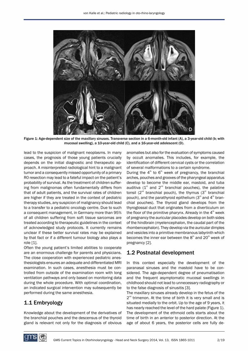
Figure 1: Age-dependent size of the maxillary sinuses. Transverse section in a 6-month-old infant (A), a 3-year-old child (b; withmucosal swelling), a 10-year-old child (C), and a 16-year-old adolescent (D).
lead to the suspicion of malignant neoplasms. In manycases, the prognosis of those young patients cruciallydepends on the initial diagnostic and therapeutic ap-proach. A misinterpreted radiological hint to a malignanttumor and a consequentlymissed opportunity of a primaryR0 resectionmay lead to a fateful impact on the patient’sprobability of survival. As the treatment of children suffer-ing from malignomas often fundamentally differs fromthat of adult patients, and the survival rates of childrenare higher if they are treated in the context of pediatrictherapy studies, any suspicion ofmalignancy should leadto a transfer to a pediatric oncology centre. Due to sucha consequent management, in Germany more than 95%of all children suffering from soft tissue sarcomas aretreated according to therapeutic guidelines in the contextof acknowledged study protocols. It currently remainsunclear if these better survival rates may be explainedby that fact or if a different tumour biology also plays arole [1].Often the young patient’s limited abilities to cooperateare an enormous challenge for parents and physicians.The close cooperation with experienced pediatric anes-thesiologists ensures an adequate and differentiatedMRIexamination. In such cases, anesthesia must be con-trolled from outside of the examination room with longventilation pathways and only based on monitoring dataduring the whole procedure. With optimal coordination,an indicated surgical intervention may subsequently beperformed during the same anesthesia.
1.1 Embryology
Knowledge about the development of the derivatives ofthe branchial pouches and the descensus of the thyroidgland is relevant not only for the diagnosis of obvious
anomalies but also for the evaluation of symptoms causedby occult anomalies. This includes, for example, theidentification of different cervical cysts or the correlationof several malformations to a certain syndrome.During the 4th to 6th week of pregnancy, the branchialarches, pouches and grooves of the pharyngeal apparatusdevelop to become the middle ear, mastoid, and tubaauditiva (1st and 2nd branchial pouches), the palatinetonsil (2nd branchial pouch), the thymus (3rd branchialpouch), and the parathyroid epithelium (3rd and 4th bran-chial pouches). The thyroid gland develops from thethyroglossal duct that originates from a diverticulum onthe floor of the primitive pharynx. Already in the 4th weekof pregnancy the auricular placodes develop on both sidesof the hindbrain (myelencephalon, the caudal part of therhombencephalon). They develop via the auricular dimplesand vesicles into a primitivemembranous labyrinth whichbecomes the inner ear between the 8th and 20th week ofpregnancy [2].
1.2 Postnatal development
In this context especially the development of theparanasal sinuses and the mastoid have to be con-sidered. The age-dependent degree of pneumatisationand the frequent asymptomatic mucosal swellings inchildhood should not lead to unnecessary radiography orto the false diagnosis of sinusitis [3].The maxillary sinuses already develop in the fetus of the2nd trimenon. At the time of birth it is very small and issituated medially to the orbit. Up to the age of 9 years, ithas nearly reached the level of the hard palate (Figure 1).The development of the ethmoid cells starts about thetime of birth in an anterior to posterior direction. At theage of about 6 years, the posterior cells are fully de-
2/19GMS Current Topics in Otorhinolaryngology - Head and Neck Surgery 2014, Vol. 13, ISSN 1865-1011
von Kalle et al.: Pediatric radiology in oto-rhino-laryngology
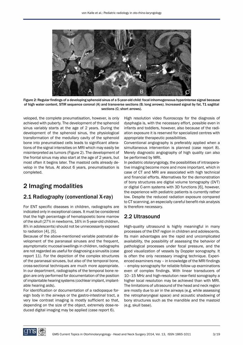
Figure 2: Regular findings of a developing sphenoid sinus of a 5-year-old child: focal inhomogeneous hyperintense signal becauseof high water content, STIR sequence coronal (A) and transverse sections (B; long arrows). Increased signal by fat, T1 sagittal
sections (C; short arrows).
veloped, the complete pneumatisation, however, is onlyachieved with puberty. The development of the sphenoidsinus variably starts at the age of 2 years. During thedevelopment of the sphenoid sinus, the physiologicaltransformation of the medullary cavity of the sphenoidbone into pneumatised cells leads to significant altera-tions of the signal intensities on MRI which may easily bemisinterpreted as tumors (Figure 2). The development ofthe frontal sinus may also start at the age of 2 years, butmost often it begins later. The mastoid cells already de-velop in the fetus. At about 6 years, pneumatisation iscompleted.
2 Imaging modalities
2.1 Radiography (conventional X-ray)
For ENT specific diseases in children, radiographs areindicated only in exceptional cases. It must be consideredthat the high percentage of hematopoietic bone marrowof the skull (27% in newborns, 16% in 5-year-old children,8% in adolescents) should not be unnecessarily exposedto radiation [4], [5].Because of the above-mentioned variable postnatal de-velopment of the paranasal sinuses and the frequent,asymptomaticmucosal swellings in children, radiographsare not regarded as useful for diagnosing a sinusitis (casereport 11). For the depiction of the complex structuresof the paranasal sinuses, but also of the temporal bone,cross-sectional techniques are much more appropriate.In our department, radiographs of the temporal bone re-gion are only performed for documentation of the positionof implantable hearing systems (cochlear implant, implant-able hearing aids).For identification or documentation of a radiopaque for-eign body in the airways or the gastro-intestinal tract, avery low contrast imaging is mostly sufficient so that,depending on the size of the object, extremely dose-re-duced digital imaging may be applied (case report 6).
High resolution video fluoroscopy for the diagnosis ofdysphagia is, with the necessary effort, possible even ininfants and toddlers, however, also because of the radi-ation exposure it is reserved for specialized centres withappropriate therapeutic possibilities.Conventional angiography is preferably applied when asimultaneous intervention is planned (case report 8).Merely diagnostic angiography of high quality can alsobe performed by MRI.In pediatric otolaryngology, the possibilities of intraopera-tive imaging becomemore and more important, which incase of CT and MRI are associated with high technicaland financial efforts. Alternatives for the demonstrationof bony structures are digital volume tomography (DVT)or digital C-arm systems with 3D functions [6], however,the experience with pediatric patients is currently ratherlow. Despite the reduced radiation exposure comparedto CT scanning, an especially careful benefit-risk analysisis therefore necessary.
2.2 Ultrasound
High-quality ultrasound is highly meaningful in manyprocesses of the ENT region in children and adolescents.Its main advantages are the rapid and uncomplicatedavailability, the possibility of assessing the behavior ofpathological processes under focal pressure, and thegood visualization of vessels by Doppler sonography. Itis often the only necessary imaging technique. Experi-enced examiners may – in knowledge of theMRI findings– employ sonography for reliable follow-up examinationseven of complex findings. With linear transducers of10–15 MHz and high-resolution near-field sonography ahigher local resolution may be achieved than with MRI.The limitations of ultrasound of the head and neck regionare mostly due to air in the airways (e.g. while assessingthe retropharyngeal space) and acoustic shadowing ofbony structures such as the mandible and the mastoid(e.g. skull base).
3/19GMS Current Topics in Otorhinolaryngology - Head and Neck Surgery 2014, Vol. 13, ISSN 1865-1011
von Kalle et al.: Pediatric radiology in oto-rhino-laryngology
2.3 Magnetic resonance imaging (MRI)
MRI is an extremely versatile tool to demonstrate normaland pathological tissue properties. Beside a multitude ofsequence techniques also high-resolution (Voxel <1mm)3D imaging with subsequent multiplanar reconstructionof the head and neck region are possible. Recent technic-al developments allow – with 3-Tesla devices – an isotrop-ic spatial resolution of 0.32 mm with high quality so thateven the neuro-epithelium of the macula utriculi can bedelineated in the vestibulum of the inner ear [7]. For anadequate choice of MR sequences and sections an exactinterdisciplinary communication about the clinicalquestions that have to be answered is essential. Thisclose cooperation helps to avoid over- as well as under-diagnosis. An examination that takes toomuch time inev-itably leads to increased movement artefacts especiallyin young children. A too short examination with “wrong”sequences may prevent a rapid diagnosis and thus leadto unnecessary subsequent examinations.Child-oriented conditions are a general prerequisite fora successful MR examination. With the parents presentand audio or video tapes available, even younger children(4–8 years) are able to keep still for a sufficient periodof time. In our department, toddlers and infants are ex-amined under sedation with Propofol, and, if necessary,airway management with laryngeal masks, and appropri-ate monitoring by a pediatric anesthesiologist.The indications for contrast-enhanced examinations alsodepend on the clinical questions. Thus the identificationof inflammatory processes, especially after an abscess,or the delineation of tumors nearly always require theapplication of contrast agents, while merely anatomicevaluations may mostly be performed without injections.Also for MR angiography, the choice of the technique andthe decision for or against contrast application dependon the question to be examined. In this context an openinterdisciplinary exchange about the newest examinationand surgery techniques seems to be indispensable inorder to fulfill the mutual requirements. According to ourexperience, a current example is the visualization ofcholesteatomas by diffusion-weightedMR sequences [8].This relatively easy examination technique requires aspecial selection and the exact spatial alignment of dif-ferent MR sequences. Only intensive interdisciplinaryexchange and close correlation of imaging findings andsurgical aspects allow a rapidly rising learning curve.
2.4 Computed tomography (CT)
For CT the same considerations on dose reduction arevalid as for radiography, however, with themain differenceof amuch higher radiation exposure. The average CT scanof the skull with an effective dose of 2.3mSv correspondsto about 35 X-rays of the skull or 115 X-rays of the thorax,and it is equivalent to a natural radiation exposure ofabout one year (http://www.imagegently.org/). Further,in comparison to adults, children have a much higherradiosensitivity beside a long average life expectancy in
which malignomas may develop [5]. Before performingCT scans, the diagnostic alternatives should therefore becarefully considered.The domain of CT is the visualization of bony and air-containing structures and their anomalies. It has signifi-cance especially for trauma diagnostics and the depictionof the middle ear and the external meatus. The inner earand the inner meatus may only be indirectly assessedwhile three dimensional MRI allows their direct visualiza-tion [9] (case report 15). All processes with soft tissueinvolvement such as tumors or sinugenic abscessesmaybe better examined by MRI and only rarely require addi-tional CT imaging of the bones (case reports 12, 14a). Ithas to bementioned that also in tertiary centres adequateMR imaging is not always available, e.g. on weekends orat inconvenient times. The examination of young unco-operative patients may be an additional challenge. Theideal constellation would include the imaging and a pos-sible surgical intervention under the same anesthesia.However, this is only possible in institutions that are lo-gistically and medically focused on children as for ex-ample the Olgahospital of the Klinikum Stuttgart, Ger-many.
3 Case reportsIn the following chapter some important particularitiesof radiology in children with ENT specific diseases will beexplained on the basis of case reports. Each of ourmutualpatients hereby represents a group of typical pediatricENT patients. For further information, we refer to specificliterature and textbooks [2], [10], [11], [12].For MRI examination of the neurocranium T1 and T2weighted sequences are commonly applied which thusalso depict the incidental findings. Water has a low signalintensity in T1weighted images, and a hyperintense signalin T2. Fat appears with high signal intensity in both se-quences.For examination of the viscerocranium, before contrastapplication STIR sequences are applied that suppressthe fat signal (low signal intensity) and show water con-taining structures with high signal intensity. After contrastapplication, fat saturated T1 sequences are applied(contrast agent with high signal intensity, water and fatwith low signal intensity).
3.1 Neck
Oncologic diseases of the neck are rare in children. Incontrast to adults, their vast majority is of mesenchymalorigin (lymphomas, sarcomas etc.). In practice, benigncystic malformations (median and lateral) and inflamma-tory processes of the lymphatic system (lymphadenitiscolli) are most frequent.In most pathological processes of the neck in children,sonography of high quality is the first and only imagingmethod. However, radiologists should always be awarethat the anatomic space of the neck begins cranially at
4/19GMS Current Topics in Otorhinolaryngology - Head and Neck Surgery 2014, Vol. 13, ISSN 1865-1011
von Kalle et al.: Pediatric radiology in oto-rhino-laryngology
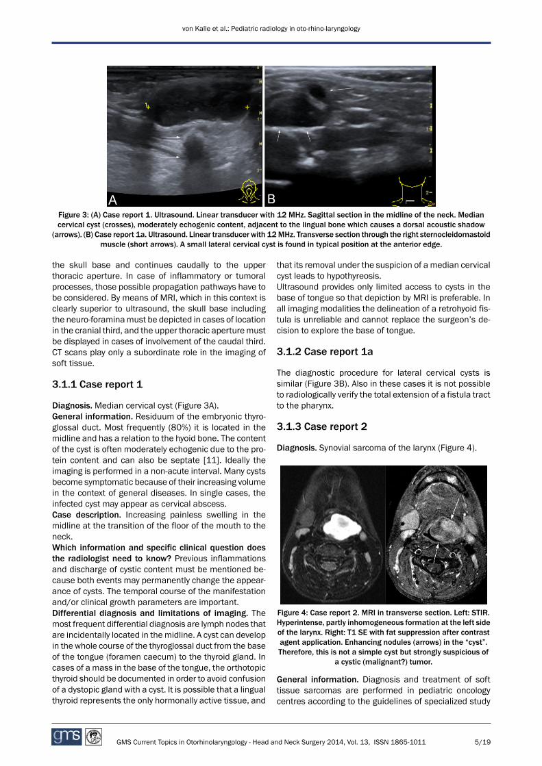
Figure 3: (A) Case report 1. Ultrasound. Linear transducer with 12 MHz. Sagittal section in the midline of the neck. Mediancervical cyst (crosses), moderately echogenic content, adjacent to the lingual bone which causes a dorsal acoustic shadow
(arrows). (B) Case report 1a. Ultrasound. Linear transducer with 12MHz. Transverse section through the right sternocleidomastoidmuscle (short arrows). A small lateral cervical cyst is found in typical position at the anterior edge.
the skull base and continues caudally to the upperthoracic aperture. In case of inflammatory or tumoralprocesses, those possible propagation pathways have tobe considered. By means of MRI, which in this context isclearly superior to ultrasound, the skull base includingthe neuro-foraminamust be depicted in cases of locationin the cranial third, and the upper thoracic aperturemustbe displayed in cases of involvement of the caudal third.CT scans play only a subordinate role in the imaging ofsoft tissue.
3.1.1 Case report 1
Diagnosis. Median cervical cyst (Figure 3A).General information. Residuum of the embryonic thyro-glossal duct. Most frequently (80%) it is located in themidline and has a relation to the hyoid bone. The contentof the cyst is often moderately echogenic due to the pro-tein content and can also be septate [11]. Ideally theimaging is performed in a non-acute interval. Many cystsbecome symptomatic because of their increasing volumein the context of general diseases. In single cases, theinfected cyst may appear as cervical abscess.Case description. Increasing painless swelling in themidline at the transition of the floor of the mouth to theneck.Which information and specific clinical question doesthe radiologist need to know? Previous inflammationsand discharge of cystic content must be mentioned be-cause both events may permanently change the appear-ance of cysts. The temporal course of the manifestationand/or clinical growth parameters are important.Differential diagnosis and limitations of imaging. Themost frequent differential diagnosis are lymph nodes thatare incidentally located in themidline. A cyst can developin the whole course of the thyroglossal duct from the baseof the tongue (foramen caecum) to the thyroid gland. Incases of a mass in the base of the tongue, the orthotopicthyroid should be documented in order to avoid confusionof a dystopic gland with a cyst. It is possible that a lingualthyroid represents the only hormonally active tissue, and
that its removal under the suspicion of a median cervicalcyst leads to hypothyreosis.Ultrasound provides only limited access to cysts in thebase of tongue so that depiction by MRI is preferable. Inall imaging modalities the delineation of a retrohyoid fis-tula is unreliable and cannot replace the surgeon’s de-cision to explore the base of tongue.
3.1.2 Case report 1a
The diagnostic procedure for lateral cervical cysts issimilar (Figure 3B). Also in these cases it is not possibleto radiologically verify the total extension of a fistula tractto the pharynx.
3.1.3 Case report 2
Diagnosis. Synovial sarcoma of the larynx (Figure 4).
Figure 4: Case report 2. MRI in transverse section. Left: STIR.Hyperintense, partly inhomogeneous formation at the left sideof the larynx. Right: T1 SE with fat suppression after contrastagent application. Enhancing nodules (arrows) in the “cyst”.Therefore, this is not a simple cyst but strongly suspicious of
a cystic (malignant?) tumor.
General information. Diagnosis and treatment of softtissue sarcomas are performed in pediatric oncologycentres according to the guidelines of specialized study
5/19GMS Current Topics in Otorhinolaryngology - Head and Neck Surgery 2014, Vol. 13, ISSN 1865-1011
von Kalle et al.: Pediatric radiology in oto-rhino-laryngology
groups: EpSSG (European Paediatric Soft-Tissue SarcomaStudy Group: http://epssg.cineca.org), CWS (CooperativeWeichteilsarkom Studiengruppe http://cws.olgahospital-stuttgart.de, http://www.kinderkrebsinfo.de), COG STS(Children's Oncology Group Soft Tissue Sarcoma Commit-tee, USA, http://www.cancer.gov/cancertopics/types/childrhabdomyosarcoma). The clinical findings of a sar-coma may be rather unspecific and mimic less harmfuldifferential diagnoses. The experienced pediatric radiolo-gist can and should be the decisive “advisor” for the ENTspecialist to find the correct diagnosis.A central requirement for the histological assessment ofa suspected sarcoma is to avoid the spreading of tumorcells into the surrounding tissue. Thus imaging is emi-nently important for the planning of the surgical approach.Case description. The 9-year-old child presented withunspecific but increasing complaints (foreign body sensa-tion when swallowing, mild hoarseness). Because of asmooth protrusion of the pharynx with displacement ofthe epiglottis, which became obvious during an examin-ation in a private practice, the emergency presentationin our hospital was indicated. Endoscopy could confirmthe findings, however, primarily a congenital laryngealcyst was suspected in the sense of an encapsulated la-ryngocele.Which information and specific clinical question doesthe radiologist need to know? The exact description ofthe clinical and endoscopic findings, and the principalsymptom.Differential diagnoses and limitations of imaging.Benigncysts are frequent in otolaryngology. Their lumen may befilled with echogenic material (see case report 1). A focalespecially nodular wall thickening should always lead tothe suspicion of a malignant disease, even in children.An appropriately careful surgicalmanagement is essential(with R0 resection whenever possible). In case of suspec-ted sarcoma, the experienced pediatric radiologist shouldemphasize the particular requirements of the histologicalconfirmation and the potential risks of iatrogenicspreading of tumour cells.The anterior and lateral wall structures of the larynx andthe trachea may be well assessed by means of ultra-sound, with only few limits caused by the aerated lumen.The exact dimensions of soft tissue lesions may best bedisplayed with high resolution MRI (slice thickness ofmax. 5mm, pixel size of <1mm) with fat saturation beforeand after contrast agent application.
3.1.4 Case report 3
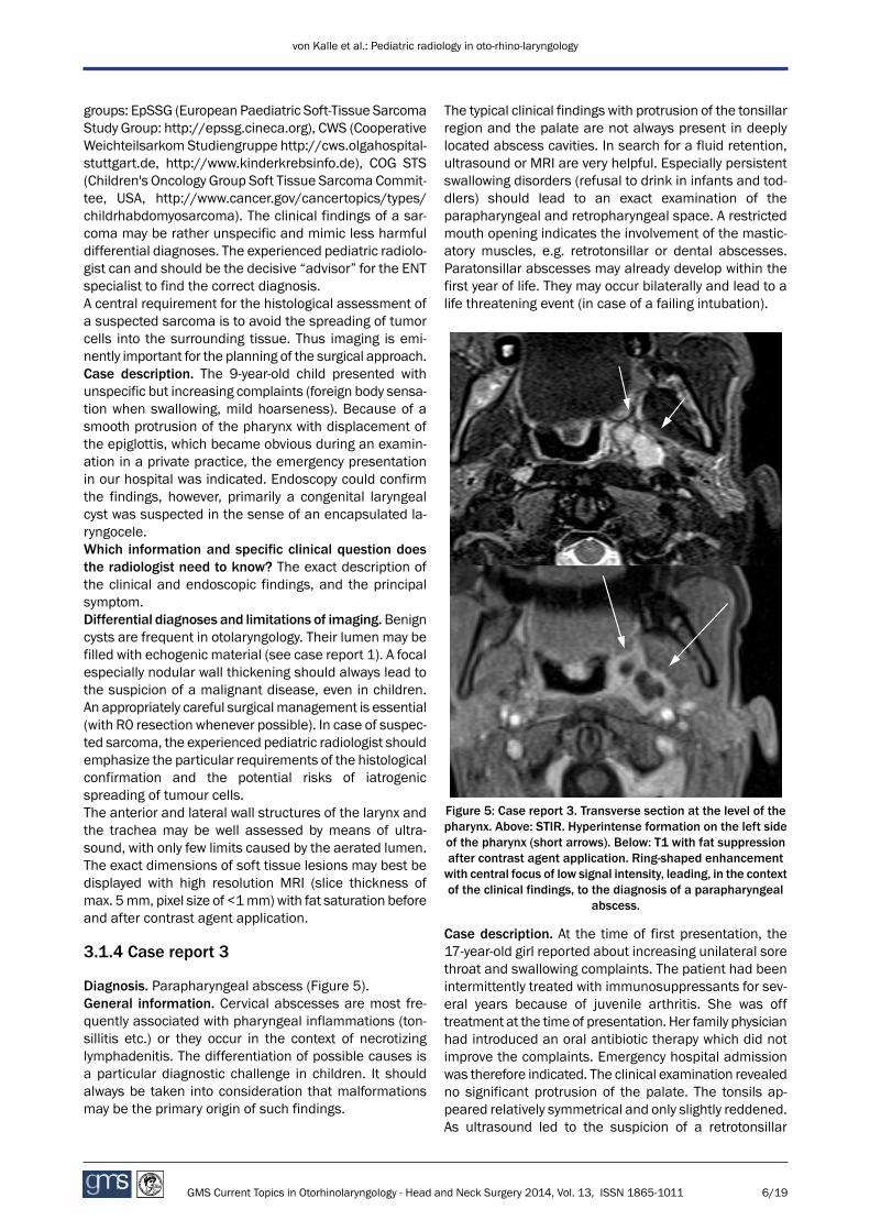
Diagnosis. Parapharyngeal abscess (Figure 5).General information. Cervical abscesses are most fre-quently associated with pharyngeal inflammations (ton-sillitis etc.) or they occur in the context of necrotizinglymphadenitis. The differentiation of possible causes isa particular diagnostic challenge in children. It shouldalways be taken into consideration that malformationsmay be the primary origin of such findings.
The typical clinical findings with protrusion of the tonsillarregion and the palate are not always present in deeplylocated abscess cavities. In search for a fluid retention,ultrasound or MRI are very helpful. Especially persistentswallowing disorders (refusal to drink in infants and tod-dlers) should lead to an exact examination of theparapharyngeal and retropharyngeal space. A restrictedmouth opening indicates the involvement of the mastic-atory muscles, e.g. retrotonsillar or dental abscesses.Paratonsillar abscesses may already develop within thefirst year of life. They may occur bilaterally and lead to alife threatening event (in case of a failing intubation).
Figure 5: Case report 3. Transverse section at the level of thepharynx. Above: STIR. Hyperintense formation on the left sideof the pharynx (short arrows). Below: T1 with fat suppressionafter contrast agent application. Ring-shaped enhancementwith central focus of low signal intensity, leading, in the contextof the clinical findings, to the diagnosis of a parapharyngeal
abscess.
Case description. At the time of first presentation, the17-year-old girl reported about increasing unilateral sorethroat and swallowing complaints. The patient had beenintermittently treated with immunosuppressants for sev-eral years because of juvenile arthritis. She was offtreatment at the time of presentation. Her family physicianhad introduced an oral antibiotic therapy which did notimprove the complaints. Emergency hospital admissionwas therefore indicated. The clinical examination revealedno significant protrusion of the palate. The tonsils ap-peared relatively symmetrical and only slightly reddened.As ultrasound led to the suspicion of a retrotonsillar
6/19GMS Current Topics in Otorhinolaryngology - Head and Neck Surgery 2014, Vol. 13, ISSN 1865-1011
von Kalle et al.: Pediatric radiology in oto-rhino-laryngology
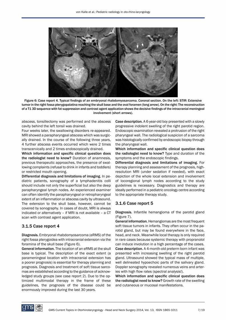
Figure 6: Case report 4. Typical findings of an embryonal rhabdomyosarcoma. Coronal section. On the left: STIR: Extensivetumor in the right fossa pterygopalatina reaching the skull base and the oval foramen (long arrow). On the right: The reconstructionof a T1 3D sequencewith fat suppression and contrast agent application shows the decisive findings of the intracranialmeningeal
involvement (short arrows).
abscess, tonsillectomy was performed and the abscesscavity behind the left tonsil was drained.Four weeks later, the swallowing disorders re-appeared.MRI showed a parapharyngeal abscess which was surgic-ally drained. In the course of the following three years,4 further abscess events occurred which were 2 timestranscervically and 2 times endoscopically drained.Which information and specific clinical question doesthe radiologist need to know? Duration of anamnesis,previous therapeutic approaches, the presence of swal-lowing complaints (refusal to drink in infants and toddlers)or restricted mouth opening.Differential diagnosis and limitations of imaging. In pe-diatric patients, sonography of a lymphadenitis collishould include not only the superficial but also the deepparapharyngeal lymph nodes. An experienced examinercan often identify the parapharyngeal or retropharyngealextent of an inflammation or abscess cavity by ultrasound.The extension to the skull base, however, cannot becovered by sonography. In cases of doubt, MRI is alwaysindicated or alternatively – if MRI is not available – a CTscan with contrast agent application.
3.1.5 Case report 4
Diagnosis. Embryonal rhabdomyosarcoma (eRMS) of theright fossa pterygoidea with intracranial extension via theforamina of the skull base (Figure 6).General information. The location of an eRMS at the skullbase is typical. The description of the exact extent (aparameningeal location with intracranial extension hasa poorer prognosis) is essential for therapy planning andprognosis. Diagnosis and treatment of soft tissue sarco-mas are established according to the guidance of acknow-ledged study groups (see case report 2). Due to the op-timized multimodal therapy in the frame of theseguidelines, the prognosis of the disease could beenormously improved during the last 30 years.
Case description. A 6-year-old boy presented with a slowlyprogressive indolent swelling of the right parotid region.Endoscopic examination revealed a protrusion of the rightpharyngeal wall. The radiological suspicion of a sarcomawas histologically confirmed by endoscopic biopsy throughthe pharyngeal wall.Which information and specific clinical question doesthe radiologist need to know? Type and duration of thesymptoms and the endoscopic findings.Differential diagnosis and limitations of imaging. Fortherapy planning and assessment of the prognosis, high-resolution MRI (under sedation if needed), with exactdepiction of the whole local extension and involvementof locoregional lymph nodes according to the studyguidelines is necessary. Diagnostics and therapy areideally performed in a pediatric oncology centre accordingto the appropriate therapy study.
3.1.6 Case report 5
Diagnosis. Infantile hemangioma of the parotid gland(Figure 7).General information.Hemangiomasare themost frequentsoft tissue tumors in infants. They often occur in the pa-rotid gland, but may be found everywhere in the face,head, and neck. Meanwhile local therapy is only requiredin rare cases because systemic therapy with propranololcan induce involution in a high percentage of the cases.Case description. A 4-month-old preterm born infant waspresented with increasing swelling of the right parotidgland. Ultrasound showed the typical mass of multiple,well delineated hypoechoic parts of the salivary gland.Doppler sonography revealed numerous veins and arter-ies with high flow rates (spectral analysis!).Which information and specific clinical question doesthe radiologist need to know?Growth rate of the swellingand cutaneous or mucosal manifestations.
7/19GMS Current Topics in Otorhinolaryngology - Head and Neck Surgery 2014, Vol. 13, ISSN 1865-1011
von Kalle et al.: Pediatric radiology in oto-rhino-laryngology
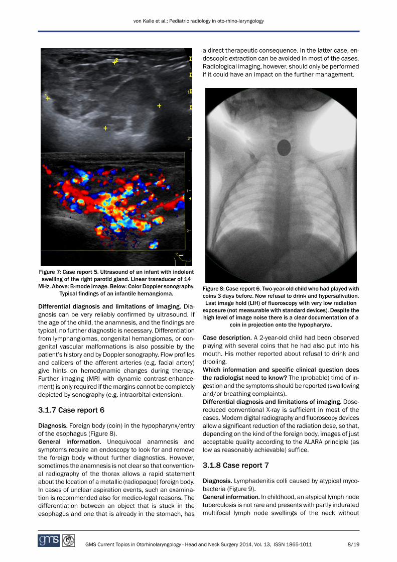
Figure 7: Case report 5. Ultrasound of an infant with indolentswelling of the right parotid gland. Linear transducer of 14
MHz. Above: B-mode image. Below: Color Doppler sonography.Typical findings of an infantile hemangioma.
Differential diagnosis and limitations of imaging. Dia-gnosis can be very reliably confirmed by ultrasound. Ifthe age of the child, the anamnesis, and the findings aretypical, no further diagnostic is necessary. Differentiationfrom lymphangiomas, congenital hemangiomas, or con-genital vascular malformations is also possible by thepatient’s history and by Doppler sonography. Flow profilesand calibers of the afferent arteries (e.g. facial artery)give hints on hemodynamic changes during therapy.Further imaging (MRI with dynamic contrast-enhance-ment) is only required if themargins cannot be completelydepicted by sonography (e.g. intraorbital extension).
3.1.7 Case report 6
Diagnosis. Foreign body (coin) in the hypopharynx/entryof the esophagus (Figure 8).General information. Unequivocal anamnesis andsymptoms require an endoscopy to look for and removethe foreign body without further diagnostics. However,sometimes the anamnesis is not clear so that convention-al radiography of the thorax allows a rapid statementabout the location of ametallic (radiopaque) foreign body.In cases of unclear aspiration events, such an examina-tion is recommended also for medico-legal reasons. Thedifferentiation between an object that is stuck in theesophagus and one that is already in the stomach, has
a direct therapeutic consequence. In the latter case, en-doscopic extraction can be avoided in most of the cases.Radiological imaging, however, should only be performedif it could have an impact on the further management.
Figure 8: Case report 6. Two-year-old child who had played withcoins 3 days before. Now refusal to drink and hypersalivation.Last image hold (LIH) of fluoroscopy with very low radiationexposure (not measurable with standard devices). Despite thehigh level of image noise there is a clear documentation of a
coin in projection onto the hypopharynx.
Case description. A 2-year-old child had been observedplaying with several coins that he had also put into hismouth. His mother reported about refusal to drink anddrooling.Which information and specific clinical question doesthe radiologist need to know? The (probable) time of in-gestion and the symptoms should be reported (swallowingand/or breathing complaints).Differential diagnosis and limitations of imaging. Dose-reduced conventional X-ray is sufficient in most of thecases.Modern digital radiography and fluoroscopy devicesallow a significant reduction of the radiation dose, so that,depending on the kind of the foreign body, images of justacceptable quality according to the ALARA principle (aslow as reasonably achievable) suffice.
3.1.8 Case report 7
Diagnosis. Lymphadenitis colli caused by atypical myco-bacteria (Figure 9).General information. In childhood, an atypical lymph nodetuberculosis is not rare and presents with partly induratedmultifocal lymph node swellings of the neck without
8/19GMS Current Topics in Otorhinolaryngology - Head and Neck Surgery 2014, Vol. 13, ISSN 1865-1011
von Kalle et al.: Pediatric radiology in oto-rhino-laryngology
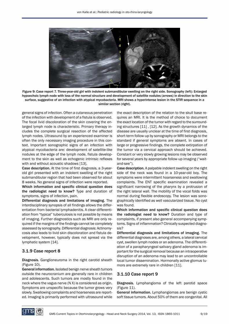
Figure 9: Case report 7. Three-year-old girl with indolent submandibular swelling on the right side. Sonography (left): Enlargedhypoechoic lymph node with loss of the normal structure and development of satellite nodules (arrows) in direction to the skinsurface, suggestive of an infection with atypical mycobacteria. MRI shows a hyperintense lesion in the STIR sequence in a
similar section (right).
general signs of infection. Often a cutaneous penetrationof the infection with development of a fistula is observed.The focal livid discoloration of the skin covering the en-larged lymph node is characteristic. Primary therapy in-cludes the complete surgical resection of the affectedlymph nodes. Ultrasound by an experienced examiner isoften the only necessary imaging procedure in this con-text. Important sonographic signs of an infection withatypical mycobacteria are: development of satellite-likenodules at the edge of the lymph node, fistula develop-ment to the skin as well as echogenic intrinsic reflexeswith and without acoustic shadows [13].Case description. At the time of first diagnosis, a 3-year-old girl presented with an indolent swelling of the rightsubmandibular region that had been observed for about8 weeks. No general signs of infection were reported.Which information and specific clinical question doesthe radiologist need to know? Type and duration ofsymptoms, signs of infection, pain.Differential diagnosis and limitations of imaging. Theinterdisciplinary synopsis of all findings allows the differ-entiation from bacterial lymphadenitis. A clear differenti-ation from “typical” tuberculosis is not possible by meansof imaging. Further diagnostics such as MRI are only re-quired if themargins of the findings cannot be completelyassessed by sonography. Differential diagnosis: Actinomy-cosis also leads to livid skin discoloration and fistula de-velopment, however, typically does not spread via thelymphatic system [14].
3.1.9 Case report 8
Diagnosis. Ganglioneuroma in the right carotid sheath(Figure 10).General information. Isolated benign nerve sheath tumorsoutside the neurocranium are generally rare in childrenand adolescents. Such tumors are mostly found in theneck where the vagus nerve (N X) is considered as origin.Symptoms are unspecific because the tumor grows veryslowly. Swallowing complaints and hoarseness are report-ed. Imaging is primarily performed with ultrasound while
the exact description of the relation to the skull base re-quires an MRI. It is the method of choice to documentthe exact location of the tumorwith regard to the surround-ing structures [11] , [12]. As the growth dynamics of thedisease are usually unclear at the time of first diagnosis,short-term follow-up by sonography or MRI belongs to thestandard if general symptoms are absent. In cases oflarge or progressive findings, the complete extirpation ofthe tumor via a cervical approach should be achieved.Constant or very slowly growing lesions may be observedfor several years by appropriate follow-up imaging (“wait-and-see”).Case description. A palpable indolent swelling on the rightside of the neck was found in a 10-year-old boy. Thesymptoms were intermittent hoarseness and swallowingcomplaints. The ENT specific examination revealed asignificant narrowing of the pharynx by a protrusion ofthe right lateral wall. The mobility of the vocal folds wasnormal during flexible endoscopy. The lesion was sono-graphically identified as well vascularized tissue. No cystwas found.Which information and specific clinical question doesthe radiologist need to know? Duration and type ofcomplaints, if present also general accompanying symp-toms. Signs of inflammation? Clinically suspected diagno-sis?Differential diagnosis and limitations of imaging. Thedifferential diagnoses are, among others, a lateral cervicalcyst, swollen lymph nodes or an adenoma. The differenti-ation of a parapharyngeal salivary gland adenoma is im-portant for the surgical removal because an intraoperativedisruption of an adenoma may lead to an uncontrollablelocal tumor dissemination. Hormonally active glomus tu-mors are extremely rare in children [11].
3.1.10 Case report 9
Diagnosis. Lymphangioma of the left parotid space(Figure 11).General information. Lymphangiomas are benign cysticsoft tissue tumors. About 50% of them are congenital. All
9/19GMS Current Topics in Otorhinolaryngology - Head and Neck Surgery 2014, Vol. 13, ISSN 1865-1011
von Kalle et al.: Pediatric radiology in oto-rhino-laryngology
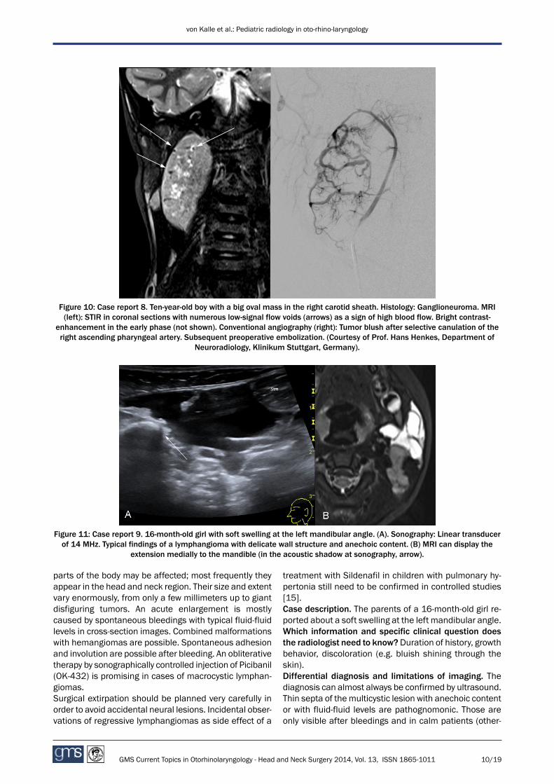
Figure 10: Case report 8. Ten-year-old boy with a big oval mass in the right carotid sheath. Histology: Ganglioneuroma. MRI(left): STIR in coronal sections with numerous low-signal flow voids (arrows) as a sign of high blood flow. Bright contrast-
enhancement in the early phase (not shown). Conventional angiography (right): Tumor blush after selective canulation of theright ascending pharyngeal artery. Subsequent preoperative embolization. (Courtesy of Prof. Hans Henkes, Department of
Neuroradiology, Klinikum Stuttgart, Germany).
Figure 11: Case report 9. 16-month-old girl with soft swelling at the left mandibular angle. (A). Sonography: Linear transducerof 14 MHz. Typical findings of a lymphangioma with delicate wall structure and anechoic content. (B) MRI can display the
extension medially to the mandible (in the acoustic shadow at sonography, arrow).
parts of the body may be affected; most frequently theyappear in the head and neck region. Their size and extentvary enormously, from only a few millimeters up to giantdisfiguring tumors. An acute enlargement is mostlycaused by spontaneous bleedings with typical fluid-fluidlevels in cross-section images. Combined malformationswith hemangiomas are possible. Spontaneous adhesionand involution are possible after bleeding. An obliterativetherapy by sonographically controlled injection of Picibanil(OK-432) is promising in cases of macrocystic lymphan-giomas.Surgical extirpation should be planned very carefully inorder to avoid accidental neural lesions. Incidental obser-vations of regressive lymphangiomas as side effect of a
treatment with Sildenafil in children with pulmonary hy-pertonia still need to be confirmed in controlled studies[15].Case description. The parents of a 16-month-old girl re-ported about a soft swelling at the left mandibular angle.Which information and specific clinical question doesthe radiologist need to know?Duration of history, growthbehavior, discoloration (e.g. bluish shining through theskin).Differential diagnosis and limitations of imaging. Thediagnosis can almost always be confirmed by ultrasound.Thin septa of themulticystic lesion with anechoic contentor with fluid-fluid levels are pathognomonic. Those areonly visible after bleedings and in calm patients (other-
10/19GMS Current Topics in Otorhinolaryngology - Head and Neck Surgery 2014, Vol. 13, ISSN 1865-1011
von Kalle et al.: Pediatric radiology in oto-rhino-laryngology
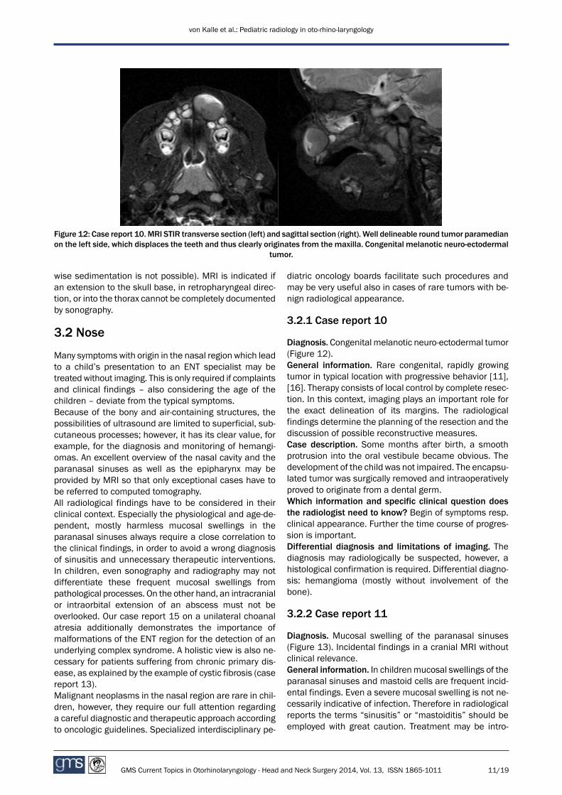
Figure 12: Case report 10. MRI STIR transverse section (left) and sagittal section (right). Well delineable round tumor paramedianon the left side, which displaces the teeth and thus clearly originates from the maxilla. Congenital melanotic neuro-ectodermal
tumor.
wise sedimentation is not possible). MRI is indicated ifan extension to the skull base, in retropharyngeal direc-tion, or into the thorax cannot be completely documentedby sonography.
3.2 Nose
Many symptoms with origin in the nasal region which leadto a child’s presentation to an ENT specialist may betreated without imaging. This is only required if complaintsand clinical findings – also considering the age of thechildren – deviate from the typical symptoms.Because of the bony and air-containing structures, thepossibilities of ultrasound are limited to superficial, sub-cutaneous processes; however, it has its clear value, forexample, for the diagnosis and monitoring of hemangi-omas. An excellent overview of the nasal cavity and theparanasal sinuses as well as the epipharynx may beprovided by MRI so that only exceptional cases have tobe referred to computed tomography.All radiological findings have to be considered in theirclinical context. Especially the physiological and age-de-pendent, mostly harmless mucosal swellings in theparanasal sinuses always require a close correlation tothe clinical findings, in order to avoid a wrong diagnosisof sinusitis and unnecessary therapeutic interventions.In children, even sonography and radiography may notdifferentiate these frequent mucosal swellings frompathological processes. On the other hand, an intracranialor intraorbital extension of an abscess must not beoverlooked. Our case report 15 on a unilateral choanalatresia additionally demonstrates the importance ofmalformations of the ENT region for the detection of anunderlying complex syndrome. A holistic view is also ne-cessary for patients suffering from chronic primary dis-ease, as explained by the example of cystic fibrosis (casereport 13).Malignant neoplasms in the nasal region are rare in chil-dren, however, they require our full attention regardinga careful diagnostic and therapeutic approach accordingto oncologic guidelines. Specialized interdisciplinary pe-
diatric oncology boards facilitate such procedures andmay be very useful also in cases of rare tumors with be-nign radiological appearance.
3.2.1 Case report 10
Diagnosis. Congenital melanotic neuro-ectodermal tumor(Figure 12).General information. Rare congenital, rapidly growingtumor in typical location with progressive behavior [11],[16]. Therapy consists of local control by complete resec-tion. In this context, imaging plays an important role forthe exact delineation of its margins. The radiologicalfindings determine the planning of the resection and thediscussion of possible reconstructive measures.Case description. Some months after birth, a smoothprotrusion into the oral vestibule became obvious. Thedevelopment of the child was not impaired. The encapsu-lated tumor was surgically removed and intraoperativelyproved to originate from a dental germ.Which information and specific clinical question doesthe radiologist need to know? Begin of symptoms resp.clinical appearance. Further the time course of progres-sion is important.Differential diagnosis and limitations of imaging. Thediagnosis may radiologically be suspected, however, ahistological confirmation is required. Differential diagno-sis: hemangioma (mostly without involvement of thebone).
3.2.2 Case report 11
Diagnosis. Mucosal swelling of the paranasal sinuses(Figure 13). Incidental findings in a cranial MRI withoutclinical relevance.General information. In childrenmucosal swellings of theparanasal sinuses and mastoid cells are frequent incid-ental findings. Even a severe mucosal swelling is not ne-cessarily indicative of infection. Therefore in radiologicalreports the terms “sinusitis” or “mastoiditis” should beemployed with great caution. Treatment may be intro-
11/19GMS Current Topics in Otorhinolaryngology - Head and Neck Surgery 2014, Vol. 13, ISSN 1865-1011
von Kalle et al.: Pediatric radiology in oto-rhino-laryngology
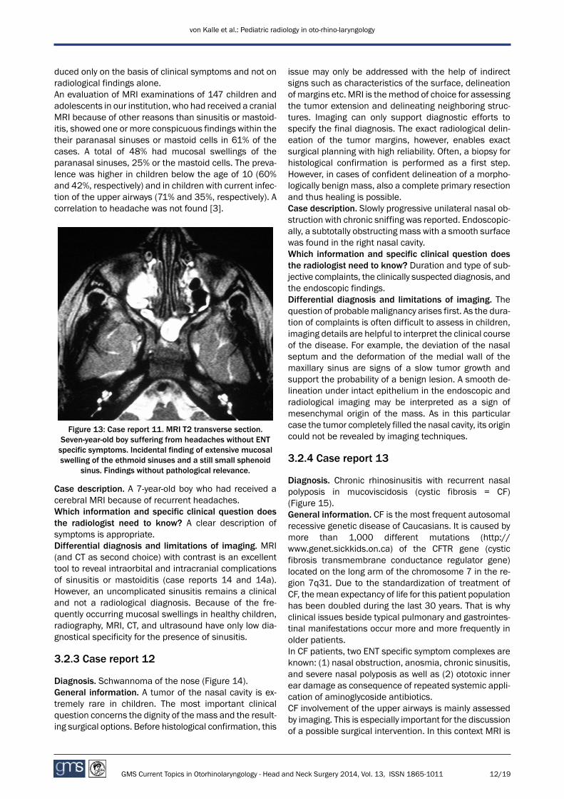
duced only on the basis of clinical symptoms and not onradiological findings alone.An evaluation of MRI examinations of 147 children andadolescents in our institution, who had received a cranialMRI because of other reasons than sinusitis or mastoid-itis, showed one or more conspicuous findings within thetheir paranasal sinuses or mastoid cells in 61% of thecases. A total of 48% had mucosal swellings of theparanasal sinuses, 25% or the mastoid cells. The preva-lence was higher in children below the age of 10 (60%and 42%, respectively) and in children with current infec-tion of the upper airways (71% and 35%, respectively). Acorrelation to headache was not found [3].
Figure 13: Case report 11. MRI T2 transverse section.Seven-year-old boy suffering from headaches without ENTspecific symptoms. Incidental finding of extensive mucosalswelling of the ethmoid sinuses and a still small sphenoid
sinus. Findings without pathological relevance.
Case description. A 7-year-old boy who had received acerebral MRI because of recurrent headaches.Which information and specific clinical question doesthe radiologist need to know? A clear description ofsymptoms is appropriate.Differential diagnosis and limitations of imaging. MRI(and CT as second choice) with contrast is an excellenttool to reveal intraorbital and intracranial complicationsof sinusitis or mastoiditis (case reports 14 and 14a).However, an uncomplicated sinusitis remains a clinicaland not a radiological diagnosis. Because of the fre-quently occurring mucosal swellings in healthy children,radiography, MRI, CT, and ultrasound have only low dia-gnostical specificity for the presence of sinusitis.
3.2.3 Case report 12
Diagnosis. Schwannoma of the nose (Figure 14).General information. A tumor of the nasal cavity is ex-tremely rare in children. The most important clinicalquestion concerns the dignity of the mass and the result-ing surgical options. Before histological confirmation, this
issue may only be addressed with the help of indirectsigns such as characteristics of the surface, delineationof margins etc. MRI is themethod of choice for assessingthe tumor extension and delineating neighboring struc-tures. Imaging can only support diagnostic efforts tospecify the final diagnosis. The exact radiological delin-eation of the tumor margins, however, enables exactsurgical planning with high reliability. Often, a biopsy forhistological confirmation is performed as a first step.However, in cases of confident delineation of a morpho-logically benign mass, also a complete primary resectionand thus healing is possible.Case description. Slowly progressive unilateral nasal ob-struction with chronic sniffing was reported. Endoscopic-ally, a subtotally obstructing mass with a smooth surfacewas found in the right nasal cavity.Which information and specific clinical question doesthe radiologist need to know? Duration and type of sub-jective complaints, the clinically suspected diagnosis, andthe endoscopic findings.Differential diagnosis and limitations of imaging. Thequestion of probablemalignancy arises first. As the dura-tion of complaints is often difficult to assess in children,imaging details are helpful to interpret the clinical courseof the disease. For example, the deviation of the nasalseptum and the deformation of the medial wall of themaxillary sinus are signs of a slow tumor growth andsupport the probability of a benign lesion. A smooth de-lineation under intact epithelium in the endoscopic andradiological imaging may be interpreted as a sign ofmesenchymal origin of the mass. As in this particularcase the tumor completely filled the nasal cavity, its origincould not be revealed by imaging techniques.
3.2.4 Case report 13
Diagnosis. Chronic rhinosinusitis with recurrent nasalpolyposis in mucoviscidosis (cystic fibrosis = CF)(Figure 15).General information. CF is the most frequent autosomalrecessive genetic disease of Caucasians. It is caused bymore than 1,000 different mutations (http://www.genet.sickkids.on.ca) of the CFTR gene (cysticfibrosis transmembrane conductance regulator gene)located on the long arm of the chromosome 7 in the re-gion 7q31. Due to the standardization of treatment ofCF, themean expectancy of life for this patient populationhas been doubled during the last 30 years. That is whyclinical issues beside typical pulmonary and gastrointes-tinal manifestations occur more and more frequently inolder patients.In CF patients, two ENT specific symptom complexes areknown: (1) nasal obstruction, anosmia, chronic sinusitis,and severe nasal polyposis as well as (2) ototoxic innerear damage as consequence of repeated systemic appli-cation of aminoglycoside antibiotics.CF involvement of the upper airways is mainly assessedby imaging. This is especially important for the discussionof a possible surgical intervention. In this context MRI is
12/19GMS Current Topics in Otorhinolaryngology - Head and Neck Surgery 2014, Vol. 13, ISSN 1865-1011
von Kalle et al.: Pediatric radiology in oto-rhino-laryngology
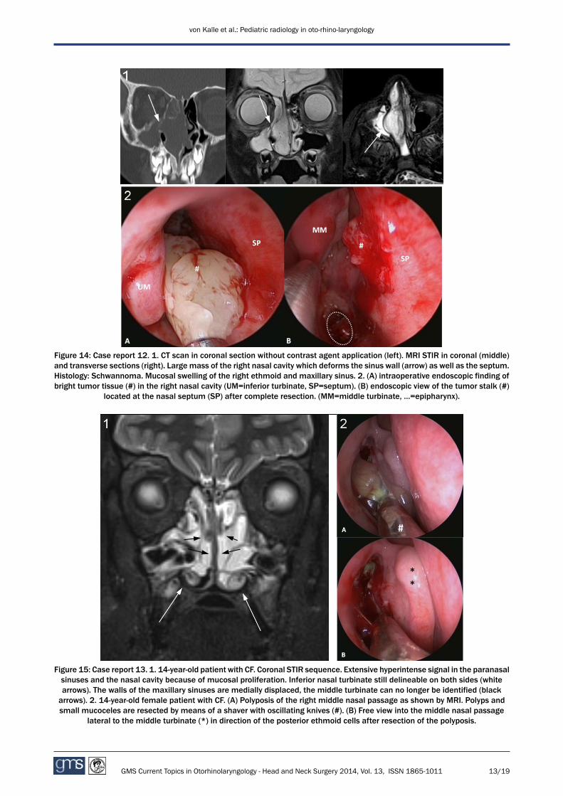
Figure 14: Case report 12. 1. CT scan in coronal section without contrast agent application (left). MRI STIR in coronal (middle)and transverse sections (right). Large mass of the right nasal cavity which deforms the sinus wall (arrow) as well as the septum.Histology: Schwannoma. Mucosal swelling of the right ethmoid and maxillary sinus. 2. (A) intraoperative endoscopic finding ofbright tumor tissue (#) in the right nasal cavity (UM=inferior turbinate, SP=septum). (B) endoscopic view of the tumor stalk (#)
located at the nasal septum (SP) after complete resection. (MM=middle turbinate, …=epipharynx).
Figure 15: Case report 13. 1. 14-year-old patient with CF. Coronal STIR sequence. Extensive hyperintense signal in the paranasalsinuses and the nasal cavity because of mucosal proliferation. Inferior nasal turbinate still delineable on both sides (whitearrows). The walls of the maxillary sinuses are medially displaced, the middle turbinate can no longer be identified (blackarrows). 2. 14-year-old female patient with CF. (A) Polyposis of the right middle nasal passage as shown by MRI. Polyps andsmall mucoceles are resected by means of a shaver with oscillating knives (#). (B) Free view into the middle nasal passage
lateral to the middle turbinate (*) in direction of the posterior ethmoid cells after resection of the polyposis.
13/19GMS Current Topics in Otorhinolaryngology - Head and Neck Surgery 2014, Vol. 13, ISSN 1865-1011
von Kalle et al.: Pediatric radiology in oto-rhino-laryngology
primarily recommended. This method has major advan-tages regarding discrimination. With the help of contrastagents it allows to differentiate well between retainedsecretion and mucosal swelling. The second significantadvantage is that it avoids radiation exposure. The CT,as an imaging alternative, should be avoided especiallyin cases of repeated examinations, as they may be re-quired in CF patients. The cumulative radiation exposuremust not be underestimated. Recent epidemiologicallong-term evaluations point to the increased lifetimecancer risk of patients who underwent a CT scan inchildhood [17].Case description. The 14-year-old girl complained aboutincreasing nasal obstruction. Endoscopic examinationrevealed a typical bilateral nasal polyposis andmedializa-tion of the lateral nasal walls. The tympanum on bothsides was aerated and without mucus retention.Which information and specific clinical question doesthe radiologist need to know? Basic knowledge of theunderlying disease of CF. Duration and type of complaintsand possible complications.Differential diagnoses and limitations of imaging. Thedocumentation of pathological alterations of the para-nasal sinuses complements the endoscopic and clinicalfindings, but is not as decisive for the indication of surgeryas in patients who do not suffer from CF.MRI of the paranasal sinuses in CF patients helps to un-derstand the differences in the development of sinusitiscompared to non-CF patients. Nearly all CF patients showan abnormal mucosal swelling, but only a small part ofthem report nasal symptoms or develop polyposis. An-other peculiarity of CF sinusitis is the primary location ofthe relevant pathological changes in themaxillary sinusesthat frequently medialize the lateral nasal walls similarto a mucocele. Often the ethmoid cells are not affected,so that in CF a sinugenic rather than a rhinogenic origin(as in non-CF) of the sinusitis may be assumed.Chronic sinusitis with nasal polyposis in children shouldalways lead to further diagnostics considering systemicand genetic causes. Beside CF, thosemay be other genet-ic defects such as primary ciliary dyskinesia (PCD) orKartagener syndrome.MRI is not able to directly display the osseous lamellae.However, with some experience they can be easily recog-nized as “negative images”. Usually sinus surgeons arenot used to this type of imaging and preferably rely on CTscans. However, in order to avoid radiation exposure inchildrenMRI is highly recommended as first line imaging.It is the surgeon’s responsibility to decide if the anatomicsituation of an individual case is sufficiently described bya certain imaging method. Generally CT scan, DVT, andMRI are not completely comparable, but all are appropri-ate for guiding a surgical intervention [18].
3.2.5 Case report 14
Diagnosis. Orbital complication of ethmoid sinusitis (Fig-ure 16).
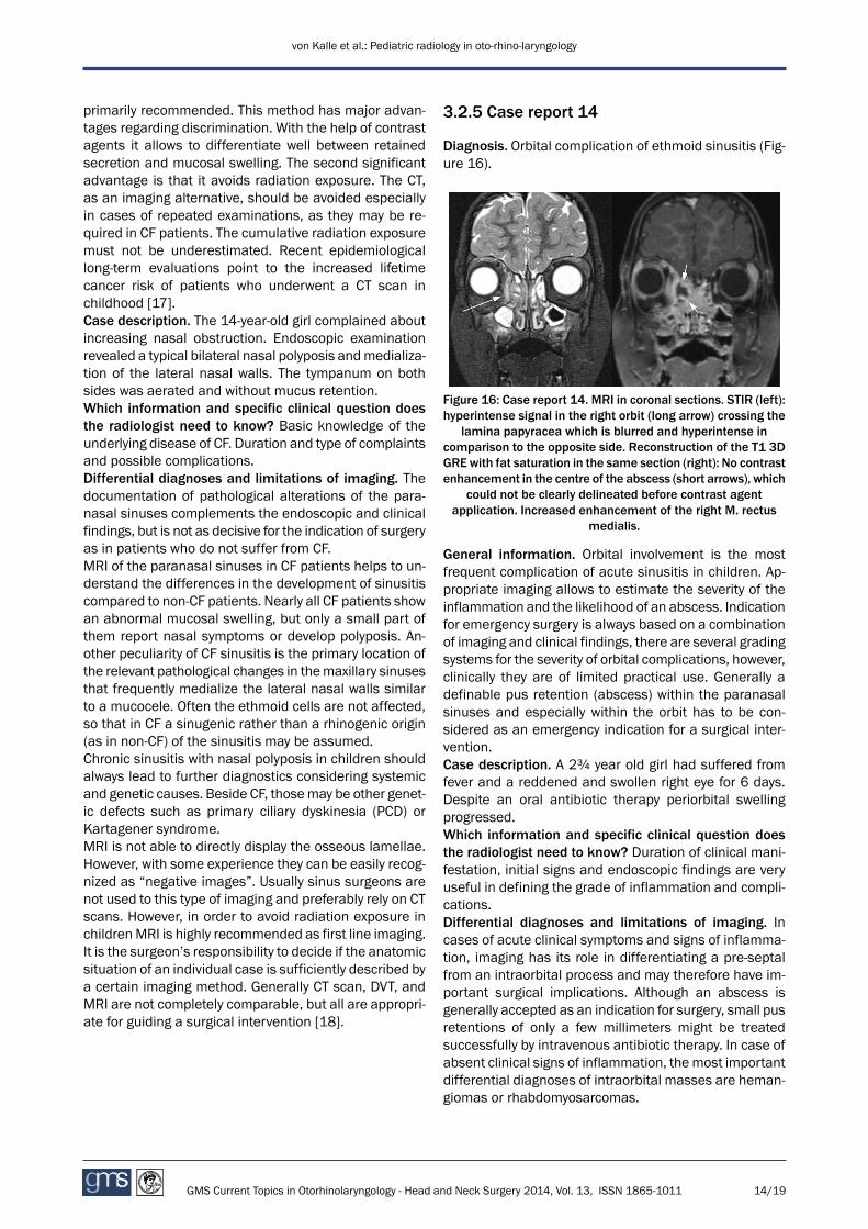
Figure 16: Case report 14. MRI in coronal sections. STIR (left):hyperintense signal in the right orbit (long arrow) crossing the
lamina papyracea which is blurred and hyperintense incomparison to the opposite side. Reconstruction of the T1 3DGREwith fat saturation in the same section (right): No contrastenhancement in the centre of the abscess (short arrows), which
could not be clearly delineated before contrast agentapplication. Increased enhancement of the right M. rectus
medialis.
General information. Orbital involvement is the mostfrequent complication of acute sinusitis in children. Ap-propriate imaging allows to estimate the severity of theinflammation and the likelihood of an abscess. Indicationfor emergency surgery is always based on a combinationof imaging and clinical findings, there are several gradingsystems for the severity of orbital complications, however,clinically they are of limited practical use. Generally adefinable pus retention (abscess) within the paranasalsinuses and especially within the orbit has to be con-sidered as an emergency indication for a surgical inter-vention.Case description. A 2¾ year old girl had suffered fromfever and a reddened and swollen right eye for 6 days.Despite an oral antibiotic therapy periorbital swellingprogressed.Which information and specific clinical question doesthe radiologist need to know? Duration of clinical mani-festation, initial signs and endoscopic findings are veryuseful in defining the grade of inflammation and compli-cations.Differential diagnoses and limitations of imaging. Incases of acute clinical symptoms and signs of inflamma-tion, imaging has its role in differentiating a pre-septalfrom an intraorbital process and may therefore have im-portant surgical implications. Although an abscess isgenerally accepted as an indication for surgery, small pusretentions of only a few millimeters might be treatedsuccessfully by intravenous antibiotic therapy. In case ofabsent clinical signs of inflammation, themost importantdifferential diagnoses of intraorbital masses are heman-giomas or rhabdomyosarcomas.
14/19GMS Current Topics in Otorhinolaryngology - Head and Neck Surgery 2014, Vol. 13, ISSN 1865-1011
von Kalle et al.: Pediatric radiology in oto-rhino-laryngology
3.2.6 Case report 14a
The additional case report 14a of an intra-cranial sinugen-ic abscess (Figure 17) demonstrates the significance ofcontrast agent application also for CT and the superiorityof MRI in the depiction of soft tissue pathologies.
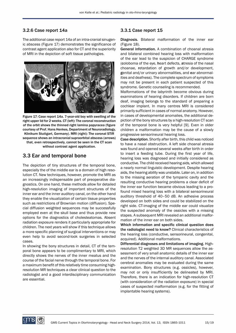
Figure 17: Case report 14a. 7-year-old boy with swelling of theright upper lid for 2 weeks. CT (left): The coronal reconstructionof the orbit shows the thinned right lamina papyracea (figurecourtesy of Prof. Hans Henkes, Department of Neuroradiology,Klinikum Stuttgart, Germany). MRI (right): The coronal STIRsequence shows an intracranial supraorbital abscess (arrows)that, even retrospectively, cannot be seen in the CT scan
without contrast agent application.
3.3 Ear and temporal bone
The depiction of tiny structures of the temporal bone,especially the of the middle ear is a domain of high reso-lution CT. New techniques, however, promote the MRI asan increasingly indispensable part of preoperative dia-gnostics. On one hand, these methods allow for detailedhigh-resolution imaging of important structures of theinner ear and the inner auditory canal, on the other handthey enable the visualization of certain tissue propertiessuch as restrictions of Brownian motion (diffusion). Spe-cial diffusion weighted sequences may be successfullyemployed even at the skull base and thus provide newoptions for the diagnostics of cholesteatomas. Absentradiation exposure renders it particularly appropriate forchildren. The next years will show if this technique allowsa more specific planning of surgical interventions or mayeven help to avoid second-look surgeries in selectedcases.In showing the bony structures in detail, CT of the tem-poral bone appears to be complimentary to MRI, whichdirectly shows the nerves of the inner meatus and thecourse of the facial nerve through the temporal bone. Foramaximumbenefit of this relatively time consuming high-resolution MR techniques a clear clinical question to theradiologist and a good interdisciplinary communicationare essential.
3.3.1 Case report 15
Diagnosis. Bilateral malformation of the inner ear(Figure 18).General information. A combination of choanal atresiaand bilateral combined hearing loss with malformationof the ear lead to the suspicion of CHARGE syndrome(coloboma of the eye, heart defects, atresia of the nasalchoanae, retardation of growth and/or development,genital and/or urinary abnormalities, and ear abnormal-ities and deafness). The complete spectrum of symptomsmay not be present in each patient suspected of thissyndrome. Genetic counseling is recommended.Malformations of the labyrinth become obvious duringexaminations of hearing disorders. If children are borndeaf, imaging belongs to the standard of preparing acochlear implant. In many centres MRI is consideredprimarily sufficient in cases of normal anatomy. However,in cases of developmental anomalies, the additional de-piction of the bony structures by a high-resolution CT scanof the temporal bone is very helpful [9]. Even in olderchildren a malformation may be the cause of a slowlyprogressive sensorineural hearing loss.Case description. Shortly after birth, this child was noticedto have a nasal obstruction. A left side choanal atresiawas found and opened several weeks after birth in orderto insert a feeding tube. During the first year of life,hearing loss was diagnosed and initially considered asconductive. The child received hearing aids, which alloweda nearly normal linguistic development. Despite hearingaids, the hearing ability was unstable. Later on, in additionto the missing aeration of the tympanic cavity and theresulting conductive hearing problems a clear deficit ofthe inner ear function became obvious leading to a pro-found mixed hearing loss with a bilateral sensorineuralauditory threshold of 40–50 dB. An adhesive processdeveloped on both sides and could be stabilized on theright side. CT-imaging of the middle ear could visualizethe suspected anomaly of the ossicles with a missingstapes. A subsequent MRI revealed an additional malfor-mation of the inner ear on both sides.Which information and specific clinical question doesthe radiologist need to know? Clinical characteristics ofthe hearing loss (conductive, sensorineural, congenital,acquired). Additional malformations.Differential diagnoses and limitations of imaging. High-resolution T2 weighted 3D MR sequences allow the as-sessment of very small anatomic details of the inner earand the nerves of the internal auditory canal. Associatedcerebral anomalies may be evaluated during the sameexamination. Bony structures (e.g. ossicles), however,may not or only insufficiently be delineated by MRI.Therefore, there is an indication for high-resolution CT(with consideration of the radiation exposure) in specialcases of suspected malformation (e.g. for the fitting ofimplantable hearing systems).
15/19GMS Current Topics in Otorhinolaryngology - Head and Neck Surgery 2014, Vol. 13, ISSN 1865-1011
von Kalle et al.: Pediatric radiology in oto-rhino-laryngology
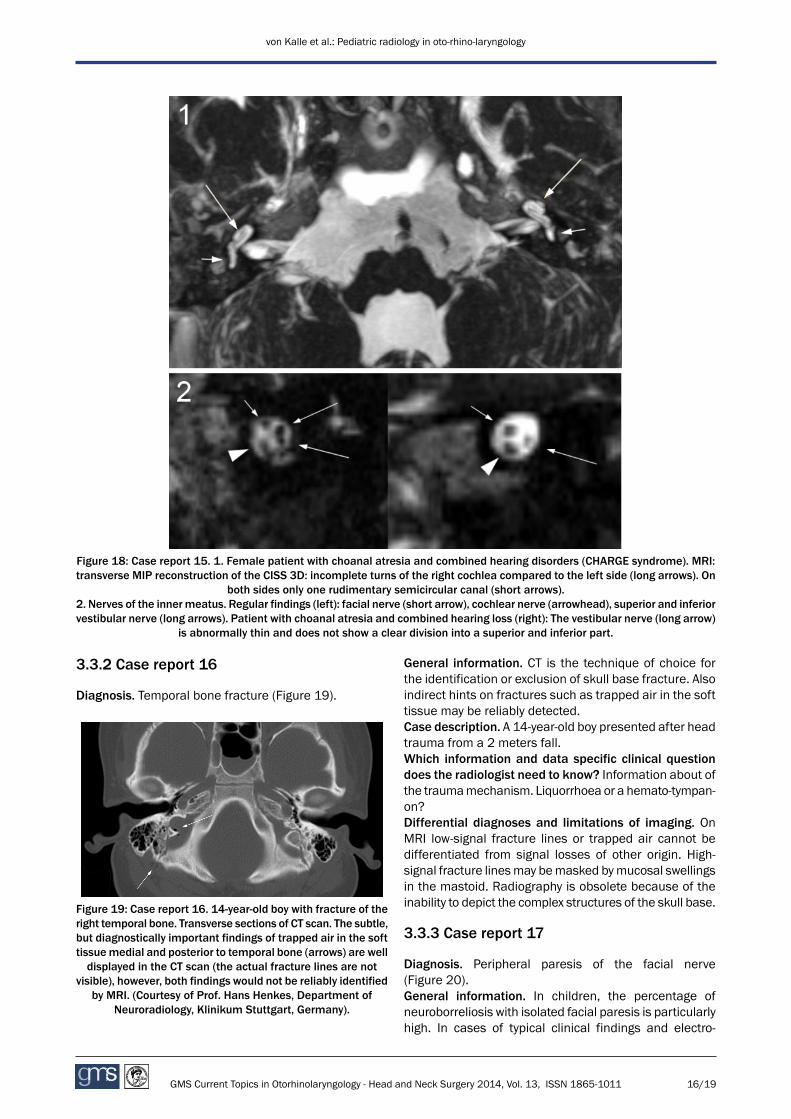
Figure 18: Case report 15. 1. Female patient with choanal atresia and combined hearing disorders (CHARGE syndrome). MRI:transverse MIP reconstruction of the CISS 3D: incomplete turns of the right cochlea compared to the left side (long arrows). On
both sides only one rudimentary semicircular canal (short arrows).2. Nerves of the innermeatus. Regular findings (left): facial nerve (short arrow), cochlear nerve (arrowhead), superior and inferiorvestibular nerve (long arrows). Patient with choanal atresia and combined hearing loss (right): The vestibular nerve (long arrow)
is abnormally thin and does not show a clear division into a superior and inferior part.
3.3.2 Case report 16
Diagnosis. Temporal bone fracture (Figure 19).
Figure 19: Case report 16. 14-year-old boy with fracture of theright temporal bone. Transverse sections of CT scan. The subtle,but diagnostically important findings of trapped air in the softtissuemedial and posterior to temporal bone (arrows) are welldisplayed in the CT scan (the actual fracture lines are not
visible), however, both findings would not be reliably identifiedby MRI. (Courtesy of Prof. Hans Henkes, Department of
Neuroradiology, Klinikum Stuttgart, Germany).
General information. CT is the technique of choice forthe identification or exclusion of skull base fracture. Alsoindirect hints on fractures such as trapped air in the softtissue may be reliably detected.Case description. A 14-year-old boy presented after headtrauma from a 2 meters fall.Which information and data specific clinical questiondoes the radiologist need to know? Information about ofthe traumamechanism. Liquorrhoea or a hemato-tympan-on?Differential diagnoses and limitations of imaging. OnMRI low-signal fracture lines or trapped air cannot bedifferentiated from signal losses of other origin. High-signal fracture linesmay bemasked bymucosal swellingsin the mastoid. Radiography is obsolete because of theinability to depict the complex structures of the skull base.
3.3.3 Case report 17
Diagnosis. Peripheral paresis of the facial nerve(Figure 20).General information. In children, the percentage ofneuroborreliosis with isolated facial paresis is particularlyhigh. In cases of typical clinical findings and electro-
16/19GMS Current Topics in Otorhinolaryngology - Head and Neck Surgery 2014, Vol. 13, ISSN 1865-1011
von Kalle et al.: Pediatric radiology in oto-rhino-laryngology
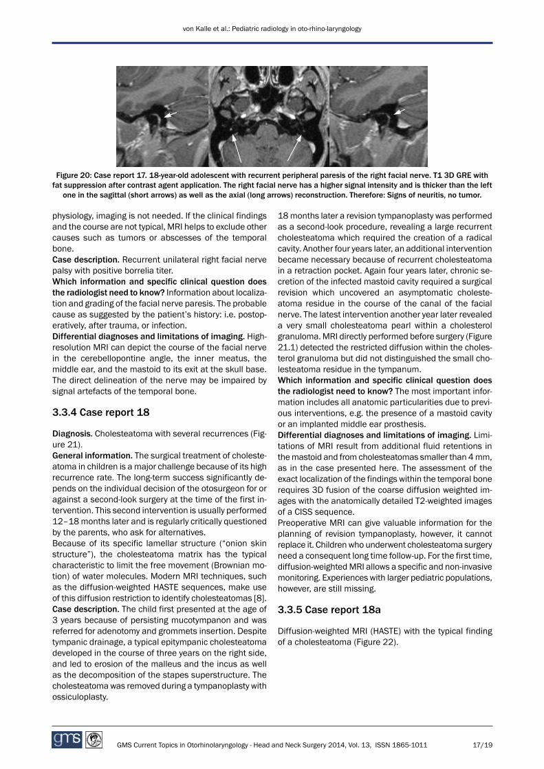
Figure 20: Case report 17. 18-year-old adolescent with recurrent peripheral paresis of the right facial nerve. T1 3D GRE withfat suppression after contrast agent application. The right facial nerve has a higher signal intensity and is thicker than the left
one in the sagittal (short arrows) as well as the axial (long arrows) reconstruction. Therefore: Signs of neuritis, no tumor.
physiology, imaging is not needed. If the clinical findingsand the course are not typical, MRI helps to exclude othercauses such as tumors or abscesses of the temporalbone.Case description. Recurrent unilateral right facial nervepalsy with positive borrelia titer.Which information and specific clinical question doesthe radiologist need to know? Information about localiza-tion and grading of the facial nerve paresis. The probablecause as suggested by the patient’s history: i.e. postop-eratively, after trauma, or infection.Differential diagnoses and limitations of imaging. High-resolution MRI can depict the course of the facial nervein the cerebellopontine angle, the inner meatus, themiddle ear, and the mastoid to its exit at the skull base.The direct delineation of the nerve may be impaired bysignal artefacts of the temporal bone.
3.3.4 Case report 18
Diagnosis. Cholesteatoma with several recurrences (Fig-ure 21).General information. The surgical treatment of choleste-atoma in children is amajor challenge because of its highrecurrence rate. The long-term success significantly de-pends on the individual decision of the otosurgeon for oragainst a second-look surgery at the time of the first in-tervention. This second intervention is usually performed12–18months later and is regularly critically questionedby the parents, who ask for alternatives.Because of its specific lamellar structure (“onion skinstructure”), the cholesteatoma matrix has the typicalcharacteristic to limit the free movement (Brownian mo-tion) of water molecules. Modern MRI techniques, suchas the diffusion-weighted HASTE sequences, make useof this diffusion restriction to identify cholesteatomas [8].Case description. The child first presented at the age of3 years because of persisting mucotympanon and wasreferred for adenotomy and grommets insertion. Despitetympanic drainage, a typical epitympanic cholesteatomadeveloped in the course of three years on the right side,and led to erosion of the malleus and the incus as wellas the decomposition of the stapes superstructure. Thecholesteatomawas removed during a tympanoplasty withossiculoplasty.
18months later a revision tympanoplasty was performedas a second-look procedure, revealing a large recurrentcholesteatoma which required the creation of a radicalcavity. Another four years later, an additional interventionbecame necessary because of recurrent cholesteatomain a retraction pocket. Again four years later, chronic se-cretion of the infected mastoid cavity required a surgicalrevision which uncovered an asymptomatic choleste-atoma residue in the course of the canal of the facialnerve. The latest intervention another year later revealeda very small cholesteatoma pearl within a cholesterolgranuloma.MRI directly performed before surgery (Figure21.1) detected the restricted diffusion within the choles-terol granuloma but did not distinguished the small cho-lesteatoma residue in the tympanum.Which information and specific clinical question doesthe radiologist need to know? The most important infor-mation includes all anatomic particularities due to previ-ous interventions, e.g. the presence of a mastoid cavityor an implanted middle ear prosthesis.Differential diagnoses and limitations of imaging. Limi-tations of MRI result from additional fluid retentions inthemastoid and from cholesteatomas smaller than 4mm,as in the case presented here. The assessment of theexact localization of the findings within the temporal bonerequires 3D fusion of the coarse diffusion weighted im-ages with the anatomically detailed T2-weighted imagesof a CISS sequence.Preoperative MRI can give valuable information for theplanning of revision tympanoplasty, however, it cannotreplace it. Childrenwho underwent cholesteatoma surgeryneed a consequent long time follow-up. For the first time,diffusion-weightedMRI allows a specific and non-invasivemonitoring. Experiences with larger pediatric populations,however, are still missing.
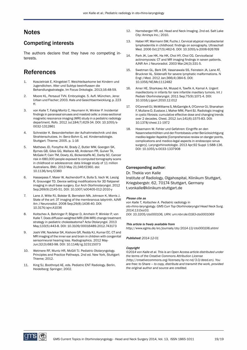
3.3.5 Case report 18a
Diffusion-weighted MRI (HASTE) with the typical findingof a cholesteatoma (Figure 22).
17/19GMS Current Topics in Otorhinolaryngology - Head and Neck Surgery 2014, Vol. 13, ISSN 1865-1011
von Kalle et al.: Pediatric radiology in oto-rhino-laryngology
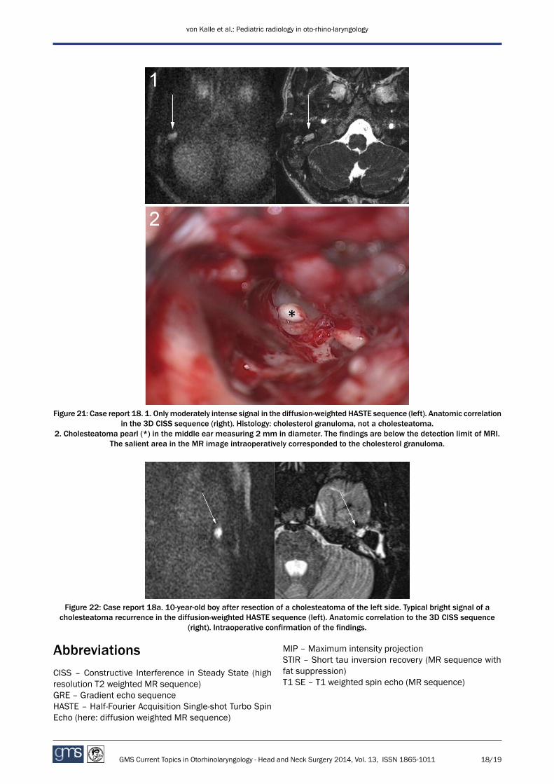
Figure 21: Case report 18. 1. Onlymoderately intense signal in the diffusion-weighted HASTE sequence (left). Anatomic correlationin the 3D CISS sequence (right). Histology: cholesterol granuloma, not a cholesteatoma.
2. Cholesteatoma pearl (*) in the middle ear measuring 2 mm in diameter. The findings are below the detection limit of MRI.The salient area in the MR image intraoperatively corresponded to the cholesterol granuloma.
Figure 22: Case report 18a. 10-year-old boy after resection of a cholesteatoma of the left side. Typical bright signal of acholesteatoma recurrence in the diffusion-weighted HASTE sequence (left). Anatomic correlation to the 3D CISS sequence
(right). Intraoperative confirmation of the findings.
AbbreviationsCISS – Constructive Interference in Steady State (highresolution T2 weighted MR sequence)GRE – Gradient echo sequenceHASTE – Half-Fourier Acquisition Single-shot Turbo SpinEcho (here: diffusion weighted MR sequence)
MIP – Maximum intensity projectionSTIR – Short tau inversion recovery (MR sequence withfat suppression)T1 SE – T1 weighted spin echo (MR sequence)
18/19GMS Current Topics in Otorhinolaryngology - Head and Neck Surgery 2014, Vol. 13, ISSN 1865-1011
von Kalle et al.: Pediatric radiology in oto-rhino-laryngology
Notes
Competing interests
The authors declare that they have no competing in-terests.
References1. Koscielniak E, Klingebiel T. Weichteilsarkome bei Kindern und
Jugendlichen. Alter und Subtyp beeinflussen dieBehandlungsstrategie. Im Focus Onkologie. 2013;16:48-59.
2. Moore KL, Persaud TVN. Embryologie. 5. Aufl. München, Jena:Urban und Fischer; 2003. Hals und Gesichtsentwicklung. p. 223ff.
3. von Kalle T, Fabig-Moritz C, Heumann H, Winkler P. Incidentalfindings in paranasal sinuses andmastoid cells: a cross-sectionalmagnetic resonance imaging (MRI) study in a pediatric radiologydepartment. Rofo. 2012 Jul;184(7):629-34. DOI: 10.1055/s-0032-1312861
4. Schneider K. Besonderheiten der Aufnahmetechnik und desStrahlenschutzes. In: Benz-Bohm G, ed. Kinderradiologie.Stuttgart: Thieme; 2005. p. 1-16
5. Mathews JD, Forsythe AV, Brady Z, Butler MW, Goergen SK,Byrnes GB, Giles GG, Wallace AB, Anderson PR, Guiver TA,McGale P, Cain TM, Dowty JG, Bickerstaffe AC, Darby SC. Cancerrisk in 680,000 people exposed to computed tomography scansin childhood or adolescence: data linkage study of 11 millionAustralians. BMJ. 2013 May 21;346:f2360. doi:10.1136/bmj.f2360
6. Hassepass F, Maier W, Aschendorff A, Bulla S, Vach W, LaszigR, Grauvogel TD. Device setting modifications for 3D flatpanelimaging in skull base surgery. Eur Arch Otorhinolaryngol. 2012Sep;269(9):2145-51. DOI: 10.1007/s00405-012-2010-y
7. Lane JI, Witte RJ, Bolster B, Bernstein MA, Johnson K, Morris J.State of the art: 3T imaging of the membranous labyrinth. AJNRAm J Neuroradiol. 2008 Sep;29(8):1436-40. DOI:10.3174/ajnr.A1036
8. Koitschev A, Behringer P, Bögner D, Amrhein P, Winkler P, vonKalle T. Does diffusion-weightedMRI (DW-MRI) change treatmentstrategy in pediatric cholesteatoma? Acta Otolaryngol. 2013May;133(5):443-8. DOI: 10.3109/00016489.2012.743173
9. Joshi VM, Navlekar SK, Kishore GR, Reddy KJ, Kumar EC. CT andMR imaging of the inner ear and brain in children with congenitalsensorineural hearing loss. Radiographics. 2012 May-Jun;32(3):683-98. DOI: 10.1148/rg.323115073
10. Wetmere RF, Muntz HR, McGill TJ. Pediatric Otolaryngology.Principles and Practice Pathways. 2nd ed. New York, Stuttgart:Thieme; 2012.
11. King SJ, Boothroyd AE, eds. Pediatric ENT Radiology. Berlin,Heidelberg: Springer; 2002.
12. Harnsberger HR, ed. Head and Neck Imaging. 2nd ed. Salt LakeCity: Amirsys Inc.; 2010.
13. Haber HP, Warmann SW, Fuchs J. Cervical atypical mycobacteriallymphadenitis in childhood: findings on sonography. UltraschallMed. 2006 Oct;27(5):462-6. DOI: 10.1055/s-2006-926769
14. Park JK, Lee HK, Ha HK, Choi HY, Choi CG. Cervicofacialactinomycosis: CT and MR imaging findings in seven patients.AJNR Am J Neuroradiol. 2003 Mar;24(3):331-5.
15. Swetman GL, Berk DR, Vasanawala SS, Feinstein JA, Lane AT,Bruckner AL. Sildenafil for severe lymphatic malformations. NEngl J Med. 2012 Jan;366(4):384-6. DOI:10.1056/NEJMc1112482
16. Amer HE, Sharkawy AA, Musad A, Tawfik A, Kamal A. Urgentmaxillectomy in infants for rare infantile maxillary tumors. Int JPediatr Otorhinolaryngol. 2011 Sep;75(9):1071-4. DOI:10.1016/j.ijporl.2010.12.012
17. O'Connell OJ,McWilliams S,McGarrigle A, O'Connor OJ, ShanahanF, Mullane D, Eustace J, MaherMM, Plant BJ. Radiologic imagingin cystic fibrosis: cumulative effective dose and changing trendsover 2 decades. Chest. 2012 Jun;141(6):1575-83. DOI:10.1378/chest.11-1972
18. Hosemann W. Fehler und Gefahren: Eingriffe an denNasennebenhöhlen und der Frontobasis unter Berücksichtigungmediko-legaler Aspekte [Comprehensive review on danger points,complications and medico-legal aspects in endoscopic sinussurgery]. Laryngorhinootologie. 2013 Apr;92 Suppl 1:S88-136.DOI: 10.1055/s-0033-1337908
Corresponding author:Dr. Thekla von KalleInstitute of Radiology, Olgahospital, Klinikum Stuttgart,Kriegsbergstr. 62, 70174 Stuttgart, [email protected]
Please cite asvon Kalle T, Koitschev A. Pediatric radiology inoto-rhino-laryngology. GMS Curr Top Otorhinolaryngol Head Neck Surg.2014;13:Doc03.DOI: 10.3205/cto000106, URN: urn:nbn:de:0183-cto0001069
This article is freely available fromhttp://www.egms.de/en/journals/cto/2014-13/cto000106.shtml
Published: 2014-12-01
Copyright©2014 von Kalle et al. This is an Open Access article distributed underthe terms of the Creative Commons Attribution License(http://creativecommons.org/licenses/by-nc-nd/3.0/deed.en). Youare free: to Share — to copy, distribute and transmit the work, providedthe original author and source are credited.
19/19GMS Current Topics in Otorhinolaryngology - Head and Neck Surgery 2014, Vol. 13, ISSN 1865-1011
von Kalle et al.: Pediatric radiology in oto-rhino-laryngology