pediatric deep neck infections: a 10-year retrospective ... · tiivistelmä...
TRANSCRIPT

!
Pediatric Deep Neck Infections: a 10-year retrospective single-centre study
Mikaela Römer
BM
Student number: 013609647
University of Helsinki, Clinicum
Helsinki University Hospital, Department of Otolaryngology- Head and Neck Surgery
Helsinki, April 01, 2015
MD Thesis
Supervisors:
Professor Antti Mäkitie, Department of Otolaryngology – Head and Neck Surgery
MD, PhD Johanna Nokso-Koivisto, Department of Otolaryngology – Head and Neck Surgery
UNIVERSITY OF HELSINKI
Faculty of Medicine
Helsinki, Finland

!

! i!
HELSINGIN!YLIOPISTO!−!HELSINGFORS!UNIVERSITET!Tiedekunta/Osasto!−!Fakultet/Sektion!–!Faculty! Faculty of Medicine!
Laitos!−!Institution!–!Department!Clinicum, Dept. of ORL-HNS!
Tekijä − Författare!–!Author! Mikaela Römer!Työn!nimi −!Arbetets!titel!–!Title! Pediatric deep neck infections: a 10-year retrospective single-centre study Oppiaine!−!Läroämne!–!Subject! Medicine!Työn!laji −!Arbetets!art!–!Level!MD Thesis!
Aika −!Datum!–!Month!and!year!April 2015
Sivumäärä MSidoantal!M!Number!of!pages! 32
Tiivistelmä −!Referat!–!Abstract!This thesis describes the characteristics and management of pediatric deep neck infections at
the Department of Otolaryngology – Head and Neck Surgery at the Helsinki University
Hospital during a 10-year period.
The cohort consisted of 62 patients. A majority (85%) of the patients underwent immediate or
late surgical intervention. Due to the low amount of conservatively treated patients (15%) no
factors suggestive for successful conservative treatment were recognized. However, initiation
of conservative treatment and close follow-up seem sufficient if the patient is in stable
condition. The mean duration for hospital stay was 4.8 days. Patients with a complicated
clinical course presented more likely with fever, cervical lymphadenopathy, trismus and
torticollis. Despite the chosen treatment modality, all patients recovered well and the risk of
serious complications was low.
Surgical drainage still remains the main treatment modality for this patient population at our
institution. Further studies are warranted to compose evidence-based treatment guidelines.
(Words: 150)
Avainsanat!–!Nyckelord!–!Keywords!Pediatric,!neck,!abscess,!parapharyngeal,!retropharyngeal,!treatment,!surgery,!outcome!Säilytyspaikka!–!Förvaringställe!–!Where!deposited! !Muita!tietoja!–!Övriga!uppgifter!–!Additional!information!!!

! !
!
ii!
Contents 1 INTRODUCTION ......................................................................................................................... 1
2 REVIEW OF THE LITERATURE ............................................................................................... 2
2.1 Background ............................................................................................................................. 2
2.1.1 Anatomy .......................................................................................................................... 2
2.1.2 Epidemiology ................................................................................................................... 4
2.1.3 Etiology ............................................................................................................................ 5
2.2 Findings .................................................................................................................................. 6
2.2.1 Symptoms ........................................................................................................................ 6
2.2.2 Signs ................................................................................................................................ 7
2.3 Diagnostics ............................................................................................................................. 8
2.3.1 Radiology / Imaging ........................................................................................................ 8
2.3.2 Microbiology ................................................................................................................... 9
2.4 Treatment .............................................................................................................................. 10
2.5 Complications ....................................................................................................................... 14
2.6 Outcome ................................................................................................................................ 15
3 AIMS OF THE THESIS .............................................................................................................. 16
4 MATERIALS AND METHODS ................................................................................................. 17
4.1 Materials ............................................................................................................................... 17
4.2 Methods ................................................................................................................................ 18
5 RESULTS .................................................................................................................................... 19
6 DISCUSSION .............................................................................................................................. 25
7 CONCLUSION ............................................................................................................................ 28
8 REFERENCES ............................................................................................................................ 29

! 1!
1 INTRODUCTION
Pediatric deep neck infections (DNI) are characterized as fairly common clinical conditions (1-5).
Pediatric DNIs are abscesses located in the deep cavities of the neck. They usually arise as
complications of upper respiratory tract infections or dental infections. Among pediatric patients the
cause is rarely a trauma of the head and neck region. Due to their location in deep spaces and
cavities of the neck and their capability of further spread towards the mediastinum and the skull
they are potentially life-threatening (1). Because of possible complications such as mediastinitis,
airway obstruction or even an aneurysm of the carotid artery it is important to recognize them early.
The diagnosis is based on clinical evaluation and radiological imaging but the specific mode of
treatment has been under wide discussion during the past 10 years. The literature does not present
clear guidelines as to whether a patient should be conservatively treated with intravenous antibiotics
or operatively treated with drainage of the abscess either transorally or externally through the neck.
Various studies have aimed at composing a treatment guideline to guide the clinician in the decision
making but no convincing treatment algorithms have been published (2,6). According to previous
research the outcome of the treatment remains good and the rate of complications will be low
regardless of the treatment modality chosen (2,4,7).
Earlier Finnish research exists on this topic, but the focus has been on patients in general rather than
mere pediatric patients (5,8-10). Therefore, it was of interest to investigate the characteristics of
pediatric deep neck abscesses, the choice of treatment and the outcome of the treatment for
pediatric DNIs in a Finnish patient population.

! !
!
2!
2 REVIEW OF THE LITERATURE
2.1 Background
!
2.1.1 Anatomy
Understanding the anatomy of the neck is helpful in understanding the localization and way of
spread of the deep neck infections. The deep neck spaces are cavities lined by muscles, muscle
fascias and other anatomical structures of the neck. They are lined by fascia cervicalis superficialis
and fascia cervicalis profunda, the superficial and deep fascia of the neck. Deep neck infections are
localized within cavities lined by fascia cervicalis profunda. Due to the build-up of these spaces
infections may spread through one cavity to the other, therefore professing the ability to lead to a
potentially wide-spread infection. Retropharyngeal processes may be complicated because this
space is directly connected to the mediastinum.
Depending on the literature the number of deep neck spaces varies from 6 to 12. They can be
divided into the parapharyngeal space, the retropharyngeal space, the prevertebral space, the
masticator space, the sublingual space, and the submandibular space (5). Some articles add six more
spaces to the list. These spaces are the danger space, the carotid space, the pretracheal space, the
peritonsillar space, the parotid space and the temporal space (11). The retropharyngeal space lies on
the posteromedial side of the parapharyngeal space. It is anteriorly lined by the pharynx and
esophagus and posteriorly by the vertebrae. This is the location of the retropharyngeal lymph nodes
of Rouvière.
The anatomy of the parapharyngeal space (Picture 1), the retropharyngeal space (Picture 2) and the
fascias of the neck (Picture 3) is presented in the following images.

! !
!
3!
Picture 1. The parapharyngeal space and close-by structures. (McGraw-Hills Access Surgery)
Picture 2. The retropharyngeal space and close-by structures. (https://www.studyblue.com/notes/note/n/block-2-clinical-correlates/deck/3439918)

! !
!
4!
Picture 3. A transversal and longitudinal view of the fascias of the neck. (5)
2.1.2 Epidemiology
Pediatric deep neck infections (DNI) are not totally uncommon and are characterized by a
potentially life-threatening clinical course (3,4,12,13). The incidence of pediatric deep neck
infections seems to be rising. Even a tenfold increase in incidence has been noted during the last
decades (13) (14). One of the reasons Pelaz et al. initially did their study was a sudden appearance
of 7 cases of pediatric DNIs during a 6-month period compared to none during the previous 4 years
(15). The reason to this rise of incidence is not exactly clear (14,16,17). Some link it to a
concomitant rise of incidence of more resistant microbes as well as the advent and wider
accessibility of advanced diagnosing possibilities such as computed tomography and magnetic
resonance imaging. The incidence was even higher though prior to the antibiotic era (18-21).
In 2014 Novis et al. published an article on a large group of pediatric deep neck infections
(N=41483). The patients were treated in the US during a 10-year time period. The patient data was
retrieved from a national database (KID – Kids’s Inpatient Database) with the aim of defining
whether the incidence of DNIs was on a rise or not. The results showed a clear rise in incidence of
!
Transversal!view!of!the!neck.! Longitudinal!view!of!the!neck.!
V.!jugularis!interna!
A.!carotis!interna!Parapharyngeal!space!
Superficial!fascia!
Prevertebral!fascia!
Pretracheal!fascia!
! !
!
5!
retropharyngeal abscesses (RPA’s) (from 0.10 to 0.22 / 10000). This was, however, not noted when
all DNIs including peritonsillar abscesses were grouped together. During this time period the
amount of operatively treated patients also diminished as conservative treatment became more
popular. (17)
Due to the anatomic development of the head and neck region, parapharyngeal and retropharyngeal
infections are more common among younger children. According to international studies the most
common age of presentation is around 3 to 6 years (2,4,14,22,23). There are some possible reasons
to this. Firstly, the lymphatic system is on a constant change in toddlers. The lymph nodes that are
typically affected in retropharyngeal or parapharyngeal abscesses usually begin atrophying and
diminishing after the age of 5. Secondly, the immune system is not fully developed in children,
which might be one of the reasons that common pathogens cause abscesses more commonly in
toddlers than adolescents. (3)
The manifestation seems to be highest during the winter months (3,4,24) which could be linked to
the larger number of upper respiratory tract infections during this time. Multiple studies present a
higher prevalence among boys than girls but no clear explanation to this has been found in the
literature (1,2,7,22,23). Duval et al. also noted that the likelihood to acquire retropharyngeal or
parapharyngeal abscesses rose with prior adenotonsillectomy (7).
2.1.3 Etiology
Pediatric deep neck infections are commonly preceded by a prodromal upper respiratory tract
infection including pharyngitis, tonsillitis or lymphadenitis (1,3,25). Lymphadenitis may be a
precursor of cellulitis and cellulitis as well as phlegmon may mature and form an abscess (7). In
2006 Abdel-Haq et al. reported that 97% of the patients in the study suffered from tonsillitis or
pharyngitis and 87% of lymphadenitis (20). A pediatric abscess is very rarely caused by direct
trauma of the head or neck (26,27). However, Daya et al. noted that a total of 30% of patients did
not have any history of a preceding illness (28). Abscesses in general are usually preceded by a
bacterial infection or a viral infection. They might present with no microbial growth (29), which is
most likely explained by a prior use of oral antibiotics.
! !
!
6!
2.2 Findings
2.2.1 Symptoms
In terms of symptoms and clinical findings pediatric deep neck infections can be difficult to
diagnose. Diagnosing difficulties arise because the symptoms are relatively un-specific and may
mimic other illnesses. Clinical conditions such as epiglottitis, tonsillitis, pharyngitis and infection
induced lymphadenitis or lymphadenopathy may present with similar symptoms (30,31). Craig et
al. compared the clinical presentation of retropharyngeal abscesses to the clinical presentation of
meningitis (32). Cellulitis, which usually is the earlier stage of an abscess, is one of the more
difficult differential diagnoses for DNIs (33).
According to the Merck Manual of Diagnosis and Therapy pediatric patients with an RPA may
present with ”odynophagia, dysphagia, fever, cervical lymphadenopathy, nuchal rigidity, stridor,
dyspnea, snoring or noisy breathing, and torticollis”. The Merck Manual reports slightly different
symptoms for parapharyngeal abscesses (PPA) than retropharyngeal abscesses. An anterior PPA
may present with trismus and swelling around the area of the mandibular angle. Posterior PPAs
however mimic RPAs and trismus is rarely present.
Pediatric patients presenting with DNIs are usually very ill and pain provoked (neck pain). This
makes the estimation of the status more difficult. Pain may also present in the form of pharyngalgia,
odynophagia or dysphagia (3,13,14,22,27,30,34). Most of the studies report fever as the most
common symptom (63-100%) closely followed by reduced neck movements or torticollis and
cervical lymphadenopathy (1,2,4,6,13,32,35). Reduced neck movements seem to be a little more
common than torticollis (3,32).
During the past decade trismus has not been a very consistent feature in the literature with a
prevalence of 8-17% (2,15,23,28). Wong et al. did not detect trismus at all (19). The prevalence of
breathing problems has been even lower with only 2-7% of the patients presenting with stridor
(2,23,28,36). A Turkish study from 2014 presented a higher percentage though (24%) which is
similar to results (23%) from the end of the 1980s (27,35).

! !
!
7!
Historically considered the list of symptoms has not varied very much. In 1939 Manuel Grodinsky
did an extensive overview study on retropharyngeal and parapharyngeal abscesses. He found that
the symptoms were a high fever (ad 40 °C), neck stiffness, edema, swallowing problems and
pharyngalgia. A rather interesting feature was the presence of a nasal voice, which does not come
up in more recent literature. (37)
Pediatric deep neck abscesses may cause secondary symptoms, too, due to their localization close to
major structures of the neck such as blood vessels or nerves. These symptoms are, however, caused
by direct pressure against the structures and they are not symptoms of the abscesses per se.
2.2.2 Signs
Majority of the patients present with either reduced neck movements or torticollis. Quite
interestingly Thomason et al. only reported a 9% finding of reduced neck movements in their
patient material (N = 245) (38). Contrary to these findings Hoffmann et al. (N=101) found that 86%
of the patients presented with reduced neck movements or torticollis (2). One difference to be noted
is that Thomason et al. observed deep neck abscesses in general whereas Hoffmann et al. focused
on parapharyngeal and retropharyngeal abscesses specifically. Torticollis is an important sign to be
remembered – according to Pelaz et al. it is ”the most important symptom to get early diagnoses to
avoid complications” (15).
Neck edema and cervical lymphadenopathy are also common (21,22,39). In an American study that
compared the clinical course of simple and more complex pediatric deep neck infections it was
noted that decreased neck movements, neck edema, pharyngalgia and airway obstruction were more
consistently present among patients that had a complex clinical course (13). In earlier transcripts
bulging of the lateral pharyngeal wall was also listed as a sign of pediatric retropharynhgeal (RPA)
and parapharyngeal (PPA) abscesses (31,35,37). In more recent literature, however, it is not a very
common finding (40).
Since deep neck abscesses might be difficult to differentiate from cellulitis it is interesting to note
whether any difference in symptoms or signs may be distinguished. In 1997 Nagy et al. reported
that patients with an abscess had fever, cervical adenopathy and pharyngeal bulging more
frequently (33). In the Epidemiology - section it was notified that RPAs and PPAs are more

! !
!
8!
common among patients below the age of 5. The symptoms appear to be similar for pediatric and
adult patients. Fever and neck mass are often present regardless of the age of the patient. However,
the occurrence of lymphadenopathy decreases with an older age whereas neck stiffness becomes
more common (41).
2.3 Diagnostics
2.3.1 Radiology / Imaging
Due to the variety of symptoms and clinical signs radiological imaging is an essential part of the
diagnosis of pediatric deep neck infections. The advent of efficacious radiological imaging such as
magnetic resonance imaging (MRI), computed tomography (CT) and contrast enhanced computed
tomography (CECT) has provided a new means of diagnostics. Earlier, before the wide-spread use
of computed tomography, ultrasound (US) and lateral neck radiographs were the foremost means of
imaging.
Computed tomography has been widely used to diagnose abscesses of the neck. In three studies
from the shift of the 1990s and 2000s the positive predictive value (PPV) of CT scans was found to
be between 40-83%. The negative predictive value (NPV) was between 53%-100% (23,42,43). In
the same studies the sensitivity of the CT scan was between 45-91% and the specificity between 60-
100% (42,43). In 1999 Stone et al. did a research in which all patients underwent a CT scan and an
explorative operation. In 73.5% of the cases the CT diagnosis of an abscess was found accurate
during surgical intervention. The false positive rate was 11.8% and the false negative rate was
14,7%, which are relatively low (44).
Suggestions have also been made concerning the diagnosis of abscesses on CT scans. Freling et al.
suggested identification of an abscess based on the presence of abnormal collections of air or large
collections of fluid (>3.5 cm in diameter) whereas Miller et al. mostly spoke of discrete
hypodensities (25,45). Malloy et al. interestingly did not recognize any correlation between surgical
drainage results and rim enhancement, abscess size or prevertebral soft tissue thickness. Therefore,
their conclusion was that no certain CT characteristics predictive of surgical drainage could be
found. (46) Smith et al. tried to investigate whether it would be possible to differentiate an abscess
from a phlegmon based on a specific spectrum of Hounsfield units. They did not find statistically
! !
!
9!
valid results. (47) A Hounsfield unit is numeric information used to define tissue density on CT
scans. Meyer et al. came to the conclusion that all susceptible patients should undergo a CT scan
regardless of symptom duration (22).
Ultrasound imaging is another imaging modality used in defining the presence of deep neck
abscesses. Kalmovich et al. reported results that show a sensitivity and PPV of 33% and 50%
respectively for ultrasound imaging. In the same study CT had a sensitivity of 58.3% (for an
abscess wall). The problem seems to be the difficult diagnosis between phlegmon and abscess on an
ultrasound scan (48). US may however be a useful complimentary tool during surgical drainage
(49).
The diagnostic criteria for abscesses on lateral neck radiographs were rather vague. According to
Lee et al. the presence of an abscess was determined based on the bulging of the retropharyngeal or
retrotracheal spaces. It is noteworthy that this bulging also occurs during expiration and normal
flexion of the neck. Since the imaging is done on pediatric patients this is a rather big cause of
unreliability. (31)
2.3.2 Microbiology
As earlier mentioned pediatric deep neck infections (DNIs) are generally caused by an upper
respiratory tract infection or less commonly by a dental infection. The microbiological cultures
mostly consist of oropharyngeal and nasopharyngeal pathogens and the flora is rich in variety
consisting of over 300 anaerobic and aerobic species. The foremost microbes represented are those
of the oropharyngeal area and maybe even microbes from the skin area (35,41). Streptococcus
pyogenes, Staphylococcus aureus and Haemophilus influenzae are mentioned among the most
common aerobic pathogens whereas anaerobic species such as Prevotella or Fusobacterium remain
more uncommon (35,50).
It is usually possible to isolate multiple organisms from an abscess. The infections may be
polymicrobial or caused by a single or two organisms (14,18,51). However, despite the presence of
pus at surgery, it is not always possible to isolate any bacterial growth from an abscess. The rate of
positive pus cultures varies from one study to the other and some present values as low as 46-61%
(2,12,19) whereas others report percentages as high as 79-97% (14,18).

! !
!
10!
A shift has been seen in the microbiological spectrum of pediatric DNI’s. In almost all recent
literature the culture results show a higher incidence of Staphylococcus aureus than Streptococcus
pyogenes (12,38,51,52). Earlier studies showed a predominance of Streptococcus species, and most
typically Streptococcus pyogenes (28,32,33). Some recent literature still shows a highest incidence
of Streptococcus pyogenes (2,4,19). Abdel-Haq et al. published two separate articles on the
microbiology of pediatric deep neck infections. In 2006 Streptococcus viridans was the most
commonly isolated organism whereas concern was made for the rising incidence of group A beta
hemolytic streptococcus (GAHBS, Streptococcus pyogenes) (20). In 2012 in contrast awareness was
risen to the rising incidence of Staphylococcus aureus and its more resistant form methicillin-
resistant Staphylococcus aureus (MRSA) (51).
Several studies compared the susceptibility of the Staphylococcus aureus species to antibiotics
during two adjacent time periods. They all noticed a striking rise in the incidence of MRSA going
from a prevalence of 0% up to 24-34% (53-55). MRSA does not pose a problem only because it is
more resistant to its nature but also because it seems to be more prone to cause complications.
During the same time-period that Wright et al. noted a risen incidence of MRSA infections, the
number of RPA (retropharyngeal abscess) caused mediastinitis rose. Other complications linked to
MRSA infections were bacteremia and the need for re-drainage, ICU treatment or intubation
(38,52). Children suffering from MRSA caused DNIs also tend to be younger in age (<2 years)
(1,56). However, the methicillin-resistant Staphylococcus aureus isolated from pediatric DNIs is
thought to be community-acquired (CA-MRSA) in nature which is in contrast to the infections
received from a hospital-environment. CA-MRSA is genetically different from hospital-based
MRSA and also less resistant to antibiotics (51,52,55).
2.4 Treatment
Throughout the years there has been wide discussion on the preferred choice of treatment for
pediatric deep neck infections. Historically, up until the advent of antibiotics in the 1940s, pediatric
deep neck abscesses were treated surgically. The discussion focused on whether an external i.e.
open surgery or transoral approach should be chosen (37). After antibiotics became more common
penicillin and penicillin-type drugs were used (35). Now, the discussion focuses on whether a

! !
!
11!
conservative or operative approach should be chosen. Depending on the article the preferences vary
and no clear consensus has been reached.
The attempt to produce treatment guidelines has not been very effective. The problem raised by
many studies is the retrospective nature of the research (26). Due to the lack of clear-cut guidelines
the choice of treatment is dependable on the ear-, nose- and throat specialist seeing the patient (2).
Therefore attempts at composing treatment protocols for pediatric deep neck infections have been
made (4,6). Saluja et al. created CT criteria and a management algorithm (per oral or parenteral
antibiotics or surgery) as shown in Picture 4 based on the CT findings in order to standardize
management (6). Johnston et al. also made an attempt to produce a treatment protocol for
retropharyngeal abscesses as seen on Picture 5 (4). The results were rather disappointing, though, as
no treatment guidelines could be composed.
Picture 4. Computed tomography (CT) criteria for initial triage of patient as having either cellulitis, phlegmon, or abscess. (6)

! !
!
12!
Bolton et al. (N=130) and Cheng et al. focused on the conservative treatment of DNIs. Bolton et al.
focused on signs suggestive of successful medical treatment whereas Cheng et al. searched for
clinical markers linked to unsuccessful medical treatment. The results were fairly similar. An
abscess size of 22 mm or less were predictive for a successful medical trial. An older age at
presentation was also statistically significant and suggestive for successful medical treatment in
both of the studies (>15 months). (2,57) Two more recent studies made in Italy and Turkey did not
find any difference between the ages of the patients undergoing surgery or medical treatment. The
Turkish study reported a good outcome for both of the groups. (27,30)
Picture 5. Treatmen protocol of children with suspected retropharyngeal abscess. (4)

! !
!
13!
Page et al. did a retrospective chart review on 162 patients with retropharyngeal abscesses. Their
research group noticed that out of 36 conservatively treated patients a total of 17 patients (47%)
required late surgical intervention which is quite a large percentage. The predictive factors for
successful surgical drainage were a symptom duration of 48 hours or more, an abscess diameter of
> 20mm and antibiotic treatment prior to the hospitalization. (14) It could be suggested that the
criteria seem quite logical – a longer duration of symptoms as well as a need for prior antibiotic
treatment are suggestive of a more complicated clinical course per se.
Neither the length of the hospital stay nor the risk for complications seem to be drastically affected
by the choice of treatment (2,4,58). Some report a slightly longer duration (1) of the hospital stay
for conservatively treated patients whereas others note no difference (3). Thus it seems that it would
be possible to wait for the initial response of the illness to conservative treatment before proceeding
to surgery. If, however, an operation would be essential for the healing process, the length of the
hospital stay would not be unnecessarily lengthened (2,4,59).
Carbone et al. reviewed eight retrospective studies that covered the conservative treatment of
pediatric DNIs. Due to the retrospective nature of the studies an insufficient strength of evidence
was noted and no conclusion could be drawn about the indications for medical treatment (26).
Wong et al. however did a case-control study where treatment groups were divided according to the
abscess size. Of those who had an abscess of less than 25 mm, 10 out of 27 conservatively treated
patients required surgical opening of the abscess after a 24-h follow-up. They were clearly younger
in age (3.66 years.) compared to those who avoided surgical intervention (5.7 years). The patients
that had an abscess of more than 25 mm were on average 4.49 years in age and primarily underwent
surgical intervention. (19)
In 2001 Kirse et al. made the claim that patients should be treated surgically due to the risk of
growth and rupture of the abscess and therefore a higher risk of complications. With surgical
intervention the duration of antibiotic treatment remained shorter, too (36). More recent studies,
though, have noticed that the risk of complications and the length of the hospital stay does not seem
to be affected by the choice of treatment.
In the end the clinical picture should be decisive for the choice of treatment. Any signs of an
unstable condition, such as respiratory distress or even torticollis, should be evaluated to assess the
need for operative treatment (15,30,32). If a surgical approach is chosen the decision of transoral

! !
!
14!
versus external approach to the abscess is made based on the localization of the abscess in relation
to the oral cavity and the great vessels of the neck (36). When a medical approach is chosen, the
antibiotics should be chosen based on the sensitivity of the causative pathogens. The pathogens are
usually those of the normal flora of the oral cavity or the airways.
2.5 Complications
Deep neck infections are potentially life-threatening and thus should be recognized. The possible
life-threatening complications include airway obstruction, mediastinitis, septic shock, abscess
rupture, jugular thrombosis or aneurysm of the carotid artery (1,7). Cheng et al. (N=178) published
results where the main complications (12/178, 9%) were a need for re-drainage (5/12), readmission
for IV antibiotic therapy (3/12) and sepsis (2/12). Life-threatening complications arose in only 2.2%
of the cases. (1) Historically pediatric DNI’s have been characterized by significant morbidity and
mortality (14).
Baldassari et al. (N=245) did a retrospective study to focus on the complications of pediatric deep
space neck abscesses. The likelihood of complications was higher for RPAs than other abscesses. A
younger age as well as the presence of Staphylococcus aureus in the pus cultures did raise the
incidence. The complications were mainly mediastinitis (N=9) and a need for intubation (N=8). (16)
Daya Hamid et al. also noted a younger mean age in the group of patients that suffered from
complications (28). In 2003 Wang et al. (N=196) highlighted characteristics of life-threatening
infections. The study did, however, not separate between pediatric and adult patients. In this study
7.7% of the patients had complications such as the ones mentioned above (airway obstruction,
mediastinitis, jugular vein thrombosis). (21)
Mediastinitis is one of the dreaded complications of retropharyngeal abscesses as there is no border
limiting the spread between the retropharyngeal space and the mediastinum. The association
between mediastinitis and methicillin-resistant Staphylococcus aureus has been investigated
(52,60). It does seem that the rise of incidence of mediastinitis might be linked to a rising incidence
of MRSA (60). Not all research, however, shares the notion of a connection between the
microbiological spectrum and a more complicated clinical course (13).
! !
!
15!
Due to the development of efficient imaging and effective antimicrobial treatment, the life-
threatening complications remain rare and the treating results of deep neck infections are generally
successful. The rate of complications remains low (4.4%-9.4%) (16,28,38,51). The highest rate of
complications (9.4%) was noted in group of patients that included a high ”urban and immigrant
population” (16). In 1988 Thompson et al. reported an astonishingly high percentage of
complications (19/65, 29%). They, however, included pneumonia (N=5) in the results which does
raise the percentage from 22% to 29%. (38) The rate is still considerably higher than the rate in
more recent study.
2.6 Outcome
The treatment outcome for pediatric deep neck infections is fairly good based on the relatively low
rate of complications. The literature suggests that children rarely present with the same illness at a
later time (2,30,61). The outcome can be evaluated based on different treatment subgroups (i.e.
immediate surgical drainage, delayed surgical drainage, and treatment with medical therapy alone
(1)). Historically the outcome has improved after the advent of efficient antibiotics, accurate
imaging and early detection (14).
Saluja et al. (2013) did not find support to the claim that immediate drainage of the abscess would
secure a better outcome (6). It does seem that first line medical treatment in well chosen patients is
effective and does not affect the outcome negatively (3,4,15,62). When Hoffmann et al. defined
whether the outcome of medical treatment was affected by age, symptom duration or leukocyte
count no influence was found (2).

! !
!
16!
3 AIMS OF THE THESIS
Even though the clinical picture of pediatric deep neck infections is rather clear (though sometimes
difficult to distinguish) the choice of treatment still remains a challenge. There are no previous
studies on pediatric deep neck infections in Finland. Also, there is a lack of clear treatment
guidelines for pediatric deep neck infections.
Therefore, the primary aim of the study was to define the clinical picture and abscess characteristics
as well as the treatment outcome of pediatric deep neck infections in a Finnish patient series. It was
of interest to observe whether certain clinical characteristics or factors would be predictive of
successful medical or surgical treatment. Realizing the limitations of a retrospective chart review
and a relatively low amount of patients the wish was to report of any characteristics suggestive of a
need of either conservative or operative treatment.
The Helsinki University Hospital (HUH) referral area currently covers an area of 1.6 M people,
which is approximately one third of the total population in Finland. Therefore, we were able to
perform an epidemiological investigation of this disease entity.
The aim of this study was to define the characteristics, treatment and outcome of pediatric deep
neck infections in the parapharyngeal, the retropharyngeal and the jugulodigastric chain of lymph
nodes.

! !
!
17!
4 MATERIALS AND METHODS
4.1 Materials
The study cohort included 62 pediatric patients between the ages of 0 to 16 years. The patient series
was identified from the Helsinki University Hospital (HUH) patient databases throughout a 10-year
study period (01.01.2001-31.12.2010). The patients were admitted to the Department of
Otolaryngology – Head and Neck Surgery primarily with a letter of referral from outpatient clinics
but also directly from regional hospitals or more rarely from the Children’s Hospital at HUH. The
laboratory results and culture results were retrieved from the laboratory database of the Hospital
District of Helsinki and Uusimaa (HUSLAB). The upper age limit was set at 16 years since this is
the upper age limit for pediatric patients at the Children’s Hospital.
The selection was made on the base of diagnostic codes and operation codes present on the patient
charts. These specific codes were J39.0 (”Retropharyngeal and parapharyngeal abscess”), J39.1
(”Other abscess of pharynx”), L02.1 (”Cutaneous abscess, furuncle and carbuncle of neck”) and
ENA32 (”Incision of deep pharyngeal infection”). The material included medical reports, surgery
reports and roentgen reports from visits at the Emergency Outpatient Clinic and Scheduled
Appointment Clinics for Ear, Nose and Throat (ENT) Diseases as well as records of inpatient care
at the ENT ward.
A total of 88 patients originally filled these criteria. 26 patients were excluded due to different
reasons. Eleven out of 26 patients had a clinically susceptible finding that pointed towards an
abscess. However, all except one had radiological imaging performed and no abscesses were found
(lymphadenitis N=7, cellulitis N=2, lymphadenopathy N=2). Three out of 26 patients were out-
patients and only needed an ENT consultation. Four out of 26 patients had a superficial abscess that
originated from the skin or other superficial parts of the head or neck. Four patients had an auricular
abscess (retroauricular n=2, preauricular n=1, periauricular n=1) and three patients had an infected
cyst of the thyroglossal duct. One patient had a ”cold abscess” – an atypical mycobacterial infection
– and was excluded from the research group. The study was performed on the remaining 62 patients
with an abscess in one of the deep neck spaces.

! !
!
18!
4.2 Methods
This study was performed as a retrospective chart review. Patient records of the 62 patients were
reviewed and the following variables were collected. The demographic data (age, sex, underlying
diseases and underlying medications) and the status prior to the arrival at the hospital (prodromal
illness and duration of fever) were checked. The clinical status of the patient as well as the clinical
findings at the arrival at the hospital (fever, pharyngalgia, difficulty swallowing, drooling, trismus,
irritability, neck edema, cervical lymphadenopathy, reduced neck movements and torticollis) were
checked. The choice of imaging modality (ultra sound, computer tomography, magnetic resonance
imaging or other) was collected. The choice of treatment was reported according to one of four
subgroups – 1) medical treatment alone, 2) first line surgical intervention, 3) an early medical trial
and a late surgical intervention and 4) an early surgical intervention and a late surgical intervention.
The presence of pus, the microbiological culture results, the rate of complications as well as the
general treatment outcome were also registered as was the length of the hospital stay.
An institutional research approval was granted for the study (02.10.2013).

! !
!
19!
5 RESULTS
A majority of the patients were boys (57%, N=35). The mean age was 7.1 years (median 5.7 years,
0.3-15.9 years). Ten patients had an underlying disease (asthma / infectious asthma (4), muscular
VSD (2), depression (1), prolonged QT-time (1), multiple diagnoses (2)). Most of the abscesses
were located in the jugulodigastric area followed by those in the parapharyngeal and the
retropharyngeal spaces. The demographic data, symptoms and clinical findings as well as treatment
and outcome across various abscess localizations are presented in Table 1.
Parapharyngeal Retropharyngeal Jugulodigastric Other N 18 7 32 5 Age, years 8.4 8.3 6.1 7.1 Symptoms, %
Fever 89 (16/18) 100 (7/7) 56 (18/32) 80 (4/5) Pharyngalgia 72 (13/18) 100 (7/7) 31 (10/32) 80 (4/5) Difficulty swallowing
61 (11/18) 43 (3/7) 25 (8/32) 0
Trismus 33 (6/18) 29 (7/7) 9 (3/32) 20 (1/5) Drooling 6 (1/18) 29 (2/7) 0 0 Irritability 67 (12/18) 43 (3/7) 31 (10/32) 80 (4/5) Neck edema 72 (13/18) 71 (5/7) 94 (30/32) 100 (5/5) Reduced neck movements
61 (11/18) 71 (5/7) 22 (7/32) 80 (4/5)
Torticollis 22 (4/18) 43 (3/7) 6 (2/32) 20 (1/5) Cervical lymphadenopathy
94 (17/18) 71 (5/7) 50 (16/32) 80 (4/5)
Duration of fever prior to hospitalization
2,7 (0-7) * 3,3 (0-7) 2,5 (0-14) ** 3,8 (0-7)
CRP count, mean 158 (33-303) 146 (41-230) 73 (9-323) 202 (34-429) Size of abscess, mm (range) *
27,5 (15-50) 37,0 (20-80) 29,4 (17-70) 21,7 (15-25)
Presence of pus Treatment, %
a) 16,5 (3/18) 29 (2/7) 9 (3/32) 20 (1/5) b) 39 (7/18) 71 (5/7) 44 (14/32) 40 (2/5) c) 28 (5/18) 0 38 (12/32) 40 (2/5) d) 16,5 (3/18) 0 9 (3/32) 0
Surgical intervention, %
Aspiration only 11 (2/18) 0 28 (9/32) 0 Transoral 67 (12/18) 71 (5/7) 9 (3/32) 40 (2/5) External 11 (2/18) 14 (1/7) 50 (16/32) 40 (2/5) Tonsillectomy 50 (9/18) 57 (4/7) 3 (1/32) 60 (3/5) Re-surgery 17 (3/18) 0 25 (8/32) 20 (1/5)
Complications, % (N) 22 (4/18) 29 (2/7) 0 0 Hospital stay, days 4,8 (1-7) 3,9 (1-10) 4,5 (0-11) 6,6 (3-16) **** * 3/18 report of fever missing ** 1/32 report of fever missing *** 5/18 report of abscess size missing **** Length of hospital stay 4,3 days, when patient with longest hospital stay excluded Treatment: a) Medical treatment only b) Early surgical intervention c) Early medical trial and late surgical intervention d) Early and late surgical intervention Table 1. Patient characteristics based on the location of the abscess.

! !
!
20!
The jugulodigastric abscesses were positioned along the sternocleid muscle or the mandibular angle
(N=19), close to the submandibular (N=6) or parotic salivary glands (N=2) or in the area of lateral
neck cysts (N=5). The group named ”Other” represents findings that showed an abscess in the
radiological imaging but were eventually not true abscesses. One of these patients ended up having
epiglottitis even though the symptoms were suggestive of a DNI.
For symptoms and clinical findings at presentation see Figure 1. Nearly half of the patients were
irritable (47%) which presented as tiredness, weepiness, or irascibleness. Drooling (5%) was not a
typical symptom and none of the 62 patients presented with breathing problems at the initial
evaluation at the emergency room.
Figure 1. Symptoms and clinical signs
Nearly all patients underwent radiological imaging (60/62). Seven out of 60 patients had no
abscess. Two patients had an early abscess on the radiograph. Ultrasound was the foremost means
of imaging (N=32). Only 7 patients that underwent an US required further evaluation by computed
tomography or magnetic resonance imaging. Twenty patients had an initial CT scan (N=11) or MRI
(N=9) done. All of the MRI scans were done during the second half of the 10-year study period. No
patients underwent a lateral neck radiograph. During the latter 5 years the amount of patients rose
by 16 and US remained the foremost imaging modality.
0 %
5 %
36 %
47 %
55 %
73 %
16 %
19 %
44 %
68 %
86 %
Breathing!problems!!
Drooling!!
Dif_iculty!swallowing!
Irritability!
Pharyngalgia!!
Fever!!
Torticollis!!
Trismus!!
Reduced!neck!movements!
Cervical!lymphadenopathy!
Neck!edema!!

! !
!
21!
In 36 of the 53 (68%) surgically treated patients an abscess was found both on the radiograph and
during operation. Seven out of 53 (13%) radiological findings were false negatives as the
radiograph showed no abscess but pus was found during incision. In addition 6 of the 53 (11%)
cases were false positives as an abscess was seen on the radiograph but could not be found during
incision. It should be noted, however, that no distinction was made between the imaging modalities
(US compared to CT scan or MRI).
A clear majority of the patients underwent either primary or secondary surgical intervention (85%,
N=53). The percentage of medically treated patients was higher during the first 5 years (22%) than
the last 5 years (10%). Table B compares the demographic data, symptoms and clinical signs as
well as treatment and outcome in the conservatively and surgically treated groups. Group B
included three types of patients. Firstly, those who underwent initial surgical intervention upon
arrival at the hospital (N=28). Secondly, patients that were initially treated with intravenous
antibiotics (mean duration 2.6 days) but later required a surgical intervention (N=19). Thirdly,
patients that underwent both early and late surgical intervention (N=6). See Figure 2 below.
Patient characteristics Group A Group B Demographic data Number of patients 9 53 Sex N
Male 5 30 Female 4 23
Age (median) 6.8 (2.6-15.7) 5.3 (0.32-15.9) Symptoms % (N)
Fever 67 (6/9) 74 (39/53) Pharyngalgia 78 (7/9) 51 (27/53) Irritability 67 (6/9) 43 (23/53) Difficulty swallowing 33 (3/9) 36 (19/53) Drooling 11 (1/9) 4 (2/53)
Clinical signs % (N) Neck edema 89 (8/9) 85 (45/53) Cervical lymphadenopathy 78 (7/9) 66 (35/53) Reduced neck movements 67 (6/9) 40 (21/53) Trismus 22 (2/9) 19 (10/53) Torticollis 11 (1/9) 17 (9/53)
Size of abscess mm 21.8 (15-30) * 31.1 (15-80) ** Duration of fever prior to hospitalization (days)
2.7 (0-7) 2.8 (0-14)
Per os antibiotic treatment prior to hospitalization % (yes)
78 (7/9) 62 (33/53)
Hospital stay (days) 3.9 (0-7) 5.0 (0-16) Group A Conservatively treated patients Group B Operatively treated patients * Missing from 1 out of 9 patients (diameter not (1)) ** Missing from 17 out of 53 patients (missing radiological report (2), diameter not mentioned on the radiological report (5), no abscess on radiograph (8), radiological evaluation Table 2. Patient characteristics based on the choice of treatment.

! !
!
22!
(a) Medical treatment alone (b) Early surgical intervention (c) Early medical intervention and late surgical intervention (d) Early surgical intervention and late surgical intervention Figure 2. Treatment modality.
Forty-six out of the 53 surgically treated patients had pus present at the surgical intervention (87%).
The distribution of the bacterial cultures can be seen Figure 3. The most commonly detected
pathogen was Staphylococcus aureus followed by Streptococcus pyogenes. None of the
Staphylococcus aureus cultures showed methicillin-resistant Staphylococcus aureus. A majority of
the cultures presented growth of one pathogen (N=22) or no growth at all (N=12). Three samples
had growth of normal flora and in a total of six samples were polymicrobial.
(a) 22 %
(b) 39 %
(c) 26 %
(d) 13 %
Treatment 2001-2005
(a) 10 %
(b) 50 %
(c) 32 %
(d) 8 %
Treatment 2006-2010

! !
!
23!
Figure 3. Microbiological culture results.
Six out of 62 patients had complications (9,6%). These were mediastinitis (N=1), pneumonia (N=2)
and a need for intubation and ICU follow-up due to breathing problems (N=3). The abscesses were
localized in the parapharyngeal (N=4) or retropharyngeal spaces (N=2). The mean age was 8.5
years. (4.9-14.2 years). Concerning the symptoms neck edema (100%), trismus (33%), torticollis
(33%) and drooling (17%) were a little more common. The mean CRP count (189) was similar to
that of RPAs and PPAs. The mean abscess size was larger 40.2 mm (21.0-80.0). The additional
diagnoses noted were primarily those of congenital neck ducts or cysts (N=5) and 4 of these were
electively removed at a later time. The patients were in the hospital for a mean of 6.7 days (3-10),
which is a little longer than the other patients.
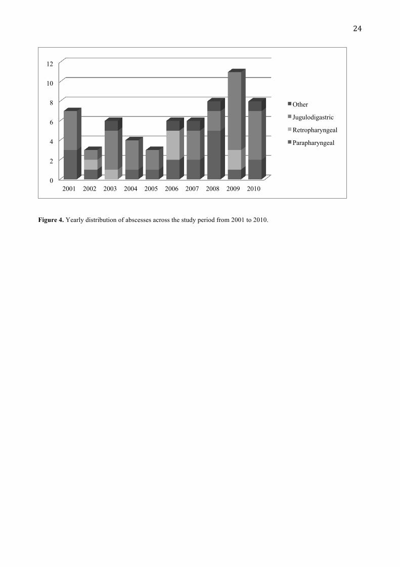
A small peak was seen in the number of pediatric deep neck infections in the fall months from
September to October (N=19, 31%). Spring (N=16), summer (N=14) and winter (N=13) came
closely after. The number of patients rose between 2006-2010 (N=39) compared to 2001-2005
(N=23) as seen of Figure 4. However, the change in patient population during this time period was
not taken into account and therefore it is not possible to take a stand on the rise of incidence for
pediatric deep neck infections in this population.
0 2 4 6 8 10 12
Staphylococcus aureus Streptococcus pyogenes
Fucobacterium species Staphylococcus epidermidis
Normal flora Streptococcus viridans
Streptococcus anginosus Haemophilus influenzae / parainfluenzae
Microaerofilic streptococcus Prevotella species
Mycobacterium tuberculosis Mycobacterium atypicans
Single pathogen Mixed infection
! !
!
24!
Figure 4. Yearly distribution of abscesses across the study period from 2001 to 2010.
0
2
4
6
8
10
12
2001 2002 2003 2004 2005 2006 2007 2008 2009 2010
Other
Jugulodigastric
Retropharyngeal
Parapharyngeal
! !
!
25!
6 DISCUSSION
Pediatric deep neck infections are potentially life-threatening complications of regular upper
respiratory tract infections or dental infections that spread into the deep cavities of the neck. New
international studies are frequently published on pediatric DNIs (6,30,63). The more recent studies
have particularly focused on the choice of treatment (2,4,26,57). However, despite the
internationally ongoing discussion there are no Finnish studies on the subject.
The aim of this study was to define the characteristics of the pediatric patients that presented with
deep neck infections, or abscesses, at the Department of Otolaryngology – Head and Neck Surgery
at the Helsinki University Hospital (HUH) during a 10-year time period. The original assumption
was that conservative treatment had become more common throughout the years. One assumption
was that the complications of pediatric deep neck infections were relatively uncommon. The focus
was not set on merely parapharyngeal and retropharyngeal abscesses but abscesses of the
jugulodigastric chain were also included in this study.
Despite the original assumption the results showed a preference for surgical intervention throughout
the 10-year time period. It was even a little more common to treat patients surgically during the
latter part of the 10-year study period (90% vs. 78%). The outcome of the treatment was good
regardless of the treatment modality that was chosen. The length of hospital stay was not affected
by the choice of treatment, which has been noted by other groups as well (2,5,58). Conservatively
treated patients had a slightly shorter mean hospital stay (3.9 days) compared with the patients that
were treated surgically (5.0 days). The median age of the patients that underwent surgical
intervention (5.3 years) was lower than the median age of the patients that were treated
conservatively (6.8 years). However, patients who were initially treated with intravenous antibiotics
and later required surgical intervention had an even lower median age (4.4 years), which has been
noted in another study too (19).
The mean age of all patients was a little higher than the one reported in the literature (7.1 years) and
even a little higher for patients with parapharyngeal (8.3 years) and retropharyngeal (8.4 years)
abscesses. It was noticed that when the abscess was located near the mandibular angle or around the
proximal parts of the sternocleid muscle the mean age was lower (4.8 years). Since these abscesses
mainly arise in necrotized lymph nodes it is possible that younger children acquire infections at
these locations in an easier manner.

! !
!
26!
The symptoms in general were compatible with those reported in the literature with neck edema,
fever, cervical lymphadenopathy, pharyngalgia and reduced neck movements being some of the
most common symptoms (1-4,6). Trismus, reduced neck movements, torticollis and cervical
lymphadenopathy were more common in patients with parapharyngeal and retropharyngeal
abscesses and for patients with a more complicated clinical course. The difference in the set of
symptoms might be explained by the different anatomical locations of the abscesses and therefore a
diverse capability to cause symptoms. Patients that presented with a parapharyngeal or
retropharyngeal abscess also presented with a higher mean CRP count (158 or 146 compared to 71).
The rate of complications was fairly high (9.6%) compared with the reported 4.4-9.4% in the
literature (16,28,38,51). One reason to the higher rate of complications could be that pneumonia
was counted as a complication. The studies referred to in the present study, only counted life-
threatening complications and excluded pneumonia (1,7). If pneumonia is left uncounted, in this
study, the rate of complications would be 6.5%, which is closer to the percentage reported in the
literature. All of these abscesses were located in the parapharyngeal or retropharyngeal areas. It
should be noted that in this study the focus was on complications of the disease and therefore
complications of the treatment (i.e. nerve trauma during surgery) were not paid attention to.
The culture results were consistent with those reported in the literature with Staphylococcus aureus
and Streptococcus pyogenes being the most commonly cultured pathogens (1,28,32,33,38,51,52). A
majority of Staphylococcus aureus (N=7) presented during the latter half of the time period. This
could mean that the pathogen is becoming more common. In contrast to the literature (16,52,55) a
risen incidence of methicillin-resistant Staphylococcus aureus (MRSA) was not noted in this study
as no cases of MRSA was present. A connection could not be established between the causative
pathogens and the complications since only 2 out of 6 patients had reliable pus cultures obtained.
They presented with streptococcus pyogenes. Elsherif et al. did, however, not note a connection in a
larger patient population either (13).
Due to the low amount of medically treated patients (15%, N=9) it would not be reliable to compare
the effectiveness, safety and outcome of medical versus surgical treatment in this study. Therefore,
it was not possible to compose a reliable treatment guideline for pediatric deep neck infections due
to a lack of supportive data, which has also been noted in other reports (4,6).

! !
!
27!
Since this study was performed as a retrospective chart review there are certain limitations to the
study. These are seen primarily in the form of an occasional lack of available data and a possible
lack of uniform variable reporting on the patient charts. These same limitations are however shared
by other researchers too. A lack of national treatment guidelines for pediatric deep neck infections
also composes a factor of unreliability in the study since the initial choice of treatment is affected
by the knowledge and experience of the medical doctor working at the ENT emergency outpatient
clinic.

! !
!
28!
7 CONCLUSION
According to this study the occurrence of pediatric deep neck infections seems to be relatively low
in the patient population of the Helsinki University Hospital referral area. Between the years of
2001 and 2010 the amount of patients was between three and eleven patients each year. The number
of parapharyngeal and retropharyngeal abscesses was even lower on a yearly level.
The management and treatment outcome of pediatric neck abscesses in our department is in line
with other reports. The use of MRI for imaging had increased which is also recommended to
diminish the radiation in children. Despite the chosen treatment modality, the children seemed to
recover well and the risk of serious complications was low. Multiresistant bacteria were not found
and therefore there is no need for primary broad-spectrum antibiotics.
On contrary to an earlier presumption, the prevalence of conservative treatment did not increase
during the studied time period. According to the results of this study surgical drainage has remained
the principal treatment modality for the patient population at the Department of Otolaryngology –
Head and Neck Surgery at the Helsinki University Hospital (HUH). Due to the low amount of
conservatively treated patients it was not possible to create a reliable treatment algorithm for
pediatric deep neck infections. However, in light of the literature and the treatment outcome of this
study it does not seem that a choice of conservative treatment would remarkably raise the
possibility of complications or extend the length of the hospital stay. Therefore, initiation of
conservative treatment of a pediatric deep neck abscess and close follow-up will be sufficient if the
patient is in stable condition.
In conclusion, further studies would be recommended in order to compose national and evidence-
based treatment guidelines for pediatric deep neck infections.

! !
!
29!
8 REFERENCES
(1) Cheng J, Elden L. Children with deep space neck infections: our experience with 178 children. Otolaryngol Head Neck Surg 2013 Jun;148(6):1037-1042.
(2) Hoffmann C, Pierrot S, Contencin P, Morisseau-Durand MP, Manach Y, Couloigner V. Retropharyngeal infections in children. Treatment strategies and outcomes. Int J Pediatr Otorhinolaryngol 2011 Sep;75(9):1099-1103.
(3) Grisaru-Soen G, Komisar O, Aizenstein O, Soudack M, Schwartz D, Paret G. Retropharyngeal and parapharyngeal abscess in children--epidemiology, clinical features and treatment. Int J Pediatr Otorhinolaryngol 2010 Sep;74(9):1016-1020.
(4) Johnston D, Schmidt R, Barth P. Parapharyngeal and retropharyngeal infections in children: argument for a trial of medical therapy and intraoral drainage for medical treatment failures. Int J Pediatr Otorhinolaryngol 2009 May;73(5):761-765.
(5) Aitasalo K. Syvät kaulainfektiot ja niiden hoito. Suomen lääkärilehti - Finlands läkartidning 2008;63(17):1595-1599.
(6) Saluja S, Brietzke SE, Egan KK, Klavon S, Robson CD, Waltzman ML, et al. A prospective study of 113 deep neck infections managed using a clinical practice guideline. Laryngoscope 2013 Aug 5.
(7) Duval M, Daniel SJ. Retropharyngeal and parapharyngeal abscesses or phlegmons in children. Is there an association with adenotonsillectomy?. Int J Pediatr Otorhinolaryngol 2008 Dec;72(12):1765-1769.
(8) Rajasuo A, Rajasuo A, Torkkeli T. Oral, pharyngeal and neck infections in specialized care. Duodecim 2014;130(6):581-586.
(9) Richardson R, Seppanen L. Deep maxillo-facial infections. Duodecim 2010;126(6):695-701.
(10) Nuutinen J. Pään ja kaulan alueen syvät infektiot. Lääkäripäivät 1999 Läkardagarna 1999.
(11) Murray AD. Deep Neck Infections . :http://emedicine.medscape.com/article/837048-overview.
(12) Cheng J, Elden L. Children with deep space neck infections: our experience with 178 children. Otolaryngology - Head & Neck Surgery 2013 Jun;148(6):1037-1042.
(13) Elsherif AM, Park AH, Alder SC, Smith ME, Muntz HR, Grimmer F. Indicators of a more complicated clinical course for pediatric patients with retropharyngeal abscess. Int J Pediatr Otorhinolaryngol 2010 Feb;74(2):198-201.
(14) Page NC, Bauer EM, Lieu JE. Clinical features and treatment of retropharyngeal abscess in children. Otolaryngol Head Neck Surg 2008 Mar;138(3):300-306.
(15) Pelaz AC, Allende AV, Llorente Pendas JL, Nieto CS. Conservative treatment of retropharyngeal and parapharyngeal abscess in children. J Craniofac Surg 2009 Jul;20(4):1178-1181.
(16) Baldassari CM, Howell R, Amorn M, Budacki R, Choi S, Pena M. Complications in pediatric deep neck space abscesses. Otolaryngol Head Neck Surg 2011 Apr;144(4):592-595.
! !
!
30!
(17) Novis SJ, Pritchett CV, Thorne MC, Sun GH. Pediatric deep space neck infections in U.S. children, 2000-2009. Int J Pediatr Otorhinolaryngol 2014 May;78(5):832-836.
(18) Cabrera CE, Deutsch ES, Eppes S, Lawless S, Cook S, O'Reilly RC, et al. Increased incidence of head and neck abscesses in children. Otolaryngol Head Neck Surg 2007 Feb;136(2):176-181.
(19) Wong DK, Brown C, Mills N, Spielmann P, Neeff M. To drain or not to drain - management of pediatric deep neck abscesses: a case-control study. Int J Pediatr Otorhinolaryngol 2012 Dec;76(12):1810-1813.
(20) Abdel-Haq NM, Harahsheh A, Asmar BL. Retropharyngeal abscess in children: the emerging role of group A beta hemolytic streptococcus. South Med J 2006 Sep;99(9):927-931.
(21) Wang LF, Kuo WR, Tsai SM, Huang KJ. Characterizations of life-threatening deep cervical space infections: a review of one hundred ninety-six cases. Am J Otolaryngol 2003 Mar-Apr;24(2):111-117.
(22) Meyer AC, Kimbrough TG, Finkelstein M, Sidman JD. Symptom duration and CT findings in pediatric deep neck infection. Otolaryngology - Head & Neck Surgery 2009 Feb;140(2):183-186.
(23) Vural C, Gungor A, Comerci S. Accuracy of computerized tomography in deep neck infections in the pediatric population. Am J Otolaryngol 2003 May-Jun;24(3):143-148.
(24) Lander L, Lu S, Shah RK. Pediatric retropharyngeal abscesses: a national perspective. Int J Pediatr Otorhinolaryngol 2008 Dec;72(12):1837-1843.
(25) Freling N, Roele E, Schaefer-Prokop C, Fokkens W. Prediction of deep neck abscesses by contrast-enhanced computerized tomography in 76 clinically suspect consecutive patients. Laryngoscope 2009 Sep;119(9):1745-1752.
(26) Carbone PN, Capra GG, Brigger MT. Antibiotic therapy for pediatric deep neck abscesses: a systematic review. Int J Pediatr Otorhinolaryngol 2012 Nov;76(11):1647-1653.
(27) Metin O, Oz FN, Tanir G, Bayhan GI, Aydin-Teke T, Gayretli-Aydin ZG, et al. Deep neck infections in children: experience in a tertiary care center in Turkey. Turk J Pediatr 2014 May-Jun;56(3):272-279.
(28) Daya H, Lo S, Papsin BC, Zachariasova A, Murray H, Pirie J, et al. Retropharyngeal and parapharyngeal infections in children: the Toronto experience. Int J Pediatr Otorhinolaryngol 2005 Jan;69(1):81-86.
(29) Schweinfurth JM. Demographics of pediatric head and neck infections in a tertiary care hospital. Laryngoscope 2006 Jun;116(6):887-889.
(30) Raffaldi I, Le Serre D, Garazzino S, Scolfaro C, Bertaina C, Mignone F, et al. Diagnosis and management of deep neck infections in children: the experience of an Italian paediatric centre. J Infect Chemother 2015 Feb;21(2):110-113.
(31) Lee SS, Schwartz RH, Bahadori RS. Retropharyngeal abscess: epiglottitis of the new millennium. J Pediatr 2001 Mar;138(3):435-437.
(32) Craig FW, Schunk JE. Retropharyngeal abscess in children: clinical presentation, utility of imaging, and current management. Pediatrics 2003 Jun;111(6 Pt 1):1394-1398.
! !
!
31!
(33) Nagy M, Pizzuto M, Backstrom J, Brodsky L. Deep neck infections in children: a new approach to diagnosis and treatment. Laryngoscope 1997 Dec;107(12 Pt 1):1627-1634.
(34) Pharisa C, Lutz N, Roback MG, Gehri M. Neck complaints in the pediatric emergency department: a consecutive case series of 170 children. Pediatr Emerg Care 2009 Dec;25(12):823-826.
(35) Thompson JW, Cohen SR, Reddix P. Retropharyngeal abscess in children: a retrospective and historical analysis. Laryngoscope 1988 Jun;98(6 Pt 1):589-592.
(36) Kirse DJ, Roberson DW. Surgical management of retropharyngeal space infections in children. Laryngoscope 2001 Aug;111(8):1413-1422.
(37) Grodinsky M. Retropharyngeal and Lateral Pharyngeal Abscesses: an Anatomic and Clinical Study. Ann Surg 1939 Aug;110(2):177-199.
(38) Thomason TS, Brenski A, McClay J, Ehmer D. The rising incidence of methicillin-resistant Staphylococcus aureus in pediatric neck abscesses. Otolaryngol Head Neck Surg 2007 Sep;137(3):459-464.
(39) Grisaru-Soen G, Komisar O, Aizenstein O, Soudack M, Schwartz D, Paret G. Retropharyngeal and parapharyngeal abscess in children--epidemiology, clinical features and treatment. Int J Pediatr Otorhinolaryngol 2010 Sep;74(9):1016-1020.
(40) Wong DK, Brown C, Mills N, Spielmann P, Neeff M. To drain or not to drain - management of pediatric deep neck abscesses: a case-control study. Int J Pediatr Otorhinolaryngol 2012 Dec;76(12):1810-1813.
(41) Coticchia JM, Getnick GS, Yun RD, Arnold JE. Age-, site-, and time-specific differences in pediatric deep neck abscesses. Arch Otolaryngol Head Neck Surg 2004 Feb;130(2):201-207.
(42) Ungkanont K, Yellon RF, Weissman JL, Casselbrant ML, Gonzalez-Valdepena H, Bluestone CD. Head and neck space infections in infants and children. Otolaryngol Head Neck Surg 1995 Mar;112(3):375-382.
(43) Boucher C, Dorion D, Fisch C. Retropharyngeal abscesses: a clinical and radiologic correlation. J Otolaryngol 1999 Jun;28(3):134-137.
(44) Stone ME, Walner DL, Koch BL, Egelhoff JC, Myer CM. Correlation between computed tomography and surgical findings in retropharyngeal inflammatory processes in children. Int J Pediatr Otorhinolaryngol 1999 Aug 5;49(2):121-125.
(45) Miller WD, Furst IM, Sandor GK, Keller MA. A prospective, blinded comparison of clinical examination and computed tomography in deep neck infections. Laryngoscope 1999 Nov;109(11):1873-1879.
(46) Malloy KM, Christenson T, Meyer JS, Tai S, Deutsch ES, Barth PC, et al. Lack of association of CT findings and surgical drainage in pediatric neck abscesses. Int J Pediatr Otorhinolaryngol 2008 Feb;72(2):235-239.
(47) Smith JL,2nd, Hsu JM, Chang J. Predicting deep neck space abscess using computed tomography. Am J Otolaryngol 2006 Jul-Aug;27(4):244-247.
(48) Kalmovich LM, Gavriel H, Eviatar E, Kessler A. Accuracy of ultrasonography versus computed tomography scan in detecting parapharyngeal abscess in children. Pediatr Emerg Care 2012 Aug;28(8):780-782.
! !
!
32!
(49) Duque CS, Guerra L, Roy S. Use of intraoperative ultrasound for localizing difficult parapharyngeal space abscesses in children. Int J Pediatr Otorhinolaryngol 2007 Mar;71(3):375-378.
(50) Brook I. Microbiology and management of peritonsillar, retropharyngeal, and parapharyngeal abscesses. J Oral Maxillofac Surg 2004 Dec;62(12):1545-1550.
(51) Abdel-Haq N, Quezada M, Asmar BI. Retropharyngeal abscess in children: the rising incidence of methicillin-resistant Staphylococcus aureus. Pediatr Infect Dis J 2012 Jul;31(7):696-699.
(52) Wright CT, Stocks RM, Armstrong DL, Arnold SR, Gould HJ. Pediatric mediastinitis as a complication of methicillin-resistant Staphylococcus aureus retropharyngeal abscess. Archives of Otolaryngology -- Head & Neck Surgery 2008 Apr;134(4):408-413.
(53) Baldassari C, Shah RK. Pediatric peritonsillar abscess: an overview. Infectious Disorders - Drug Targets 2012 Aug;12(4):277-280.
(54) Wright CT, Stocks RM, Armstrong DL, Arnold SR, Gould HJ. Pediatric mediastinitis as a complication of methicillin-resistant Staphylococcus aureus retropharyngeal abscess. Archives of Otolaryngology -- Head & Neck Surgery 2008 Apr;134(4):408-413.
(55) Ossowski K, Chun RH, Suskind D, Baroody FM. Increased isolation of methicillin-resistant Staphylococcus aureus in pediatric head and neck abscesses. Arch Otolaryngol Head Neck Surg 2006 Nov;132(11):1176-1181.
(56) Bolton M, Wang W, Hahn A, Ramilo O, Mejias A, Jaggi P. Predictors for successful treatment of pediatric deep neck infections using antimicrobials alone. Pediatr Infect Dis J 2013 Sep;32(9):1034-1036.
(57) Bolton M, Wang W, Hahn A, Ramilo O, Mejias A, Jaggi P. Predictors for successful treatment of pediatric deep neck infections using antimicrobials alone. Pediatr Infect Dis J 2013 Sep;32(9):1034-1036.
(58) Courtney MJ, Mahadevan M, Miteff A. Management of paediatric retropharyngeal infections: non-surgical versus surgical. ANZ J Surg 2007 Nov;77(11):985-987.
(59) Courtney MJ, Miteff A, Mahadevan M. Management of pediatric lateral neck infections: Does the adage "... never let the sun go down on undrained pus ..." hold true? Int J Pediatr Otorhinolaryngol 2007 Jan;71(1):95-100.
(60) Shah RK, Chun R, Choi SS. Mediastinitis in infants from deep neck space infections. Otolaryngol Head Neck Surg 2009 Jun;140(6):936-938.
(61) Mutlu M, Dereci S, Aslan Y. Deep neck abscess in neonatal period: case report and review of literature. Int J Pediatr Otorhinolaryngol 2014 Apr;78(4):577-582.
(62) Georget E, Gauthier A, Brugel L, Verlhac S, Remus N, Epaud R, et al. Acute cervical lymphadenitis and infections of the retropharyngeal and parapharyngeal spaces in children. BMC Ear, Nose & Throat Disorders 2014;14:8.
(63) Novis SJ, Pritchett CV, Thorne MC, Sun GH. Pediatric deep space neck infections in U.S. children, 2000-2009. Int J Pediatr Otorhinolaryngol 2014 May;78(5):832-836.