pediatric brain tumors-posterior fossa - school of medicine brain tumors... · lsuhsc new orleans...
TRANSCRIPT
LSUHSC New Orleans Department of Neurosurgery
Pediatric Brain Tumors:Lecture 1‐Posterior Fossa
Jerome M. Volk III, PGY‐6LSU Department of Neurosurgery
LSUHSC New Orleans Department of Neurosurgery
March 28 & 29, 2014
LSUHSC New Orleans Department of Neurosurgery
Epidemiology
• Compromise ~20% of all pediatric cancers• Incidence of 2.4‐3.5/100,000 children
– Roughly 3,000 new diagnoses in the US annually
• Location:– 60‐65% are located in the Infratentorial region:
•
• •

LSUHSC New Orleans Department of Neurosurgery
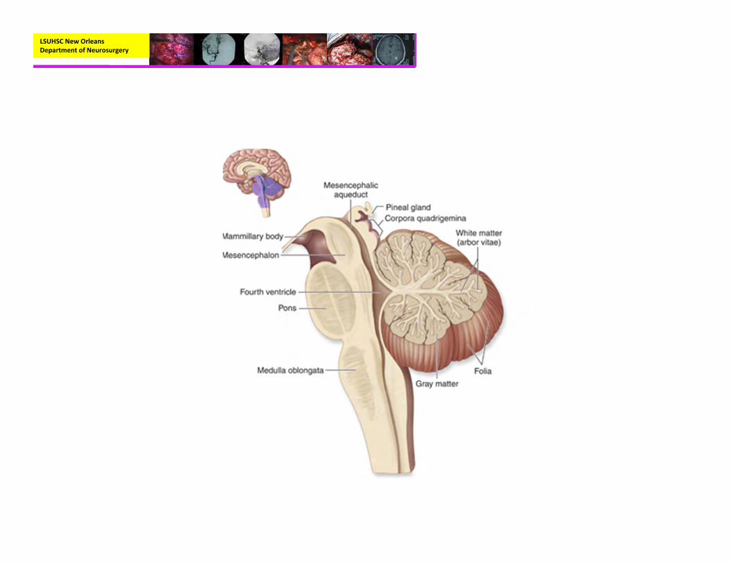
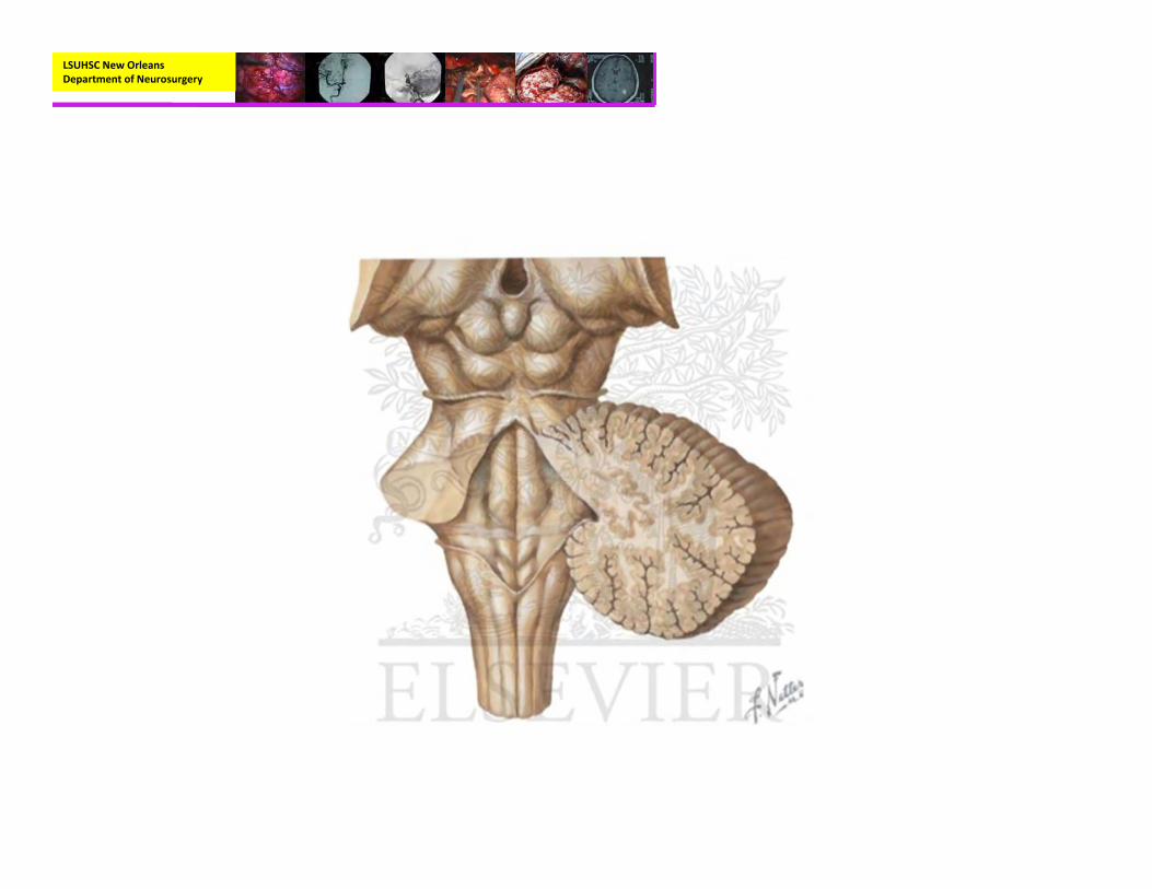
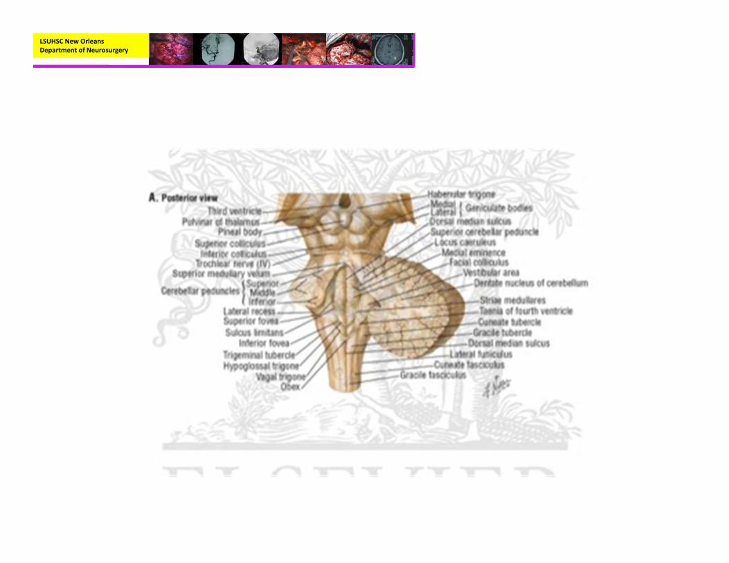
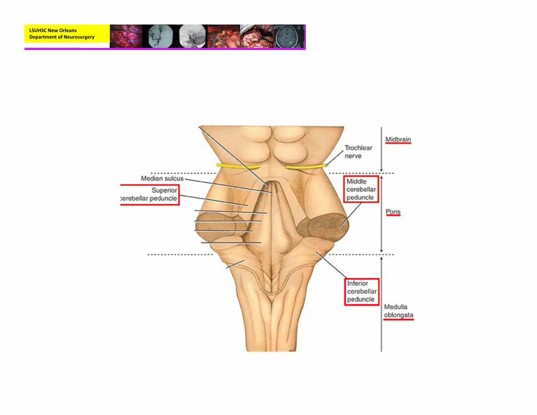
Anatomy
LSUHSC New Orleans Department of Neurosurgery

LSUHSC New Orleans Department of Neurosurgery
Anatomy
LSUHSC New Orleans Department of Neurosurgery
LSUHSC New Orleans Department of Neurosurgery
LSUHSC New Orleans Department of Neurosurgery
LSUHSC New Orleans Department of Neurosurgery
Pilocytic Astrocytoma
• Most common pediatric brain tumor is astrocytoma– With half being found in the posterior fossa
• Cerebellar astrocytomas make up 40% of pilocytic atrocytomas
• Usually occur in the latter half of the first decade– Mean age of 7 years old– Rarely found in children less than 1 year of age
LSUHSC New Orleans Department of Neurosurgery
Pilocytic Astrocytoma
• Median duration of symptoms before diagnosis is 5‐9 months
• Symptoms:– Increased ICP (headache, N/V, head size)– Cerebellar deficits
• Signs:– Papilladema (84%)– Ataxia– Hydrocephalus (85%)
LSUHSC New Orleans Department of Neurosurgery
Pilocytic Astrocytoma
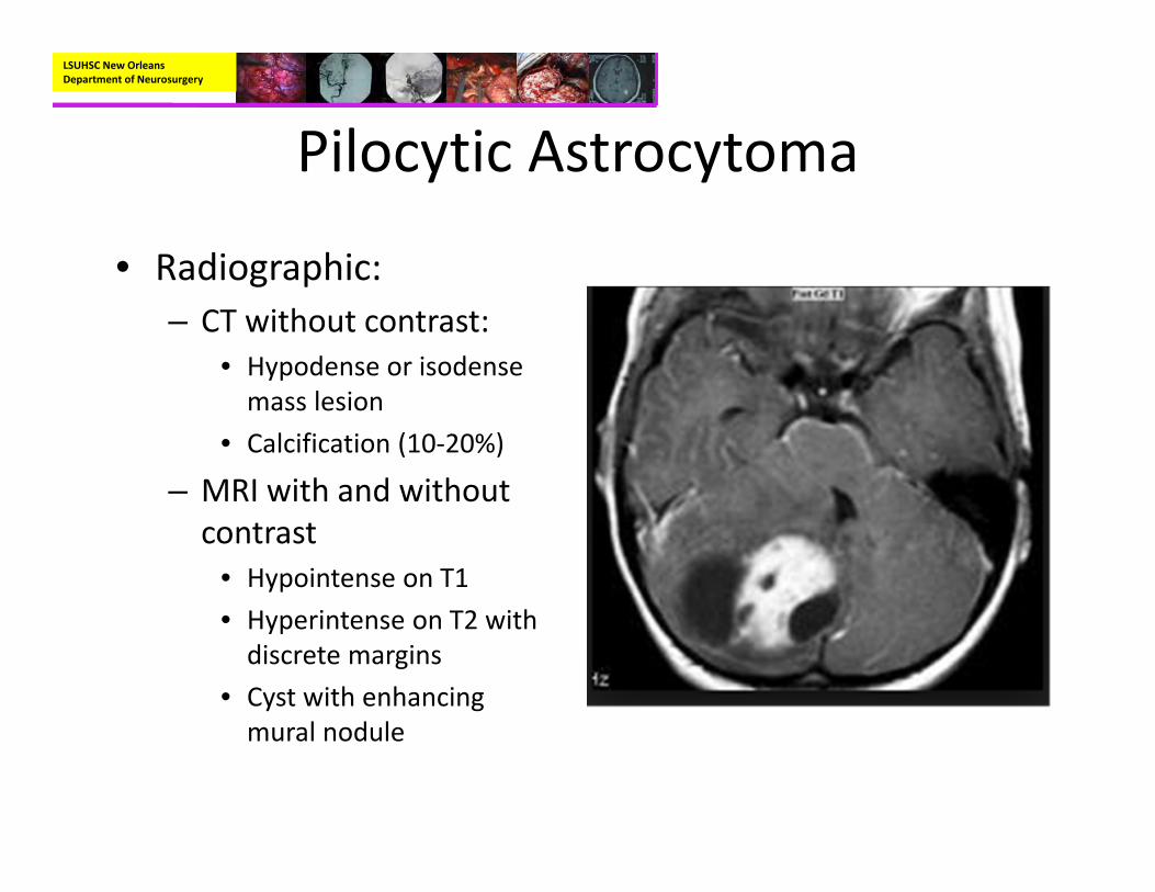
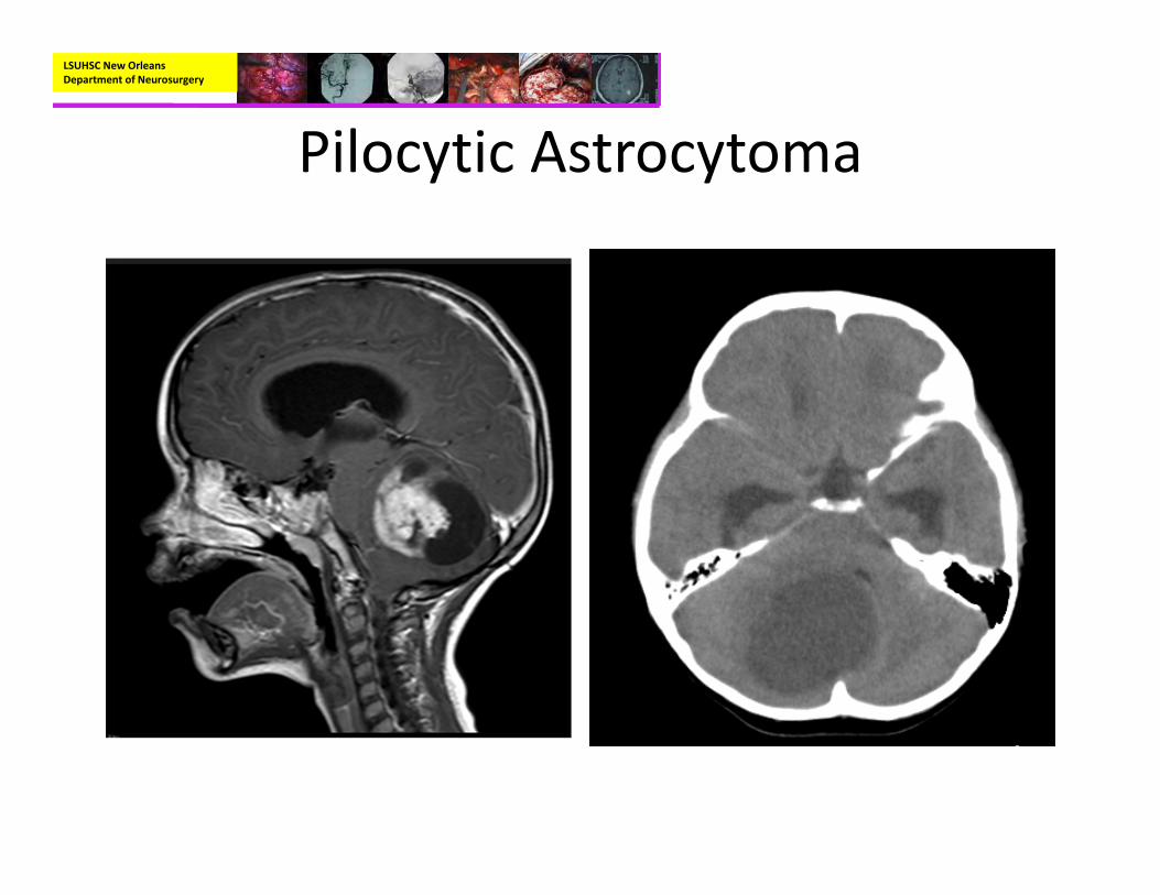
• Radiographic:– CT without contrast:
• Hypodense or isodense mass lesion
• Calcification (10‐20%)
– MRI with and without contrast
• Hypointense on T1• Hyperintense on T2 with discrete margins
• Cyst with enhancing mural nodule
LSUHSC New Orleans Department of Neurosurgery
Pilocytic Astrocytoma
LSUHSC New Orleans Department of Neurosurgery
Pilocytic Astrocytoma
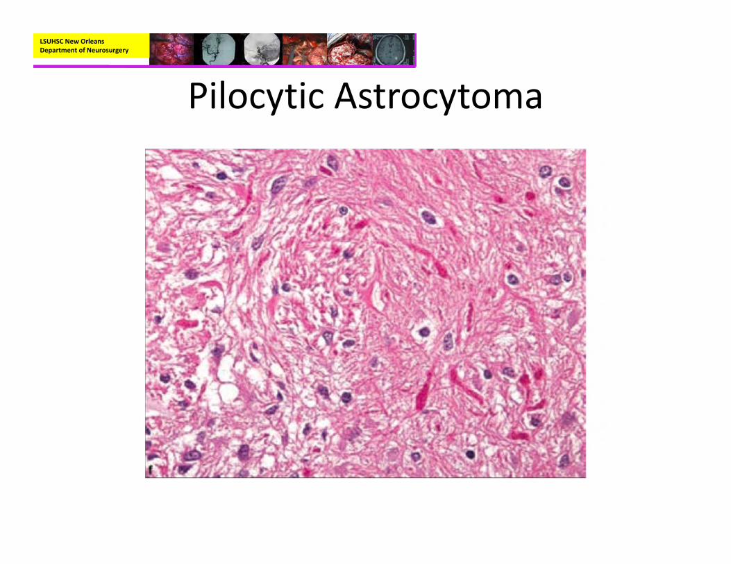
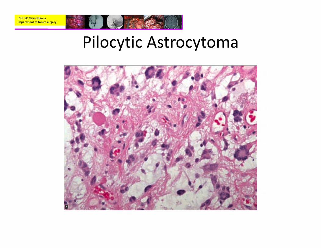
• Pathology– Biphasic pattern with areas of compact, bipolar cells next to loosely structured areas of multipolar cells and microcysts
– Features:• Rosenthal fibers• Microcysts• Endothelial proliferation• Eosinophilic Granular bodies
LSUHSC New Orleans Department of Neurosurgery
Pilocytic Astrocytoma
• Pathology– Immunohistochemistry:
• GFAP positive, S‐100 Positive
– Genetics:• Increased incidence in NF1 (usually seen with optic nerve or brainstem involvement).

LSUHSC New Orleans Department of Neurosurgery
Pilocytic Astrocytoma

LSUHSC New Orleans Department of Neurosurgery
Pilocytic Astrocytoma
LSUHSC New Orleans Department of Neurosurgery
Pilocytic Astrocytoma
LSUHSC New Orleans Department of Neurosurgery
Pilocytic Astrocytoma
LSUHSC New Orleans Department of Neurosurgery
Pilocytic Astrocytoma
• Treatment:– The single best treatment is gross total resection
• Children with no visible residual tumor do not require adjuvant therapy
– Cases with large amount of residual tumor should have a repeat craniotomy for removal of residual tissue.
LSUHSC New Orleans Department of Neurosurgery
Pilocytic Astrocytoma
• Treatment:– Subtotal Resection
• In those cases of residual tumor being left because of involvement with eloquent structures (brainstem), some authors argue to follow with serial imaging
– 40‐50% of residual tumors will regress or not progress– If tumors do progress, most of the time they progress within 2 years (can be up to 8 years).
• If at all possible re‐resection is still the best treatment
LSUHSC New Orleans Department of Neurosurgery
Pilocytic Astrocytoma
• Treatment:– Adjuvant Therapy:
• Radiotherapy:– Not recommended as a first line treatment because it can predispose the patient to malignant degeneration
– In those cases of progression after subtotal resection in eloquent areas radiosurgery can be a helpful option
» Tumor control achieved in roughly 60% of cases
LSUHSC New Orleans Department of Neurosurgery
Pilocytic Astrocytoma
• Treatment:– Adjuvant
• Chemotherapy:– There is no consensus on the use of chemotherapy in pilocytic astrocytoma.
– For those cases of recurrence, a regimen of vincristine and carboplatin can be used
– In cases of disseminated leptomeningeal pilocytic astrocytoma, high dose cyclophosphamide has been used with much success.
LSUHSC New Orleans Department of Neurosurgery
Pilocytic Astrocytoma
• Outcome:– Long term event‐free survival is greater than 90% with gross total resection
– Survival rates at 5‐,10‐, 25‐ year= 90%, 89%, 85%.– Tumor recurrence with GTR is ~10%– Malignant transformation is rare in absence of radiotherapy
– Does not follow Collin’s Law.
LSUHSC New Orleans Department of Neurosurgery
Medulloblastoma
• A primitive neuroectodermal tumor (PNET)• First described in 1925• Most common malignant brain tumor of childhood.– ~20% of all pediatric brain tumors– ~30% of posterior fossa tumors
• Median age 5‐7 years old• 250‐350 new cases of Medulloblastoma in the US each year
LSUHSC New Orleans Department of Neurosurgery
Medulloblastoma
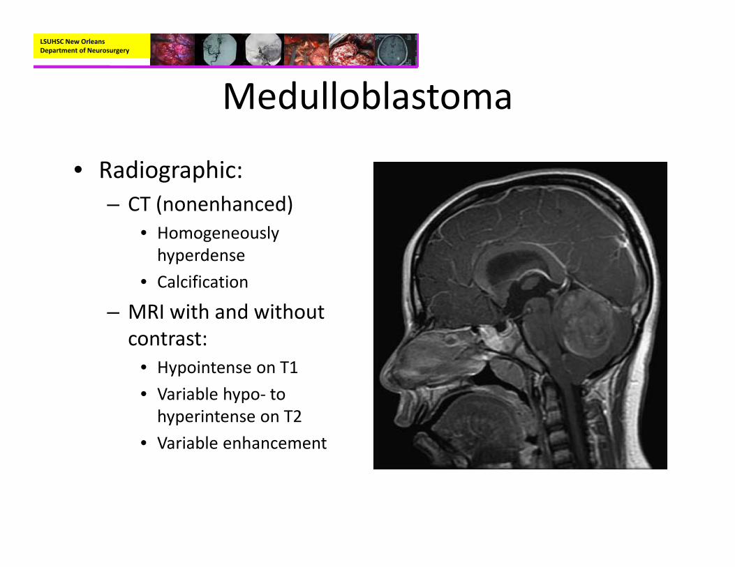
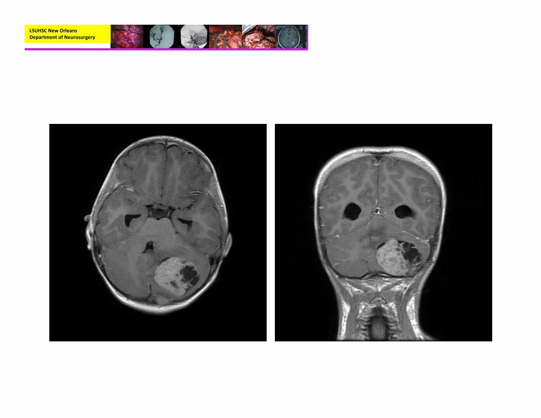
• Radiographic:– CT (nonenhanced)
• Homogeneously hyperdense
• Calcification
– MRI with and without contrast:
• Hypointense on T1• Variable hypo‐ to hyperintense on T2
• Variable enhancement
LSUHSC New Orleans Department of Neurosurgery
LSUHSC New Orleans Department of Neurosurgery
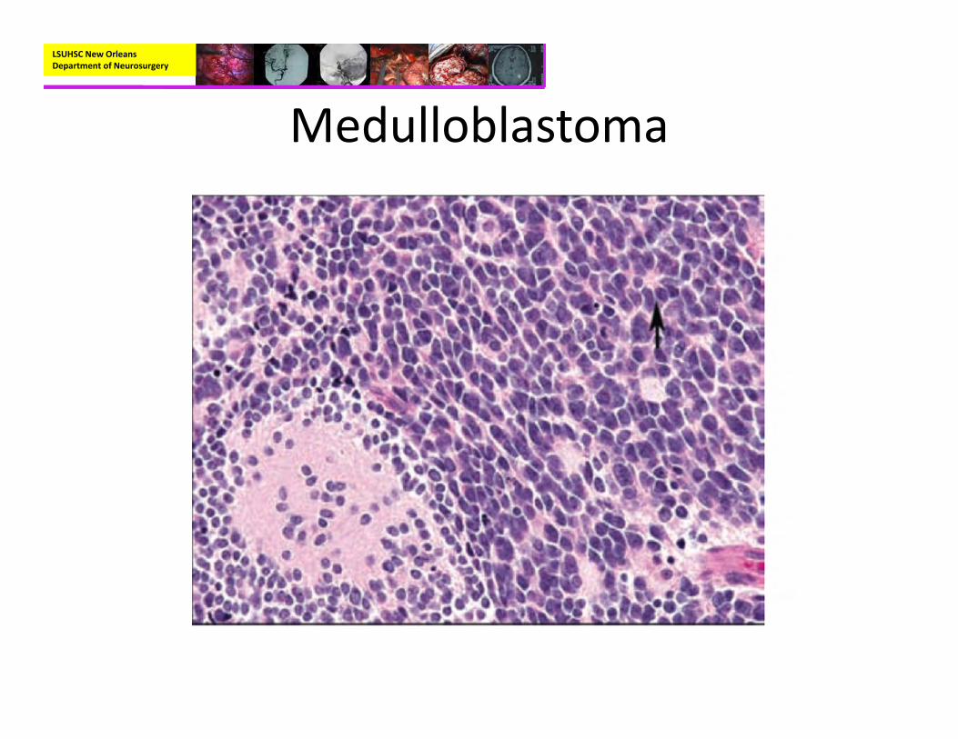
Medulloblastoma• Pathology:
– Small blue cell neoplasm with high nuclear to cytoplasmic ratio
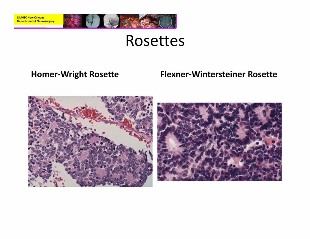
– Homer Wright Rosettes in about a third or cases– Immunohistochemisty:
• Syntapotphysin positive, Neuron‐specific enolase positive, GFAP positive (variable)
– Genetics:• Most commonly involve defect in chromosome 17• Can be seen with Gorlin’s syndrome and Turcot’s syndrome
LSUHSC New Orleans Department of Neurosurgery
Medulloblastoma
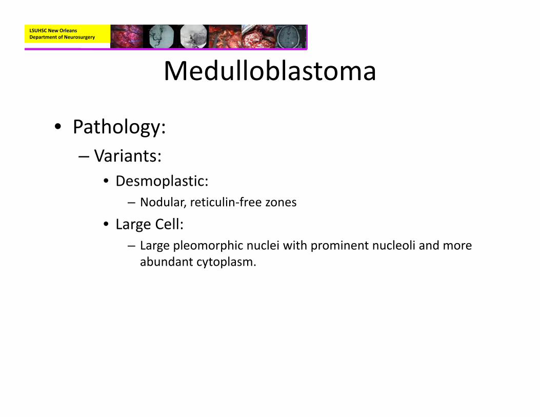
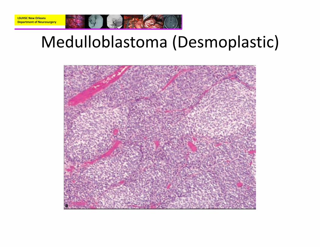
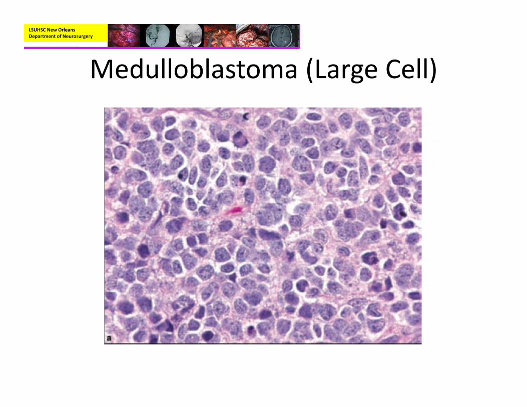
• Pathology:– Variants:
• Desmoplastic:– Nodular, reticulin‐free zones
• Large Cell:– Large pleomorphic nuclei with prominent nucleoli and more abundant cytoplasm.
LSUHSC New Orleans Department of Neurosurgery
Medulloblastoma
LSUHSC New Orleans Department of Neurosurgery
Medulloblastoma
LSUHSC New Orleans Department of Neurosurgery
Medulloblastoma
LSUHSC New Orleans Department of Neurosurgery
Medulloblastoma
LSUHSC New Orleans Department of Neurosurgery
Medulloblastoma (Desmoplastic)
LSUHSC New Orleans Department of Neurosurgery
Medulloblastoma (Large Cell)
LSUHSC New Orleans Department of Neurosurgery
Medulloblastoma
• Treatment:– Survival from medulloblastoma correlates strongly with three factors:
• Age at diagnosis• Extent of dissemination at the the time of diagnosis• Extent of surgical resection
– Like with most tumors of the posterior fossa the amount of resection is directly related to survival
LSUHSC New Orleans Department of Neurosurgery
Medulloblastoma
• Treatment:– Adjuvant:
• Radiotherapy:– Radiation was noted to be a treatment of medulloblastoma as early as 1925.
– Craniospinal radiation has been the standard of care for medulloblastoma because of their tendency to disseminate through the CSF pathways.
– The dose has been able to be lowered with the addition of chemotherapy to almost 23.4 Gy
LSUHSC New Orleans Department of Neurosurgery
Medulloblastoma
• Treatment:– Adjuvant:
• Chemotherapy:– The addition of chemotherapy to the treatment regimen of medulloblastoma has led to an increase in the survival rates as well as a decrease in the dose of radiation given.
– 4 cycles of cyclophosphamide, cisplatin, and vincristine with stem cell support have been used after maximum resection and radiotherapy, resulting in a similarly improved outcome but with substantial reduction in the duration of treatment (4 months vs. 12 months).
LSUHSC New Orleans Department of Neurosurgery
Medulloblastoma• Outcome:
– The addition of chemotherapy has led to an increase in 5 year survival of 85% and 70% in average and high risk patients respectively.
– Patients younger than 3 years generally have a poor prognosis with an estimated 5‐ year PFS of 30% to 40%, attributable to an increased incidence of neuroaxis dissemination, inability or physician/patient reluctance to administer full‐dose radiotherapy, and the possible presence of different genetic alterations.
LSUHSC New Orleans Department of Neurosurgery
Medulloblastoma
• Gorlin’s Syndrome (Nevoid basal cell carcinoma):– Characterized bye occurrence of multiple nevoid basal cell carcinomas, skeletal anomalies, developmental delay, calcification of dura
• 5% develop Medulloblastomas (usually younger in age)• Usually associated with desmoplastic medulloblastomas
– Autosomal dominant disorder– Activation of tumor suppressor at chromosome 9q
LSUHSC New Orleans Department of Neurosurgery
Medulloblastoma
• Gorlin’s syndrome:– The gene name is the PTCH 1 gene (also the genetic test name)
– Better prognosis than non‐syndromic medulloblastoma
– This is critical to diagnose because radiation can cause formation of basal cell carcinoma in the field as well as malignant degeneration of residual tumor
– Treat with resection and chemotherapy regimen.
LSUHSC New Orleans Department of Neurosurgery
Ependymoma
• Third most common pediatric brain tumor• Mean age at diagnosis is 4‐6 years
– 1/3 of which are diagnosed before age 3.
• Arise from the ependymal cells that line the ventricle of the brain and spinal cord– Most commonly located infratentorial (15% of all posterior fossa tumors).
• Floor of the 4th ventricle (60%)• Lateral aspect of the 4th ventricle (30%)• Roof of the 4th ventricle (10%)
LSUHSC New Orleans Department of Neurosurgery
Ependymoma
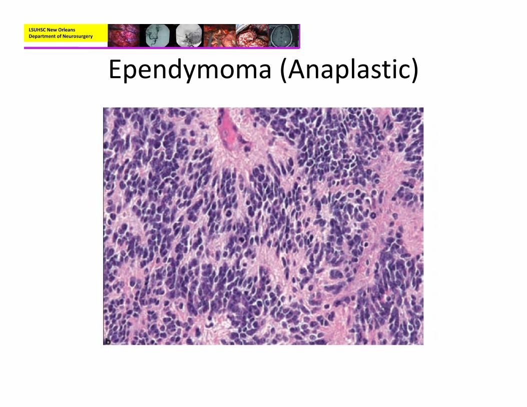
• Types of Ependymoma:– Classic– Papillary– Myxopapillary– Clear Cell– Tanycytic– Giant Cell– Anaplastic
LSUHSC New Orleans Department of Neurosurgery
Ependymoma
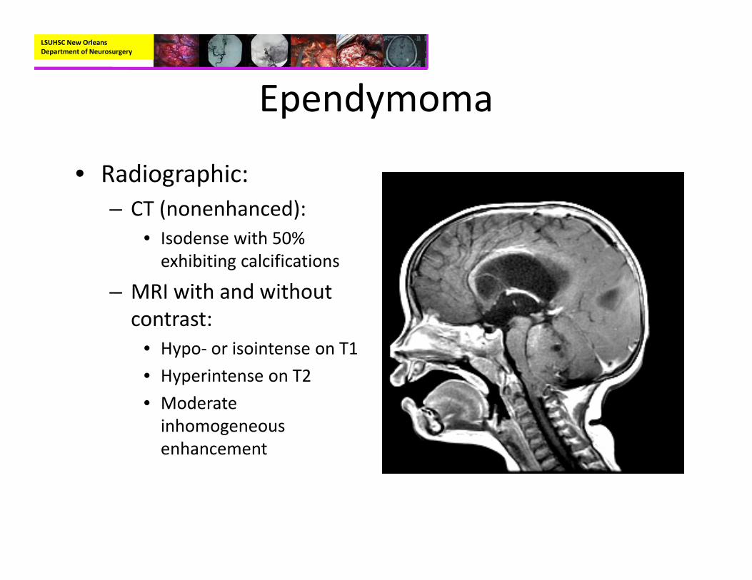
• Radiographic:– CT (nonenhanced):
• Isodense with 50% exhibiting calcifications
– MRI with and without contrast:
• Hypo‐ or isointense on T1• Hyperintense on T2• Moderate inhomogeneous enhancement

LSUHSC New Orleans Department of Neurosurgery
Ependymoma
LSUHSC New Orleans Department of Neurosurgery
Ependymoma
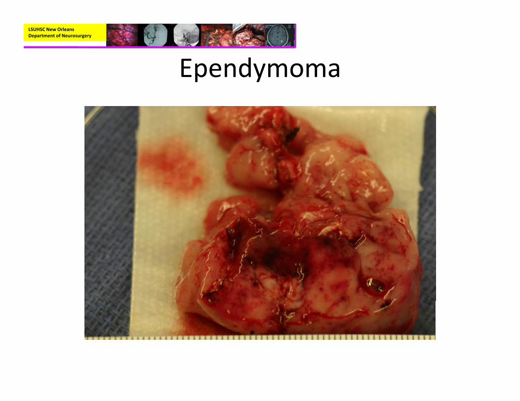
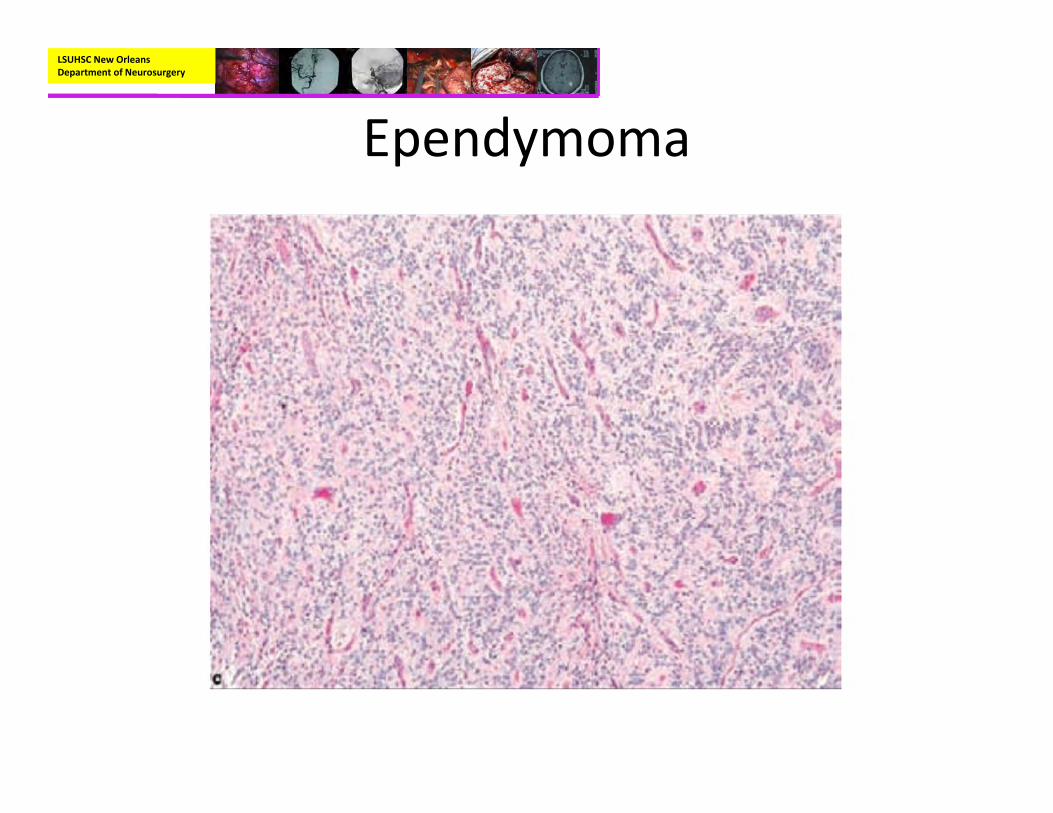
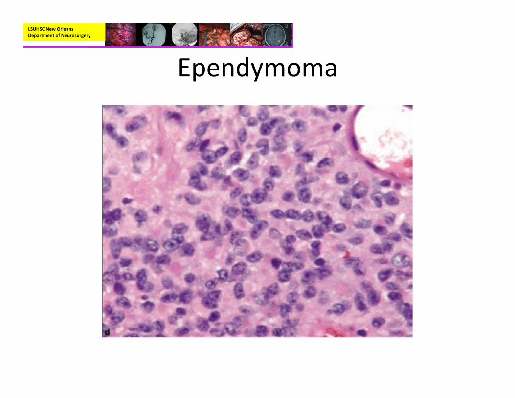
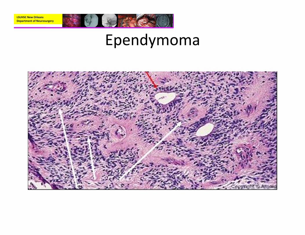
• Pathology:– Mostly well‐circumscribed, soft tan‐red mass on gross inspection.
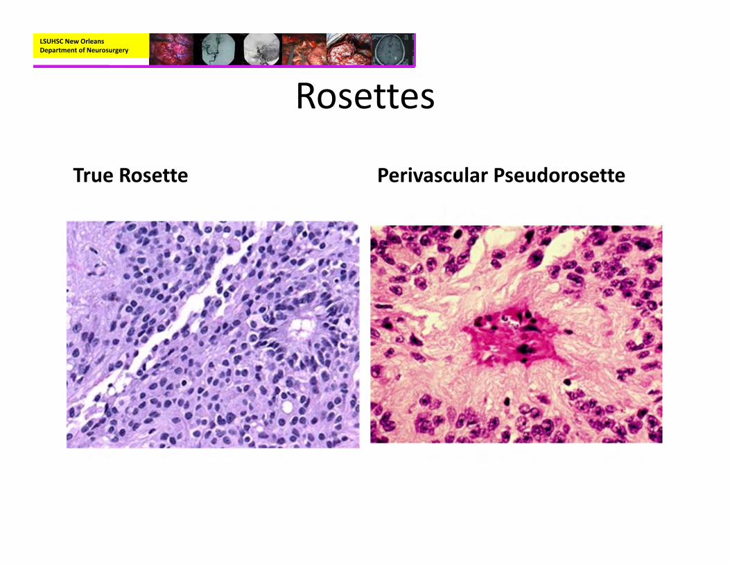
– Characteristically have perivascular pseudorosettes
– True Rosettes seen in 5‐10% of cases– Usually arranged in sheets or spindled, astrocyte‐like, epitheliod, and signet ring‐shaped cells with round nuclei containing small nucleoli
LSUHSC New Orleans Department of Neurosurgery
Ependymoma
• Pathology:– Immunohistochemistry
• GFAP positive, S‐100 Positive, EMA positive
– Genetics:• Deletion of chromosome 22q• NF2 patients commonly have spinal ependymomas not intracranial.
• Can be seen with Li‐Fraumeni syndrome (p53) and Turcot syndrome (APC gene)
LSUHSC New Orleans Department of Neurosurgery
Ependymoma
LSUHSC New Orleans Department of Neurosurgery
Ependymoma
LSUHSC New Orleans Department of Neurosurgery
Ependymoma
LSUHSC New Orleans Department of Neurosurgery
Ependymoma
LSUHSC New Orleans Department of Neurosurgery
Ependymoma
LSUHSC New Orleans Department of Neurosurgery
Ependymoma (Anaplastic)
LSUHSC New Orleans Department of Neurosurgery
Rosettes
Homer‐Wright Rosette Flexner‐Wintersteiner Rosette
LSUHSC New Orleans Department of Neurosurgery
Rosettes
True Rosette Perivascular Pseudorosette
LSUHSC New Orleans Department of Neurosurgery
Ependymoma
• Treatment:– The single most important determinant of outcome is extent of resection.
• A Near‐total resection (<1.5cm3) is prognostically equal to GTR
• 5 year survival for GTR varies between 60‐80%, with STR being ~20%.
– If it is safe the best treatment for residual tumor is re‐operation.
LSUHSC New Orleans Department of Neurosurgery
Ependymoma
• Treatment:– Adjuvant:
• Radiotherapy:– Postoperative radiation is standard of care in patients with ependymoma that are older than 3 years of age.
– Studies have shown that pt’s treated with GTR and radiation by far have the lowest mortality and the highest progression free survival.
• Chemotherapy:– While some centers do use chemotherapy as an adjuvant to surgery, it is NOT considered the standard of care for treatment of intracranial ependymoma
LSUHSC New Orleans Department of Neurosurgery
Ependymoma
• Treatment:– Adjuvant:
• Chemotherapy:– Cisplatin appears to be the most used agent; however, even if the tumor does respond to chemotherapy, there has been no study to show increased survival
– Chemotherapy has been shown to be beneficial as an agent to be used in between 1st and 2nd craniotomies.
» Some authors have noted that the borders became more well defined.
– Ependymomas express the MDR‐1 gene, which mediates drug resistance.
LSUHSC New Orleans Department of Neurosurgery
Ependymoma
• Outcomes:– Children less than 3 have a poorer prognosis due to that fact that they only receive lower doses of radiation if any at all
• One study reports a 5 year survival 22% for children under 3, 75% in older children.
LSUHSC New Orleans Department of Neurosurgery
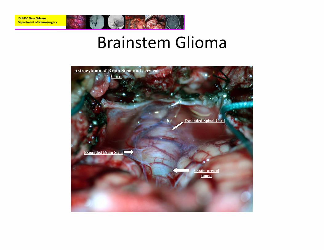
Brainstem Glioma
• Represent 10‐20% of all CNS tumors in children
• 150‐300 new cases annually in the US• Peak presentation at 7‐9 years• Classic triad of physical findings (all three seen in 1/3 of cases):– Cranial nerve palsies– Ataxia– Long tract signs
LSUHSC New Orleans Department of Neurosurgery
Brainstem Glioma
LSUHSC New Orleans Department of Neurosurgery
Brainstem Glioma
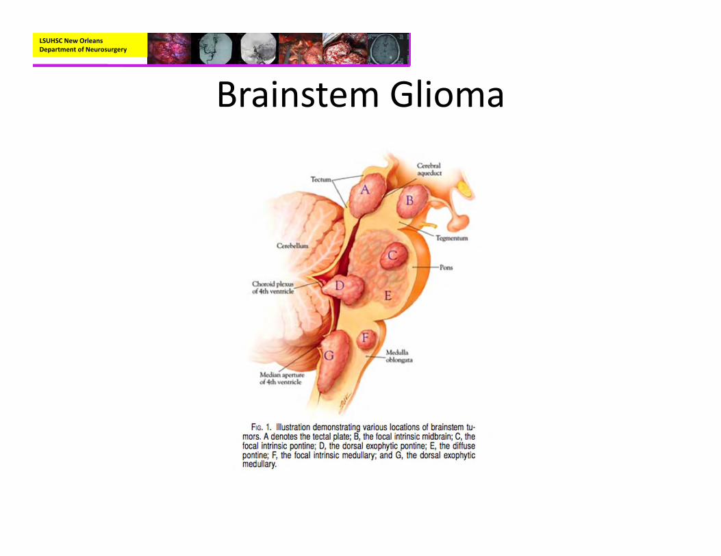
• Types:– Diffuse intrinsic brainstem Glioma:
• Most commonly located in the Pons• Account for 80% of brainstem tumors• Symptoms 2.5 months before diagnosis, ~75% had at least 1 cranial neuropathy, and 25% were alive after 2 years.

LSUHSC New Orleans Department of Neurosurgery
Brainstem Glioma
• Types:– Focal Tumors
• Occupy less than 50% of brainstem subregion.• Often clearly distinguishable from surrounding brainstem.
• Dorsal Exophytic Tumors:– ~20% of brainstem tumors.– Typically fills the 4th ventricle

LSUHSC New Orleans Department of Neurosurgery
• Types:– Focal Tumors:
• Cervicomedullary Tumors:– Majority arise in upper cervical cord and grow rostrally
• Midbrain Tumors:– Carry the best prognosis– Can be located tegmental region or tectal region
LSUHSC New Orleans Department of Neurosurgery
Brainstem Glioma
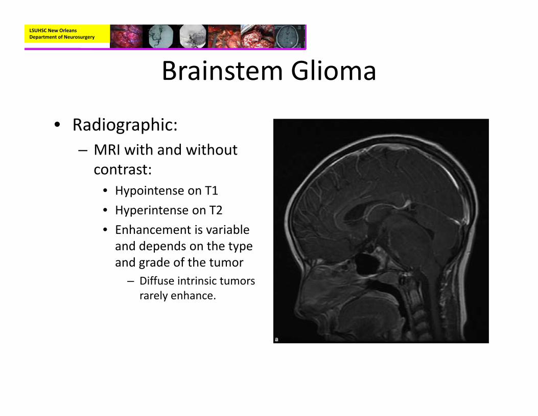
• Radiographic:– MRI with and without contrast:
• Hypointense on T1• Hyperintense on T2• Enhancement is variable and depends on the type and grade of the tumor
– Diffuse intrinsic tumors rarely enhance.
LSUHSC New Orleans Department of Neurosurgery
Brainstem Glioma
LSUHSC New Orleans Department of Neurosurgery
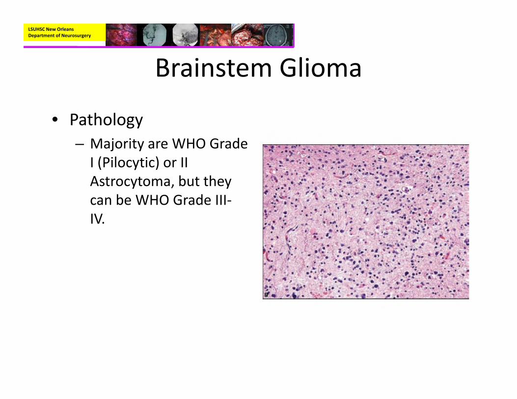
Brainstem Glioma
• Pathology– Majority are WHO Grade I (Pilocytic) or II Astrocytoma, but they can be WHO Grade III‐IV.
LSUHSC New Orleans Department of Neurosurgery
Brainstem Glioma
LSUHSC New Orleans Department of Neurosurgery
Brainstem Glioma
• Treatment:– Surgery is the main treatment for focal tumors (either biopsy or resection) with overall survival of > 90% at 5 years in most studies
– Surgery is not an viable option in the diffuse intrinsic tumors.• However, most studies do advocate a biopsy if the imaging is not typical, or in patients < 2 years of age.
• Also, with the molecular subclassification of diffuse intrinsic tumors being studied, some authors believe that biopsy of all lesions will become a possibility.
LSUHSC New Orleans Department of Neurosurgery
Brainstem Glioma
• Treatment:– Adjuvant:
• Radiotherapy:– Is the mainstay of treatment for diffuse intrinsic tumors, usually of a dose of 54 Gy.
» Transient improvement in neurologic function but not in overall survival
– Can be offered as an adjuvant after subtotal resection in the focal tumor group.

LSUHSC New Orleans Department of Neurosurgery
Brainstem Glioma
• Treatment:– Adjuvant:
• Chemotherapy:– There has been little evidence that chemotherapy offers any impact on the prognosis
– Chemotherapy is withheld for those patients that have high grade lesions (GBM or PNET)
LSUHSC New Orleans Department of Neurosurgery
Acoustic Schwannoma
• Can be unilateral or bilateral.– If there are bilateral acoustic schwannomas this is diagnostic for NF2
– Unilateral Acoustic schwannoma and a first degree relative with NF2 is diagnostic of NF2
• Can be sporadic or associated with NF2.– Sporadic variety is very rare in pediatric population
• Account for 0.8% of pediatric brain tumors

LSUHSC New Orleans Department of Neurosurgery
Acoustic Schwannoma
• NF2 associated schwannomas present with auditory complaints only 30% of the time.
• As opposed to the sporadic variant, NF2 associated schwannomas grow faster and have increased invasion of the nerve.

LSUHSC New Orleans Department of Neurosurgery
Acoustic Schwannoma
• Radiographic:– CT (nonenhanced):
• Iso‐ or slightly hypodense to adjacent brain
– MRI with and without contrast:
• Hypo‐ to isointense on T1• Hyperdense on T2• Enhance intensely• Cystic degeneration ~20%
LSUHSC New Orleans Department of Neurosurgery
Acoustic Schwannoma
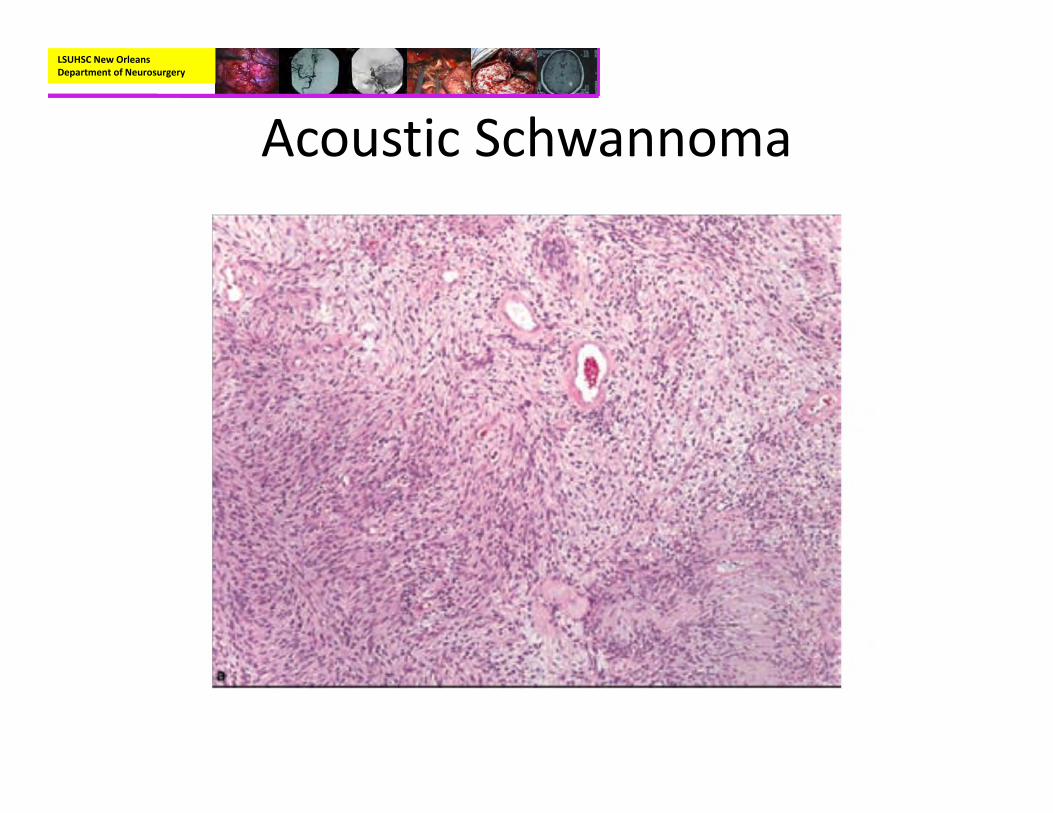
• Pathology– Gross
• Circumscribed masses, frequently encapsulated, sometimes cystic.
• Light tan, yellow, or red in color depending on amount of hemorrhage
– Cells with spindled nuclei– Biphasic with Antoni A pattern (dense) and Antoni B pattern (loose)
– Verocay bodies‐nuclear palisading
LSUHSC New Orleans Department of Neurosurgery
Acoustic Schwannoma
• Pathology:– Immunohistochemistry:
• S‐100 positive
– Genetics:• Can be associated with NF2 gene (merlin protein)
LSUHSC New Orleans Department of Neurosurgery
Acoustic Schwannoma

LSUHSC New Orleans Department of Neurosurgery
Acoustic Schwannoma
LSUHSC New Orleans Department of Neurosurgery
Acoustic Schwannoma
• Treatment:– One of the main goals in treating the pediatric population is to maintain hearing for as long as possible so that they do not have interruptions in educational or social development.
• Attempts at hearing preservation are less successful in NF2 associated schwannomas.
– Some argue that surgery should be reserved for those cases that show tumor growth on serial imaging
LSUHSC New Orleans Department of Neurosurgery
Acoustic Schwannoma
• Treatment:– Another option is to only offer resection of acoustic schwannoma when no useful hearing remains.
– And still another option is to offer surgery when there is significant brainstem compression, even if there is no auditory complaints.
– Hearing preservation surgery should be offered in those patients with residual hearing.
• However, rates of successful hearing preservation are poor
LSUHSC New Orleans Department of Neurosurgery
Acoustic Schwannoma
• Treatment:– In those cases of bilateral acoustic schwannomas that are both growing.
• Approach the side with the worst hearing first.

LSUHSC New Orleans Department of Neurosurgery
Acoustic Schwannoma
• Treatment:– Radiosurgery:
• Up to 20% of NF2 patients who have undergone radiosurgery to their acoustic require further treatment within 8 years of follow‐up, with good control in only 50% of cases.
– Chemotherapy:• Bevacizumab (Avastin), which is a blockade agent against the VEGF pathway, has been shown in studies to improve hearing and decrease the tumor volume in NF2 patients.
LSUHSC New Orleans Department of Neurosurgery
Atypical Teratoid‐Rhabdoid tumor (ATRT)
• Represented as a separate entity in the WHO classification of 1993– Before 1993, ATRT’s were grouped with PNET’s
• Account for 1% of pediatric brain tumors– However, in children less than 3, ATRT’s account for 10‐20% of tumors
LSUHSC New Orleans Department of Neurosurgery
ATRT
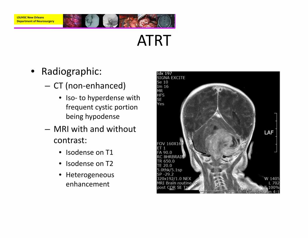
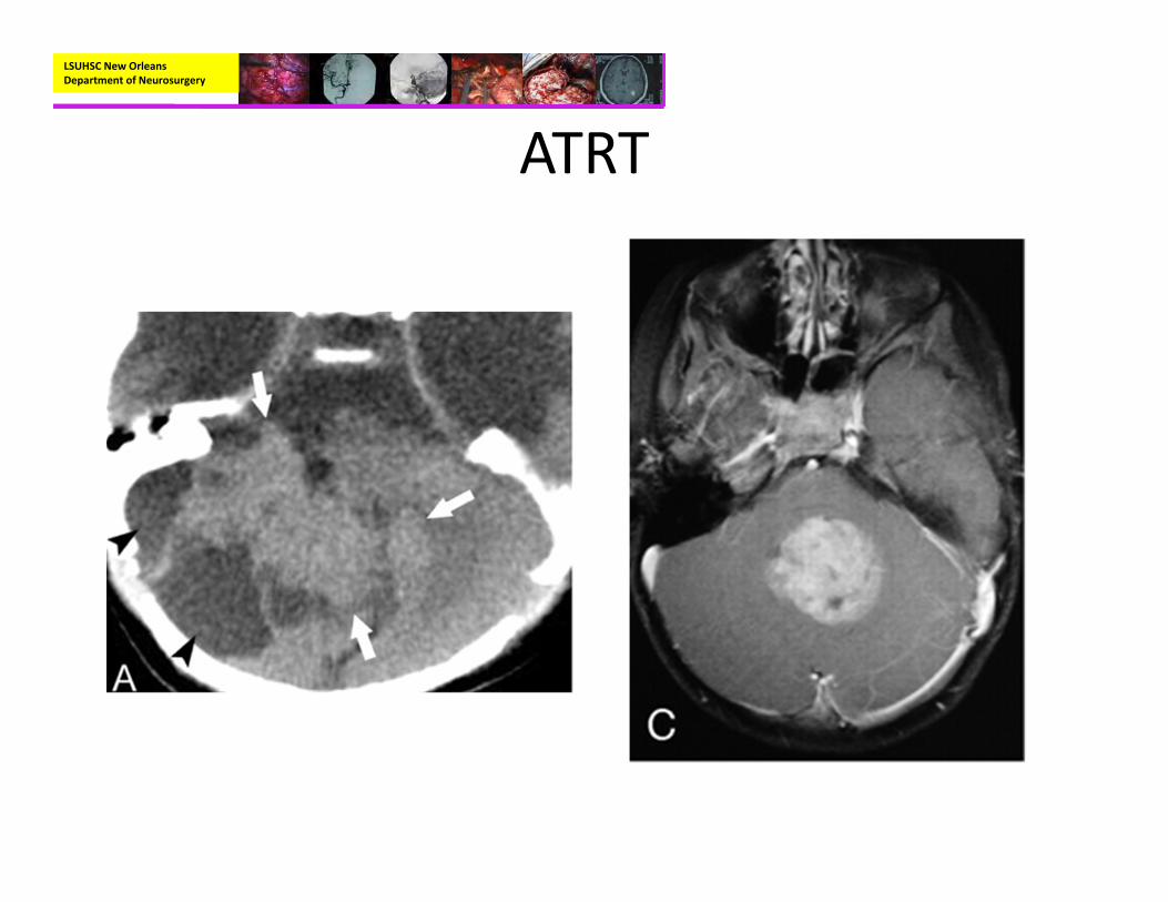
• Radiographic:– CT (non‐enhanced)
• Iso‐ to hyperdense with frequent cystic portion being hypodense
– MRI with and without contrast:
• Isodense on T1• Isodense on T2• Heterogeneous enhancement
LSUHSC New Orleans Department of Neurosurgery
ATRT
LSUHSC New Orleans Department of Neurosurgery
ATRT
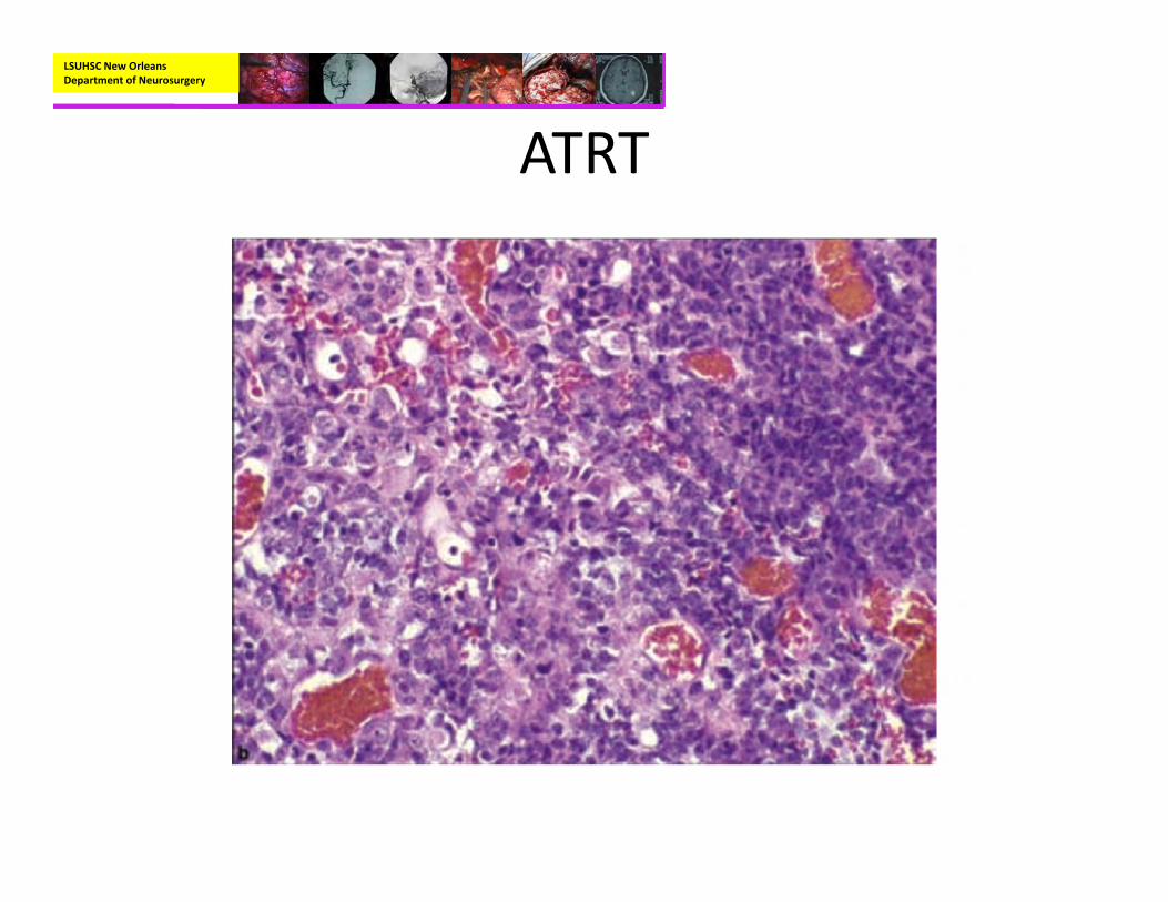
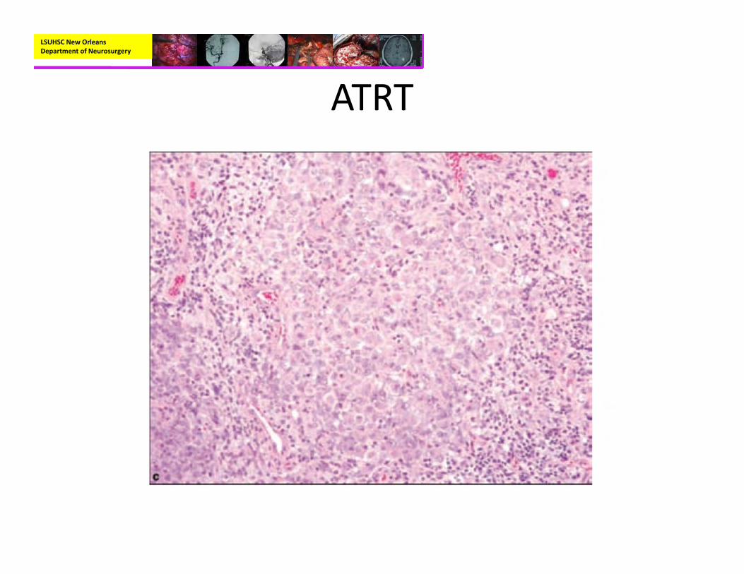
• Pathology:– Rhabdoid cells with eccentric nuclei, prominent nucleoli, and cytoplasmic pink body inclusions.
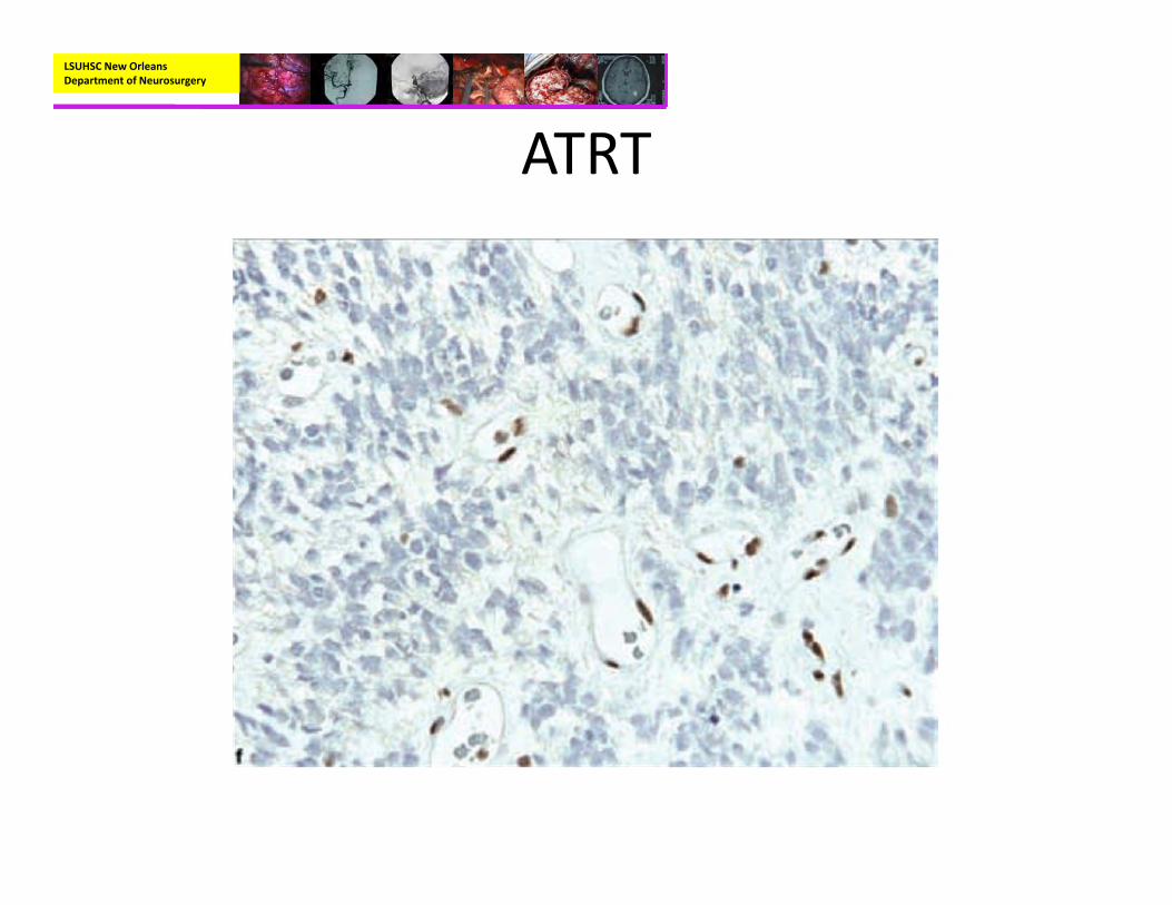
– Immunohistochemisty:• EMA and Vimentin positive• BAF47/SNF5‐negative staining in tumor cells with positive immunoreactivity for endothelial cells
LSUHSC New Orleans Department of Neurosurgery
ATRT
• Pathology:– Genetics:
• ATRT is characterized by loss of the long arm of chromosome 22 which results in loss of the hSNF5/INI‐1 gene.
• INI‐1 is important in maintenance of the mitotic spindle and cell cycle control
LSUHSC New Orleans Department of Neurosurgery
ATRT
LSUHSC New Orleans Department of Neurosurgery
ATRT

LSUHSC New Orleans Department of Neurosurgery
ATRT
LSUHSC New Orleans Department of Neurosurgery
ATRT

LSUHSC New Orleans Department of Neurosurgery
ATRT
• Treatment:– The standard treatment protocol consists of surgical resection, radiotherapy, chemotherapy.
• Median time to death reported to be roughly 6 months to 18 months
• Therefore, some physicians do offer comfort care measures initially.
– Surgical resection is still the cornerstone of treatment.
LSUHSC New Orleans Department of Neurosurgery
ATRT
• Treatment:– Adjuvant:
• Radiotherapy:– Radiotherapy has been shown to increase survival– Some studies have advocated for the use of radiotherapy in children less than 3 because of the improved survival.
• Chemotherapy:– There is no standard chemotherapy treatment regimen– The most commonly used regiment is the Intergroup Rhabdomyosarcoma III (IRS‐III) therapy.
LSUHSC New Orleans Department of Neurosurgery
ATRT
• Treatment:– Adjuvant:
• Chemotherapy:– The IRS III therapy consists of weekly vincristine during radiation, actinomycin‐D, doxorubicin, and triple intrathecal chemotherapy with hydrocortisone, methotrexate, and cytosine arabinoside.
» Intrathecal chemotherapy has shown improved survival in those patients that did not receive radiation.
– Since 2008, the Children’s Oncology group has been working on a new regiment with enrollment still going on.
LSUHSC New Orleans Department of Neurosurgery
References• Albright, A., Pollack, I., Adelson, P. Principles and Practice of Pediatric Neurosurgery. 2nd Edition.
2008. Thieme.• Vogel, H. Cambridge Illustrated Surgical Pathology‐Nervous System. 2009. Cambridge Press.• Prayson, R. Foundations in Diagnostic Pathology‐Neuropathology. 2005. Elsevier• Osborn, A. Diagnostic Neuroradiology.1994. Mosby• Ogiwara, H., et al. Long‐term Follow‐up of Pediatric Benign Cerebellar Astrocytomas. Neurosurgery
70:40–48, 2012• Hadjipanayis, C., et al. Stereotactic radiosurgery for pilocytic astrocytomas when multimodal
therapy is necessary. J Neurosurg 97:56–64, 2002.• Plotkin, S., et al. Hearing Improvement after Bevacizumab in Patients with Neurofibromatosis Type
2. N Engl J Med 2009;361:358‐67.• Shepard, T., et al. Management of Hearing in Pediatric NF2. Otology & Neurotology 33:1066‐1070.
2012.
LSUHSC New Orleans Department of Neurosurgery
References• Cage, T., et al. A systematic review of treatment outcomes in pediatric patients with intracranial
ependymomas. J Neurosurg Pediatrics 11:673–681, 2013.• Pejavar, S., et al. Pediatric intracranial ependymoma: the roles of surgery, radiation and
chemotherapy. J Neurooncol (2012) 106:367–375.• Samkari, A., et al. Medulloblastoma/Primitive Neuroectodermal Tumor and Germ Cell
Tumors. The Uncommon but Potentially Curable Primary Brain Tumors. Hematol Oncol Clin N Am 26 (2012) 881–895.
• Packer, R., et al. Treatment of Children With Medulloblastomas With Reduced‐Dose Craniospinal Radiation Therapy and Adjuvant Chemotherapy: A Children’s Cancer Group Study. Journal of Clinical Oncology, Vol 17, No 7 (July), 1999: pp 2127‐2136.
• Buscariollo, D., et al. Survival Outcomes in Atypical Teratoid Rhabdoid Tumor for Patients Undergoing Radiotherapy in a Surveillance, Epidemiology, and End Results Analysis. Cancer 2012;118:4212‐9.
• Biegel, J. Molecular genetics of atypical teratoid/rhabdoid tumors. Neurosurg Focus 20 (1):E11, 2006.
• Zimmerman, M., et al. Continuous remission of newly diagnosed and relapsed central nervous system atypical teratoid/rhabdoid tumor. Journal of Neuro‐Oncology (2005) 72: 77–84.
LSUHSC New Orleans Department of Neurosurgery
References• Ginn, K., Gajjar, A. Atypical teratoid rhabdoid tumor: current therapy and future directions. Front.
Oncol., 12 September 2012.• Raabe, E., et al. New Strategies in Pediatric Gliomas: Molecular Advances in Pediatric Low‐
Grade Gliomas as a Model. Clin Cancer Res 2013;19:4553‐4558.• Guillamo, J., et al. Brain stem gliomas. Current Opinion in Neurology 2001, 14:711‐715.• Dellaretti, M., et al. Correlation among magnetic resonance imaging findings, prognostic
factors for survival, and histological diagnosis of intrinsic brainstem lesions in children. J Neurosurg Pediatrics 8:050309–050403, 2011.
• Sufit, A., et al. Diffuse intrinsic pontine tumors: a study of primitive neuroectodermal tumors versus the more common diffuse intrinsic pontine gliomas. J Neurosurg Pediatrics 10:81–88, 2012.
• Klimo, P., et al. Management and outcome of focal low‐grade brainstem tumors in pediatric patients: the St. Jude experience. J Neurosurg Pediatrics 11:274–281, 2013.
• Frazier, J., et al. Treatment of diffuse intrinsic brainstem gliomas: failed approaches and future strategies. J Neurosurg Pediatrics 3:020590–200690, 2009.
• Schofield, D., et al. Correlation of Loss of Heterozygosity at Chromosome 9q with Histological Subtype inMedulloblastomas. American Journal ofPathology, Vol. 146, No. 2, February 1995.
LSUHSC New Orleans Department of Neurosurgery
References• Smucker, P., Smith, J. Multifocal desmoplastic medulloblastoma in an African‐American child with
nevoid basal cell carcinoma (Gorlin) syndrome. J Neurosurg (4 Suppl Pediatrics) 105:315–320, 2006.• Muzio, L. Nevoid basal cell carcinoma syndrome (Gorlin syndrome). Orphanet Journal of Rare
Diseases 2008, 3:32.• Amlashi, S., et al. Nevoid Basal Cell Carcinoma Syndrome: Relation with Desmoplastic
Medulloblastoma in Infancy A Population‐Based Study and Review of the Literature. Cancer 2003;98:618–24.
• Cowan, R., et al. The gene for the naevoid basal cell carcinoma syndrome acts as a tumour‐suppressor gene in medulloblastoma. BritishJoumalofCancer(1997)76(2),141‐145.
• Mazzoni, A., et al. Sporadic acoustic neuroma in pediatric patients. International Journal of Pediatric Otorhinolaryngology (2007) 71, 1569—1572.
• Tysome, J., et al. Surgical Management of Vestibular Schwannomas and Hearing Rehabilitation in Neurofibromatosis Type 2. Otology & Neurotology 33:466‐472. 2012
LSUHSC New Orleans Department of Neurosurgery
Thank you