pdf(2199k) - wiley online library
TRANSCRIPT

Br. J. Pharmac. Chernother. (1968), 32, 546-566.
IONIC INFLUENCES ON SUCCINYLCHOLINE BLOCKADEOF THE MAMMALIAN NEUROMUSCULAR JUNCTION
BY
SHIRLEY E. FREEMAN
From the A ustralian Defence Scientific Service, Defence Standards Laboratories, Melbourne,A ustralia
(Received October 23, 1967)
Recent studies from a number of laboratories have been concerned with the delineationof factors affecting presynaptic events in neuromuscular transmission. In particular itseems that substances which block neuromuscular transmission by processes that includedepolarization of the motor end-plate have important effects on the motor nerve terminals.These effects are not only produced by drugs such as succinylcholine and decamethonium,but are also shown by acetylcholine and its stable analogues.Hubbard, Schmidt & Yokota (1965) noted that acetylcholine reduced the quantal
content of end-plate potentials (e.p.p.s.) without affecting miniature end-plate potential(m.e.p.p.) frequency. It appeared that acetylcholine did not depolarize the site of trans-mitter release, although the threshold of motor nerve terminals in paralysed preparationswas reduced by acetylcholine. They concluded that acetylcholine depolarized theterminals at a site more proximal than that of transmitter release. Riker (1966) hasproduced evidence for a transitory depolarization of the motor nerve terminals byacetylcholine following close arterial injection in the cat. It is likely that Hubbard et al.(1965) did not observe this effect because of the slower rate of diffusion of acetylcholineinto the isolated diaphragm preparation. Riker (1966) suggests that the motor nerveterminals undergo sequentially two stages of block in response to acetylcholine-depolarization and stabilization.
This is a reformulation of the proposals of Jenden, Kamijo & Taylor (1954) andThesleff (1955a) for a two-phase block of neuromuscular transmission by decomethoniumand acetylcholine. The earlier proposals were, however, based on the assumption thatthese drugs had a postsynaptic site of action only.The similarity of action at the neuromuscular junction of acetylcholine, decamethonium
and succinylcholine has been noted by most workers in the field. In particular one maycompare the similarity of results obtained by Standaert & Adams (1965) with thoseobtained by Riker (1966). Standaert & Adams investigated prejunctional effects ofsuccinylcholine in the intact cat; Riker used similar techniques to study the effects ofacetylcholine. The emphasis which Standaert & Adams (1965) have placed on pre-junctional effects of succinylcholine receives support from the observation by Edwards &Ikeda (1962) that succinylcholine reduces the quantum content of e.p.p.s. Standaert& Adams (1965) remark, however, that while their work demonstrates unequivocally

IONS AND SUCCINYLCHOLINE BLOCKADE
that prejunctional effects of succinylcholine exist, they were not able to evaluate therelative importance of pre- and postjunctional effects in the development of neuromuscularblockade.The recent work of Gibberd (1966) draws attention to the importance of the metabolic
status of the nerve-muscle preparation when such experiments are carried out. Wholeanimal preparations have the advantage of maintaining the metabolic status of thejunction, but they also have the disadvantage for studies of succinylcholine action thatthe pseudocholinesterase of blood rapidly hydrolyses the compound. Not only doesthis complicate quantitative work, but it leads also to succinylmonocholine accumulation,which itself has some action on the neuromuscular junction.
Because of this it was thought worthwhile to study the kinetics of succinylcholineblockade, using the phrenic nerve-diaphragm preparation of the rat (Bulbring, 1946). Inspite of the limitations inherent in the measurement of twitch tension of a preparation,it has been possible to obtain quantitative information about the rate of development ofneuromuscular blockade by succinylcholine in conditions of ionic stress.The results obtained indicate that succinylcholine has effects on both pre- and post-
junctional membranes. The rate of onset of succinylcholine blocks seems to dependlargely but not entirely on a presynaptic inhibition of transmitter release.
METHODS
Phrenic nerve-diaphragm preparations were dissected from male hooded rats of the Wistar strain.The rats ranged in weight from 250 to 350 g; the average weight was 300 g. The rats were stunnedby a blow on the head, and killed by exsanguination. The use of anaesthetic agents was avoided,because of the likelihood of residual effects on the neuromuscular junction.
Left and right hemidiaphragms were dissected out of the animal, and mounted in separate organbaths. (Bulbring, 1946; Beani, Bianchi & Ledda, 1962). The left hemidiaphragm was routinelydissected before the right; it took between 6 and 9 min after stunning the animal before the twopreparations were placed in an oxygenated solution; the total time to set up the two preparationsin the two organ baths was 17-18 min.
Semi-isometric contractions were measured by strain gauges; the output of the strain gauges was
recorded on a Beckman type R dynograph recorder. Supramaximal rectangular pulses of 0.15 msec
duration and a frequency of 14/min were used to stimulate the preparations, either by direct musclestimulation or by the phrenic nerve. The force of contraction was determined by calibrating thestrain gauges with weights. The twitch tension developed by the preparations varied between 3 and6 g; while that of most preparations fell within a range of 4.5 to 5.5 g.
All preparations maintained full contractility over the usual experimental period of approximately5 hr. At the end of an experiment all preparations were able to sustain a 10 sec tetanus (700impulses), and showed post-tetanic potentiation. The tetanic contraction was 3.5-5 times the heightof the twitch tension; post-tetanic potentiation was approximately 160% of the control tension.
The preparations were left for approximately 30 min in the organ bath before the experimentcommenced. During this time they were washed four times with bathing solution to remove as
much plasma, and therefore pseudocholinesterase, as possible, which might have caused some
hydrolysis of succinylcholine close to the neuromuscular junction. In general, preparations did notshow any change in force of contraction during this period.The temperature within the organ baths was monitored continuously with a thermistor. The
temperature during any one experiment never varied by more than 0.5° C.
The normal bathing solution had the following composition: NaCl 115 mM, KCI 4.63 mm, CaCl21.5 mM, MgCl2 1.0 mm, NaH2PO4 1.2 mM, NaHCO3 22 mm, glucose 22 mm. When bubbled with
547

SHIRLEY E. FREEMAN
95% oxygen and 5%° carbon dioxide the pH was 7.4 at 370 C. To compensate osmotically forremoval of NaCI, 1.57 mm sucrose was added to the solution for every 1 mm NaCi omitted (Gage& Quastel, 1966). The level of Ca++ was lower than has been used by some other workers; thiswas in conformity with the finding of Van Breeman, Daniel & Van Breeman (1966) that in ratplasma only 1.5 mm Ca++ is not bound to albumin.
Succinylcholine chloride was used and in most experiments the concentration of the drug in theorgan bath was 8.7 x 10⁻⁶ M. This was the minimum quantity that brought about complete neuro-muscular blockade at 30° C. The drug was added to the organ bath by rapid injection into the streamof bubbles of oxygen and carbon dioxide which aerated the solution. Experiments with dyes showedthat mixing was complete within 10 sec.
Preliminary experiments carried out at 36 C showed great variation in the rate of developmentof succinylcholine blockade; there was also a rapid onset of tachyphylaxis. These variations madea quantitative approach to the problem impossible. It was found that these effects were markedlyreduced at 29°-300 C.
32
4,
4
5
6
Fig. 1. Recorder trace of the development of succinylcholine blockade. Panel 1 shows initialsuccinylcholine block (8.7 x 10⁻⁶ M); succinylcholine added at arrow. Panels 2-4 show partialrecovery in the continued presence of succinylcholine. Panel 2 at 30 min; panel 3 at 60 min;panel 4 at 120 min. Panel 5 shows succinylcholine block after 150 min in succinylcholinefollowed by 30 min wash. Time marker 1 min intervals.
.A-
548
I
!Hill ii H-1111111i'llilliliMPIll 'I I

IONS AND SUCCINYLCHOLINE BLOCKADE
The kinetic approach to the rate of development of succinyicholine blockade, and the statisticalprocedures used will be detailed in the results section.
RESULTS
Figure 1 illustrates the decline in twitch tension of a diaphragm following the additionof 8.7 x 10⁻⁶ M succinylcholine to the organ bath. After a latent period of some 4 minthe twitch tension declined exponentially to zero. The rate of onset of blockade wasestimated by plotting the twitch tension at 12 sec intervals on semi-logarithmicco-ordinates. A straight line was fitted by eye through the exponential portion of thegraph to estimate the time to half decay of twitch tension; the length of the latent period(latency) was taken as the intercept of this line with a line drawn through the points whenthe twitch tension did not alter. This procedure is illustrated in Fig. 2, curve A. Thelength of the latent period (latency), and the time to half decay of twitch tension (t!) havebeen used as parameters of succinylcholine blockade throughout this study.
40
30
20
EEC0._
C
._
v
10
5
2
0 000 0 0
4 8 12Time (min)
Fig. 2. Semilogarithmic plot of succinylcholine blockade, using data shown in Fig. 1. CurveA (L-tZ2), initial block, succinylcholine 8.7 x 1O-6 M. Curve B (O--O), succinylcholineblock after 150 min exposure to succinylcholine followed by 30 min wash. Succinylcholineadded at zero time. In this and Fig. 3 20 mm twitch tension is equivalent to 5 g.
549

SHIRLEY E. FREEMAN
In the present experiments preparations showed little spontaneous recovery in thecontinued presence of succinylcholine, as may be seen from Fig. 1. Neuromuscularblockade was complete 10 min after adding succinylcholine to the organ bath, a further15 min elapsed before there was any response to nerve stimulation. Fifty-two minutesafter the commencement of blockade the preparation had recovered 20% of the controltwitch tension; at 100 min this had risen to 50%, wvxere it remained constant until 150min, when succinylcholine was washed out. Recovery of twitch tension was completein 5 min. After a further 30 min succinylcholine was re-admitted; neuromuscularblockade proceeded without latency, and with an increase in r! to 5.4 min, comparedwith an initial value of 1.4 min (Figs. 1 and 2). However, the second dose failed tocause complete block.
Figure 3 illustrates the effect of repeated succinylcholine block, using the companionhemidiaphragm to the one used for Figs. 1 and 2. The preparation was washed for25-35 min between each block. It may be seen that the first three blocks followed onalmost identical time course; the fourth and fifth blocks showed an increased tr bit
40
30
20
E
r- 10 o0~~~~~~~
LI~~~~~~~~~~~~~~~~~~~~f4
0
0
2 D3A D
B
l I L \I4 8 12
Time (min)
Fig. 3. Effect of repeated succinyicholine blockade. Succinylcholine block was obtained in theorder A-E. Semilogarithmic plots of blocks A-C follow a similar time course. Blocks D andE show an increase in tO. Companion hemidiaphragm to that illustrated in Figs. 1 and 2.
550

IONS AND SUCCINYLCHOLINE BLOCKADE
little or no change in latency. This response to succinylcholine was typical at 290-300 C,and permitted a quantitative approach to the problem.These results differ from those of most workers who have carried out experiments
with depolarizing neuromuscular blocking drugs at 36°-37° C. Gibberd (1966), incommon with earlier workers (for references see Taylor & Nedergaard, 1965), found thatpreparations showed spontaneous recovery after an initial block with decamethonium,with the slow development of a secondary block. This phenomenon has been describedas Phase I and Phase II block (Jenden, Kamijo & Taylor, 1954). Failure to obtain PhaseI and Phase II blocks in the present experiments is likely to be the result of the choiceof experimental conditions which avoided factors promoting the onset of tachyphylaxis.Further, decamethonium blockade is more susceptible to tachyphylaxis than is succinyl-choline (Freeman, unpublished).A statistical analysis of the data was undertaken to determine the most appropriate
experimental procedure for testing the effects of ionic or other environmental stresseson the preparation. Table 1 shows that the first succinylcholine block obtained on the
TABLE 1
THE EFFECT OF TEMPERATURE ON LATENCY AND tj OF SUCCINYLCHOLINE BLOCKADEFigures shown are + S.E. of the mean. Figures in parenthesis refer to the number of observations. The
concentration of succinylcholine was 8-7 x 10-s M.
29°-30° C 20-5o-21.5' C
Latency t Latency t4Left hand preparation 4-5±0-2 (28) 224±0-2 (28) 3-2±0-2 (20) 0-494±0-01 (20)Right hand preparation 4-5±0-2 (25) 2-1 ±02 (25)
left hemidiaphragm followed an almost identical time course to the first block using theright preparation. To test whether the variation between animals was greater than thevariation between right and left preparations a linear correlation was calculated. It wasfound that there was no significant correlation between left and right preparations whenconsidering the length of latency. Thus the variation between paired preparations wassimilar to the variation between animals. When, however, the time to half block (ti) wasconsidered it was found that there was a highly significant linear correlation between rightand left preparations (P<0.001). Further, the slope of the regression was not significantlydifferent from one. Consequently it is legitimate to estimate t2 of one preparation byuse of the companion preparation.
It was found, however, that the first block offered an even better estimate of the secondblock than was provided by the use of paired preparations, because in this instance bothlatency and t-,. were highly significantly correlated (P<0.001). The slope of the regressionfor both latency and t1 was not significantly different from one.
A consideration of the relation between second and third blocks showed that therewas no significant linear correlation between either the latency or t-. The scatter inresults however was not great, as can be seen from the means + S.E. of ten pairs of results.The second block had a mean latency of 4.4 + 0.4 min, and a mean tI of 2.5 + 0.5 min; thethird block had a mean latency of 4.3 +0.3 min, and a mean t-k of 3.8+1.1 min. Thuswhile it was not possible to estimate the time course of the third block in terms of the
551

SHIRLEY E. FREEMAN
second, significant qualitative information could usually be obtained from third and evenfourth blocks.
In planning experiments the first block was usually taken as control for the second;however, paired preparations were always used, and occasionally the left preparationacted as control for the right.
Dose-response relationshipsThe constancy of the data presented in the previous section encouraged the hope that
it might be possible to obtain a dose-response curve on a single hemidiaphragm. Thisattempt was only partially successful, because the development of tachyphylaxis at or afterthe third block tended to augment or diminish the effect of change in dose, dependingwhether a high or low dose was given first.The results of one typical experiment are shown in Fig. 4. Paired preparations were
blocked successively with increasing or decreasing doses of succinylcholine. It may beseen from Fig. 4A that a " reasonable" dose-response curve was obtained when thesequence of blocks started with a low dose. Fig. 4B illustrates the marked tachyphylaxisthat developed when the highest dose (2.6 x 10⁻⁵ M) was introduced first. High levelsof succinylcholine promote the development of tachyphylaxis.
40 -
30 -
Fig. ~ ~4A A A
20
~~10~
C
U~~~~~~~~~~~~
2 B
Fig. 4A
4 8 12Time (min)
552

IONS AND SUCCINYLCHOLINE BLOCKADE
40
30
20
c 100C
'5
2
Fig. 4B
4 8 12Time (min)
Fig. 4. A: Dose response data. Semilogarithmic plots of succinylcholine blocks obtained in the orderA-D. A (K- *), succinylcholine 5.8 x I 10M, latency 4.8 min, ti 5.2 min; B A --A), succinyl-choline 8.7 x 10⁶ M latency 5.0 min, t 3.2 min; C (0 O). succinylcholine 1.7 x 10⁻⁵ M, latency3.2 min, t 0.9 min; D (L]-Il ), succinylcholine 2.5 x 10⁻⁵ M, latency 2.6 minm, d 0.60 min. B:Semilogarithmic plots of succinylcholine blocks obtained in the order A-D. A ( * *), succinyl-choline 2.6 x 10⁻⁵ M, latency 2.6 min, t 0.78 min; B (0 0), succinylcholine 1.7 x I0-1 M, latency3.9 min, H 1.3 min; C (A A), succinylcholine 8.7x 10- M, latency 5.4 min, t 6.5 min;D (C-- 3), succinylcholine 5.8 x 1')6 M, latency 3.8 min, 4 30.7 min.
It can be noted from Fig. 4 (A and B) that latency was reduced concurrently with t 2at high levels of succinylcholine.
Rate of recovery from succinylcholinJ blockadeThe recovery of twitch tension on washing succinylcholine out of the organ bath was
very rapid at 29°-30° C. The rate of reckovery could be described by a single exponentialterm, without latency. Recovery was complete in approximately 5 min; the half-timeof recovery was approximately 1 min. A typical curve is illustrated in Fig. 5, whichalso shows the reduced rate of recovery at 210 C.
553

SHIRLEY E. FREEMAN
The preparation had not recovered entirely when normal twitch tension returned,because the readmission of succinylcholine within 15 min of wash-out led to a decreasein tj. It was possible to correlate delayed recovery with a long-lasting depression of theability of the preparation to sustain a 10 sec tetanus, and with a loss of post-tetanicpotentiation (PTP).
40
30
20
I-,
C0C
ElU
2
210 C
4 8 12Time (min)
Fig. 5. Rate of recovery from succinylcholine blockade. Somilogarithmic plots show recovery at29.50 C (U U), and recovery at 21° C (C C). At the lower temperature the gain ofthe recorder was reduced by half.
It was noted, in common with Zaimis (1953) that a preparation that was partiallyblocked with succinylcholine would not sustain a tetanus. As the preparation recoveredon washing there was a slow return of tetanic contraction (Zaimis, 1953, Fig. 5). Eightto ten minutes elapsed before the tetanus was sustained. Post-tetanic potentiation (PTP)was replaced by post-tetanic depression between 3 aKdiinin after wash-out of succinyl-choline. There was a gradual recovery of PTP which followed a somewhat slower timecourse than the recovery of tetanic tension. Fi recovery of PTP took approximately20 min.The slow recovery of PTP after succinylcholin -alls the findings of Standaert &
Adams (1965) and Riker (1966), using succinkchchine and acetylcholine respectively, in
554

IONS AND SUCCINYLCHOLINE BLOCKADE
the intact cat. These authors noted a suppression of post-tetanic repetition in the nerveterminals at the neuromuscular junction, which persisted for 15-20 min after a blockingdose of either drug. Standaert & Adams (1965) state that PTP is almost entirely depen-dent on the occurrence of post-tetanic repetition in the motor nerve; each potentiatedcontraction is a brief tetanic contraction rather than a simple twitch.The response of the muscle to direct stimulation was depressed during neuromuscular
blockade. This elevation of threshold is no doubt caused by the spread of depolariza-tion around the end-plate region, similar to that occurring with decamethonium block(Bums & Paton, 1951; Creese, Dillon, Marshall, Sabawala, Schneider, Taylor & Zinn,1957) and could be overcome by increasing the stimulating voltage. The threshold ofthe muscle to direct stimulation was determined with the bath emptied of bathing solu-tion. Maximal contractions were obtained with stimulating voltages of 3-5 V. It wasnecessary to increase the voltage to 75-80 V to overcome the threshold increase duringneuromuscular blockade.
It is noteworthy that during neuromuscular blockade the directly stimulated musclewould sustain a tetanus, and showed PTP.
It was noted that when three or four periods of tetanic stimulation were applied duringrecovery from succinylcholine block there was a two- to three-fold increase in t-k onsubsequent succinylcholine block. This increase in t-1- was associated with some loss oflatency.
Temperature effectsSuccinylcholine blockade differs from that produced by curariform drugs in that the
rate of onset of block increases with decreasing temperature (Bigland, Goetzee, Maclagan& Zaimis, 1958), whilst curare and its congeners block more slowly at low temperature(Holmes, Jenden & Taylor, 1951; Beani, Bianchi & Ledda, 1962). Experiments whichwere carried out at 20°-210 C showed a shortening of the latent period, and a decreasein t-l compared with results obtained at 290-30° C. Table 1 shows a comparison ofthe values obtained for the two parameters. The change in latency with drop in tempera-ture was proportionately less than the change in t-1 which shows a very high temperaturecoefficient. It was also noted that tachyphylaxis did not occur at this temperature. Re-iterative blockade with succinylcholine interspersed with 30 min wash periods yieldedidentical data for five successive blocks.The rate of recovery was considerably slower at 210 C than at 290 C (Fig. 5). Recovery,
after a latent period of approximately 1 min, occurred as a sum of two exponentialterms. There was an initial rapid phase followed by a slow phase; recovery of twitchtension was not complete until 14-16 min after the commencement of the wash.
Temperature effects were also studied by changing the temperature during the progressof a block. It was found that it was possible to alleviate a block by raising the tempera-ture, and vice versa. The rate of onset or recovery could also be altered as would beexpected from the above data. There was no apparent time lag when a warm succinyl-choline solution was replaced by a cool one.
Additional experiments were carried out in which the first one or two blocks wereapplied at one temperature, and subsequent blocks were applied at a higher or lower
555

SHIRLEY E. FREEMAN
temperature. The results obtained depended on the order of the temperaturechange.When the initial block was carried out at 360 C subsequent blocks at 290-300 C showed
normal latency, but tj was increased three- to four-fold over the values shown in Table 1.It would appear that incubation at 36° C favours the development of tachyphylaxis.Low temperature has been found to diminish the end-plate potential of frog muscle,
because of a decrease in transmitter output from the nerve terminals (Takeuchi, 1958). Thisfinding was confirmed for mammalian muscle by Hofmann, Parsons & Feigen (1966), whonoted a marked decline in sustained transmitter release at 240-260 C. The end-platemembrane seems to be more sensitive to acetylcholine at low temperature (Beani, Bianchi& Ledda, 1962); however, these authors also report a decreased release of acetylcholineat low temperatures. These findings indicate that succinyicholine augments the inhibitionof transmitter release which is found at low temperature.
Frequency of sti'nulatioeiThe finding that succinylcholine reduces the presynaptic output of acetylcholine
(Edwards & Ikeda, 1962) could result from an interference by succinylcholine with theacetylation of choline. Such a " hemicholinium-like " action has been ascribed todecamethylene-bis-(hydroxyethyl)dimethylammonium salts (Bowman & Hemsworth, 1965)and could be a feature common to all depolarizing neuromuscular blocking drugs.
This is unlikely to apply to succinylcholine blockade, for the rate of development ofblockade was insensitive to the frequency of stimulation over a four-fold range offrequency. This effect was tested using the first block at 14 pulses/min as control forthe second, which was recorded at 60 pulses/min. In a second series of experiments thefirst block of a companion hemidiaphragm was used as control for the high frequencyblock. In neither situation was there any difference between the control and the highfrequency block. It was regularly noted, however, that subsequent blocks after a blockat high frequency showed tachyphylaxis. This made a bridging experiment impossible,because a high frequency block between two controls invariably lengthened tl of thethird block.The range of frequencies tested in these experiments may not have been extensive
enough to say that no reduction in the acetylation of choline occurred. It is clearhowever that it cannot be an important aspect of succinylcholine block.
HypoxiaThe importance of the metabolic status of the diaphragm muscle in determining the
rate of succinylcholine blockade is clear both from the preceding results, and those ofGibberd (1966). Because of this it was of interest to evaluate the effect of periods ofhypoxia. Hypoxia was induced by switching the stream of oxygen and carbon dioxideto a mixture of 95% nitrogen and 5o/% carbon dioxide.
The experiments were arranged so that the hypoxic period was placed between thefirst and second succinylcholine block; the companion hemidiaphragm was blockedserially without hypoxia, to check for any spontaneous tachyphylaxis. It was found that
556

IONS AND SUCCINYLCHOLJNE BLOCKADE
hypoxia led to a lengthening of t-2 by an average of 70% of the control value; somepreparations showed a decrease in latency, but this was not a constant finding.
Periods of hypoxia (<14 min) which led to a diminution in twitch tension of 30%or less did not affect subsequent succinylcholine block.
Alteration of the external ionic environment
In assessing the relative importance of pre- and postsynaptic actions of succinylcholine,use has been made of the known sensitivity of the nerve terminals to alterations in theexternal ionic environment. Ionic changes which alter miniature end-plate potentialfrequency and/or the quantum content of the end-plate potential have been found tomodify the rate of onset of succinylcholine blockade. It will be appreciated that becauseof the use of twitch tension to monitor the onset of block, it was only possible to changethe ionic environment within the limits of what would be tolerated by the hemidiaphragmpreparation. Minor ionic changes have had marked effects on the rate of onset ofblockade, and have thrown light on the mechanism involved. Because exposure to thetest solutions was restricted to periods of 10-15 min before addition of succinylcholineit is likely that the effects noted reflect membrane changes rather than changes in internalionic levels.
Reduction in the external Na+ level
Experiments were carried out in which NaI was replaced either by sucrose or by Li+.The Na+ level was reduced to 81 mm or 109 mm by both procedures. Experiments werecarried out in the usual way, with either the first succinylcholine block acting as controlfor the second, low-Na+ block, or with the companion hemidiaphragm acting as thecontrol. The test solution was allowed to equilibrate with the preparation for 10-15 minbefore addition of succinylcholine. In no instance did the substitution of sucrose forNa+ cause any alteration in latency or t-l of the treated diaphragm, nor was the twitchtension altered by this procedure.One may assume either that this modest degree of Na+ deprivation has had no effect
on the transmission process, or that two opposing effects have balanced out. A decreasein the amplitude of the nerve spike (Hodgkin & Katz, 1949) and a reduction in theamplitude of the end-plate potential (del Castillo & Katz, 1955) would be expected.These effects, which decrease the efficiency of the transmission process, may be offset byan increase in miniature end-plate potential frequency, which Gage & Quastel (1966)have used as an index of transmitter release. Substitution of Na+ with Li+ mightreasonably be expected to overcome the first two effects (Gallego & Lorente de No, 1951),while leaving the increase in the m.e.p.p. frequency untouched (Gage & Quastel, 1966).
Substitution of 29 mm of the external Na+ with Li+L was without effect on the rateof onset of succinylcholine blockade. However, substitution of 59 mM Na+ with Li+produced an effect which depended on the time of exposure to Li+. Succinylcholineblock after exposure for 9 min followed a time course identical to the control; afterexposure to Li+ for 20 min there was a reduction in twitch tension to 60% of thecontrol level, and succinylcholine block proceeded almost three times as fast as usual.Washing the preparation in the normal bathing solution for 30 min restored both the
557
. SHIRLEY E. FREEMAN
twitch tension and the normal rate of succinylcholine block. Prolonged exposure to thisconcentration of Li+ appears to have a depressant effect on the preparation, in commonwith the findings of Onodera & Yamakawa (1966) using the sartorius preparation of thefrog.
A lteration of the external Ca+± levelThe rate of onset of succinylcholine blockade proved to be extremely sensitive to
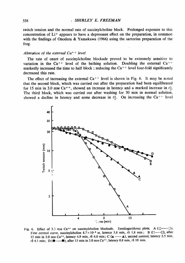
variation in the Ca+ + level of the bathing solution. Doubling the external Ca-markedly increased the time to half block; reducing the Ca++ level four-fold significantlydecreased this rate.The effect of increasing the external Ca++ level is shown in Fig. 6. It may be noted
that the second block, which was carried out after the preparation had been equilibratedfor 15 min in 3.0 mM Ca+ +, showed an increase in latency and a marked increase in tl.The third block, which was carried out after washing for 30 min in normal solution,showed a decline in latency and some decrease in tl. On increasing the Ca+ level
40
30
20
I--
EE 10C0
C
._-3~5
2
4 0 12r.ne (min)
Fig. 6. Effect of 3.0 nLw Ca++ on succinylcholine blockade. Semilogarithmic plots. A (0-O).First control curve, succinylcholine 8.7 x 10F M, latency 3.8 min, t 1.8 min; B (L -U), after15 min in 3.0 mn Ca++, latency 4.9 min, t 6.0 min; C (A - A), second control, latency 2.5 min.td 4.1 min; D (O - ), after 15 min in 3.0 mmv Ca++, latency 0.8 min, t4 10 min.
558

IONS AND SUCCINYLCHOLINE BLOCKADE
again, the fourth block occurred virtually without latency, and with a further increase intj. The comparative irreversibility of the Ca++ effect was noted consistently, and meantthat it was only possible to evaluate this effect by a comparison of first and second blocks.
Ten such experiments showed that there was no change in latency between the firstand second (high Ca++) block; the mean values were 4.2 and 4.1 min. However therewas a significant (P<0.05) increase in t-l from 2.3 to 6.7 min. It was further noted thatthere was a highly significant linear correlation between the to of the control block, andt4 of the high Ca++ block. The slope of the regression, b, equalled 5.09, and thecorrelation was significant at P<0.005. In other words, slow control blocks were particu-larly susceptible to the action of high Ca++ solutions. Data derived from third controlblocks, and fourth high Ca++ blocks showed similar relationships.
It appeared likely that exposure to high Ca++ solutions had permanently altered thestate of the junction. This was confirmed in experiments in which preparations werebathed in high Ca++ solutions for 17-20 min, then washed in normal solution for 10min before the addition of succinylcholine. This exposure to high Ca++ (3.0 mM) causeda mean lengthening of t- between first and second blocks from 3.1 to 9.4 min. There wasno change in latency.The antagonism between succinylcholine and Ca++ was further demonstrated when
the external Ca++ level was doubled in a preparation which was 95% blocked withsuccinylcholine. The force of contraction was doubled within 30 sec of adding Ca++to the bath. Further elevation of external Ca++ brought about further alleviation of theblock. The results obtained with this type of experiment varied somewhat; it was alwayspossible to bring about some degree of alleviation of the block by increasing the externalCa++ level; however, it was not possible to alleviate it completely.
Reduction in the external Ca++ level to 0.38 mm (0.25 x normal) brought about asignificant decrease in both latency and t!. The latency fell from 4.8 +0.3 min (s.E. often observations) to 2.8 + 0.2 min (S.E. of fourteen observations); the difference wassignificant at P<0.001. The time to half block fell from a control value of 3.3 + 0.3 minto 1.4 +0.1 min; the difference was significant at P<0.00I.The sensitivity of the rate of succinylcholine blockade to small changes in the level
of external Ca++ contrasts with the apparent insensitivity of this phenomenon to lowNa+ solutions. One must presume that as the safety margin in the transmission processis reduced by succinylcholine, Ca++ dependent mechanisms become rate-determining.The antagonistic effect of raised Ca++ solutions indicates a presynaptic action forsuccinylcholine, because del Castillo & Stark (1952) and Takeuchi (1963) have notedthat raised Ca++ does not alter the sensitivity of the end-plate region. The alteration ofthe amplitude of the end-plate potential in response to variation in external Ca++ (delCastillo & Stark, 1952) was brought about by Ca++ changes similar in magnitude to thoseused in the present study, and suggests that succinylcholine blockade is alleviated byhigh Ca++ solutions because of an increase in acetylcholine release.
Na+-Ca++ interactionsKelly (1965) and Gage & Quastel (1966) have noted an antagonism between the effects
of Na+ and Ca++ on transmitter release. It is noteworthy therefore that the effects of
559
SHIRLEY E. FREEMAN
high or low Ca++ on the rate of development of succinylcholine blockade could not bealtered by decreasing the Nat concentration.The increase in t- brought about by 3.0 mm Ca++ was compared with t-1 in a com-
panion diaphragm immersed in 3.0 mm Ca++ and 81 mm Na+ ; the osmotic pressure wasmaintained with sucrose. Although there was some variability in the response of paireddiaphragms it was not possible to detect any change in tI as a result of the reduced Na+level. In other experiments the rate of onset of succinylcholine blockade was comparedin the presence of 0.38 mm Ca++, and 0.38 mm Ca++ and 70 mM Na++. Again it wasfound that reduction of external Na+ did not alter the Ca++ effect. It was notedhowever, that succinylcholine blockade in the presence of low Ca++, low Na+ solutionspromoted the development of tachyphylaxis to subsequent succinylcholine blocks, whilethis did not appear after succinylcholine block in the presence of low Ca++ alone.
A lteration of the external K + levelThe rate of onset of succinylcholine blockade was accelerated by increasing the K+
level of the external medium. After one succinylcholine block in normal solution thediaphragms were washed, and then transferred to a solution containing twice the usualK+ level (9.2 mM). After 15 min succinylcholine was added to the organ bath. It wasfound that there was a significant decline in latency and t-1 in high K+ solutions. Latencyfell from a control value of 3.7+0.2 min (S.E. of twelve observations) to 3.0+0.2 min(S.E. of fourteen observations); the difference was significant at P<0.02. The time tohalf block fell from 2.6 + 0.2 min to 1.7 + 0.3 min; the difference was significant atP<0.05. Tachyphylaxis did not develop in the presence of high K+. Thus these resultsclosely resemble the effects of low Ca++ solutions.
It was noteworthy that high K+ solutions would reduce or reverse tachyphylaxis causedby high Ca++ solutions or repeated succinylcholine block.Reducing the K+ level of the medium to 1.2 mm for 10-15 min before succinylcholine
block increased tl by a factor of 2 or 3, but had little effect on the length of latency.The results resembled closely those obtained with high Ca++ solutions. Although the lowK+ series was not as extensive as the 3.0 mM Ca++ series it was possible to relate theincrease in t-2 to the initial t2, as was possible in the former series. It was also notedthat the increase in t-2 in low Kt solutions was irreversible (compare with Fig. 6).
K+-Ca++ interactionsBecause increases in the external levels of K+ and Ca++ have opposing effects, it was
of interest to determine whether the increase in tr brought about by 3.0 mM Ca++ wasantagonized by 9.2 mm K+. These experiments were carried out in the same way asthe low Na+, high Ca++ experiments; that is, companion preparations were first blockedin normal solution, then transferred either to high K+, high Ca++ or to high Ca++solutions. It was not possible to detect any difference in latency or t-k between the twogroups. Thus when they are exhibited together K+ does not modify the effect of raisedCa+ +.
560

IONS AND SUCCINYLCHOLINE BLOCKADE
A Iteration of the external Mg+' levelDoubling the external Mg++ level (2.0 mm Mg+) for 10-15 min before succinyl-
choline block reduced the latency and t{ in a fashion similar to that found with lowCa++ or high K+ solutions. Latency was reduced from 4.7 + 0.4 min (S.E. of nine observa-tions) to 3.8 +0.3 min (S.E. of nine observations); the difference was significant at P<0.05.The time to half block was reduced from 2.4 + 0.2 min to 1.4+0.1 min; the differencewas significant at P<O.OC4.
Tachyphylaxis did not occur in the presence of high Mg++, nor was there any altera-tion in the twitch tension of preparations. As was found with high K+ solutions, tachy-phylaxis was partially reversed by raising the external Mg+ + level. This was welldemonstrated in experiments in which the Mg++ level was raised during succinylcholineblock in a preparation in which tachyphylaxis had already developed. Increasing theMg++ level three- or four-fold caused an immediate increase in the rate of blockade, asmay be seen in Fig. 7. It was characteristically found, however, that blockade did notproceed to completion, but levelled off at between 15 and 20% of the initial value. This
40
30 _
20 -
EEC10 _0C
4.,
15
2
4 8 12Time (min)
Fig. 7. Effect of raising the Mg+ + level during succinylcholine blockade in a preparation withtachyphylaxis caused by repeated succinyicholine block. Mg+ + level raised to 3.0 mm atarrow. Latency 4.2 min, tj initial part of block 5.8 min; i I after adding Mg++ 2.5 min.
561

SHIRLEY E. FREEMAN
can be compared with curve B in Fig. 2, which illustrates another preparation in whichtachyphylaxis had been induced.Exposure of preparations to low Mg++ solutions (0.25 mM) for periods of 20 min was
without effect on either latency or to-. Longer periods of exposure did not alter latency,but caused an increase in t- which resembled that induced by high Ca++ solutions in allrespects.
DISCUSSION
The present study has outlined factors which affect the kinetics of onset of succinyl-choline blockade of the hemidiaphragm preparation and which are involved in thedevelopment of tachyphylaxis. Experiments carried out at 35°-36° C showed a greatvariability of response to succinyicholine ; it seems that in vitro the preparation undergoesa progressive change which reduces its sensitivity to the drug. Consequently the earlypart of this investigation was concerned to develop a standardized preparation, andconcurrently to establish what factors are involved in the development of tachyphylaxis.Briefly, tachyphylaxis is promoted by incubation at body temperature, high levels ofsuccinylcholine, prolonged exposure to succinylcholine, high rates of stimulation, repeatedperiods of tetanic stimulation, hypoxia, high levels of external Ca++, low levels of Kor Mg++, and low Ca++-low Na+ solutions.
It is not affected by the time of incubation of the preparation at 290-300 C beforesuccinylcholine block and it is diminished or abolished by low temperature, low Ca++,high K+ or high Mg++ solutions. The occurrence of tachyphylaxis in the intact animal(Zaimis, 1953) suggests that it is a property of the drug, rather than a by-product of theartificial environment.The marked sensitivity of the preparation to external Ca++ changes suggests that
interactions between this ion and succinylcholine are important both in determining therate of succinylcholine blockade and the onset of tachyphylaxis. It is possible to correlatetranslocation of membrane Ca++ with most of the factors that promote tachyphylaxis.Thus depletion of membrane Ca++ has been suggested as a factor in the hypoxic failureof neuromuscular transmission (Hubbard & L0yning, 1966). Post-tetanic potentiation ofthe end-plate potential is likely to be associated with a residual change in ionized calciumconcentration at an important membrane site (Gage & Hubbard, 1966; Katz & Miledi,1965). K+ may potentiate succinylcholine block by depletion of membrane Ca++ (Shanes& Bianchi, 1959; Koketsu & Miyamoto, 1961), rather than by virtue of the slightdepolarization caused by this level of K+ (9.2 mM), or the moderate increase in m.e.p.p.frequency (Gage & Quastel, 1966). Mg++ is also likely to potentiate succinylcholineblock by a presynaptic inhibition of acetylcholin- release, because postsynaptically itsactions potentiate those of Ca++ (Takeuchi, 1963).One may postulate a competition between succinylcholine and Ca++ in the presynaptic
membrane, leading to a reduction in acetylcholine release. On removal of succinyl-choline from the organ bath Ca++ restitution may be delayed, with a consequent delayedrecovery of tetanic tension and post-tetanic potentiation. This factor could account forthe prolonged suppression of post-tetanic repetition in the nerve terminals that wasobserved by Standaert & Adams (1965), and which Riker (1966) also noted after acetyl-
562
IONS AND SUCCINYLCHOLINE BLOCKADE
choline blockade. As these authors noted, the suppression was too prolonged to beassociated with the continued presence of the drug.The final state of membrane Ca++ after succinylcholine block may differ from the
initial state, in that a more stable calcium complex may be formed. Tachyphylaxis wouldensue if succinylcholine were less able to displace Ca++ from this complex than initially.
This discussion suggests that the development of tachyphylaxis is a presynapticphenomenon. It is likely, however, that postsynaptic changes are also involved, forGibberd (1966) was able to correlate tachyphylaxis to decamethonium in the diaphragmwith a reduction in muscle K+. Stevenson (1960) also demonstrated loss of muscle K+in dogs subjected to succinylcholine relaxation.The form of the curve of twitch tension against time during succinylcholine blockade
is worthy of comment. The latent period before the exponential decline in twitch tensionrecalls the period during which succinylcholine causes twitch potentiation in frog muscle(Nastuk & Karis, 1964). It seems to coincide with the period of d&-olarization of theend-plate, before repolarization and block (Thesleff, 1955a, b). The difference in responseof the two preparations may reside in the greater depolarization caused by succinylcholinein the frog sartorius than in the rat diaphragm (Thesleff, 1955a, b), because hexafluore-nium, which reduces this depolarization, converts the frog muscle response to one whichresembles that of the rat diaphragm (Nastuk & Karis, 1964).The functional reserve of the transmission process will also lead to latency, because a
substantial number of receptors (either pre or postsynaptic) must be occupied before thetransmission process is affected (Paton & Waud, 1967).
Diffusion delays could also become rate-determining in conditions of very rapidsuccinylcholine block, because Creese (1954) observed a t-4 of 1 min for the extracellularexchange of Na+ in the diaphragm. The preparation used in the present study were ofa thickness comparable with those of Creese (mean thickness 0.64 mm compared with0.60 mm in Creese's series).The hypothesis is proposed that the latent period is the result of tile combined effects
of depolarization and reduction in transmitter release. The safety margin for transmissionis increased by depolarization, because the further depolarization necessary to trigger anaction potential is smaller. Thus a reduction in transmitter release will not affect thetransmission process until either the postsynaptic membrane becomes unresponsive orthe reduction in transmitter release becomes overwhelming.
If desensitization of the postsynaptic membrane were the principal factor in maintain-ing blockade, then increases in the external Ca++ level would be expected to intensify thiseffect, rather than relieve it (Nastuk, 1966). The importance of presynaptic factors isconfirmed by the findings of Karczmar, Kim & Koketsu (1961) that tetraethylammonium,which increases the release of acetylcholine, will alleviate an established decamethoniumblock. Further, antagonism of succinylcholine blockade has been found to be a propertyof a number of drugs which promote acetylcholine release (Freeman, unpublished).
It may be relevant that the concentration of succinylcholine used by Thesleff (1956)when studying postsynaptic depolarization was considerably greater than was used byEdwards & Ikeda (1962), and in the present study. It may be that presynaptic effectspredominate at levels of succinylcholine which are just sufficient to cause block.
563

SHIRLEY E. FREEMAN
It is likely that a unifying concept of succinylcholine action may be possible. Excita-tion followed by stabilization of the membrane may occur at both pre- and postsynapticsites. Species differences and differences between muscle types may reflect differencesin the intensity of these sequential actions. Facilitatory actions of succinylcholine will pre-dominate when the excitatory phase is well developed; block without facilitation willoccur, as in the rat diaphragm, when stabilization predominates.The effects observed in the present study may reflect the differential sensitivity of the
presynaptic membrane to ionic changes rather than a fundamentally dissimilar mechan-ism on either side of the junction.
SUMMARY
1. The kinetics of succinylcholine blockade of the rat phrenic nerve-diaphragmpreparation have been studied. A standardized procedure has been developed whichpermits of a quantitative and reproducible estimate of the rate of onset of blockade.
2. Blockade was found to occur in two stages. Using a succinylcholine level of8.7 x 10-6 M there was an initial latent period of some 4 min duration, which was followedby an exponential decline in twitch tension to zero, with a half-time of approximately2 min.
3. In common with the results of other workers, it was found that the preparationdeveloped tachyphylaxis to repeated succinylcholine block. Factors promoting tachy-phylaxis included incubation at 370 C, high levels of succinylcholine, prolonged exposureto succinylcholine, high rates of stimulation, repeated periods of tetanic stimulation,hypoxia, raised levels of external Ca+-, low levels of K+ or Mg++, low Ca+-low Na+ solutions. Tachyphylaxis was diminished or abolished by incubation at300 C, low Ca++, high K+ or high Mg++ solutions.
4. The rate of onset of blockade was increased by low temperature, low Ca+ +, highK+ or high Mg++ solutions. It was markedly decreased by doubling the external Ca+-level. It seemed to be insensitive to reduction in the external Na+ level.
5. It is suggested that at the concentration of succinylcholine used in these experiments(8.7 x 10⁻⁶ M), which was just sufficient to block the preparation completely at 30° C,succinylcholine has an action which is largely but not entirely presynaptic. There seemsto be competition between succinylcholine and Ca + in the presynaptic membrane, whichresults in a decreased release of acetylcholine.
I should like to thank Dr. T. E. B. Keen for his constant support and encouragement. I also wishto acknowledge the assistance of Mr. G. L. White and Mr. R. Turner with statistical procedures.
564

IONS AND SUCCINYLCHOLINE BLOCKADE
REFERENCES
BEANI, L., BIANCHI, C. & LEDDA, F. (1962). The effect of tubocurarine on acetylcholine release from motornerve terminals. J. Physiol., Lond., 174, 172-183.
BIGLAND, B., GOETZEE, B., MACLAGAN, J. & ZAIMIS, E. (1958). The effect of lowered muscle temperatureon the action of neuromuscular blocking drugs. J. Physiol., Lond., 141, 425-434.
BOWMAN, W. C. & HEMSWORTH, B. A. (1965). Effects of some polymethylene bis-(hydroxyethyl)dimethyl-ammonium salts on neuromuscular transmission. Br. J. Pharmac. Chemother., 25, 392-404.
BULBRING, E. (1946). Observations on the isolated phrenic nerve diaphragm preparation of the rat. Br.J. Pharmac. Chemother., 1, 38-61.
BURNS, B. D. & PATON, W. D. M. (1951). Depolarization of the motor end-plate by decamethonium andacetylcholine. J. Physiol., Lond., 115, 41-73.
CREESE, R. (1954). Measurement of cation fluxes in rat diaphragm. Proc. R. Soc. B., 142. 497-513.CREESE, R., DILLON, J. B., MARSHALL, J., SABAWALA, B., SCHNEIDER, D. J., TAYLOR, D. B. & ZINN, D. E.
(1957). Effect of neuromuscular blocking agents on isolated human intercostal muscles. J. Pharmac.,exp. Ther., 119, 485-494.
DEL CASTILLO, J. & STARK, L. (1952). The effect of calcium ions on the motor end-plate potentials. J.Physiol., Lond., 116, 507-515.
DEL CASTILLO, J. & KATZ, B. (1955). Local activity at a depolarized nerve-muscle junction. J. Ph'siol.,Lond., 128, 396-412.
EDWARDS, C. & IKEDA, K. (1962). Effect of 2-PAM and succinylcholine on neuromuscular transmissionin the frog. J. Pharmac. exp. Ther., 138, 322-327.
GAGE, P. W. & HUBBARD, J. 1. (1966). An investigation of the post-tetanic potentiation of end-platepotentials at a mammalian neuromuscular junction. J. Physiol., Lond., 184, 353-375.
GAGE, P. W. & QUASTEL, D. M. J. (1966). Competition between sodium and calcium ions in transmitterrelease at mammalian neuromuscular junctions. J. Physiol., Lond., 185, 95-123.
GALLEGO, A. & LORENTE DE N6, R. (1951). On the effect of ammonium and lithium ions upon the frognerve deprived of sodium. J. gen. Physiol., 35, 227-244.
GIBBERD, F. B. (1966). Action of decamethonium on rat diaphragm. Br. J. Pharmac. Chemother., 28,128-136.
HODGKIN, A. L. & KATZ, B. (1949). The effect of sodium ions on the electrical activity of the giant axonof the squid. J. Physiol., Lond., 108, 37-77.
HOFMANN, W. W., PARSONS, R. L. & FEIGEN, G. A. (1966). The effects of temperature and drugs on mam-malian motor nerve terminals. Am. J. Physiol., 211, 135-140.
HOLMES, P. E. B., JENDEN, D. J. & TAYLOR, D. B. (1951). The analysis of the mode of action of curare onneuromuscular transmission; the effect of temperature changes. J. Pharmac. exp. Ther., 103, 382-402.
HUBBARD, J. I. & LOYNING, Y. (1966). The effects of hypoxia on neuromuscular transmission in a mam-malian preparation. J. Physiol., Lond., 185, 205-223.
HUBBARD, J. I., SCHMIDT, R. F. & YOKOTA, T. (1965). The effect of acetylcholine upon mammalian motornerve terminals. J. Physiol., Lond., 181, 810-829.
JENDEN, D. J., KAmuo, K. & TAYLOR, D. B. (1954). The action of decamethonium on the isolated rabbitlumcrical muscle. J. Pharmac. exp. Ther., 111, 229-240.
KARCZMAR, A. G., KIM, K. C. & KOKETSU, K. (1961). End-plate effects and antagonism to d-tubocurarineand decamethonium of tetraethylammonium and methoxyambenonium (WIN8078). J. Phuarma.exp. Ther., 134, 199-205.
KATZ, B. & MILEDI, R. (1965). The effect of calcium on acetylcholine release from motor nerve endings.Proc. R. Soc. B., 161, 496-503.
KELLY, J.S. (1965). Antagonism between Na+ and Ca+ at the neuromuscular junction. Nature, Lond..205, 296-297.
KOKETSU, K. & MIYAMOTO, S. (1961). Significance of membrane calcium in Ca-free and K-rich media.Nature, Lond., 189, 403-404.
NASTUK, W. L. & KARIS, J. H. (1964). The blocking action of hexafluorenium on neuromuscular trans-mission and its interaction with succinylcholine. J. Pharmac. exp. Ther., 144, 236-252.
NASTUK, W. L. (1966). Fundamental aspects of neuromuscular transmission. Ann. N. Y. Acad. Sci., 135,111-135.
ONODERA, K. & YAMAKAWA, K. (1966). The effects of lithium on the neuromuscular junction of the frog.Jap. J. Physiol., 16, 541-550.
PATON, W. D. M. & WAUD, D. R. (1967). The margin of safety of neuromuscular transmission. J. Physiol.,Lond., 191, 59-90.
RIKER, W. F. (1966). Actions of acetylcholine on mammalian motor nerve terminal. J. Pharmac. exp.Ther., 152, 397-416.
SHANES, A. M. & BIANCHI, C. P. (1959). Radiocalcium release by stimulated and potassium-treated sar-torius muscles of the frog. J. gen. Physiol., 43, 481-493.
565

566 SHIRLEY E. FREEMAN
STANDAERT, F. G. & ADAM,, J. E. (1965). The actions of succinylcholine on the mammalian motor nerveterminal. J. Pharmac. exp. Tner.. 149, 113-123.
STEVENSON, D. E. (1960). Changes in the blood electrolytes of anaesthetized dogs caused by suxamethonium.Br. J. Anaesth., 32, 364-371.
TAKEUCHI, N. (1958). The effect of temperature on the neuromuscular junction of the frog. Jap. J. Physiol.8, 390-404.
TAKEUCHI, N. (1963). Effects of calcium on the conductance change of the end-plate membrane during theaction of transmitter. J. Physio., Lond., 167, 141-155.
TAYLOR, D. B. & NEDERGAARD, 0. A. (1965). Relation between structure and action of quaternary ammo-nium neuromuscular blocking agents. Physiol. Rev., 45, 523-554.
THELEFF, S. (1955a). The mode of neuromuscular block caused by acetylcholine, nicotine, decamethoniumand succinylcholine. Acta p'zysiol. scand., 34, 218-231.
TfE LEFF, S. (1955b). The effects of acetylcholine, decamethonium and succinylcholine on neuromusculartransmission in the rat. Acta physiol. scand., 34, 386-392.
VAN BREEMAN, C., DANIEL, E. E. & VAN BREEMAN, D. (1966). Calcium distribution and exchange in therat uterus. J. gen. Physiol., 49, 1265-1298.
ZAIMIS, E. (1953). Motor end-plate differences as a determining factor in the mode of action of neuro-muscular blocking substances. J. Physio!., Lond., 122, 238-251.