pdf (1.1 mb) - jpis - journal of periodontal & implant science
TRANSCRIPT
www.jpis.org
Journal of Periodontal& Implant ScienceJPIS
pISSN 2093-2278eISSN 2093-2286
Copyright © 2011 Korean Academy of PeriodontologyThis is an Open Access article distributed under the terms of the Creative Commons Attribution Non-Commercial License (http://creativecommons.org/licenses/by-nc/3.0/).
A short-term clinical study of marginal bone level change around microthreaded and platform-
switched implantsHee-Jung Yun, Jung-Chul Park, Jeong-Ho Yun, Ui-Won Jung, Chang-Sung Kim, Seong-Ho Choi, Kyoo-Sung Cho*
Department of Periodontology, Yonsei University College of Dentistry, Seoul, Korea
Purpose: The marginal bone levels around implants following restoration are used as a reference for evaluating implant suc-cess and survival. Two design concepts that can reduce crestal bone resorption are the microthread and platform-switching concepts. The aims of this study were to analyze the placement of microthreaded and platform-switched implants and their short-term survival rate, as well as the level of bone around the implants.Methods: The subjects of this study were 27 patients (79 implants) undergoing treatment with microthreaded and platform-switched implants between October 2008 and July 2009 in the Dental Hospital of Yonsei University Department of Periodon-tology. The patients received follow-up care more than 6 months after the final setting of the prosthesis, at which time peri-apical radiographs were taken. The marginal bone level was measured from the reference point to the lowest observed point of contact between the marginal bone and the fixture. Comparisons were made between radiographs taken at the time of fix-ture installation and those taken at the follow-up visit.Results: During the study period (average of 11.8 months after fixture installation and 7.4 months after the prosthesis delivery), the short-term survival rate of microthreaded and platform-switched implants was 100% and the marginal bone loss around implants was 0.16±0.08 mm, the latter of which is lower than the previously reported values. Conclusions: This short-term clinical study has demonstrated the successful survival rates of a microthread and platform-switched implant system, and that this system is associated with reduced marginal bone loss.
Keywords: Alveolar bone loss, Dental implants.
J Periodontal Implant Sci 2011;41:211-217 • http://dx.doi.org/10.5051/jpis.2011.41.5.211
Research Article
INTRODUCTION
Since Brånemark found that osseointegration occurred be-tween titanium and bone in the mid-1960s [1], several studies have investigated titanium dental implants and their clinical applications. The functional and esthetic restoration of eden-tulous areas using dental implants is now considered a desir-able treatment option. The advantages of implant restoration relate not only to esthetic demands but also to avoiding the
involvement of the adjacent teeth. In addition, implant res-toration is more comfortable for the patient than conven-tional dentures and prevents the resorption of the remaining bone that occurs with dentures. As a result, implant treatment has become common and several new implant systems have been developed and are now available in the marketplace. Consequently, dentists are now able to choose an implant that is most appropriate for the condition of each patient.
The ability of the dentist, as well as the quality and quantity
Received: Jun. 13, 2011; Accepted: Aug. 24, 2011*Correspondence: Kyoo-Sung ChoDepartment of Periodontology, Yonsei University College of Dentistry, 134 Shinchon-dong, Seodaemun-gu, Seoul 120-752, Korea E-mail: [email protected], Tel: +82-2-2228-3188, Fax: +82-2-392-0398
Journal of Periodontal& Implant ScienceJPISMarginal bone loss around microthreaded and platform-switched implants 212
of available bone, are the primary factors for successful im-plant therapy. Atwood evaluated changes in the volume of bone after loss of teeth [2], and in 1985, Lekholm and Zarb [3] classified the quality and quantity of remaining bone at the planned implant site. Taking these factors into account, pre-dictable treatments can be assured if the dentist selects the implant system with a high survival rate; the design and fea-tures of the implant surface should also be considered. Al-though it is difficult to define the survival and success of im-plants, the success rate is currently defined as the proportion of implants that conform to the success criteria after a spe-cific period, and the survival rate as the proportion of im-plants that do not need to be removed at certain points in time [4].
The resulting crestal bone levels around implants following restoration have been a topic of discussion and used as a ref-erence for evaluating implant success and survival for many years [5,6]. Achieving esthetically pleasing implant therapy is crucially affected by the height of the supracrestal soft-tissue portion, since this is highly relevant to the level of bony sup-port around the fixture [7].
There are many suggested causes for early implant bone loss. Changes in crestal bone height have been attributed to implant loading and concentration of forces, the counter-sinking procedure during implant placement procedures, and localized soft-tissue inflammation, among others [8]. Implant design can affect occlusal overload and the crestal module, which is the implant body that receives the stress from the implant after loading. The implant system should be designed so that it can best distribute stress to the peri-implant bone in a manner that supports a restoration in function and en-courages osseous attachment [9]. Two design concepts that can reduce crestal bone resorption are the microthread and platform-switching concepts. These features are incorporat-ed into the Osstem GS III implant system (Osstem Implant Co., Seoul, Korea), together with a tapered body, self-tapping
ability, and internal connection, and a resorbable blast media (RBM) surface. The tapered body is good for ensuring initial stability and controlling the depth and path of insertion [10], and implants with an RBM surface are reportedly associated with a high success rate [11].
The aims of this study were to analyze the placement of microthreaded and platform-switched implants and their short-term survival rate, as well as the effect of the micro-threads and platform-switching on the level of bone around the implants.
MATERIALS AND METHODS
The subjects of this study were patients undergoing treat-ment with Osstem GS III implants (Fig. 1) between October 2008 and July 2009 in the Department of Periodontology of the Dental Hospital of Yonsei University. This study was ap-proved by the Institutional Review Board at Yonsei Dental Hospital (IRB number 2-2009-0025). Overall, 27 patients (15 males, 12 females) were included in this investigation. The subjects’ ages ranged from 19 to 77 years (mean, 58.6±13.5 years). In total, 79 implants were inserted (Table 1). The pres-ence of systemic disease among the patients was evaluated using a questionnaire. Bone quality and quantity were evalu-ated during the operation in accordance with the Lekholm and Zarb index. Among the 79 implants, 30 were inserted into the maxilla and 49 were inserted into the mandible. Eleven implants were placed in the anterior teeth area and 68 were placed in the posterior teeth area. Thus, most of the implants were placed in the posterior mandible (Table 2). Hypertension was the most common general disorder in this patient group.
The patients were followed for more than 6 months after the final setting of the prosthesis, at which time periapical radiographs were taken using the parallel cone technique with an Extension Cone Paralleling device. All films were de-
Figure 1. Illustration of GS III implant.
GS III
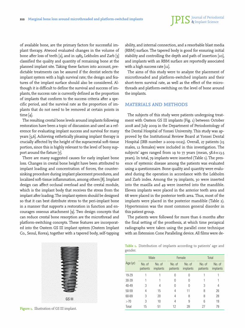
Table 1. Distribution of implants according to patients’ age and gender.
Age (yr)Male Female Total
No. of patients
No. of implants
No. of patients
No. of implants
No. of patients
No. of implants
19-29 1 1 0 0 1 130-39 1 1 0 0 1 140-49 3 4 0 0 3 450-59 4 15 4 11 8 2660-69 3 20 4 8 8 28>70 3 10 4 9 6 19Total 15 51 12 28 27 79

Journal of Periodontal& Implant ScienceJPIS Hee-Jung Yun et al. 213
veloped using the same automatic processor in accordance with the manufacturer’s instructions.
This study was carried out retrospectively using the patients’ charts, from which the following information was collected: age, gender, distribution of the implants, general health dis-order, reasons for tooth loss, bone quality and quantity, and implant diameter and length. Most of the teeth had been lost because of periodontal problems, although in some cases the cause was unknown. Type D3, B bone was common in the maxilla, and type D2, B bone in the mandible, according to the Lekholm and Zarb index (Tables 3 and 4). The distribu-tions of implant length and diameter are given in Tables 5 and 6.
Survival rateThe survival rate was evaluated according to the criteria re-
ported by Buser et al. [12] as follows:1. The absence of persistent subjective complaints, such as
pain, foreign body sensation, and/or dysesthesia.2. The absence of recurrent peri-implant infections with
suppuration.3. The absence of mobility.4. The absence of continuous radiolucency around the im-
plant.5. The possibility for restoration.
Measurement of changes in marginal bone levelAfter digitization, all files were transferred to a personal
computer and examined on the same monitor. The Starpacs System (Infinitt Co., Seoul, Korea) was used as the image-anal-ysis software. The marginal bone level was measured (to the nearest 0.01 mm) from the reference point to the lowest ob-served point of contact between the marginal bone and the fixture. The reference point of the fixture was the top of the fixture (Fig. 2). The amounts of bone loss on the mesial and distal sides of the implants were measured and the average value was used. Calibration was performed with a known distance between screws (1.6 mm) as the reference length [13]. The radiographs were magnified to enable precise measure-
Figure 2. The reference point with the periapical radiograph. Arrow 1 marks the reference and arrow 2 marks the lowest observed point of contact between the marginal bone and the fixture.
2 Cm
1
2
Table 5. Distribution of implant length.
Length (mm)
Maxilla MandibleTotal
Anterior Posterior Anterior Posterior
7 0 0 0 5 58.5 0 0 0 6 610 0 8 0 9 1711.5 4 16 2 21 4313 0 2 5 1 8Total 4 26 7 42 79
Table 6. Distribution of implant diameter.
Diameter (mm)
Maxilla MandibleTotal
Anterior Posterior Anterior Posterior
3.5 2 0 1 2 54 2 3 6 11 224.5 0 10 0 10 205 0 13 0 19 32Total 4 26 7 42 79
Table 2. Distribution of implants according to position.
Maxilla Mandible Total
Incisor 2 4 6Canine 2 3 51st premolar 7 5 122nd premolar 3 8 111st molar 7 15 222nd molar 9 14 23Total 30 49 79
Values are presented as no. of implants.
Table 3. Distribution of bone quality.
Bone quality D1 D2 D3 D4 Unknown Total
Maxilla 0 0 22 5 3 30Mandible 4 24 10 1 10 49Total 4 24 32 6 13 79
Table 4. Distribution of bone quantity.
Bone quantity A B C D Unknown Total
Maxilla 0 19 8 0 3 30Mandible 0 27 10 2 10 49Total 0 46 18 2 13 79
Journal of Periodontal& Implant ScienceJPISMarginal bone loss around microthreaded and platform-switched implants 214
ments. Only the amount of vertical bone loss was measured. Comparisons were made between radiographs taken at the time of fixture installation and those taken at the follow-up visit more than 6 months after final prosthesis delivery.
Statistical analysisThe change in marginal bone level around microthreaded
and platform-switched implants was analyzed using paired t-testing. Statistical software (SPSS ver. 16.0, SPSS Inc., Chica-go, IL, USA) was used for statistical analysis. The data are pre-sented as mean±SD values, and the level of statistical signifi-cance was set at P<0.05.
RESULTS
Survival rateNo implant was lost during the observation period (11.8±1.8
months on average), and none of the patients reported sub-jective complaints after implant installation. No peri-implant infection, implant mobility, or radiolucency around the im-plant was detected. Therefore, according to the survival crite-ria reported by Buser et al. [12], the implant survival rate in our cohort was 100%.
Changes in marginal bone levelThe mean follow-up time was 11.8 months after fixture in-
stallation and 7.4 months after prosthesis delivery. The radio-graphic analysis revealed a significant marginal bone loss of 0.16±0.08 mm at the follow-up visit (P<0.05; Table 7). More bone was resorbed at the distal than the mesial side.
DISCUSSION
Many recent studies have found no differences in the suc-cess and failure rates among various root-formed osseous integrated dental implant systems [14]. Implant failure is di-vided into early failure (occurring before loading) and late failure (destruction of osseointegration). The reported per-centages of late failures have varied widely, from 2.1 to 11.3% [10]. In the present study, the survival rate of microthreaded and platform-switched implants was 100%. However, be-
cause the study period was short, further investigation is re-quired to evaluate the survival rate over longer durations.
Albrektsson et al. [5] suggested the following success crite-ria: 1) the change in marginal bone level in the first year should be less than 1 to 1.5 mm, and 2) ongoing annual bone loss should be less than 0.2 mm. In their 15-year study, Adell et al. [15] reported a crestal bone loss of 1.2 mm for Brånemark System implants for the first year. In the present study, the marginal bone loss was 0.16±0.08 mm, which is lower than previously reported data.
Two design concepts that can reduce crestal bone resorp-tion are the microthread system and the platform-switching concept. The microthread system enhances the contact area between implant and bone. A study of the mechanical prop-erties of bone [16] found it to be more resistant to compres-sive forces than tensile and shear forces (its resistances to the latter were reportedly 30% and 65% lower, respectively, than its resistance to compression). The crestal module design is particularly important with regard to minimizing bone loss, because it can decrease the shear force exerted on the crestal bone [17]. Therefore, it has been hypothesized that bone loss slows down at the first thread of the implant fixture when the force changes from a crestal shear force to a compressive force induced by the thread itself [18]. In addition, correla-tions were found between the amount of bone loss and the length of the machined surface for various implant systems, thus relating bone loss to the level of the first thread [19].
Hansson utilized a 3D mathematical model and axisym-metric finite element analysis to determine the ideal rough surface. He hypothesized that the surface roughness or the retentive elements such as microthreading increases the re-sistance of marginal bone to bone loss by improving the in-terlocking force between the implant surface and the crestal bone [20]. Abrahamsson and Berglundh [21] suggested that the microthread configuration offered improved conditions for osseointegration in a study using dogs. In that study, the degree of bone-implant contact within the marginal portion of the implants was significantly higher for the test (micro-thread) implants (81.8%) than for the control implants (72.8%).
The results of a study that used two types of Astra implants (one with the microthreads on the coronal portion of the fix-ture and one without) suggested that microthreads have the effect of maintaining the marginal bone loss in the presence of loading forces [17]. The amount of peri-implant bone loss was significantly greater around implants without micro-threads than around those with microthreads during the ex-amination period.
The platform-switching concept was developed to control bone loss after implant placement. This refers to the use of an abutment of a smaller diameter connected to an implant
Table 7. Marginal bone level.
Baselinea) F/Ub) Change between baseline and F/U
Mean bone loss (mm) 0.05 0.21 0.16Standard deviation (mm) 0.02 0.10 0.08
F/U, follow-up.a)At the time of fixture installation, b)At >6 months follow-up after prosthesis delivery.

Journal of Periodontal& Implant ScienceJPIS Hee-Jung Yun et al. 215
neck of a larger diameter. This connection shifts the perime-ter of the implant-abutment junction (IAJ) inward, toward the central axis (the middle of the implant), in order to im-prove the force distribution. Quirynen et al. [22] suggested that bacterial leakage occurs through the microgap of the IAJ. Ericsson et al. [23] found histologic evidence that an inflam-matory cell infiltration is located 1 to 1.5 mm adjacent to the IAJ after implant placement. To protect the underlying bone from this inflammatory cell infiltration and microbiologic invasion, 1 mm of healthy connective tissue is needed to es-tablish a biologic seal comparable to that around natural teeth [23,24]. This movement of the IAJ is also believed to shift in-flammatory cell infiltration to the central axis of the implant and away from the adjacent crestal bone, which is thought to restrict crestal bone resorption [8]. Indeed, Hurzeler et al. [25] reported that the concept of platform switching does appear to limit crestal resorption and preserve the peri-implant bone level. They found that the amount of bone loss was signifi-cantly lower in the platform-switching group.
Lopez-Mari et al. [26] found that platform switching is ca-pable of reducing or eliminating crestal bone loss to 1.56± 0.70 mm. It also appears to help to maintain the width and height of crestal bone and the crestal peak between adjacent implants, and reduces circumferential bone loss. It was con-cluded that the implant design modifications involved in platform switching offer multiple advantages and potential applications, including situations in which a larger implant is desirable but the prosthetic space is limited, and some im-plants are desirable in the anterior zone where preservation of the crestal bone can lead to improved esthetics .
From a review of the literature, Kwon et al. [27] concluded that the marginal bone loss associated with a flat-top implant is 1.0 to 1.3 mm at 1 year post-implantation, even in the pres-ence of an improved surface [28-30]. In contrast, the margin-al bone loss with a microthread, conical seal, and platform-switched design was found to be 0.11 to 0.24 mm [17,31]. Those authors concluded that the marginal bone levels of the sub-jects in their study (0.16 to 0.17 mm) were comparable to those of previous studies. Similarly, in the present study, the mean amount of marginal bone loss was small, and it can therefore be assumed that microthreaded and platform-switched im-plants have the ability to reduce marginal bone loss because of certain features of the implant design.
Adell et al. [32] stated that the success of implants should be evaluated 1 year after prosthesis installation, because by then almost all crestal bone loss following abutment installation would have ceased. Additional long-term studies are required to confirm that the microthreaded and platform-switched implant system has considerable potential to reduce crestal bone resorption.
Radiographic analysis can lead to false conclusions when analyzing small, peri-implant bone level changes. Bragger et al. [13] and Siegele and Soltesz [33] suggested that the implant thread is a useful aid to radiograph interpretation. In the present study, calibrations were performed with the aid of a fixture with a known length. The accuracy of using the thread pitch distance as an internal reference is reported as being within 0.3 mm [34].
The findings of this study suggest that the microthreaded and platform-switched implant system is associated with successful short-term survival rates and reduces marginal bone loss. Further long-term, post-implantation studies are required.
CONFLICT OF INTEREST
No potential conflict of interest relevant to this article was reported.
ACKNOWLEDGEMENTS
This research was supported by Basic Science Research Program through the National Research Foundation of Ko-rea (NRF) funded by the Ministry of Education, Science and Technology (2009-0073534).
REFERENCES
1. Brånemark PI, Adell R, Breine U, Hansson BO, Lindström J, Ohlsson A. Intra-osseous anchorage of dental prosthe-ses. I. Experimental studies. Scand J Plast Reconstr Surg 1969;3:81-100.
2. Atwood DA. Some clinical factors related to rate of resorp-tion of residual ridges. 1962. J Prosthet Dent 2001;86:119-25.
3. Lekholom U, Zarb GA. Patient selection and preparation. In: Brånemark PI, Zarb GA, Albrektsson T, editors. Tissue-integrated prostheses: osseointegration in clinical den-tistry. Chicago: Quintessence; 1985. p.199-220.
4. Ahlqvist J, Borg K, Gunne J, Nilson H, Olsson M, Astrand P. Osseointegrated implants in edentulous jaws: a 2-year longitudinal study. Int J Oral Maxillofac Implants 1990; 5:155-63.
5. Albrektsson T, Zarb G, Worthington P, Eriksson AR. The long-term efficacy of currently used dental implants: a re-view and proposed criteria of success. Int J Oral Maxillofac Implants 1986;1:11-25.
6. Berglundh T, Persson L, Klinge B. A systematic review of the incidence of biological and technical complications in implant dentistry reported in prospective longitudinal studies of at least 5 years. J Clin Periodontol 2002;29 Suppl
Journal of Periodontal& Implant ScienceJPISMarginal bone loss around microthreaded and platform-switched implants 216
3:197-212.7. Chang M, Wennström JL, Odman P, Andersson B. Im-
plant supported single-tooth replacements compared to contralateral natural teeth. Crown and soft tissue dimen-sions. Clin Oral Implants Res 1999;10:185-94.
8. Lazzara RJ, Porter SS. Platform switching: a new concept in implant dentistry for controlling postrestorative crestal bone levels. Int J Periodontics Restorative Dent 2006;26:9-17.
9. Schrotenboer J, Tsao YP, Kinariwala V, Wang HL. Effect of microthreads and platform switching on crestal bone stress levels: a finite element analysis. J Periodontol 2008; 79:2166-72.
10. Friberg B, Grondahl K, Lekholm U. A new self-tapping Brånemark implant: clinical and radiographic evaluation. Int J Oral Maxillofac Implants 1992;7:80-5.
11. Gonshor A, Goveia G, Sotirakis E. A prospective, multi-center, 4-year study of the ACE Surgical resorbable blast media implant. J Oral Implantol 2003;29:174-80.
12. Buser D, Weber HP, Lang NP. Tissue integration of non-submerged implants. 1-year results of a prospective study with 100 ITI hollow-cylinder and hollow-screw implants. Clin Oral Implants Res 1990;1:33-40.
13. Brägger U, Häfeli U, Huber B, Hämmerle CH, Lang NP. Evaluation of postsurgical crestal bone levels adjacent to non-submerged dental implants. Clin Oral Implants Res 1998;9:218-24.
14. Esposito M, Grusovin MG, Coulthard P, Thomsen P, Worthington HV. A 5-year follow-up comparative analysis of the efficacy of various osseointegrated dental implant systems: a systematic review of randomized controlled clinical trials. Int J Oral Maxillofac Implants 2005;20:557-68.
15. Adell R, Lekholm U, Rockler B, Brånemark PI. A 15-year study of osseointegrated implants in the treatment of the edentulous jaw. Int J Oral Surg 1981;10:387-416.
16. Guo E. Mechanical properities of cortical bone and can-cellous bone tissue. 2nd ed. Boca Raton: CRC Press; 2001. p.1-23.
17. Lee DW, Choi YS, Park KH, Kim CS, Moon IS. Effect of mi-crothread on the maintenance of marginal bone level: a 3-year prospective study. Clin Oral Implants Res 2007;18: 465-70.
18. Oh TJ, Yoon J, Misch CE, Wang HL. The causes of early implant bone loss: myth or science? J Periodontol 2002; 73:322-33.
19. Jung YC, Han CH, Lee KW. A 1-year radiographic evalua-tion of marginal bone around dental implants. Int J Oral Maxillofac Implants 1996;11:811-8.
20. Hansson S. The implant neck: smooth or provided with retention elements. A biomechanical approach. Clin Oral
Implants Res 1999;10:394-405.21. Abrahamsson I, Berglundh T. Tissue characteristics at mi-
crothreaded implants: an experimental study in dogs. Clin Implant Dent Relat Res 2006;8:107-13.
22. Quirynen M, Bollen CM, Eyssen H, van Steenberghe D. Microbial penetration along the implant components of the Brånemark system. An in vitro study. Clin Oral Im-plants Res 1994;5:239-44.
23. Ericsson I, Persson LG, Berglundh T, Marinello CP, Lindhe J, Klinge B. Different types of inflammatory reactions in peri-implant soft tissues. J Clin Periodontol 1995;22:255-61.
24. Waerhaug J. Subgingival plaque and loss of attachment in periodontosis as evaluated on extracted teeth. J Periodon-tol 1977;48:125-30.
25. Hürzeler M, Fickl S, Zuhr O, Wachtel HC. Peri-implant bone level around implants with platform-switched abut-ments: preliminary data from a prospective study. J Oral Maxillofac Surg 2007;65(7 Suppl 1):33-9.
26. López-Marí L, Calvo-Guirado JL, Martín-Castellote B, Go-mez-Moreno G, López-Marí M. Implant platform switch-ing concept: an updated review. Med Oral Patol Oral Cir Bucal 2009;14:e450-4.
27. Kwon HJ, Lee DW, Park KH, Kim CK, Moon IS. Influence of the tooth- and implant-side marginal bone level on the interproximal papilla dimension in a single implant with a microthread, conical seal, and platform-switched design. J Periodontol 2009;80:1541-7.
28. Calandriello R, Tomatis M, Vallone R, Rangert B, Gottlow J. Immediate occlusal loading of single lower molars using Brånemark System Wide-Platform TiUnite implants: an interim report of a prospective open-ended clinical multi-center study. Clin Implant Dent Relat Res 2003;5 Suppl 1: 74-80.
29. Glauser R, Lundgren AK, Gottlow J, Sennerby L, Portmann M, Ruhstaller P, et al. Immediate occlusal loading of Brånemark TiUnite implants placed predominantly in soft bone: 1-year results of a prospective clinical study. Clin Implant Dent Relat Res 2003;5 Suppl 1:47-56.
30. Vanden Bogaerde L, Pedretti G, Dellacasa P, Mozzati M, Rangert B, Wendelhag I. Early function of splinted im-plants in maxillas and posterior mandibles, using Bråne-mark System Tiunite implants: an 18-month prospective clinical multicenter study. Clin Implant Dent Relat Res 2004;6:121-9.
31. Wennström JL, Ekestubbe A, Gröndahl K, Karlsson S, Lindhe J. Implant-supported single-tooth restorations: a 5-year prospective study. J Clin Periodontol 2005;32:567-74.
32. Adell R, Lekholm U, Rockler B, Brånemark PI, Lindhe J, Er-iksson B, et al. Marginal tissue reactions at osseointegrat-ed titanium fixtures (I). A 3-year longitudinal prospective

Journal of Periodontal& Implant ScienceJPIS Hee-Jung Yun et al. 217
study. Int J Oral Maxillofac Surg 1986;15:39-52.33. Siegele D, Soltesz U. Numerical investigations of the in-
fluence of implant shape on stress distribution in the jaw bone. Int J Oral Maxillofac Implants 1989;4:333-40.
34. Hollender L, Rockler B. Radiographic evaluation of osseo-
integrated implants of the jaws. Experimental study of the influence of radiographic techniques on the measurement of the relation between the implant and bone. Dentomax-illofac Radiol 1980;9:91-5.