pd-l1 expression , ana maria waaga-gasser , martin gasser ... · pd-l1 expression murugabaskar...
TRANSCRIPT
1
Novel Roles of c-Met in the Survival of Renal Cancer Cells through the Regulation of HO-1 and PD-L1 Expression
Murugabaskar Balana,b, Eduardo Mier y Terana, Ana Maria Waaga-Gasserc, Martin Gasserc, Toni
K. Choueirib,d, Gordon Freemanb,d and Soumitro Pala,b
aDivision of Nephrology, Boston Children’s Hospital, MA 02115; and bHarvard Medical School, Boston, MA 02115; cDepartment of Surgery, Molecular Oncology and Immunology, University of
Wurzburg, 97080 Wurzburg, Germany; dDana Farber Cancer Institute, Boston, MA 02115
*Address for correspondence: Soumitro Pal, Division of Nephrology, Children’s Hospital, 300 Longwood Ave, Boston, MA 02115; e-mail: [email protected], Phone: 617-919-2989, Fax: (617) 730-0130
Running Title: c-Met-mediated regulation of HO-1 and PD-L1
Key Words: c-Met, HGF, HO-1, PD-L1 and apoptosis CAPSULE Background: Tyrosine kinase receptor c-Met plays critical roles in the growth of RCC. Results: c-Met-mediated Ras activation and HO-1 over-expression promote anti-apoptotic signals, and increase PD-L1 expression, which inhibits immune cell-mediated killing of RCC cells. Conclusion: c-Met-induced HO-1 and PD-L1 expression promotes RCC cell survival. Significance: HO-1 and PD-L1 can serve as novel therapeutic targets for c-Met-induced RCC. ABSTRACT The receptor tyrosine kinase c-Met is over-expressed in renal cancer cells and can play major role in the growth and survival of tumor. We investigated how the c-Met-mediated signaling through binding to its ligand hepatocyte growth factor (HGF) can modulate the apoptosis and immune escape mechanism(s) of renal cancer cells by the regulations of novel molecules heme oxygenase-1 (HO-1) and programmed cell death 1 ligand (PD-L1). We found that HGF/c-Met-mediated signaling activated the Ras/Raf pathway and down-regulated cancer cell apoptosis; and it was associated with the over-expression of cytoprotective HO-1 and anti-apoptotic Bcl-2/Bcl-xL. c-Met-induced HO-1 over-expression was regulated at the transcriptional level. Next, we observed that c-Met induction markedly up-
regulated the expression of the negative co-stimulatory molecule PD-L1, and this can be prevented following treatment of the cells with pharmacological inhibitors of c-Met. Interestingly, HGF/c-Met-mediated signaling could not induce PD-L1 at the optimum level when either Ras or HO-1 was knocked down. To study the functional significance of c-Met-induced PD-L1 expression, we performed a co-culture assay using mouse splenocytes (expressing PD-L1 receptor PD-1) and murine renal cancer cells (RENCA, expressing high PD-L1). We observed that the splenocyte-mediated apoptosis of cancer cells during co-culture was markedly increased in the presence of either c-Met inhibitor or PD-L1 neutralizing antibody. Finally, we found that both c-Met and PD-L1 are significantly up-regulated and co-localized in human renal cancer tissues. Together, our study suggests a novel mechanism(s) by which c-Met can promote increased survival of renal cancer cells through the regulation of HO-1 and PD-L1. INTRODUCTION c-Met is a receptor tyrosine kinase that binds with its specific ligand hepatocyte growth factor (HGF). It is composed of a 45-kDa extracellular α-chain and a disulfide-linked 145-kDa membrane spanning β-chain; and it contains HGF binding extra-cellular region and a catalytic tyrosine kinase domain in the intracellular region (1). Upon
http://www.jbc.org/cgi/doi/10.1074/jbc.M114.612689The latest version is at JBC Papers in Press. Published on February 2, 2015 as Manuscript M114.612689
Copyright 2015 by The American Society for Biochemistry and Molecular Biology, Inc.
by guest on February 3, 2019http://w
ww
.jbc.org/D
ownloaded from

2
binding with HGF, c-MET activates a wide range of pathways, including those involved in angiogenesis, proliferation, cell cycle progression, migration and invasion (2). Although c-Met is important in the control of tissue homeostasis under normal physiological conditions, it becomes aberrantly activated in human cancers via mutation, amplification or protein over-expression (3). It has been shown that c-Met is over-expressed in both clear cell and papillary renal cell carcinoma (RCC) (4, 5). pVHL-defective clear cell RCC cells are hypersensitive to HGF, leading to enhanced growth (6). Interestingly, both HGF and c-Met are located on chromosome 7, which is often amplified in clear cell RCC, and pVHL-defective cells are more dependent on c-Met for survival than are isogenic cells in which pVHL function has been restored (7). c-MET inhibitors are already being tested in papillary renal cancer because some hereditary papillary renal cancers are linked to germline c-Met mutations (8). HGF binding to c-Met results in receptor homodimerization and phosphorylation of two tyrosine residues (Y1234 and Y1235) located within the catalytic loop of the tyrosine kinase domain. Subsequently, tyrosines 1349 and 1356 in the carboxy-terminal tail become phosphorylated; and they recruit signaling effectors that include the adaptor proteins GRB2, SHC and CRK (9). These events promote the activation of multiple signaling pathways, like, Ras-Raf-ERK, PI-3K-Akt and Src-FAK. For activation of the mitogen activated protein kinase (MAPK) cascades, c-Met activation stimulates the activity of guanine nucleotide exchanger Son of Sevenless (SOS) via binding with SHC and GRB2, leading to the activation of Ras-Raf-ERK pathway (10). There is a growing appreciation that c-Met activation can confer resistance to the inhibitors of other targets (like EGFR) in RCC (11). Thus, the blockade of c-Met-mediated pathway(s), alone or in conjunction with other inhibitor, may act as promising and novel therapeutics in RCC (12). However, the downstream effector molecules for c-Met-induced tumor-promoting pathways need to be thoroughly examined. Heme oxygenase-1 (HO-1) is a stress-inducible cytoprotective molecule. It catalyses the
degradation of potent inflammatory agent heme into carbon monoxide (CO), billiverdin and ferrous iron (13). However, HO-1 also plays an important role in cancer growth (14). We have demonstrated that HO-1 is often over-expressed in RCC following activation of the Ras pathway, and it protects cancer cells from chemotherapeutic drug-induced apoptosis (15). It can promote tumor angiogenesis (16). Interestingly, HGF also induces HO-1 in renal tubular cells (17). The expression of HO-1 is tightly controlled primarily at the transcriptional level (18, 19). The transcription factors that regulate the HO-1 gene expression (like Nrf2 and Bach-1) also play a role in tumor progression (20). However, the correlation between HGF/c-Met signaling and the cytoprotective HO-1 expression in RCC is not well established. Renal tumors show increased T cell infiltration, yet T cells are rendered incapable of tumor rejection (21). This suggests that RCC cells have acquired the ability to evade host anti-tumor immunity, possibly by suppressing T cell activation. It is well demonstrated that the signaling through either positive or negative co-stimulatory molecules regulates the T cell effector function. Engagement of B7-1 expressed on antigen presenting cells (APCs) and CD28 expressed on T cells provides positive stimulatory signal for T cell activation (22). Potent negative co-stimulatory molecules are programmed death-1 (PD-1) receptor (or CD279) expressed on immune cells such as activated T cells, B cells and mature DCs, and its ligand programmed death-1 ligand (PD-L1) (or B7-H1 or CD274) (23). Both PD-1 and PD-L1 belong to cell surface glycoprotein B7 family of co-stimulatory molecules that govern T cell function and proliferation. Although PD-L1 expression is prominent in macrophage-lineage cells and activated T cells, recent studies have reported increased expression of PD-L1 in cancer cells (24). Blockade of PD-L1 resulted in increased anti-tumor responses in murine models (25, 26). Infiltrating T cells in renal tumor express PD-1 and are associated with poor outcome for patients (27). Similarly, PD-L1 expression on renal cancer cells hinders prognosis and indicates tumor aggressiveness (28, 29). Therefore, PD-L1 expressed in renal tumors may play a significant role in down-regulating host immune responses. It
by guest on February 3, 2019http://w
ww
.jbc.org/D
ownloaded from

3
has been identified that HGF/c-met signaling axis can be crucial for tumor progression and metastasis (3, 9); however, it is not known whether this pathway can regulate PD-L1 in RCC. In this study, we report that the activation of c-Met protects renal cancer cells from apoptosis and immune cell-mediated killing through the up regulation of HO-1 and PD-L1. c-Met-induced PD-L1 up-regulation is dependent on the Ras signaling pathway and HO-1 induction. We observed that the splenocyte-mediated apoptosis of cancer cells was markedly increased in the presence of either c-Met inhibitor or PD-L1/PD-1 neutralizing antibody. c-Met and PD-L1 are significantly increased and co-expressed in human renal cancer tissues. Together, for the first time, our data suggest that c-Met-induced HO-1/PD-L1 expression can play a pivotal role in the survival as well as immune escape of renal cancer cells. EXPERIMENTAL PROCEDURES Reagents: The recombinant human and mouse HGF were purchased from e-bioscience. Gene-specific siRNAs for Ras, HO-1 and PD-L1 along with control siRNA were purchased from Qiagen. Cells were transfected with siRNA using Lipofectamine 2000 (Invitrogen). The c-Met inhibitor XL-184 was obtained from selleckchem (30). PI-3K inhibitor LY294002 was obtained from Calbiochem. Cobalt Protoporphyrine (CoPP) and Zinc Protoporphyrine (ZnPP) were purchased from Frontier Scientific. Plasmid DNAs were transfected using Effectene Transfection reagent (Qiagen). Mouse anti-PD-L1 and Mouse anti-PD-1 monoclonal antibodies were developed in the laboratory of Dr. Gordon Freeman (Dana Farber Cancer Institute, Boston, MA). Mouse IgG isotype control antibody was obtained from BD Biosciences. Cell Lines: Human renal cancer cell lines (786-0, ACHN, CAKI-1) and mouse kidney cancer cell line RENCA were obtained from the ATCC. 786-O and RENCA cells were grown in RPMI 1640 medium; ACHN cells were grown in EMEM medium; and CAKI-1 cells were grown in McCoy’s 5A medium. All the media were supplemented with 10% fetal bovine serum (FBS) (Invitrogen).
Tissue Samples: Tissue samples of human renal cell carcinoma (RCC) were obtained from surgical specimens of patients who underwent surgery at the University Hospital (Wurzburg, Germany). The protocol to obtain tissue samples was approved by the review board of the hospital. Tumor tissues were graded (stages I through IV) according to Robson staging system. Plasmids: The human HO-1 promoter-luciferase plasmid was a gift from J. Alam (Alton Ochsner Medical Foundation, New Orleans, LA) (15). The plasmid phHO4luc was constructed by cloning the human HO-1 promoter sequence (bp -4067 to +70 relative to the transcription start site) into the luciferase reporter gene vector pSKluc. The human PD-L1 promoter-luciferase plasmid pGL3-PD-L1p was obtained from the laboratory of Dr. Margaret A. Shipp (Dana Farber Cancer Institute, Boston, MA). The pGL3-PD-L1p was constructed by cloning the promoter sequence (-281 to +43 base pair) relative to the transcription start site of human PD-L1 gene into the promoter-less pGL3 luciferase vector (Promega). All Ras expression constructs encode mutant versions of the transforming human Ha-Ras(12V). The pDCR-ras(12V), pDCR-ras(12V,35S), pDCR-ras(12V,37G), and pDCR-ras(12V,40C) mammalian constructs encode effector domain mutants of Ha-Ras(12V), in which expression is under the control of the cytomegalovirus promoter (31). Western Blot Analysis: Protein samples were run on SDS polyacrylamide gel and transferred to a polyvinylidene difluoride membrane (Millipore Corp.). The membranes were incubated with anti-phospho-MET, anti-c-Met, anti-HO-1, anti-Ras, anti-Bcl-xL, and anti-Bcl-2, anti-GAPDH (Cell Signaling), anti-Nrf2, anti-Bach-1, anti-Sp1, anti-phospho-RKIP, anti-RKIP (Santa Cruz Biotechnology); or anti-β-Actin (Sigma-Aldrich); and subsequently incubated with peroxidase-linked secondary antibody. Reactive bands were detected using chemiluminescent substrate (Pierce). Measurement of Active/GTP bound Ras: The active/GTP-bound form of Ras in the cell lysates was measured by utilizing an EZ-detect Ras
by guest on February 3, 2019http://w
ww
.jbc.org/D
ownloaded from
4
activation kit (Thermo Scientific). This kit utilizes specific Ras-binding domain (RBD) of Raf-1 that can specifically bind active GTP-bound form of Ras. The cell lysates were incubated with GST-Raf-1-RBD and glutathione resin. The eluted samples were separated by SDS-polyacrylamide gel electrophoresis, transferred to a polyvinylidene difluoride (PVDF) membrane, and probed with anti-Ras antibody. Cell Proliferation Assay: Cell proliferation was measured by MTT cell proliferation assay (ATCC) following manufacturer’s protocol. Briefly, cells were plated in 96-well plates. Following treatment, 10µl of MTT reagent was added to each well. When purple crystals of formazan became clearly visible under microscope, 100µl of detergent was added, and the cells were incubated in the dark for 4h. Absorbance was measured at 570 nm and corrected against blanks, which consisted of culture medium processed in the same way as above in the absence of cells. The reading at 570 nm is directly proportional to cell proliferation (number of viable cells). The cell proliferative ability was also assessed by BrdU incorporation assay utilizing BrdU Flow kit (BD Pharmingen). Anti-BrdU-FITC-stained cells were analyzed by flow cytometry. Analysis of PD-L1 cell surface expression: Renal cancer cells were trypsinized, washed, and resuspended in phosphate buffered saline containing 2% FBS. The cells were then incubated with either APC-conjugated human PD-L1 antibody or PE-conjugated mouse PD-L1 antibody (eBioscience). Following incubation and washing, the stained cells were analyzed by flow cytometry. Apoptosis Assay: Cellular apoptosis was measured by Annexin-V and propidium iodide (PI) staining using an allophycocyanin (APC) Annexin-V apoptosis detection kit (eBioscience), according to the manufacturer’s protocol. Following staining, the cells were analyzed by flow cytometry on a FACSCalibur. Luciferase Assay: Cancer cells (0.5 ×10⁵) were transfected with either HO-1 or PD-L1 promoter-luciferase plasmid. The cells were then treated with HGF/vehicle in the absence or presence of
XL-184/vehicle as described. Cells were harvested after 48 h of transfection, and luciferase activity was quantified using a standard assay kit (Promega) in a luminometer. To assess the transfection efficiency, cells were co-transfected with β-galactosidase gene under the control of the cytomegalovirus immediate early promoter, and β-galactosidase activity was measured by a standard assay system (Promega). Preparation of Nuclear Extracts: Nuclear extracts were prepared from the cells using a nuclear extraction kit (Active Motif) following manufacturer’s protocol. After treatments, cells were collected in phosphate buffered saline (PBS) in the presence of phosphatase inhibitors. The cells were resuspended in hypotonic buffer and incubated on ice for 15 min. Cytoplasmic fractions were collected after adding detergent and centrifuging the cells. The nuclear pellets were resuspended in complete lysis buffer and incubated on ice for 30 min. The nuclear fractions were collected by centrifuging the cellular extracts for 10 min. RENCA/splenocytes co-culture: Splenocytes were isolated from the spleens obtained from BALB/c mice following standard protocol. Briefly, extracted spleen was placed into RPMI medium with 10% FBS, and minced under aseptic condition. Large chunks of tissues in the suspension were removed using 40µm nylon mesh cell strainer and splenocytes were pelleted by centrifugation, resuspended in RPMI medium and then utilized in co-culture experiments. To perform co-culture, freshly isolated splenocytes (1 × 10⁶ cells) were added to the culture of RENCA cells grown in 6-well plates. Following 72 h of treatment as described, cell culture medium containing the splenocytes and dead RENCA cells was removed. The adherent RENCA cells were then trypsinized, washed with PBS and subsequently cellular apoptosis was analyzed by Flow cytometry as described above. Immunohistochemistry: Acetone-fixed frozen RCC tissue sections were first stained with anti-PD-L1 (Biosciences), and then incubated with a species-specific Cy3-conjugated secondary antibody. In a second step, the sections were stained with anti-c-Met (Abcam), and then
by guest on February 3, 2019http://w
ww
.jbc.org/D
ownloaded from

5
incubated with a species-specific AlexaFluor488-conjugated secondary antibody. To prevent non-specific binding between first and second staining, the sections were blocked with Dako double-staining block. Specimens were washed thoroughly in between incubations, and then mounted with medium containing DAPI (4’6-diamidino-2-phenylindole-dihydrochloride) (Sigma-Aldrich). The sections were visualized under fluorescence microscope. Co-localization of c-Met and PD-L1 was evaluated by merged images. Statistical analysis: Statistical significance was determined by Student’s t test. Differences with p < 0.05 were considered statistically significant. RESULTS
c-Met-mediated signaling promotes Ras activation, induces cell proliferation and inhibits apoptosis of renal cancer cells: We have demonstrated that hyper-activation of the Ras pathway plays a major role in mediating growth-promoting signals in renal cancer cells (32). Here, we checked how the c-Met-induced signaling can alter Ras activation in 786-0 and ACHN renal cancer cells. First, we observed that the treatment of 786-0 and ACHN (data not shown) renal cancer cells with the c-Met ligand HGF significantly induced c-Met phosphorylation; and when the cells were pre-treated with the specific c-Met inhibitor XL-184, HGF-induced c-Met phosphorylation was blocked (Fig-1A). Next, we found that HGF treatment markedly increased the level of active GTP-bound form of Ras compared with vehicle-treated control, while there was no significant change in the expression of total Ras (Fig-1B, top two lanes). It has been established that Raf kinase inhibitory protein (RKIP) acts as an endogenous inhibitor of the Raf-1/MEK pathway, which channels important signals from active Ras (33). Non-phosphorylated RKIP normally inhibits Raf and therefore blocks Raf-mediated signaling events. Here, we observed that HGF treatment increased RKIP phosphorylation, while there was no significant change in the expression of total RKIP (Fig-1B, bottom two lanes).
As the activation of Ras induces proliferative signals, we examined the effect of c-Met signaling on the proliferation of renal cancer cells. Cells were treated with HGF in the absence or presence of XL-184 and proliferation was measured by MTT assay. HGF treatment significantly increased cell proliferation compared with vehicle-treated cells, and the blockade of c-Met activation reduced the proliferative effect (Fig-1C). We also confirmed through BrdU incorporation assay that the HGF treatment increased the proliferative ability of renal cancer cells (Fig-1D). Next, we examined the effect of c-Met-mediated signaling in regulating renal cancer cell death. Cells were treated with different combinations of rapamycin (RAPA) and HGF in the absence or presence of c-Met inhibitor. Following treatments, cells were stained with annexin V and propidium iodide and analyzed by flow cytometry to check the apoptotic index. As shown in Fig-1E, treatment with RAPA increased cellular apoptosis, while that of HGF significantly inhibited apoptosis compared with vehicle-treated control. The percentage of RAPA-induced total apoptotic cells (early + late) was decreased from (17.87 + 7.12) = 24.99% to (3.91 + 3.16) = 7.07% following HGF treatment; however, this effect of HGF was markedly prevented following XL-184 treatment. In XL-184 pre-treated cells, RAPA-induced total apoptotic cells increased from 7.07% to (17.01 + 5.78) = 22.79% even in the presence of HGF (bottom panels). We found that the anti-apoptotic molecules Bcl-2 and Bcl-xL were significantly increased in HGF-treated cells compared with vehicle-treated controls (Fig-1E, right panel). Together, the c-Met-induced and Ras-mediated signals can play important roles in mediating proliferation and inhibiting apoptosis of renal cancer cells. Induction of c-Met promotes HO-1 over-expression to mediate survival of renal cancer cells: As discussed earlier, we have shown that activation of Ras induces the over-expression of cytoprotective HO-1, which promotes survival of renal cancer cells through the inhibition of apoptosis (15). Here, we examined if the c-Met pathway is involved in regulating HO-1 in renal cancer cells. We observed that the treatment of both 786-0 and ACHN (data not shown) cells with
by guest on February 3, 2019http://w
ww
.jbc.org/D
ownloaded from

6
the c-Met ligand HGF significantly increased HO-1 expression compared with vehicle-treated control (Fig-2A); and the treatment with XL-184 blocked c-Met-mediated HO-1 induction (Fig-2B).
Next, we studied if the c-Met activation can regulate HO-1 expression at the transcriptional level. By utilizing HO-1 promoter-luciferase construct, we observed that the HGF treatment markedly increased HO-1 promoter activity compared with vehicle-treated control; and c-Met/HGF-induced HO-1 transcriptional activation was blocked in the presence of XL-184 (Fig-2C). It is known that the transcription factors Bach1 and Nrf2 tightly control the HO-1 expression in a negative and positive manner respectively (18, 34). Here, we wished to study if c-Met-mediated signaling can modulate the expression of Bach1 and Nrf2 in terms of nuclear versus cytoplasmic localization. As shown in Fig-2D, HGF treatment decreased Bach1 in the nuclear fraction and increased Nrf2 nuclear translocation compared with vehicle-treated controls. We also found that the HGF treatment increased Bach1 and decreased Nrf2 expression in the cytoplasmic fraction.
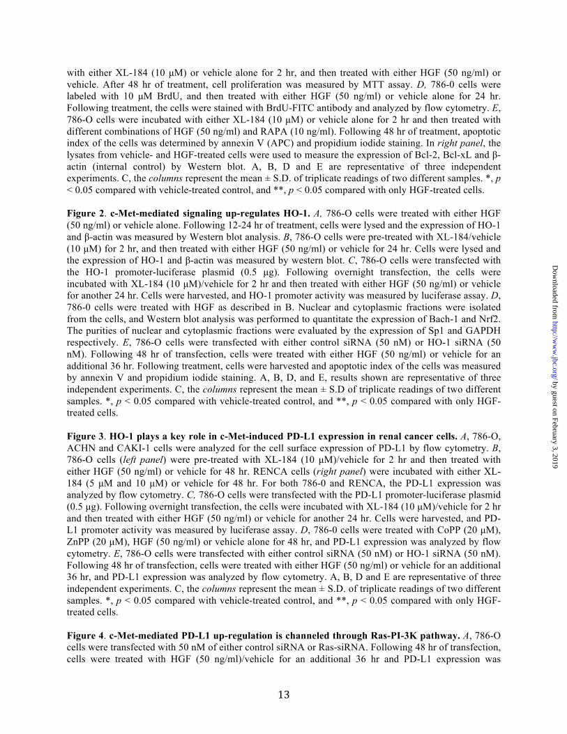
Finally, we sought to determine if HO-1 is involved in c-Met-induced survival of renal cancer cells. 786-0 and ACHN (data not shown) cells were first transfected with HO-1 siRNA to knock-down HO-1, and then they were treated with either HGF or vehicle alone. The apoptotic indexes of the cells were analyzed as described before. We observed that when HO-1 was knocked down, the HGF treatment could not inhibit apoptosis (early + late) of cancer cells compared with control cells (Fig-2E). Together, our results suggest that the induction of c-Met can protect renal cancer cells from apoptotic cell death through up-regulation of HO-1. c-Met activation promotes the expression of immuno-suppressive PD-L1, and HO-1 plays a key role in this process: Renal cancer cells can evade host anti-tumor immunity, possibly by suppressing T cell activation (21). As HO-1 may regulate immune functions (35, 36), we checked if the induction of c-Met and HO-1 can modulate the expression of the negative co-stimulatory molecule PD-L1 in renal cancer cells. 786-O cells were treated with either HGF or vehicle alone in the absence or presence of the c-Met inhibitor XL-184, and then the expression of PD-L1 was
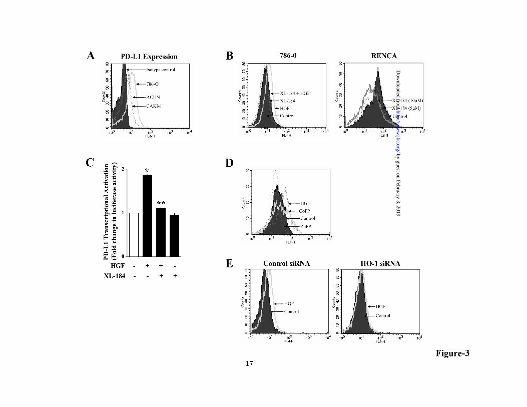
analyzed by flow cytometry. We first confirmed that PD-L1 is significantly expressed in renal cancer cell lines 786-0, Caki-1 and ACHN (Fig-3A). In contrast, the expression (data not shown) of PD-1 (receptor for PD-L1) was very low-moderate on these cells. As shown in Fig-3B (left panel), HGF treatment markedly increased PD-L1 expression on 786-0 cells compared with vehicle-treated control, and this effect was blocked in the presence of XL-184. We also found that XL-184 treatment significantly reduced the expression of PD-L1 on murine renal cancer cells RENCA (Fig-3B, right panel). We demonstrated that c-Met-/HGF-induced PD-L1 over-expression was regulated at the transcriptional level as observed by luciferase assay, and this was inhibited by XL-184 treatment (Fig-3C). Next, we wished to study if HO-1 is involved in c-Met-induced PD-L1 up-regulation in renal cancer cells. We checked how the treatment with either the HO-1 inducer CoPP or the HO-1 inhibitor ZnPP can alter PD-L1 expression. Interestingly, we observed that similar to HGF treatment, CoPP also increased PD-L1 on renal cancer cells, while ZnPP decreased PD-L1 expression (Fig-3D). Next, the cancer cells were transfected with either HO-1 siRNA or control siRNA. Following transfection, cells were treated with either HGF or vehicle alone, and then the PD-L1 expression was analyzed. We found that in HO-1 siRNA-transfected cells, HGF could not increase PD-L1 expression up to a similar level as that observed in control cells (Fig-3E). Together, our observations clearly suggest that c-Met activation promotes the up-regulation of the negative co-stimulatory molecule PD-L1 in renal cancer cells, and the over-expressed HO-1 can play an important role in this process. Ras-PI-3K signaling pathway is involved in c-Met-induced PD-L1 up-regulation: As demonstrated earlier, HGF-c-Met interaction promotes Ras activation (Fig-1B); and the active Ras can induce HO-1 in renal cancer cells (15). Here, we examined if Ras and its downstream effector molecules are involved in c-Met-induced PD-L1 expression. We observed that siRNA-mediated knock-down of Ras inhibited c-Met-induced PD-L1 expression in renal cancer cell (Fig-4A). Raf, Rho and PI-3K are critical effector molecules of Ras-induced signaling pathways. We next wished to determine which effector is
by guest on February 3, 2019http://w
ww
.jbc.org/D
ownloaded from
7
functional for c-Met-induced and Ras-mediated PD-L1 expression. We used three effector domain mutants of Ras. Ras(12V,35S) retains full-length Raf-1 binding activity, Ras(12V,37G) retains Rho binding activity, and Ras(12V,40C) retains PI-3K binding activity (31, 37). 786-0 cells were transfected with one of these Ras effector domain mutants (or empty vector); and following transfection, the expression of PD-L1 was analyzed. We observed that the cells transfected with Ras(12V,40C) showed the highest level of PD-L1 expression compared with control cells (transfected with either empty vector or Ras-12V). There was minimal change in PD-L1 expression in either Ras(12V,35S) or Ras(12V,37G)-transfected cells (Fig-4B). These findings suggest that the signals for c-Met-induced and Ras-mediated PD-L1 up-regulation can be primarily channeled through the PI-3K pathway. We confirmed our observation by utilizing a specific PI-3K inhibitor, LY294002. As shown in Fig-4C, HGF/c-Met-induced PD-L1 over-expression was blocked by LY294002. We also checked if c-Met-induced PD-L1 can regulate the proliferation of renal cancer cells. However, we observed that c-Met-induced proliferation was not PD-L1-dependent, as the HGF treatment increased cell proliferation even in PD-L1 knock-down cells compared with control (Fig-4D). c-Met-induced increase in PD-L1 protects renal cancer cells from immune cell-mediated cytotoxicty: As discussed earlier, renal tumor infiltrating immune cells are rendered dysfunctional and they express high levels of PD-1 (27). Here, we sought to determine the functional significance of c-Met-induced PD-L1 over-expression in terms of modulating immune cell-mediated cytotoxicity of cancer cells. To this end, RENCA cells were co-cultured with freshly isolated splenocytes (syngenic; BALB/c), and treated with either HGF or vehicle alone in the absence or presence of c-Met inhibitor (XL-184). RENCA cells grown alone in culture and treated with either HGF or vehicle served as an internal control. Following treatments, the apoptotic indices of RENCA cells were analyzed by flow cytometry. As expected (data not shown), there was increased apoptosis of cancer cells when they were co-cultured with splenocytes. We observed
that the treatment with HGF reduced splenocyte-mediated cell killing compared with vehicle-treated control (Fig-5A, top two panels). The total apoptotic cells (early + late) following HGF treatment decreased from 21.03% (8.53 + 12.50) to 10.01% (4.50 + 5.51) compared with vehicle-treated control. However, in the presence of XL-184, HGF treatment could not lower splenocyte-mediated cell killing; the total apoptotic cells increased from 10.01% (4.50 + 5.51) to 20.58% (8.41 + 12.17) (Fig-5A, upper right and bottom right panels).
We next tested the effect of PD-L1 blockade in modulating c-Met-mediated down-regulation of renal cancer cell immunocytotoxicity. The co-cultured cells (RENCA and splenocytes) were treated with HGF/vehicle in the absence or presence of PD-L1 neutralizing antibody. Following treatment, apoptotic indices of cancer cells were analyzed by flow cytometry. As shown in Fig-5B (upper two panels), HGF treatment decreased splenocyte-mediated cancer cell apoptosis; the total apoptotic cells (early + late) decreased from 16.85% (4.54 + 12.31) to 9.35% (2.27 + 7.08). However, the HGF treatment could not decrease splenocyte-mediated cancer cell apoptosis in the presence of PD-L1 neutralizing antibody (Fig-5B, upper right and bottom right panels). We found that the total apoptotic cells increased from 9.35% (HGF-treated group) to 21.49% (8.02 + 13.47) in the presence of PD-L1 neutralizing antibody. We also observed (data not shown) a similar result in the presence of PD1 neutralizing antibody. As an alternate approach, we also confirmed that the siRNA-mediated knock-down of PD-L1 in RENCA cells markedly increased immune cell mediated killing of cancer cells (co-culture assay) even in the presence of HGF treatment. As shown in Fig-5C (upper two panels), HGF treatment decreased splenocyte-mediated cancer cell apoptosis in control siRNA-transfected cells; the total apoptotic cells (early + late) decreased from 19.59% (13.24 + 6.35) to 12.25% (6.18 + 6.07). However, the HGF treatment could not decrease splenocyte-mediated cancer cell apoptosis in PD-L1 siRNA-transfected cells (Fig-5C, upper right and bottom right panels). The total apoptotic cells increased from 12.25% (HGF-treated group) to 27.27% (18.53 + 8.74) in PD-L1 siRNA-transfected cells, and this effect was at the
by guest on February 3, 2019http://w
ww
.jbc.org/D
ownloaded from
8
comparable level as observed with PD-L1 neutralizing antibody. Together, we suggest that c-Met-induced up-regulated PD-L1 on cancer cells plays a major role to inhibit immune cell-mediated cytotoxicity of renal cancer cells. c-Met and PD-L1 are over-expressed and co-localized in human renal cancer tissues: We checked the expression of c-Met and PD-L1 in human renal cancer tissues. As shown in Fig-6 (top panels), we found that both the proteins were low-moderately expressed in normal renal tissue; however, both PD-L1 and c-Met were markedly over-expressed and co-localized in renal cancer tissues compared with normal (Fig-6, middle and bottom panels). DISCUSSION
c-Met is over-expressed in renal cancer, and it can play a major role in the growth and survival of tumor cells. However, the mechanistic pathway(s) and the effector molecule(s) for c-Met-induced tumor growth are not well defined. In this study, we show that the induction of c-Met in renal cancer cells activates the Ras signaling pathway, and prevents cellular apoptosis through over-expression of the cytoprotective molecule HO-1. In addition, we also demonstrate for the first time that c-Met-/HO-1-induced signaling regulates the expression of the negative co-stimulatory molecule PD-L1 on renal cancer cells, and thereby it prevents immune cell-mediated killing of tumor cells.
The Ras protein is often hyper-activated in renal cancer cells without being mutated, and it promotes tumor growth and progression (38). The induction of receptor tyrosine kinases can play significant role in Ras activation (39). Here, we find that the induction of c-Met promotes the activation of Ras signaling pathway in renal cancer, and is also associated with increased phosphorylation mediated suppression of its negative regulator RKIP. In our previous study (15), we have demonstrated that the activation of Ras pathway induces over-expression of the cytoprotective and anti-apoptotic molecule HO-1 in renal cancer cells to protect them from the killing effects of chemotherapeutic agents.
However, we could not link the upstream signal(s) associated with Ras-induced HO-1 over-expression. Here, we identify that the induction of c-Met, which is over-expressed in renal cancer cells, plays a vital role in Ras-mediated HO-1 expression; and this pathway promotes cell survival through reduced apoptosis. Importantly, along with c-Met, increased serum level of the c-Met ligand HGF has also been suggested to be of prognostic significance in RCC (40). As discussed earlier, the expression of HO-1 is tightly regulated at the transcriptional level. We observe that the c-Met/HGF-mediated signaling can modulate the critical balance between the transcription factors Nrf2 (positive regulator) and Bach-1 (negative regulator) to induce HO-1 over-expression. c-Met-induced protection of renal cancer cells from apoptosis is markedly reduced when we knocked down HO-1. In addition to promoting cell survival, HO-1 expression has been reported to mediate immune-modulation in certain inflammatory disease conditions (35, 36, 41). HGF/c-Met signaling axis also plays important roles in the functional regulation of immune cells and exhibiting immunoregulatory properties (42-44). HGF treatment has been shown to induce regulatory T cells and suppress CNS autoimmunity in a murine model (45). HGF can inhibit dendritic cell function (46). It has been reported that the expression of the negative co-stimulatory molecule PD-L1 is an indicator of tumor aggressiveness in RCC patients (28). In this study, we find that the HGF treatment markedly increased PD-L1 expression in renal cancer cells; and the c-MET inhibitor XL-184 down-regulated PD-L1. c-Met-induced PD-L1 expression is channeled through the Ras-PI-3K pathway. Interestingly, we observe that the over-expressed HO-1 plays a major role in regulating c-Met-induced PD-L1 expression in renal cancer cells. However, the detailed mechanism(s) by which HO-1 regulates PD-L1 needs to be explored. It has been reported by Thompson et al that the expression of PD-1, the PD-L1 receptor on RCC tumor infiltrating cells, is associated with poor outcome for patients (27). Here, we show that c-Met-induced PD-L1 expression significantly reduces the cytotoxicity of splenocytes; and this
by guest on February 3, 2019http://w
ww
.jbc.org/D
ownloaded from

9
can be prevented by utilizing PD-L1/PD-1 neutralizing antibody. Our data provides a logical correlation to the observation of Uzzo et al and Rayman et al that the RCC tumor infiltrating PD-1 expressing T cells are dysfunctional (21, 47). Targeted PD-L1 antibody therapy can be beneficial in the treatment of some specific cancer types (48, 49). However, the molecular insights into PD-L1 expression and its regulation in RCC cells are limited. Our data points out the potential of exploring c-Met inhibitors and PD-L1 targeted therapy, either alone or in combination, for the treatment of renal cancer patients. As pathophysiological significance, we observe that both c-Met and PD-L1 are over-expressed and co-localized in human renal cancer tissues. However, we did not find (data not shown) any interaction/complex formation (in vitro) between
c-Met and PD-L1. Also, the c-Met-induced proliferation of renal cancer cells was not PD-L1-dependent. Thus, we suggest that c-Met-induced over-expressed PD-L1 on renal cancer cells is primarily involved in immune escape of tumors through its interaction with PD-1 expressed on T or other immune cells. In summary, this study explores a novel c-Met-induced pathway for the survival of renal cancer cells through the regulation of anti-apoptotic HO-1 and negative co-stimulatory molecule PD-L1. Our study for the first time, demonstrates that the c-Met-induced signaling promotes PD-L1 over-expression in renal cancer cells, and it plays a major role for immune escape of tumor cells. Together, HO-1 and PD-L1 can serve as novel therapeutic targets in c-Met-induced renal cancer.
REFERENCES
1. Ponzetto, C., Bardelli, A., Zhen, Z., Maina, F., dalla Zonca, P., Giordano, S., Graziani, A., Panayotou, G., and Comoglio, P. M. (1994) A multifunctional docking site mediates signaling and transformation by the hepatocyte growth factor/scatter factor receptor family. Cell 77, 261-271
2. Birchmeier, C., Birchmeier, W., Gherardi, E., and Vande Woude, G. F. (2003) Met, metastasis, motility and more. Nature reviews. Molecular cell biology 4, 915-925
3. Gherardi, E., Birchmeier, W., Birchmeier, C., and Vande Woude, G. (2012) Targeting MET in cancer: rationale and progress. Nature reviews. Cancer 12, 89-103
4. Natali, P. G., Prat, M., Nicotra, M. R., Bigotti, A., Olivero, M., Comoglio, P. M., and Di Renzo, M. F. (1996) Overexpression of the met/HGF receptor in renal cell carcinomas. International journal of cancer. Journal international du cancer 69, 212-217
5. Miyata, Y., Kanetake, H., and Kanda, S. (2006) Presence of phosphorylated hepatocyte growth factor receptor/c-Met is associated with tumor progression and survival in patients with conventional renal cell carcinoma. Clinical cancer research : an official journal of the American Association for Cancer Research 12, 4876-4881
6. Nakaigawa, N., Yao, M., Baba, M., Kato, S., Kishida, T., Hattori, K., Nagashima, Y., and Kubota, Y. (2006) Inactivation of von Hippel-Lindau gene induces constitutive phosphorylation of MET protein in clear cell renal carcinoma. Cancer research 66, 3699-3705
7. Kuroda, N., Tamura, M., Shiotsu, T., Nakamura, S., Taguchi, T., Tominaga, A., Hes, O., Michal, M., Kawada, C., Shuin, T., and Lee, G. H. (2010) Chromosomal abnormalities of clear cell renal cell carcinoma: frequent gain of chromosome 7. Pathology international 60, 9-13
8. Bellon, S. F., Kaplan-Lefko, P., Yang, Y., Zhang, Y., Moriguchi, J., Rex, K., Johnson, C. W., Rose, P. E., Long, A. M., O'Connor, A. B., Gu, Y., Coxon, A., Kim, T. S., Tasker, A., Burgess, T. L., and Dussault, I. (2008) c-Met inhibitors with novel binding mode show activity against several hereditary papillary renal cell carcinoma-related mutations. The Journal of biological chemistry 283, 2675-2683
by guest on February 3, 2019http://w
ww
.jbc.org/D
ownloaded from

10
9. Gao, C. F., and Vande Woude, G. F. (2005) HGF/SF-Met signaling in tumor progression. Cell research 15, 49-51
10. Maina, F., Pante, G., Helmbacher, F., Andres, R., Porthin, A., Davies, A. M., Ponzetto, C., and Klein, R. (2001) Coupling Met to specific pathways results in distinct developmental outcomes. Molecular cell 7, 1293-1306
11. Gusenbauer, S., Vlaicu, P., and Ullrich, A. (2013) HGF induces novel EGFR functions involved in resistance formation to tyrosine kinase inhibitors. Oncogene 32, 3846-3856
12. Gibney, G. T., Aziz, S. A., Camp, R. L., Conrad, P., Schwartz, B. E., Chen, C. R., Kelly, W. K., and Kluger, H. M. (2013) c-Met is a prognostic marker and potential therapeutic target in clear cell renal cell carcinoma. Annals of oncology : official journal of the European Society for Medical Oncology / ESMO 24, 343-349
13. Agarwal, A., and Nick, H. S. (2000) Renal response to tissue injury: lessons from heme oxygenase-1 GeneAblation and expression. Journal of the American Society of Nephrology : JASN 11, 965-973
14. Was, H., Dulak, J., and Jozkowicz, A. (2010) Heme oxygenase-1 in tumor biology and therapy. Current drug targets 11, 1551-1570
15. Banerjee, P., Basu, A., Datta, D., Gasser, M., Waaga-Gasser, A. M., and Pal, S. (2011) The heme oxygenase-1 protein is overexpressed in human renal cancer cells following activation of the Ras-Raf-ERK pathway and mediates anti-apoptotic signal. The Journal of biological chemistry 286, 33580-33590
16. Nath, K. A. (2006) Heme oxygenase-1: a provenance for cytoprotective pathways in the kidney and other tissues. Kidney international 70, 432-443
17. Kamimoto, M., Mizuno, S., Matsumoto, K., and Nakamura, T. (2009) Hepatocyte growth factor prevents multiple organ injuries in endotoxemic mice through a heme oxygenase-1-dependent mechanism. Biochemical and biophysical research communications 380, 333-337
18. Ryter, S. W., Alam, J., and Choi, A. M. (2006) Heme oxygenase-1/carbon monoxide: from basic science to therapeutic applications. Physiological reviews 86, 583-650
19. Suzuki, H., Tashiro, S., Hira, S., Sun, J., Yamazaki, C., Zenke, Y., Ikeda-Saito, M., Yoshida, M., and Igarashi, K. (2004) Heme regulates gene expression by triggering Crm1-dependent nuclear export of Bach1. The EMBO journal 23, 2544-2553
20. Kim, T. H., Hur, E. G., Kang, S. J., Kim, J. A., Thapa, D., Lee, Y. M., Ku, S. K., Jung, Y., and Kwak, M. K. (2011) NRF2 blockade suppresses colon tumor angiogenesis by inhibiting hypoxia-induced activation of HIF-1alpha. Cancer research 71, 2260-2275
21. Uzzo, R. G., Rayman, P., Kolenko, V., Clark, P. E., Bloom, T., Ward, A. M., Molto, L., Tannenbaum, C., Worford, L. J., Bukowski, R., Tubbs, R., Hsi, E. D., Bander, N. H., Novick, A. C., and Finke, J. H. (1999) Mechanisms of apoptosis in T cells from patients with renal cell carcinoma. Clinical cancer research : an official journal of the American Association for Cancer Research 5, 1219-1229
22. Chen, L., and Flies, D. B. (2013) Molecular mechanisms of T cell co-stimulation and co-inhibition. Nature reviews. Immunology 13, 227-242
23. Keir, M. E., Butte, M. J., Freeman, G. J., and Sharpe, A. H. (2008) PD-1 and its ligands in tolerance and immunity. Annual review of immunology 26, 677-704
24. Blank, C., Gajewski, T. F., and Mackensen, A. (2005) Interaction of PD-L1 on tumor cells with PD-1 on tumor-specific T cells as a mechanism of immune evasion: implications for tumor immunotherapy. Cancer immunology, immunotherapy : CII 54, 307-314
25. Pilon-Thomas, S., Mackay, A., Vohra, N., and Mule, J. J. (2010) Blockade of programmed death ligand 1 enhances the therapeutic efficacy of combination immunotherapy against melanoma. Journal of immunology 184, 3442-3449
26. Hirano, F., Kaneko, K., Tamura, H., Dong, H., Wang, S., Ichikawa, M., Rietz, C., Flies, D. B., Lau, J. S., Zhu, G., Tamada, K., and Chen, L. (2005) Blockade of B7-H1 and PD-1 by monoclonal antibodies potentiates cancer therapeutic immunity. Cancer research 65, 1089-1096
by guest on February 3, 2019http://w
ww
.jbc.org/D
ownloaded from

11
27. Thompson, R. H., Dong, H., Lohse, C. M., Leibovich, B. C., Blute, M. L., Cheville, J. C., and Kwon, E. D. (2007) PD-1 is expressed by tumor-infiltrating immune cells and is associated with poor outcome for patients with renal cell carcinoma. Clinical cancer research : an official journal of the American Association for Cancer Research 13, 1757-1761
28. Thompson, R. H., Gillett, M. D., Cheville, J. C., Lohse, C. M., Dong, H., Webster, W. S., Krejci, K. G., Lobo, J. R., Sengupta, S., Chen, L., Zincke, H., Blute, M. L., Strome, S. E., Leibovich, B. C., and Kwon, E. D. (2004) Costimulatory B7-H1 in renal cell carcinoma patients: Indicator of tumor aggressiveness and potential therapeutic target. Proceedings of the National Academy of Sciences of the United States of America 101, 17174-17179
29. Thompson, R. H., Kuntz, S. M., Leibovich, B. C., Dong, H., Lohse, C. M., Webster, W. S., Sengupta, S., Frank, I., Parker, A. S., Zincke, H., Blute, M. L., Sebo, T. J., Cheville, J. C., and Kwon, E. D. (2006) Tumor B7-H1 is associated with poor prognosis in renal cell carcinoma patients with long-term follow-up. Cancer research 66, 3381-3385
30. Yakes, F. M., Chen, J., Tan, J., Yamaguchi, K., Shi, Y., Yu, P., Qian, F., Chu, F., Bentzien, F., Cancilla, B., Orf, J., You, A., Laird, A. D., Engst, S., Lee, L., Lesch, J., Chou, Y. C., and Joly, A. H. (2011) Cabozantinib (XL184), a novel MET and VEGFR2 inhibitor, simultaneously suppresses metastasis, angiogenesis, and tumor growth. Molecular cancer therapeutics 10, 2298-2308
31. Khosravi-Far, R., White, M. A., Westwick, J. K., Solski, P. A., Chrzanowska-Wodnicka, M., Van Aelst, L., Wigler, M. H., and Der, C. J. (1996) Oncogenic Ras activation of Raf/mitogen-activated protein kinase-independent pathways is sufficient to cause tumorigenic transformation. Molecular and cellular biology 16, 3923-3933
32. Datta, D., Contreras, A. G., Basu, A., Dormond, O., Flynn, E., Briscoe, D. M., and Pal, S. (2009) Calcineurin inhibitors activate the proto-oncogene Ras and promote protumorigenic signals in renal cancer cells. Cancer research 69, 8902-8909
33. Yeung, K., Seitz, T., Li, S., Janosch, P., McFerran, B., Kaiser, C., Fee, F., Katsanakis, K. D., Rose, D. W., Mischak, H., Sedivy, J. M., and Kolch, W. (1999) Suppression of Raf-1 kinase activity and MAP kinase signalling by RKIP. Nature 401, 173-177
34. Balan, M., and Pal, S. (2014) A novel CXCR3-B chemokine receptor-induced growth-inhibitory signal in cancer cells is mediated through the regulation of Bach-1 protein and Nrf2 protein nuclear translocation. The Journal of biological chemistry 289, 3126-3137
35. Chora, A. A., Fontoura, P., Cunha, A., Pais, T. F., Cardoso, S., Ho, P. P., Lee, L. Y., Sobel, R. A., Steinman, L., and Soares, M. P. (2007) Heme oxygenase-1 and carbon monoxide suppress autoimmune neuroinflammation. The Journal of clinical investigation 117, 438-447
36. Tzima, S., Victoratos, P., Kranidioti, K., Alexiou, M., and Kollias, G. (2009) Myeloid heme oxygenase-1 regulates innate immunity and autoimmunity by modulating IFN-beta production. The Journal of experimental medicine 206, 1167-1179
37. Datta, D., Flaxenburg, J. A., Laxmanan, S., Geehan, C., Grimm, M., Waaga-Gasser, A. M., Briscoe, D. M., and Pal, S. (2006) Ras-induced modulation of CXCL10 and its receptor splice variant CXCR3-B in MDA-MB-435 and MCF-7 cells: relevance for the development of human breast cancer. Cancer research 66, 9509-9518
38. Fujita, J., Kraus, M. H., Onoue, H., Srivastava, S. K., Ebi, Y., Kitamura, Y., and Rhim, J. S. (1988) Activated H-ras oncogenes in human kidney tumors. Cancer research 48, 5251-5255
39. Margolis, B., and Skolnik, E. Y. (1994) Activation of Ras by receptor tyrosine kinases. Journal of the American Society of Nephrology : JASN 5, 1288-1299
40. Tanimoto, S., Fukumori, T., El-Moula, G., Shiirevnyamba, A., Kinouchi, S., Koizumi, T., Nakanishi, R., Yamamoto, Y., Taue, R., Yamaguchi, K., Nakatsuji, H., Kishimoto, T., Izaki, H., Oka, N., Takahashi, M., and Kanayama, H. O. (2008) Prognostic significance of serum hepatocyte growth factor in clear cell renal cell carcinoma: comparison with serum vascular endothelial growth factor. The journal of medical investigation : JMI 55, 106-111
by guest on February 3, 2019http://w
ww
.jbc.org/D
ownloaded from

12
41. Ke, B., Shen, X. D., Ji, H., Kamo, N., Gao, F., Freitas, M. C., Busuttil, R. W., and Kupiec-Weglinski, J. W. (2012) HO-1-STAT3 axis in mouse liver ischemia/reperfusion injury: regulation of TLR4 innate responses through PI3K/PTEN signaling. Journal of hepatology 56, 359-366
42. Mizuno, S., and Nakamura, T. (2012) Improvement of sepsis by hepatocyte growth factor, an anti-inflammatory regulator: emerging insights and therapeutic potential. Gastroenterology research and practice 2012, 909350
43. Singhal, E., Kumar, P., and Sen, P. (2011) A novel role for Bruton's tyrosine kinase in hepatocyte growth factor-mediated immunoregulation of dendritic cells. The Journal of biological chemistry 286, 32054-32063
44. Skibinski, G. (2003) The role of hepatocyte growth factor/c-met interactions in the immune system. Archivum immunologiae et therapiae experimentalis 51, 277-282
45. Benkhoucha, M., Santiago-Raber, M. L., Schneiter, G., Chofflon, M., Funakoshi, H., Nakamura, T., and Lalive, P. H. (2010) Hepatocyte growth factor inhibits CNS autoimmunity by inducing tolerogenic dendritic cells and CD25+Foxp3+ regulatory T cells. Proceedings of the National Academy of Sciences of the United States of America 107, 6424-6429
46. Baek, J. H., Birchmeier, C., Zenke, M., and Hieronymus, T. (2012) The HGF receptor/Met tyrosine kinase is a key regulator of dendritic cell migration in skin immunity. Journal of immunology 189, 1699-1707
47. Rayman, P., Wesa, A. K., Richmond, A. L., Das, T., Biswas, K., Raval, G., Storkus, W. J., Tannenbaum, C., Novick, A., Bukowski, R., and Finke, J. (2004) Effect of renal cell carcinomas on the development of type 1 T-cell responses. Clinical cancer research : an official journal of the American Association for Cancer Research 10, 6360S-6366S
48. Nomi, T., Sho, M., Akahori, T., Hamada, K., Kubo, A., Kanehiro, H., Nakamura, S., Enomoto, K., Yagita, H., Azuma, M., and Nakajima, Y. (2007) Clinical significance and therapeutic potential of the programmed death-1 ligand/programmed death-1 pathway in human pancreatic cancer. Clinical cancer research : an official journal of the American Association for Cancer Research 13, 2151-2157
49. Curiel, T. J., Wei, S., Dong, H., Alvarez, X., Cheng, P., Mottram, P., Krzysiek, R., Knutson, K. L., Daniel, B., Zimmermann, M. C., David, O., Burow, M., Gordon, A., Dhurandhar, N., Myers, L., Berggren, R., Hemminki, A., Alvarez, R. D., Emilie, D., Curiel, D. T., Chen, L., and Zou, W. (2003) Blockade of B7-H1 improves myeloid dendritic cell-mediated antitumor immunity. Nature medicine 9, 562-567
ACKNOWLEDGEMENTS We wish to thank Dr. David Briscoe for helpful discussion and also for providing unused murine tissue (spleens from BALB/c mice). We thank Evelyn Flynn for technical assistance. This work was supported by National Institutes of Health Grant R01 CA131145 and R21 CA172946 (to S. P.) FIGURE LEGENDS Figure 1. Induction of c-Met activates Ras, promotes proliferation and inhibits apoptosis of renal cancer cells. A, 786-O cells were incubated with either XL-184 (10 µM) or vehicle alone for 2 hr and then treated with either HGF (50 ng/ml) or vehicle for 15 min. Following treatment, cells were lysed and the cell lysates were used to measure the levels of phospho-c-Met, c-Met and β-actin (internal control) by Western blot analysis. B, 786-O cells were treated with HGF as described in A. Cell lysates were prepared utilizing a Ras activation kit as described under “Experimental Procedures”, and the expression of GTP bound Ras was subsequently analyzed by Western blot. Cell lysate were also used to measure the expression of total Ras, phospho-RKIP, and total RKIP by Western blot. C, 786-O cells were incubated
by guest on February 3, 2019http://w
ww
.jbc.org/D
ownloaded from
13
with either XL-184 (10 µM) or vehicle alone for 2 hr, and then treated with either HGF (50 ng/ml) or vehicle. After 48 hr of treatment, cell proliferation was measured by MTT assay. D, 786-0 cells were labeled with 10 µM BrdU, and then treated with either HGF (50 ng/ml) or vehicle alone for 24 hr. Following treatment, the cells were stained with BrdU-FITC antibody and analyzed by flow cytometry. E, 786-O cells were incubated with either XL-184 (10 µM) or vehicle alone for 2 hr and then treated with different combinations of HGF (50 ng/ml) and RAPA (10 ng/ml). Following 48 hr of treatment, apoptotic index of the cells was determined by annexin V (APC) and propidium iodide staining. In right panel, the lysates from vehicle- and HGF-treated cells were used to measure the expression of Bcl-2, Bcl-xL and β-actin (internal control) by Western blot. A, B, D and E are representative of three independent experiments. C, the columns represent the mean ± S.D. of triplicate readings of two different samples. *, p < 0.05 compared with vehicle-treated control, and **, p < 0.05 compared with only HGF-treated cells. Figure 2. c-Met-mediated signaling up-regulates HO-1. A, 786-O cells were treated with either HGF (50 ng/ml) or vehicle alone. Following 12-24 hr of treatment, cells were lysed and the expression of HO-1 and β-actin was measured by Western blot analysis. B, 786-O cells were pre-treated with XL-184/vehicle (10 µM) for 2 hr, and then treated with either HGF (50 ng/ml) or vehicle for 24 hr. Cells were lysed and the expression of HO-1 and β-actin was measured by western blot. C, 786-O cells were transfected with the HO-1 promoter-luciferase plasmid (0.5 µg). Following overnight transfection, the cells were incubated with XL-184 (10 µM)/vehicle for 2 hr and then treated with either HGF (50 ng/ml) or vehicle for another 24 hr. Cells were harvested, and HO-1 promoter activity was measured by luciferase assay. D, 786-0 cells were treated with HGF as described in B. Nuclear and cytoplasmic fractions were isolated from the cells, and Western blot analysis was performed to quantitate the expression of Bach-1 and Nrf2. The purities of nuclear and cytoplasmic fractions were evaluated by the expression of Sp1 and GAPDH respectively. E, 786-O cells were transfected with either control siRNA (50 nM) or HO-1 siRNA (50 nM). Following 48 hr of transfection, cells were treated with either HGF (50 ng/ml) or vehicle for an additional 36 hr. Following treatment, cells were harvested and apoptotic index of the cells was measured by annexin V and propidium iodide staining. A, B, D, and E, results shown are representative of three independent experiments. C, the columns represent the mean ± S.D of triplicate readings of two different samples. *, p < 0.05 compared with vehicle-treated control, and **, p < 0.05 compared with only HGF-treated cells. Figure 3. HO-1 plays a key role in c-Met-induced PD-L1 expression in renal cancer cells. A, 786-O, ACHN and CAKI-1 cells were analyzed for the cell surface expression of PD-L1 by flow cytometry. B, 786-O cells (left panel) were pre-treated with XL-184 (10 µM)/vehicle for 2 hr and then treated with either HGF (50 ng/ml) or vehicle for 48 hr. RENCA cells (right panel) were incubated with either XL-184 (5 µM and 10 µM) or vehicle for 48 hr. For both 786-0 and RENCA, the PD-L1 expression was analyzed by flow cytometry. C, 786-O cells were transfected with the PD-L1 promoter-luciferase plasmid (0.5 µg). Following overnight transfection, the cells were incubated with XL-184 (10 µM)/vehicle for 2 hr and then treated with either HGF (50 ng/ml) or vehicle for another 24 hr. Cells were harvested, and PD-L1 promoter activity was measured by luciferase assay. D, 786-0 cells were treated with CoPP (20 µM), ZnPP (20 µM), HGF (50 ng/ml) or vehicle alone for 48 hr, and PD-L1 expression was analyzed by flow cytometry. E, 786-O cells were transfected with either control siRNA (50 nM) or HO-1 siRNA (50 nM). Following 48 hr of transfection, cells were treated with either HGF (50 ng/ml) or vehicle for an additional 36 hr, and PD-L1 expression was analyzed by flow cytometry. A, B, D and E are representative of three independent experiments. C, the columns represent the mean ± S.D. of triplicate readings of two different samples. *, p < 0.05 compared with vehicle-treated control, and **, p < 0.05 compared with only HGF-treated cells. Figure 4. c-Met-mediated PD-L1 up-regulation is channeled through Ras-PI-3K pathway. A, 786-O cells were transfected with 50 nM of either control siRNA or Ras-siRNA. Following 48 hr of transfection, cells were treated with HGF (50 ng/ml)/vehicle for an additional 36 hr and PD-L1 expression was
by guest on February 3, 2019http://w
ww
.jbc.org/D
ownloaded from

14
analyzed by flow cytometry. B, 786-O cells were transfected with either one of the three Ras effector domain mutant plasmids Ras(12V, 35S), Ras(12V, 37G), Ras(12V, 40C), or active Ras plasmid Ras(12V) or empty vector alone. Following 48 hr of transfection, cells were analyzed for PD-L1 expression by flow cytometry. C, 786-O cells were incubated with either LY294002 (10 µM) or vehicle for 2 hr and then treated with HGF (50 ng/ml)/vehicle for 36 hr. Following treatment, cells were analyzed for PD-L1 expression by flow cytometry. D, 786-0 cells were transfected with 50 nM of either control siRNA or PD-L1 siRNA. Following 48 hr of transfection, the cells were treated with either HGF (50 ng/ml) or vehicle alone. After 24 hr of treatment, cell proliferation was measured by MTT assay. A-C, representative data of three independent experiments. D, the columns represent the mean ± S.D. of triplicate readings of three different samples. *, p < 0.05 compared with control siRNA-transfected and vehicle-treated control; and NS, not significant compared with control siRNA-transfected and HGF-treated cells. Figure 5. c-Met-induced increase in PD-L1 protects renal cancer cells from immune cell-mediated cytotoxicity. A, The RENCA cells/splenocytes co-culture (as described in “Experimental Procedures”) was pre-treated with either XL-184 (10µM) or vehicle for 4 hr. Following incubation, co-cultured cells were treated with either HGF (50 ng/ml) or vehicle. Following 72 hr of treatment, the adherent RENCA cells were harvested and apoptotic index of the cells was analyzed by annexin V and propidium iodide staining. B, The RENCA cells/splenocytes co-culture was incubated with either PD-L1 antibody (10 µg/ml) or isotype antibody for 8 hr, and then treated with either HGF (50 ng/ml) or vehicle. Following 72 hr of treatment, the adherent RENCA cells were harvested and apoptotic index of the cells was analyzed by annexin V and propidium iodide staining. C, RENCA cells were transfected with 50 nM of either PD-L1 siRNA or control siRNA. Following 48 hr of transfection, cells were utilized in the RENCA cells/splenocytes co-culture and treated with either HGF (50 ng/ml) or vehicle. Following 48 hr of treatment, the adherent RENCA cells were harvested and apoptotic index of the cells was analyzed by annexin V and propidium iodide staining. The Western blot (right panel) represents the siRNA-mediated knockdown of PD-L1 compared with β-actin control. A-C, representative data of three independent experiments. Figure 6. c-Met and PD-L1 are over-expression and co-localized in human renal cancer tissues. Representative photomicrographs illustrating double fluorescence labeling of both PD-L1 (red) and c-Met (green) in normal renal and RCC tissues. DAPI (blue) was used to visualize nuclei. Merged images are shown to indicate co-localization (yellow) of PD-L1 and c-Met. Scale bar, 200 µm. Results are representative of four different tissues.
by guest on February 3, 2019http://w
ww
.jbc.org/D
ownloaded from


Toni K. Choueiri, Gordon Freeman and Soumitro PalMurugabaskar Balan, Eduardo Mier Y. Teran, Ana Maria Waaga-Gasser, Martin Gasser,
HO-1 and PD-L1 ExpressionNovel Roles of c-Met in the Survival of Renal Cancer Cells through the Regulation of
published online February 2, 2015J. Biol. Chem.
10.1074/jbc.M114.612689Access the most updated version of this article at doi:
Alerts:
When a correction for this article is posted•
When this article is cited•
to choose from all of JBC's e-mail alertsClick here
by guest on February 3, 2019http://w
ww
.jbc.org/D
ownloaded from