pathophysiology of heart failure. essential functions of the heart essential functions of the heart...
TRANSCRIPT

PATHOPHYSIOLOGY OF HEART PATHOPHYSIOLOGY OF HEART FAILUREFAILURE

• Essential functions of the heartEssential functions of the heart
• to cover metabolic needs of body tissueto cover metabolic needs of body tissue (oxygen, substrates) by adequate blood supply(oxygen, substrates) by adequate blood supply
• to receive all blood comming back from the tissue to receive all blood comming back from the tissue to the heartto the heart
• Essential conditions for fulfilling these functionsEssential conditions for fulfilling these functions
• normal structure and functions of the heartnormal structure and functions of the heart
• adequate filling of the heart by bloodadequate filling of the heart by blood

Essential functions of the heart are secured Essential functions of the heart are secured by integration of electrical and mechanicalby integration of electrical and mechanical
functions of the heartfunctions of the heart
Cardiac output (CO) = heart rate (HR) x stroke vol.(SV)Cardiac output (CO) = heart rate (HR) x stroke vol.(SV)
- - changes of the heart ratechanges of the heart rate
- changes of stroke volume- changes of stroke volume
• Control of HR:Control of HR:
- autonomic nervous system- autonomic nervous system
- - hormonal(humoral) controlhormonal(humoral) control
• Control of SVControl of SV:: - preload- preload
- contractility- contractility
- afterload- afterload

Adaptive mechanisms of the heart to increased load
• Frank - Starling mechanism
• Ventricular hypertrophy – increased mass of contractile elements → ↑strength
of contraction
• Increased sympathetic adrenergic activity – increased HR, increased contractility
• Incresed activity of R–A–A system

Causes leading to changes of number and size of cardiomyocytes

PreloadPreload
Stretching the myocardial fibers during diastole by increasing end-Stretching the myocardial fibers during diastole by increasing end-
diastolic volume → ↑force of contraction during systole = diastolic volume → ↑force of contraction during systole = Starling´s lawStarling´s law
preloadpreload = = diastolic muscle sarcomere length leading to increased diastolic muscle sarcomere length leading to increased tension in muscle before its contraction (Fig.2,3)tension in muscle before its contraction (Fig.2,3)
- venous return to the heart is important → end-diastolicvenous return to the heart is important → end-diastolic volume is influenced
- stretching of the sarcomere maximises the number stretching of the sarcomere maximises the number of actin-myosin bridges responsible for development of forceof actin-myosin bridges responsible for development of force
- - optimal sarcomere length 2.2 μm∼optimal sarcomere length 2.2 μm∼


Myocardial contractilityMyocardial contractility
Contractility of myocardiumContractility of myocardium Changes in ability of myocardium to develop the force Changes in ability of myocardium to develop the force by contraction that occur independently on the by contraction that occur independently on the changes in myocardial fibre lengthchanges in myocardial fibre length
Mechanisms involved in changes of contractilityMechanisms involved in changes of contractility
• ↑ ↑ amount of created cross-bridges in the sarcomereamount of created cross-bridges in the sarcomere by ↑ of [Ca by ↑ of [Ca ++++]]ii concentration concentration
- - catecholamines → ↑[Cacatecholamines → ↑[Ca++++]]ii→ ↑ contractility→ ↑ contractility
- - inotropic drugs → ↑[Cainotropic drugs → ↑[Ca++++]]ii→ ↑ contractility→ ↑ contractility
↑ ↑ contractilitycontractility → → shifting the entire ventricular function shifting the entire ventricular function curve upward and to the leftcurve upward and to the left
↓ ↓ contractilitycontractility → → shiffting the entire ventricular functionshiffting the entire ventricular function curve (hypoxia, acidosis) downward and to the rightcurve (hypoxia, acidosis) downward and to the right

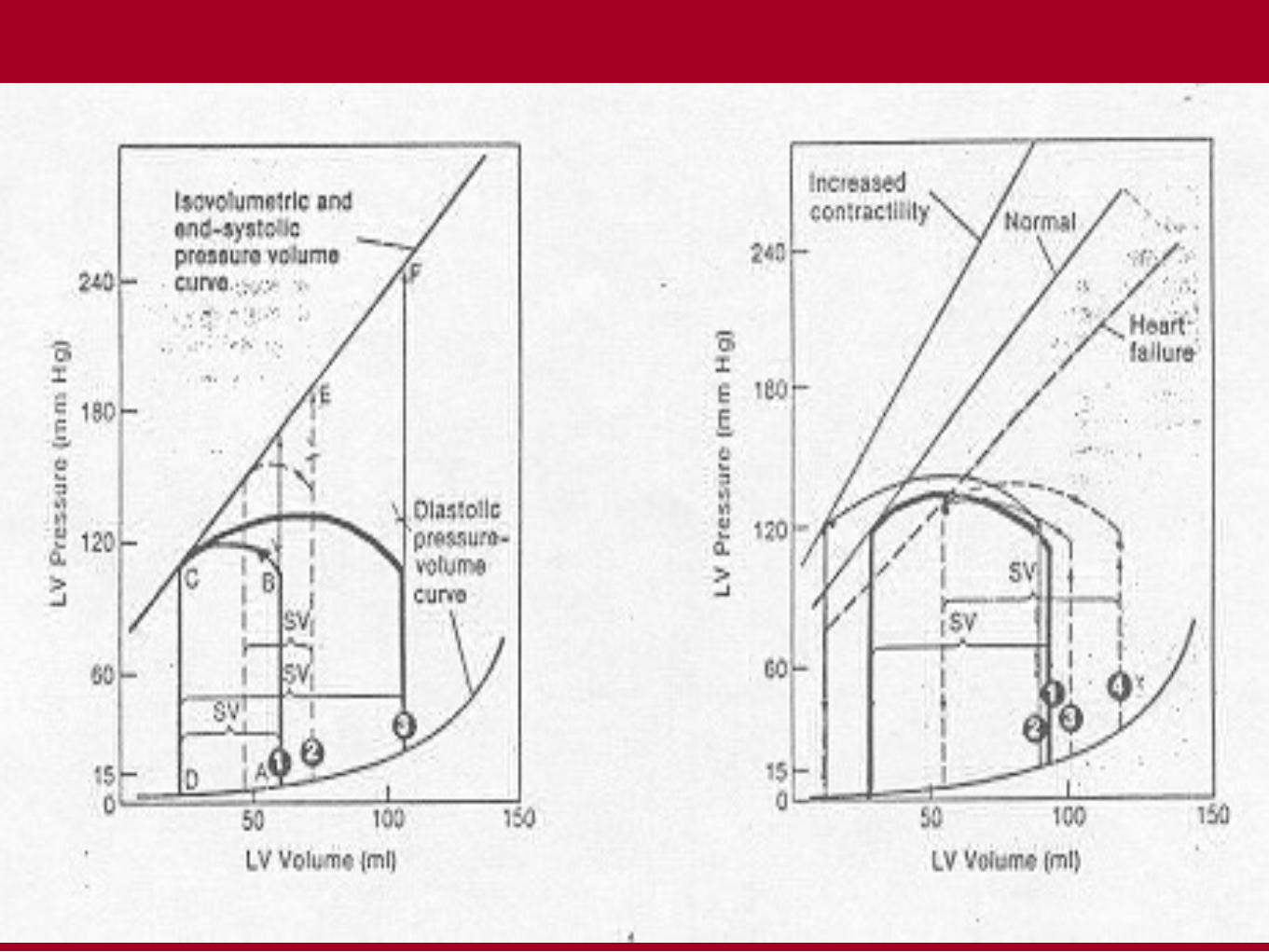
The pressure – volume loop
• It is the relation between ventricular volume and pressure
• This loop provides a convenient framework for understanding
the response of individual left ventricular contractions
to alterations in preload, afterload, and contractility
• It is composed of 4 phases:
- filling of the ventricle
- isovolumic contraction of ventricle
- isotonic contraction of ventricle(ejection of blood)
- isovolumic relaxation of ventricle

Pressure – volume loops recorded under different conditions
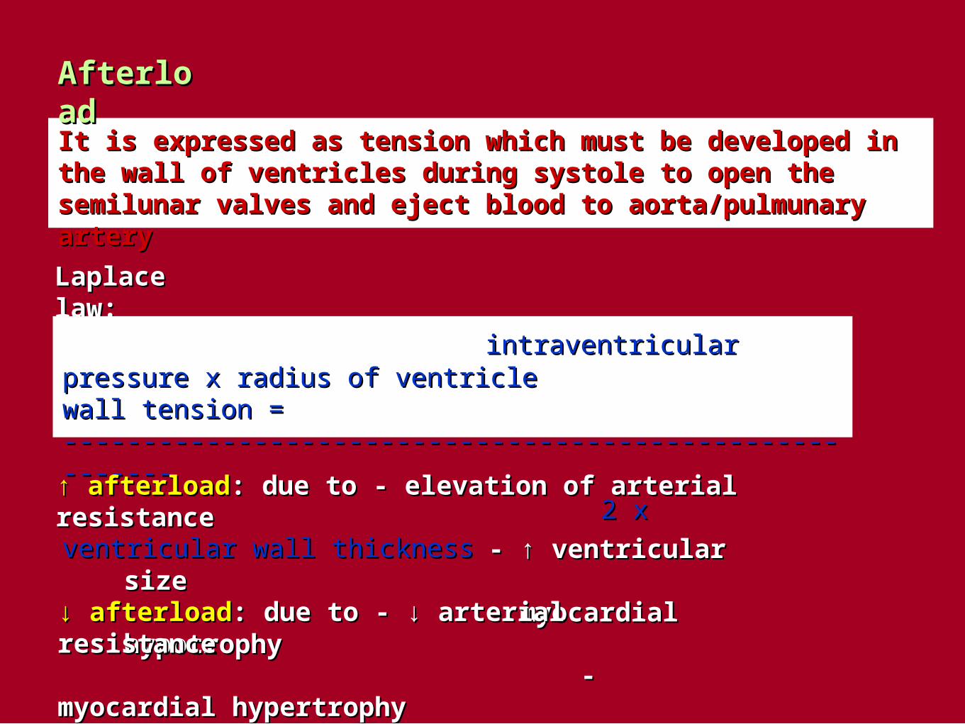
It is expressed as tension which must be developed in the wall of It is expressed as tension which must be developed in the wall of ventricles during systole to open the semilunar valves and eject ventricles during systole to open the semilunar valves and eject blood to aorta/pulmunary arteryblood to aorta/pulmunary artery
Laplace law:Laplace law:
intraventricular pressure x radius of ventricleintraventricular pressure x radius of ventriclewall tension = --------------------------------------------------------wall tension = --------------------------------------------------------
2 x ventricular wall thickness2 x ventricular wall thickness
↑ ↑ afterloadafterload:: due to - elevation of arterial resistancedue to - elevation of arterial resistance - ↑ ventricular size- ↑ ventricular size - myocardial hypotrophy- myocardial hypotrophy
↓ ↓ afterloadafterload:: due to - ↓ arterial resistance due to - ↓ arterial resistance - myocardial hypertrophy- myocardial hypertrophy - ↓ ventricular size- ↓ ventricular size
AfterloadAfterload

Heart failureHeart failure
DefinitionDefinition
It is the pathophysiological process in whichIt is the pathophysiological process in which
the heart as a pump is unable to meetthe heart as a pump is unable to meet
the metabolic requirements of the tissue for the metabolic requirements of the tissue for
oxygen and substrates despite the venousoxygen and substrates despite the venous
return to heart is either normal or increasedreturn to heart is either normal or increased
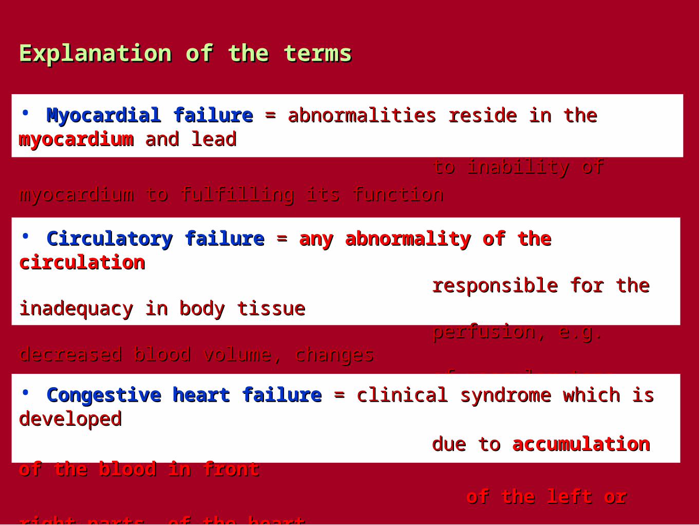
Explanation of the termsExplanation of the terms
• Myocardial failure Myocardial failure = abnormalities reside in the = abnormalities reside in the myocardium myocardium and lead and lead
to inability of myocardium to fulfilling its function to inability of myocardium to fulfilling its function
• Circulatory failureCirculatory failure = = any abnormality of the circulationany abnormality of the circulation responsible for the inadequacy in body tissueresponsible for the inadequacy in body tissue perfusion, e.g. decreased blood volume, changes perfusion, e.g. decreased blood volume, changes of vascular tone, heart functiones disordersof vascular tone, heart functiones disorders
• Congestive heart failureCongestive heart failure = clinical syndrome which is developed = clinical syndrome which is developed
due to due to accumulation of the blood in frontaccumulation of the blood in front
of the left or right parts of the heartof the left or right parts of the heart
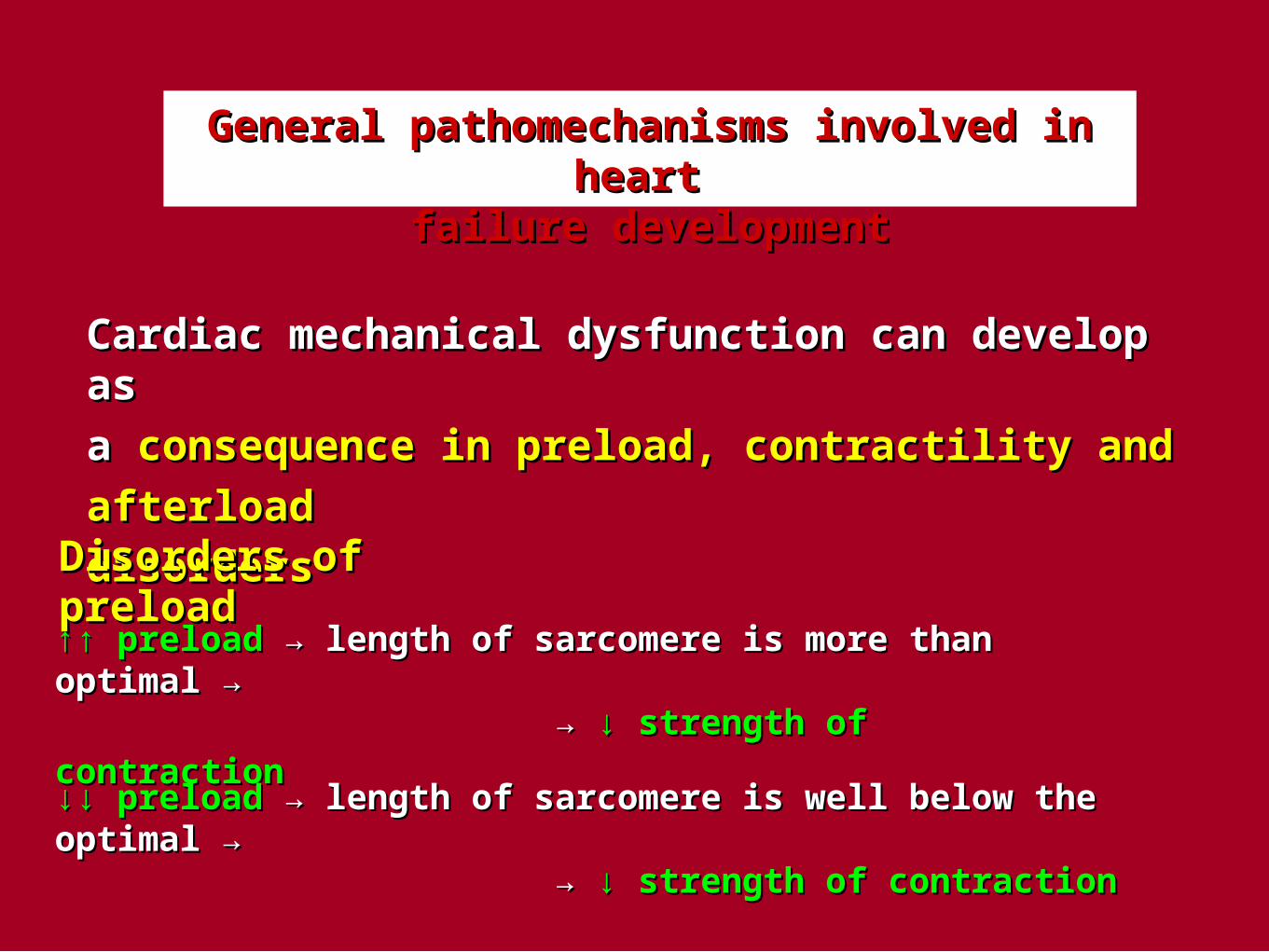
General pathomechanisms involved in heart General pathomechanisms involved in heart failure developmentfailure development
Cardiac mechanical dysfunction can develop as Cardiac mechanical dysfunction can develop as
a a consequence in preload, contractility and afterload consequence in preload, contractility and afterload
disordersdisorders
Disorders of preloadDisorders of preload
↑↑ ↑↑ preloadpreload → → length of sarcomere is more than optimal → length of sarcomere is more than optimal →
→ → ↓ ↓ strength of contractionstrength of contraction
↓↓ ↓↓ preloadpreload → → length of sarcomere is well below the optimal → length of sarcomere is well below the optimal → → → ↓ ↓ strength of contractionstrength of contraction


Important:Important: failing ventricle requires higher end-diastolic volume failing ventricle requires higher end-diastolic volume
to achieve the same improvement of CO that normalto achieve the same improvement of CO that normal
ventricle achieves with lower ventricular volumesventricle achieves with lower ventricular volumes
Disorders of contractilityDisorders of contractility
In the most forms of heart failure the contractility of myocardium In the most forms of heart failure the contractility of myocardium
is decreased (ischemia, hypoxia, acidosis, inflammation, toxins, is decreased (ischemia, hypoxia, acidosis, inflammation, toxins,
metabolic disorders... )metabolic disorders... )
Disorders of afterload due to:Disorders of afterload due to:
• fluid retention in the bodyfluid retention in the body
• ↑ ↑ arterial resistancearterial resistance
• valvular heart diseases ( stenosisvalvular heart diseases ( stenosis ))
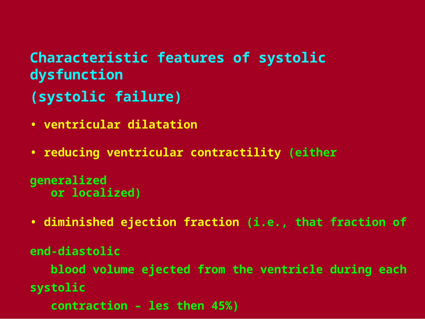
Characteristic features of systolic dysfunction
(systolic failure)
• ventricular dilatation
• reducing ventricular contractility (either generalized or localized)
• diminished ejection fraction (i.e., that fraction of end-diastolic
blood volume ejected from the ventricle during each systolic
contraction – les then 45%)
• in failing hearts, the LV end-diastolic volume (or pressure)
may increse as the stroke volume (or CO) decreases

Characteristic features of diastolic dysfunctions(diastolic failure)
• ventricular cavity size is normal or small
• myocardial contractility is normal or hyperdynamic
• ejection fraction is normal (>50%) or supranormal
• ventricle is usually hypertrophied
• ventricle is filling slowly in early diastole (during the period
of passive filling)
• end-diastolic ventricular pressure is increased

Causes of heart pump failureCauses of heart pump failure
A. MECHANICAL ABNORMALITIESA. MECHANICAL ABNORMALITIES
1. Increased pressure load1. Increased pressure load
– – central (aortic stenosis, aortic coarctation...)central (aortic stenosis, aortic coarctation...)
–– peripheral (systemic hypertension)peripheral (systemic hypertension)
2. Increased volume load2. Increased volume load– – valvular regurgitationvalvular regurgitation
– hypervolemia
3. Obstruction to ventricular filling3. Obstruction to ventricular filling –– valvular stenosisvalvular stenosis
–– pericardial restrictionpericardial restriction

B. MYOCARDIAL DAMAGEB. MYOCARDIAL DAMAGE
1. Primary1. Primary
a) cardiomyopathya) cardiomyopathy
b) myocarditisb) myocarditis
c) toxicity (e.g. alcohol)c) toxicity (e.g. alcohol)
d) metabolic abnormalities (e.g. hyperthyreoidismd) metabolic abnormalities (e.g. hyperthyreoidism))
2. Secondary2. Secondary
a) oxygen deprivation (e.g. coronary heart disease)a) oxygen deprivation (e.g. coronary heart disease)
b) inflammation (e.g. increased metabolic demands)b) inflammation (e.g. increased metabolic demands)
c) chronic obstructive lung diseasec) chronic obstructive lung disease

C. ALTERED CARDIAC RHYTHMC. ALTERED CARDIAC RHYTHM
1. ventricular flutter and fibrilation1. ventricular flutter and fibrilation
2. extreme tachycardias2. extreme tachycardias
3. extreme bradycardias3. extreme bradycardias

Pathomechanisms involved in heart failurePathomechanisms involved in heart failure
A. Pathomechanisms involved in myocardial failureA. Pathomechanisms involved in myocardial failure
1.1.Damage of cardiomyocytesDamage of cardiomyocytes → ↓ → ↓ contractility, contractility, ↑↓ ↑↓ compliancecompliance
Consequences:Consequences:• • defect in ATP production and utilisationdefect in ATP production and utilisation
• • changes in contractile proteinschanges in contractile proteins
• • uncoupling of excitation – contraction processuncoupling of excitation – contraction process
• • ↓ ↓ number of cardiomyocytesnumber of cardiomyocytes
• impairment of relaxation of cardiomyocytes with decrease impairment of relaxation of cardiomyocytes with decrease compliance of myocardiumcompliance of myocardium
• impaired of sympato-adrenal system (SAS) → ↓ number of impaired of sympato-adrenal system (SAS) → ↓ number of
ββ11-adrenergic receptors on the surface of cardiomycytes-adrenergic receptors on the surface of cardiomycytes

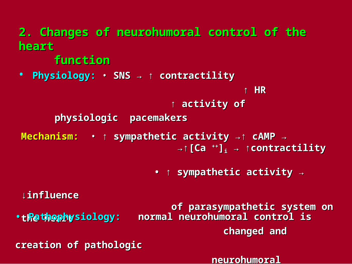
2. Changes of neurohumoral control of the heart 2. Changes of neurohumoral control of the heart functionfunction
• Physiology:Physiology: • • SNSSNS → ↑ contractility→ ↑ contractility
↑ ↑ HRHR
↑ ↑ activity of physiologic pacemakersactivity of physiologic pacemakers
Mechanism:Mechanism: • • ↑ sympathetic activity →↑ cAMP →↑ sympathetic activity →↑ cAMP → →↑ →↑[Ca [Ca ++++]]ii → ↑contractility → ↑contractility
• ↑ • ↑ sympathetic activity → ↓influencesympathetic activity → ↓influence of parasympathetic system on the heartof parasympathetic system on the heart
•• Pathophysiology:Pathophysiology: normal neurohumoral control isnormal neurohumoral control is
changed and creation of pathologicchanged and creation of pathologic
neurohumoral mechanisms are presentneurohumoral mechanisms are present
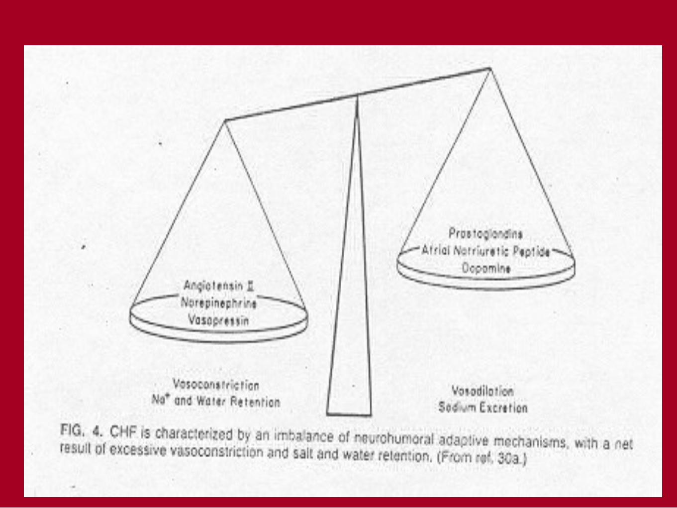

Chronic heart failure (CHF) is characterized by an imbalance of Chronic heart failure (CHF) is characterized by an imbalance of
neurohumoral adaptive mechanisms with a net results of excessive neurohumoral adaptive mechanisms with a net results of excessive
vasoconstriction and salt and water retentionvasoconstriction and salt and water retention
Catecholamines :Catecholamines : - concentration in blood :- concentration in blood :
- norepinephrin – 2-3x higher at the rest than in healthy subjectsnorepinephrin – 2-3x higher at the rest than in healthy subjects
- - circulating norepinephrin is increased much more circulating norepinephrin is increased much more during equal load in patients suffering from CHF than during equal load in patients suffering from CHF than in healthy subjectin healthy subject
- ↓ ↓ number of beta 1 – adrenergic receptors →number of beta 1 – adrenergic receptors → →↓ →↓ sensitivity of cardiomyocytes to catecholamines →sensitivity of cardiomyocytes to catecholamines → → ↓ → ↓ contractilitycontractility
System rennin – angiotensin – aldosteronSystem rennin – angiotensin – aldosteron
heart failureheart failure →↓ CO →↓ kidney perfusion → stim. Of RAA system→↓ CO →↓ kidney perfusion → stim. Of RAA system
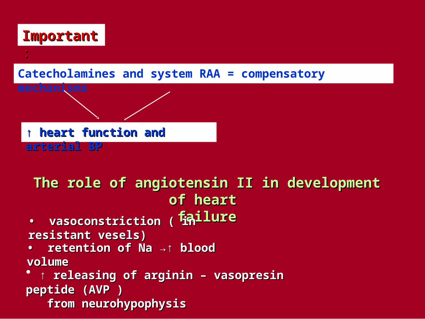
Important:Important:
Catecholamines and system RAA = compensatory mechanisms
↑ ↑ heart function and arterialheart function and arterial BPBP
The role of angiotensin II in development of heart The role of angiotensin II in development of heart failurefailure
• • vasoconstriction ( in resistant vesels)vasoconstriction ( in resistant vesels)
• • retention of Na →↑ blood volumeretention of Na →↑ blood volume
• ↑ ↑ releasing of arginin – vasopresin peptide (AVP ) releasing of arginin – vasopresin peptide (AVP ) from neurohypophysisfrom neurohypophysis

• ↑ • ↑ sensitivity of vessel wall to norepinephrinesensitivity of vessel wall to norepinephrine
• mitogenic effect on smooth muscles in vessels and mitogenic effect on smooth muscles in vessels and
on cardiomyocytes → hypertrophyon cardiomyocytes → hypertrophy
• • constriction of vas efferens ( in glomerulus )constriction of vas efferens ( in glomerulus )
• • ↑ ↑ sensation of thirstsensation of thirst
• ↑ • ↑ secretion of aldosteron from adrenal glandsecretion of aldosteron from adrenal gland
• • mesangial conctraction → ↓glomerular filtration ratemesangial conctraction → ↓glomerular filtration rate
• facilitation of norepinephrine releasing from facilitation of norepinephrine releasing from sympathetic nerve endingssympathetic nerve endings


Pathophysiology of diastolic heart failurePathophysiology of diastolic heart failure
• systolic heart failuresystolic heart failure = = failure of ejecting function of the heartfailure of ejecting function of the heart
• diastolic heart failurediastolic heart failure = = failure of filling the ventricles,failure of filling the ventricles,
↑ ↑ resistance to filling of ventriclesresistance to filling of ventricles
But, which of the cardiac cycle is real diastole ?But, which of the cardiac cycle is real diastole ?
Diastolic failure is a widely recognized clinical entity

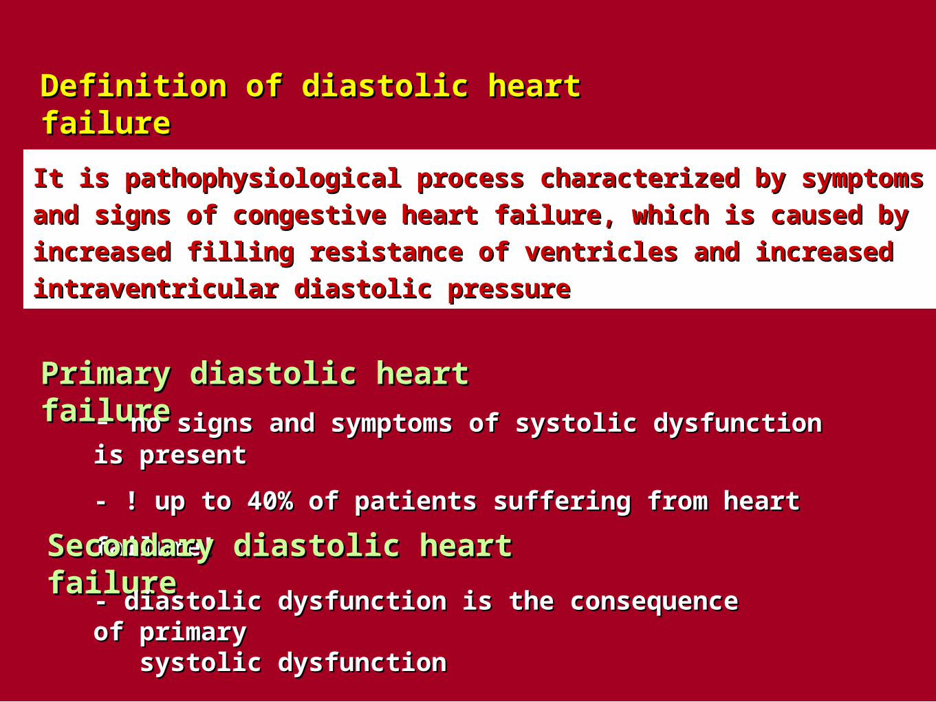
Definition of diastolic heart failureDefinition of diastolic heart failure
It is pathophysiological process characterized by symptoms and signs of It is pathophysiological process characterized by symptoms and signs of
congestive heart failure, which is caused by increased filling resistance congestive heart failure, which is caused by increased filling resistance
of ventricles and increased intraventricular diastolic pressureof ventricles and increased intraventricular diastolic pressure
Primary diastolic heart failurePrimary diastolic heart failure
- no signs and symptoms of systolic dysfunction is presentno signs and symptoms of systolic dysfunction is present
- ! up to 40% of patients suffering from heart failure!- ! up to 40% of patients suffering from heart failure!
Secondary diastolic heart failureSecondary diastolic heart failure
- diastolic dysfunction is the consequence of primary- diastolic dysfunction is the consequence of primary systolic dysfunctionsystolic dysfunction

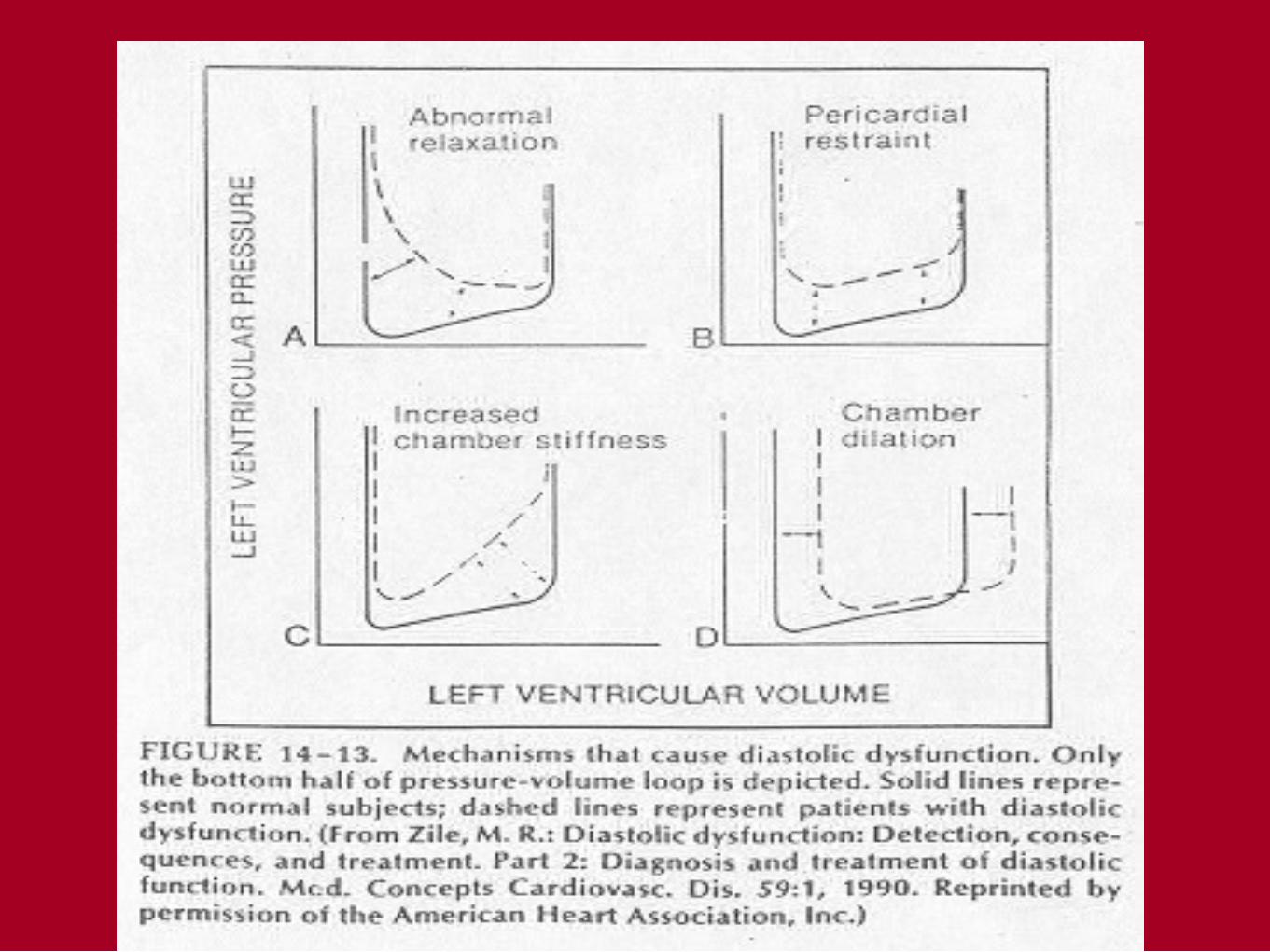
Main causes and pathomechanisms of diastolicMain causes and pathomechanisms of diastolic heart failureheart failure
1. structural disorders1. structural disorders →↑passive chamber stiffness→↑passive chamber stiffness
a)a)intramyocardialintramyocardial
– – e.g. myocardial fibrosis, amyloidosis, hypertrophy, e.g. myocardial fibrosis, amyloidosis, hypertrophy,
myocardial ischemia...myocardial ischemia...
b) extramyocardialb) extramyocardial – e.g. constrictive pericarditis – e.g. constrictive pericarditis
2. functional disorders2. functional disorders → ↓ relaxation of chambers→ ↓ relaxation of chambers e. g. myocardial e. g. myocardial
ischemia, advanced hypertrophy of ventricles, ischemia, advanced hypertrophy of ventricles,
failing myocardium, asynchrony in heart failing myocardium, asynchrony in heart
functionsfunctions

Causes and mechanism participating on impairedCauses and mechanism participating on impaired ventricular relaxationventricular relaxation
a)a) physiological changesphysiological changes in chamber relaxation due to: in chamber relaxation due to:
– – prolonged ventricular contraction prolonged ventricular contraction
Relaxation of ventricles is not impairedRelaxation of ventricles is not impaired !
b) pathological changesb) pathological changes in chamber relaxation due to: in chamber relaxation due to:
Impaired relaxation processImpaired relaxation process
• • delayed relaxation (retarded)delayed relaxation (retarded)
• • incomplete (slowed) relaxationincomplete (slowed) relaxation


• • Consequences of impaired ventricular relaxationConsequences of impaired ventricular relaxation
- filling of ventricles isfilling of ventricles is more dependent on diastasis more dependent on diastasis and onand on thethe systole of atrias than in healthy subjectssystole of atrias than in healthy subjects
Symptoms and signs:Symptoms and signs:
• • exercise intoleranceexercise intolerance = = early sign of diastolic failureearly sign of diastolic failure
• ↓ • ↓ coronary blood flow during diastolecoronary blood flow during diastole
• • Causes and mechanisms involved in developmentCauses and mechanisms involved in development of ventricular stiffnessof ventricular stiffness
• • ventricular complianceventricular compliance = passive property of ventricle= passive property of ventricle
Source of compliance: Source of compliance: cardiomyocytes and other heart cardiomyocytes and other heart
tissue to stretching tissue to stretching

↓ ↓ Ventricular compliance is caused by structural abnormalities Ventricular compliance is caused by structural abnormalities
localized in myocardium and in extramyocardial tissuelocalized in myocardium and in extramyocardial tissue
a) Intramyocardial causesa) Intramyocardial causes : myocardial fibrosis, hypertrophy of : myocardial fibrosis, hypertrophy of
ventricular wall,restrictive cardiomyopathyventricular wall,restrictive cardiomyopathy
b. Extramyocardial causesb. Extramyocardial causes :: constrictive pericarditis constrictive pericarditis
The role of myocardial remodelling in genesis ofThe role of myocardial remodelling in genesis of heart failureheart failure
• • adaptive remodelling of the heartadaptive remodelling of the heart
• • pathologic remodelling of the heartpathologic remodelling of the heart

Main causes and mechanisms involved in Main causes and mechanisms involved in pathological remodelation of the heartpathological remodelation of the heart
1.Increased amount and size1.Increased amount and size of myocytesof myocytes == hypertrophyhypertrophy
Due to:Due to: - ↑ volume and/or pressure load - ↑ volume and/or pressure load (excentric, concentric hypertrophy(excentric, concentric hypertrophy))
- hormonal stimulation of cardiomyocytes by - hormonal stimulation of cardiomyocytes by norepinephrine, angiotenzine IInorepinephrine, angiotenzine II
2. Increased % of2. Increased % of non-myocytic cellsnon-myocytic cells in myocardium in myocardium and their influence on structure and function of heartand their influence on structure and function of heart
a.a. endothelial cellsendothelial cells – – endothelins : endothelins : mitogenic ability → mitogenic ability → → → stimulation growth of smooth muscle cells of vessels, fibroblastsstimulation growth of smooth muscle cells of vessels, fibroblasts
b.b. fibroblastsfibroblasts - ↑ production of kolagens - ↑ production of kolagens

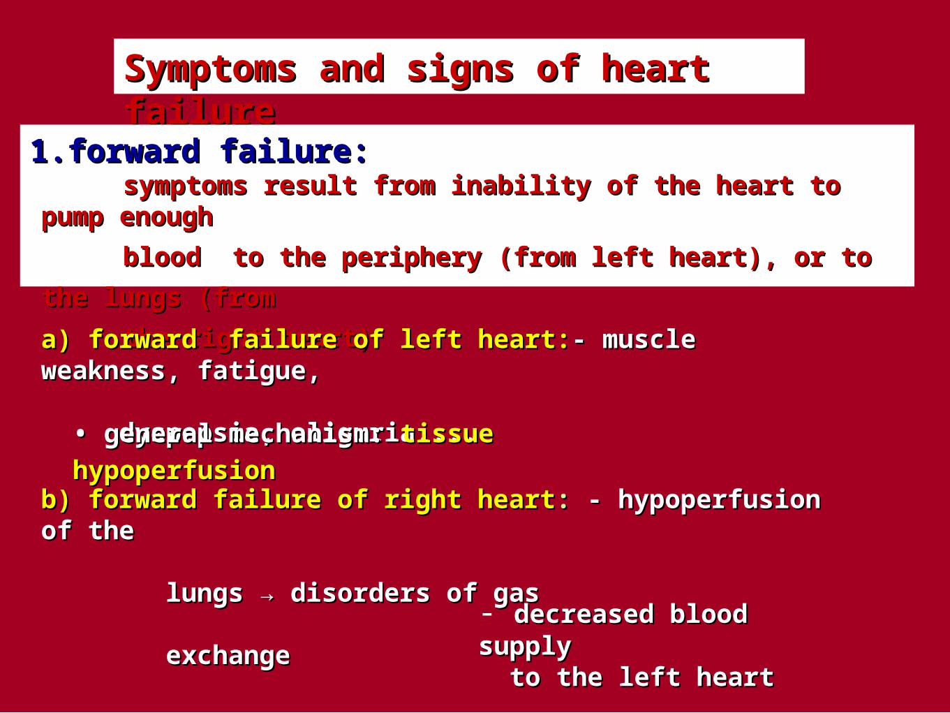
Symptoms and signs of heart failureSymptoms and signs of heart failure
1.1.forward failure:forward failure: symptoms result from inability of the heart to pump enough symptoms result from inability of the heart to pump enough
blood to the periphery (from left heart), or to the lungs (from blood to the periphery (from left heart), or to the lungs (from
the right heart)the right heart)
a) forward failure of left heart:a) forward failure of left heart:-- muscle weakness, fatigue, muscle weakness, fatigue, dyspepsia, oliguria....dyspepsia, oliguria....
• • general mechanism:general mechanism: tissue hypoperfusiontissue hypoperfusion
b) forward failure of right heartb) forward failure of right heart:: - hypoperfusion of the - hypoperfusion of the lungs → disorders of gaslungs → disorders of gas exchangeexchange
- decreased blood supply decreased blood supply to the left heartto the left heart

2. backward failure:2. backward failure: – symptoms result from inability of the heart to accept symptoms result from inability of the heart to accept
the blood comming from periphery and from lungsthe blood comming from periphery and from lungs
a.a.backward failure of left heart:backward failure of left heart:
– – increased pulmonary capillary pressureincreased pulmonary capillary pressure → dyspnoea → dyspnoea
and tachypnoea, pulmonary edema (cardiac asthma) → and tachypnoea, pulmonary edema (cardiac asthma) →
→ → arterial hypoxemia and hypercapnia....arterial hypoxemia and hypercapnia....
b. backward failure of right heart:b. backward failure of right heart:
– increased pressure in systemic venous systemincreased pressure in systemic venous system → →
→ → peripheral edemas, hepatomegaly, ascites →↑nocturnal diuresis....peripheral edemas, hepatomegaly, ascites →↑nocturnal diuresis....