pathophysiology of external breathing. hypoxia
DESCRIPTION
PATHOPHYSIOLOGY OF EXTERNAL BREATHING. HYPOXIA. Professor Yu.I. Bondarenko. Respiratory insufficiency – it is such pathological state, when the tension O 2 in blood arterial is reduced (arterial hypoxemia) and the tension CO 2 - PowerPoint PPT PresentationTRANSCRIPT

PATHOPHYSIOLOGY OF EXTERNAL
BREATHING. HYPOXIA
Professor Yu.I. Bondarenko

Respiratory insufficiencyRespiratory insufficiency – it is such – it is such pathological state, when the tension Opathological state, when the tension O22
in blood arterial is reduced (arterial in blood arterial is reduced (arterial hypoxemia) and the tension COhypoxemia) and the tension CO22
exceeds 50 mm Hg (hypercapnia). exceeds 50 mm Hg (hypercapnia). Most just characteristic of respiratory Most just characteristic of respiratory insufficiency is degree arterial insufficiency is degree arterial hypoxemia.hypoxemia.
Many Many specialitsspecialits consider as consider as respiratory respiratory insufficiencyinsufficiency also such state, when the also such state, when the respiratory parameters of blood are respiratory parameters of blood are within of physiological within of physiological variationvariation, but it is , but it is provided with excessive provided with excessive acaction of tion of external breath, which exhausts and external breath, which exhausts and limits reserve possibilities of an limits reserve possibilities of an organism.organism.


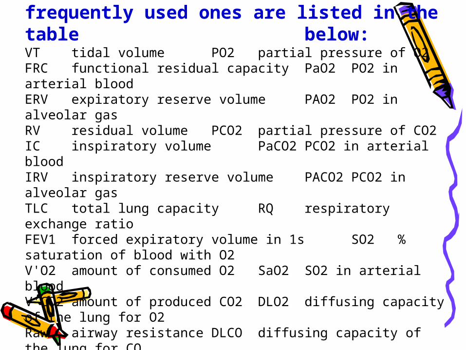
In pulmonary function studies a number of abbreviations and symbols have become standardized. Some frequently used ones are listed in the table
below: VT tidal volume PO2 partial pressure of O2FRC functional residual capacity PaO2 PO2 in arterial bloodERV expiratory reserve volumePAO2 PO2 in alveolar gasRV residual volume PCO2 partial pressure of CO2IC inspiratory volume PaCO2 PCO2 in arterial bloodIRV inspiratory reserve volume PACO2 PCO2 in alveolar gasTLC total lung capacity RQ respiratory exchange ratioFEV1 forced expiratory volume in 1s SO2 % saturation of blood with O2V'O2 amount of consumed O2 SaO2 SO2 in arterial bloodV'CO2 amount of produced CO2 DLO2 diffusing capacity of the lung for O2Raw airway resistance DLCO diffusing capacity of the lung for CO


Forms of respiratory
insufficiency
•Acute respiratory insufficiency
•Chronic respiratory insufficiency

Acute respiratory insufficiency
Acute respiratory insufficiency it is such state, when syndrome
develops fast, within minutes, of hours or day and has tendency to progress.
Fast develops arterial hypoxemia, hypercapnia, develops acidosis, there are disorders of the central nervous system. All this can be completed coma and death.

Asphyxia This state, threatening for life, when in blood don’t enter
oxygen, and from blood the carbonic gas is not removed Asphyxia occurs, as a rule, owing to sharp contraction or
complete closing of respiratory ways. a) external compression of respiratory ways; b) presence in its of foreign bodies; c) narrowing larynx (allergic edema); d) presence in respiratory ways and alveolars of liquid (sink,
aspiration of vomit mass); e) swelling of lung; f) double-side pneumothorax. g) strong oppression of respiratory centre; h) disturbance of impulses transfer in neuro-muscular synapses
(on respiratory muscles); i) traumas of thorax Duration acute asphyxia the person – 3-4 mines
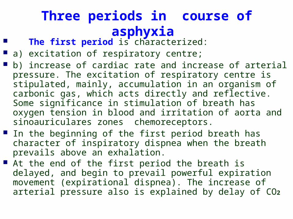
Three periods in course of asphyxia The first period is characterized: a) excitation of respiratory centre; b) increase of cardiac rate and increase of arterial pressure.
The excitation of respiratory centre is stipulated, mainly, accumulation in an organism of carbonic gas, which acts directly and reflective. Some significance in stimulation of breath has oxygen tension in blood and irritation of aorta and sinoauriculares zones chemoreceptors.
In the beginning of the first period breath has character of inspiratory dispnea when the breath prevails above an exhalation.
At the end of the first period the breath is delayed, and begin to prevail powerful expiration movement (expirational dispnea). The increase of arterial pressure also is explained by delay of CO2
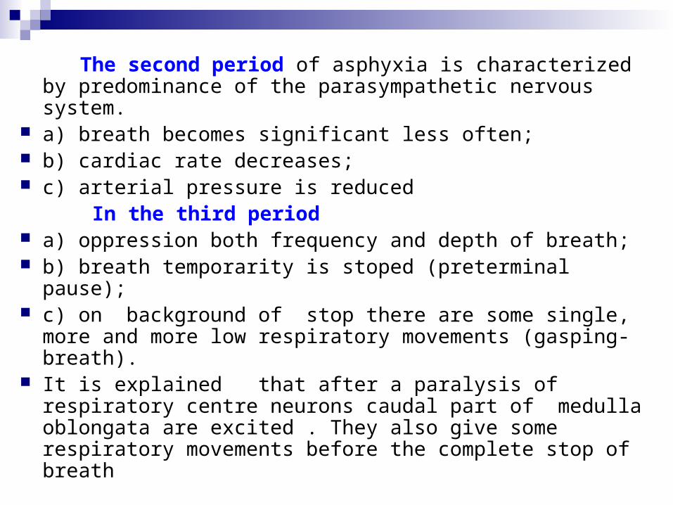
The second period of asphyxia is characterized by predominance of the parasympathetic nervous system.
a) breath becomes significant less often; b) cardiac rate decreases; c) arterial pressure is reduced In the third period a) oppression both frequency and depth of breath; b) breath temporarity is stoped (preterminal pause); c) on background of stop there are some single, more and
more low respiratory movements (gasping-breath). It is explained that after a paralysis of respiratory centre
neurons caudal part of medulla oblongata are excited . They also give some respiratory movements before the complete stop of breath

The chronic respiratory insufficiency
The chronic respiratory insufficiency is characterized by slower increase of hypoxemia and hypercapnia, and they do not reach such degree, as of acute insufficiency due to inclusion of compensatory mechanisms (erythrocytosis, increase of hemoglobin in erytrocytes)
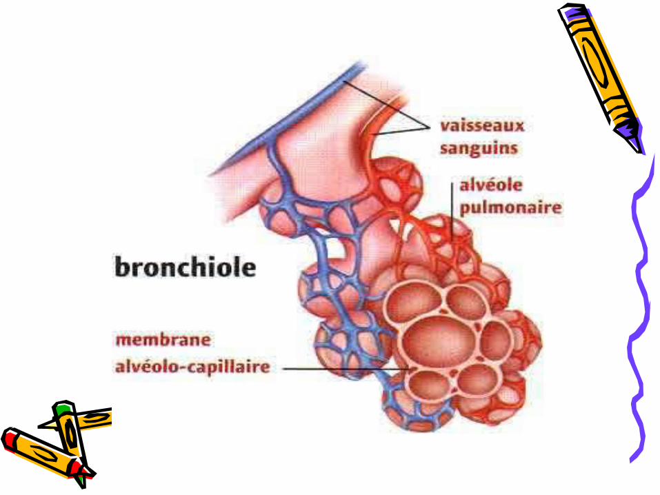
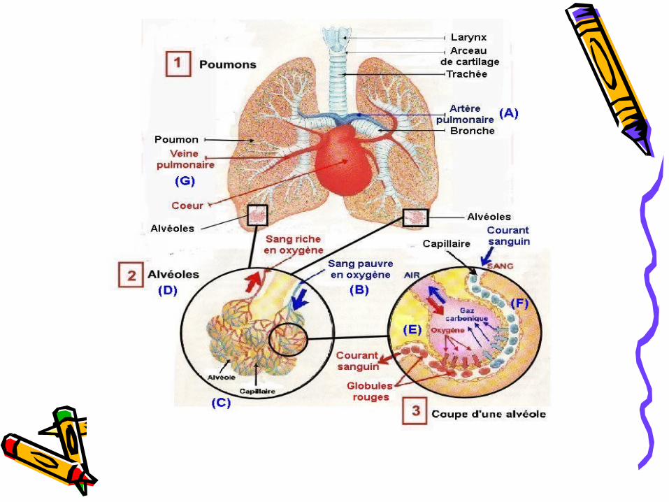
It is known, that the external breath is provided with three processes – ventilating lung, diffusion of gases (О2 and CO2) through alveolar wall and perfusion of blood through lung capillaries. The disorder of any of these processes can serve as the reason of respiratory insufficiency.
In pathogenesis is distinguished two forms of respiratory insufficiency – ventilation and alveolar-respiratory

Pathogenetic classification of respiratory insufficiency
Ventilative Diffusive Perfusive Combined

Ventilative respiratory insufficiency
Obstructive Restrictive Dysregulative (disorder of central regulation of breath)

Ventilative respiratory insufficiency
The essence of ventilative insufficiency is that in the alveolars for unit time enter less air than in norm.
This state is called alveolar hypoventilation. Outlung reasons of ventilative insufficiency : Disturbance of respiratory centre function a) effects of medical drugs; b) cerebral-brain traumas with epidural or subdural hematoma; c) malignant tumors of brain; d) absceses of brain, meningitis; e) disorder of brain circulation blood. Disorder of the motoneurons function of spinal cord, which
innervation respiratory muscles (tumor of spinal cord, syringomyelia, poliomyelitis).

Ventilative respiratory insufficiency Disorder of innervation of breathing muscles:
а) lesion of nerves – due to avitaminosis, inflammation, trauma; b) blockade of impulses transfer in nervous – muscular synapses
– myasthenia, action of myorelaxantes; c) lesion of respiratory muscles-myositis, dystrophies, periodic
paralysis, hypocaliemia, hypophosphoremia. Limitation of thorax mobility: a) inherent or ecquired deformation of ribs and vertebral; b) ossification of costales cartilages; c) grown of preular parts, ascites, meteorism, obesity; d) pain due to neuralgia of intercostales nerves. Disorder of thorax integrity and pleural cavity (pneumothorax).

Obstructive insufficiency
• Obstruction of respiratory ways is narrowing their lumen and increase of resistance to movement of air
• The damage can be located in upper respiratory ways (with diameter of 2 mm and more) and in lower respiratory ways (diameter – up to 2 mm)

Obstructive respiratory insufficiency
Upper respiratory ways is understood cavity of mouth, nasal passage, pharynges, larynges, trachea, large bronchus.
Obstruction it may be is caused internal and externals mechanical trauma.
Internal trauma most frequently it arises as complication of trachea-intubation, less often – after operation on larynges.
External mechanical trauma – fractures of lower jaw, cervical cartilages, larynges cartilages, epiglossus, trachea, damage of tongue basis, mouth, neck.
The mechanism of obstruction is spasm, edema also paralysis of voice slot, damage or off set of larynges cartilages, hematoma, edema of mucous membrane or serrounding tissues.

Internal trauma
a) Burns and inhalating of poisoning gases. In these cases develops edema of mucous upper respiratory paths.
b) Bleeding in respiratory ways is observed after operation
on head and neck, after tonsillectomy, tracheostomy. Sometimes bleeding happens spontaneously, for example from nose. The bleeding especially is dangerous when the patient is in coma or in narcosis, that is when the drainage of respiratory paths is impossible.
c) Aspiration of foreign body is observed in children in the
age from 6 months to 4 years more often. In the adult aspiration of foreign body occurs, usually, during take food, especially in state of alcoholic intoxication.
d) Obstruction lower respiratory ways – necrotic Ludvig`s
angina (suprogenis necrotic flegmona of oral bottom cavity of an infectious origin), subpharynges abscess, which is caused aerobic and anaerobic microflora, аngioneurotic edema, which develops as response on allergen and is accompanied nettle-rash, asthma, rhinitis.

Obstruction of lower respiratory ways
a) liquid aspiration – vomit mass, blood, water;
b) allergy response mainly on medical preparations – antibiotics and protein substitutes.
It develops immediately, during 30 minutes and appears hardly expressed laryngo- and bronchospasm.
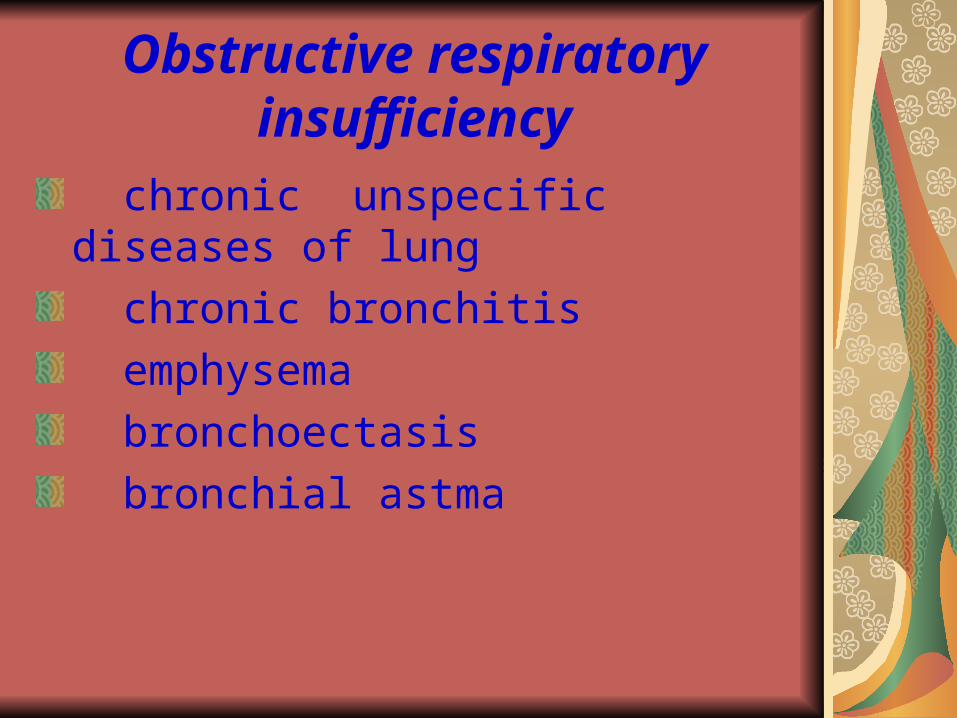
Obstructive respiratory insufficiency
chronic unspecific diseases of lung
chronic bronchitis
emphysema
bronchoectasis
bronchial astma

EMPHYSEMA Emphysema is an illness, in which rupture interalveolar septums and lungs
capillaries. By basis it is considered degraded collagen and elastic fibres of proteolytic
enzymes, which are produced phagocytes under influence of the external factors – microorganisms, dust particles, tobacco smoke.
In etiology of emphysema some role is attached importance of hereditary predisposition due to synthesis of defective collagen and elasthyne, insufficient synthesis of proteolytic enzymes inhibitors.
The mechanism of obstruction due to emphysema Walls of bronchioles very thin and pliable. The lumen them is
supported transpulmonaris more pressure. The more elasticity lung, the should be transpulmonaris more pressure to overcome elastic recoil. Bronchioles for want of that will be in an extended state. When the elasticity lung is reduced, it is enough for their stretch low transpulmonaris pressure. The force, which acts on walls bronchioles from within, decreases and also their lumen is narrowed. The decrease of lumen conducts to sharp increase of resistance to movement of air. As a result of it the breath is difficulty. But even more exhalation is difficulty. For want of emphysema it becomes active. The pressure in pleural cavity increases, and bronchioles are compressed from the outside of lungs fabric. With the cource of time bronchioles compress completely, and the exhalation becomes impossible. Air becomes isolated in alveoles.

The mechanism of obstruction due to bronchial
asthma a) аccumulation of viscous glasslike mucus in
bronchus. It is connected with hypertrophy of mucous
glands and hyperproduction by them mucus (hypercrinia). The viscous mucus is difficultly discharge and congest (mucostasis). The important role in the mechanism of mucostasis plays hyperplasia of goblet cells, which substitude cells of ciliated epithelium.
b) edema of mucous, spasm of smooth bronchus muscules.
c) increased reactivity of bronchial muscles on specific and unspecific stimulus.
The highest degree hyperreactivity is observed at once time or after an attack. Strong stimulus, which provokes bronchoconstriction in the patients of bronchial asthma, is the physical load.

Restrictive insufficiency
• This form of respiratory insufficiency arises, when the extensibility lung is reduced, that is when it not capable easily to be straightened.
• To carry out a breath, it is necessary to increase transpulmonary pressure, and it can be made at the expense of increase of respiratory muscles action
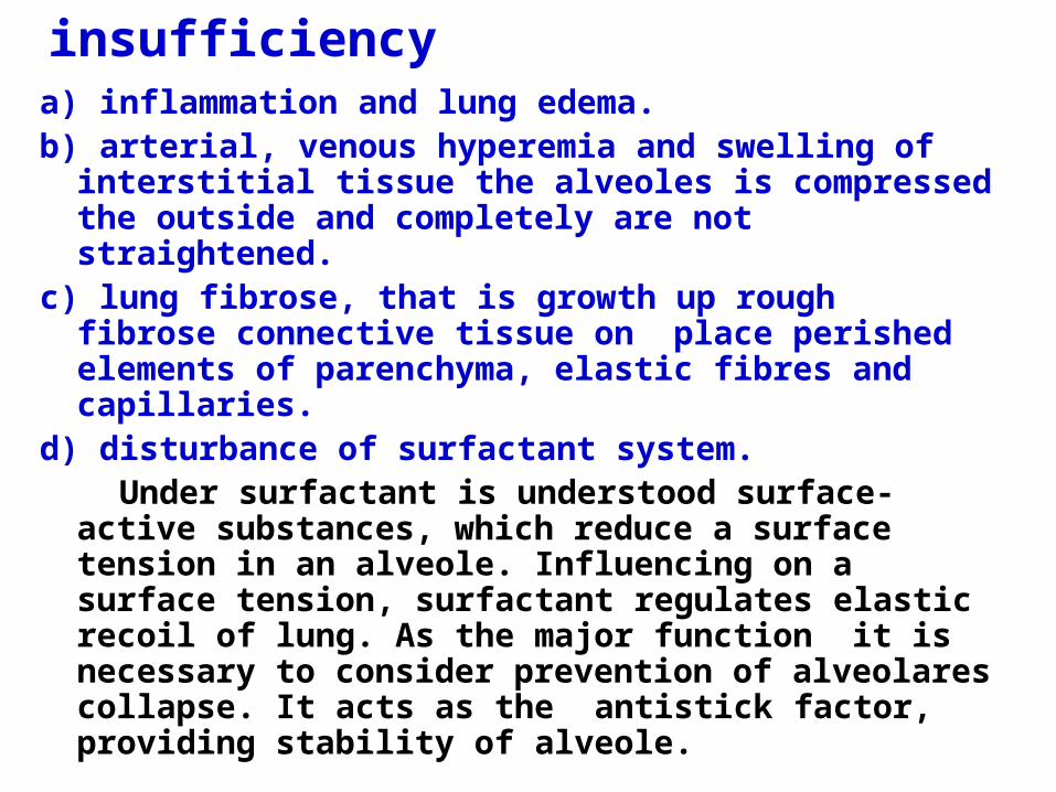
Restrictive insufficiencya) inflammation and lung edema. b) arterial, venous hyperemia and swelling of
interstitial tissue the alveoles is compressed the outside and completely are not straightened.
c) lung fibrose, that is growth up rough fibrose connective tissue on place perished elements of parenchyma, elastic fibres and capillaries.
d) disturbance of surfactant system. Under surfactant is understood surface-
active substances, which reduce a surface tension in an alveole. Influencing on a surface tension, surfactant regulates elastic recoil of lung. As the major function it is necessary to consider prevention of alveolares collapse. It acts as the antistick factor, providing stability of alveole.

The deficiency of surfactant Insufficient synthesis it or excessive remove
from a surface of alveoles. The insufficient synthesis is characterized for illness of hyaline membranes in newborn, for want of which destroy intraalveolares septum and in alveoles is stored hyaline with epithelial cells and blood form elements.
Ecquired decrease of surfactant is observed due to asphyxia, acidosis, pneumonia, pollution of air. The defect of surfactant predetermines high surface tension of alveoles and high resistance lung for want of expansion by their inhaled air.
Besides, by cause of restrictive insufficency may be аthelectasis ( fall of alveoles and stopping of their ventilation), рneumothorax, deformation of thorax, paralysis of respiratory muscles.

Disregulative respiratory insufficiency
Respiratory center dysfunction a) hypoxia b) hypoglycemia c) brain trauma d) compression (edema, tumor, hematoma) e) disorder of brain circulation f) intoxication (narcotic, muscarine, toxic
metabolic product) g) inflammation and dystrohpy Disorder of impulse transmission to the
respiratory muscle

Disorder of the central regulation of breath
Influence of reflex and humoral factors Direct influences on the respiratory
centre changes function. Those disorders of breath regulation,
which limit alveolar ventilation, can serve as the reason of respiratory insufficiency.

Breath disturbances of central genesis.
Bradypnea – rare breath. It can arise reflexly in case of arterial pressure increase(reflex from baroreceptors of aorta and carotide bodies), and also due to hyperoxia (reflex from hemoreceptors of the same zones).
The deep and rare breath arises due to narrowing of upper respiratory ways. It is named stenotic.
The reason of the bradypnea is a direct lesion of neurons of respiratory centre due to long time hypoxia, under influence of narcotic, due to organic changes in a brain (inflammation, insult) or functional disorders of the central nervous system (neurosis, hysteria)

Polypnea (tachypnea) – friquent surface breath. It arises reflexly in fever, hysteria, pain in area of thorax, peritoneum, pleura.
Hyperpnea – deep and friquent breath. It has compensatory character, however excessive stimulation of respiratory centre in pathological conditions (decrease of partial pressure O2 , anemia, acidosis) provokes rather intensive breath, which can lead to remowal of CO2 from organism and paralysis of respiratory centre. Owing to diabetic mellitus there is so-called noisy breath (Kussmaul’s breath), due to metabolic acidosis.
Apnea – temporary stopping of breath. This manifestation of inhibition neurons of respiratory centre under influence hypoxia or intoxication, owing to organic lesion of brain. Apnea occurs affer hyperventilation, when tension of CO2 in blood is dropped lower threshold for respiratory centre of level, and also during fast rise of arterial pressure (reflex from baroreceptors of vessels).

Periodic breathing This such disorder of respiratory rhythm, when
the periods of respiratory movements alternate with periods of apnea.
There are two main types of periodic breath – Cheyne-Stokes and Biot’s.
In the first case the amplitude of respiratory movements cyclic is increased up to expressed hyperpnea and decreases to apnea. Owing to Biot’s breath amplitude of respiratory movements is constant.

Dyspnea. This sensation of air lack and simultaneously necessity to
strengthen breath. а) insufficient oxygenation of blood in lung (low partial
pressure of oxygen in inhaled air, difficulty of ventilation lung, disorder of lungs hemodynamics);
b) disorder of gases transport by blood (insufficiency of blood flow, anemia, inactivation of hemoglobin);
c) metabolic acidosis (diabetis mellitus); d) functional and organic lesion of brain (hysteria,
encephalitis, insult). In arise of dyspnea the large role belongs to reflexes from respiratory ways, parenchyma of lung, aorta and carotide bodies.

Diffusive respiratory insufficiency
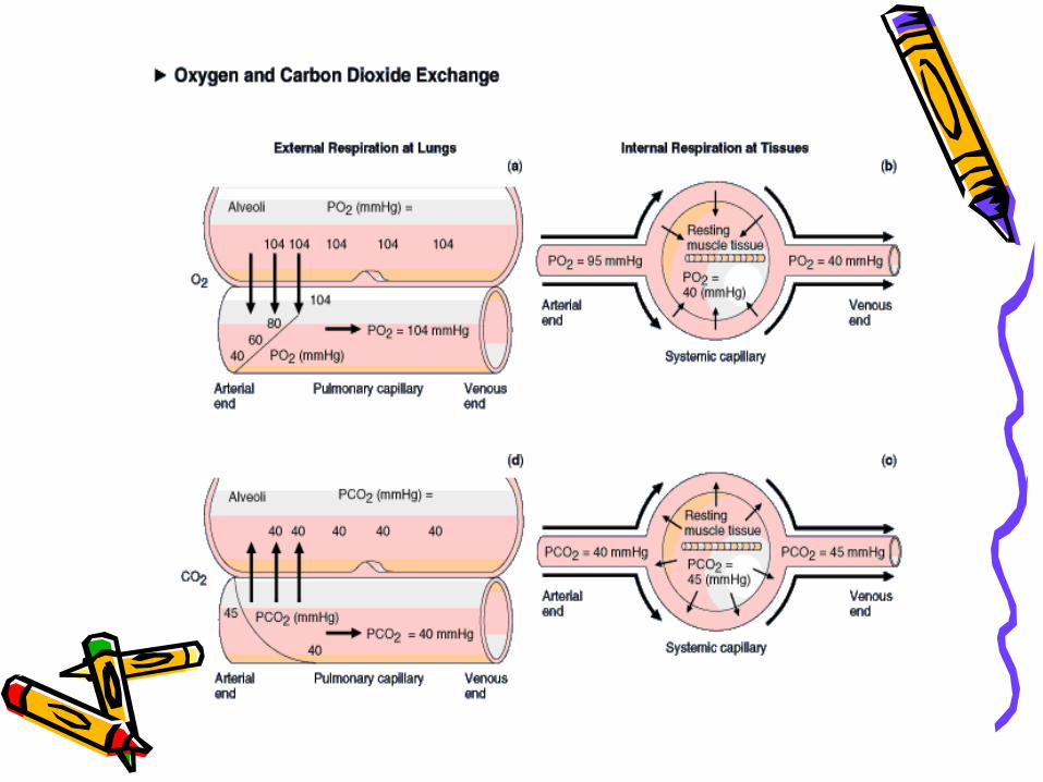
Decrease of the area alveolar capillary membrane; Thickening of the capillary membrane; Decrease of transmembrane gradient of partial
pressure in the alveoli-blood systen; Decrease of the volume of the capillary blood in the
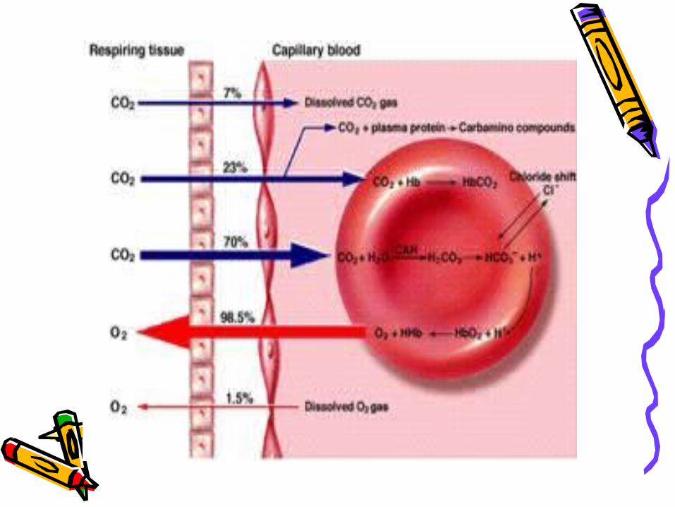
lung; Decrease of the speed of reaction between O2 and Hb
and oxygen capacity of blood; Decrease of alveolar ventilation; Decrease of exposition and contact of erythrocytes with
the alveolar air

Pathological processes connected with disorder of diffuse in lung
Damage of membranes (membranogenic poisoning);
Inflammation and accumulation of exudate; Sclerosis; Fibrosis; Edema; Anemia and Hb inactivation; Emphysema; Cardiac insufficiency

Perfusive of respiratory insufficiency
Disorders of pulmonare blood circulation; Systemic disorders of hemodynemic; Local disorders (hyperemia, venous
congestion, ischemia, thrombosis embolism);
Inflammation

Alveolar-respiratory insufficiency
This type of respiratory insufficiency arises in that case, when is reduced gas exchange between alveolar air and blood
There are two variants of insufficiency:
1) due to inadequacy of ventilation and perfusion of lung
2) owing to difficulty diffusion of gases through alveolar wall

HYPOXIA Hypoxia is typical pathological process, which
arises owing to insufficient oxygen supply of tissues or insufficient use it by tissues.
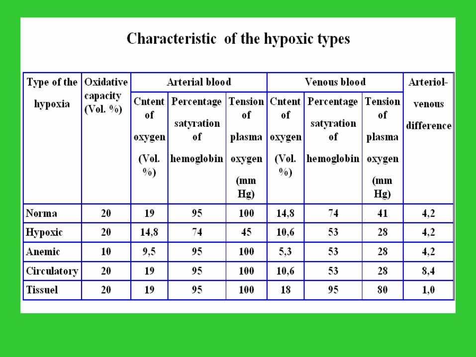
Hypoxic Hemic Circulatory Histotoxic

Hypoxic hypoxia The essence of this hypoxia is oxygen
tension reduction in arterial blood, the hemoglobin saturation with oxygen decrease and as a result the contents of oxygen decrease. Thus hypoxemia develops.
Decrease of the partial oxygen pressure in inhaled air is main reason of oxygen tension reduction in blood.
The second varient of hypoxic hypoxia stipulated respiratory insufficiency which arise as a result external breath disorders – ventilation, diffusion and perfusion. Respiratory hypoxia is result of gas metabolism disorder in the lung for normal partial oxygen pressure of atmospheric air.

Alveolar hypoventilation
а) oppression of respiratory centre – overdose of drugs, brain swelling, brain
insult (stroke); b) obstruction of the air passages –
dyphteria, laryngospasm, larynx swelling, getting into tracheal of the foreign
body; c) thorax damage; d) diseases of respiratory muscles and
intercostals nerves.

Hemic hypoxia Basis of this hypoxia type is decrease of
blood oxygen capacity. There are two variants: anemic and toxic In case of anemic form, the total level of
circulating hemoglobin decrease– owing to blood loss, erythrocytes hemolysis, oppression of blood formation.
Toxic form arises in case of hemic toxines poisoning. The general content of hemoglobin in blood remains normal, but the contents of functionally active hemoglobin decreases. The part of hemoglobin turns into such compounds, which are not capable to execute oxygen transport function.

Circulatory hypoxia This is such hypoxia, which is stipulated by
circulation blood speed decrease, that is oxygen delivery to tissues slowing down.
It arises owing to general blood flow slowing in case of cardio-vascular insufficiency, or owing to local blood supply disorders. Pure circulatory hypoxia does not happen really. It is observed only as a local phenomenon in some organs ischemia.


Histotoxic hypoxia In this hypoxia mainly is the tissues
inability of oxygen utilisating. The main index is the low arterioal-venous
difference. Neither, the contents of oxygen in blood, nor it’s delivery to tissues are not disordered, but tissue use it less for one time unit, than it is necessary for maintenance their energy needs.
The reason of this hypoxia is decrease of respiratory enzymes activity.

Mechanism of histotoxic hypoxia
There are three enzymic systems participate in electron transport from substrat to molecular oxygen
Pyridindependent dehydrogenases Flavindependent dehydrogenases, Cytochromes.
Any of these systems blockade will result in disorder of electron transport throughout on a respiratory chain and will cause histotoxic hypoxia.
Respiratory chain enzymes are oppressed with cyanides, monoiodacetatis, drugs, spirit, formaldehyde, aceton, ethylurethane, sulfurhydrogen, cocain, carbon oxide and other substances. Typical example of hystotoxical hypoxia – cyanic poisoning, which oppresses cytochromoxydase. The cytochromes oxidazing – restoring processes get blocked with local anestetics (novocaine). Flavine enzymes lose their activity in case of riboflavine (Vit B2) deficiency. Pyridine enzymes activity is slowing in case of nicotine acid deficiency.


Metabolism in state of hypoxia Features of protein metabolism disorders:
Inhibition of synthesis and simultaneously acceleration of fibers disintegration, increase of residual blood nitrogen contents, ammonia accumulation.
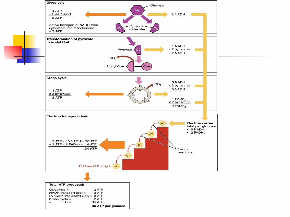
The АТP decrease in cells underlie of hypoxic damaging effect upon the organism. The contents other macroergic compound – creatinephosphate decreases in brain and heart very fast. Major changes of carbohydrate metabolism are the following: anaerobic glycolysis increase, glycogen stocks exhausting, accumulation of pyruvic and lactic acids, metabolic acidosis.
The fatty metabolism in case of hypoxia is characterized by such features: intensive fat disintegration in depot, accumulation of fatty acids in tissues, ketoacids accumulation in blood.
Cerebral and heart activity disorder in state of hypoxia Nervous system is the most sensitive to
oxygen starvation. Brain cortex neurons can work perfectly in conditions of complete blood flow termination only for 5-6 minutes. Oblongata brain neurons maintain the complete blood supply termination for 20-30 minutes, spinal cord neurons - up to 60 minutes. Heavy and long hypoxia causes a defect of blood supply and breath centres, reflex activity disorder.
The myocardium is very sensitive to oxygen starvation. It takes on the second place after the nervous system. Increasing heavy hypoxia oppresses contract and rhythmic heart activity. It due to inhibition of oxidation and energy deficiency.

Compensatory responses in state of hypoxia
The compensatory responses of an organism, which are directed to hypoxia removal are divided into four groups:
Respiratory Hemodynamic Bloodly Tissuel
The respiratory responses The respiratory responses appear as dyspnea,
with acceleration and deepening of breathing. It is named high-altitude, or hypoxic, or compensatory.
Dispnea arises in reply to an hemoreceptors aortae arc and sinocarotide zones irritation with hypoxic blood. Due to dyspnea pulmonary ventilation is increased. The compensatory significance of lung hyperventilation is not absolute, because after excessive and long hyperventilation occurs hypocapnia. It has an negatively effect upon breath regulation, as carbonic acid intensively removal from blood physiological irritant of respiratory centre. Sudden unconsciousness during the height rise can be explained just by respiratory centre paralysis.

The hemodynamic compensator responses
а) tachycardia – result of sympathetic tonus increase
b) stock blood volume increase at the expense of adrenaline action upon myocardium adrenoreceptors
c) cardiac output increase as a result of tachycardia and stock volume increase
d) the blood flow acceleration - it is connected in main to cardiac output increase
e) blood circulation centralization that is peripheral vessels narrowing and vessels of the vital organs extension owing to redistribution of blood to brain, heart, lung in fact of simultaneous blood supply limitation of skin, muscles, intestines, spleen
Bloodly compensatory responses:
erythrocytosis – first of all due to output of blood from depot, and later due to blood forming stimulation
the increase of hemoglobin in erythrocytes increase of hemoglobin affinity to oxygen in
lung (shift of oxyhemoglobin dissociation curve to the left) and decrease it this similarity in tissues (shift of oxyhemoglobin dissociation curve to the right)
Tissuel compensatory responses are the decrease of metabolism, activation of glycolysis, activation of respiratory chain enzymes
Adaptation to hypoxia Hypoxia – is not only a damaging, but it is also a training factor. During hypoxia training period adaptation is formed first. In case of long hypoxia certain made the emergency adaptation turns into long-term adaptation arise. Adapted organism spend very economically energetic and plastic resources.
In case of the hypoxia training termination the state of desadaptation occurs. The positive properties of adapted organism bought during trainings, are getting gradually lost.It is important not infringe mode of trainings at all stages of adaptive process. The mode disorder is dangerous. Dangerous life disorders arise in such cases, which increase sensitivity to hypoxia. Adaptation, desadaptation, failure adaptation is very important problem. It has practical significance in human selection and preparation for certain professional mastering (divers, spaceman) and for examination the territories with extremal life conditions (space, ocean, mountain).
