pathology pathology: is the study of diseases. diseases are the deviations from normal. the concept...
TRANSCRIPT
Medical Pathology
Pathology
Pathology: is the study of diseases.Diseases are the deviations from normal.The concept of diseases:• For the pathologist: structural changes that are
accompanied by functional changes.
The Scope of Human Pathology• Pathology deals with recognition of diseases,
their causes (etiology), and their progression.• Pathologists study structural changes (gross, or
microscopic), etiology and mechanisms of diseases (pathogenesis)
• Most diseases can be placed in one of these categories:
1. Inflammatory2. Neoplastic 3. Degenerative conditions4. Developmental conditions

InflammationInflammation: Local defense and protective response
against cell injury or irritation or Local vascular and cellular reaction, against an irritant.
Irritating or injurious agents (Irritant)
Living: • Bacteria,• Fungi,• Virus,• Parasite• or their
toxins
Non-Living: • Chemical• Physical
Inflammation is designated by adding the suffix (itis) to the end of the name of the inflamed organ or tissue.
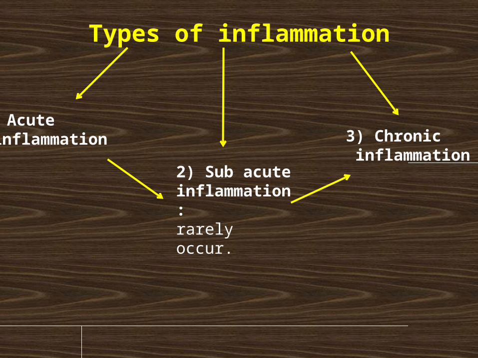
Types of inflammation
1) Acute inflammation 3) Chronic
inflammation2) Sub acute inflammation: rarely occur.
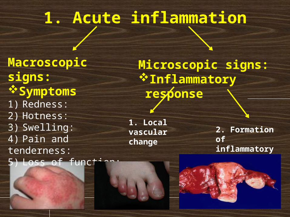
1. Acute inflammation
Microscopic signs:Inflammatory response
Macroscopic signs:Symptoms1) Redness: 2) Hotness: 3) Swelling: 4) Pain and tenderness: 5) Loss of function:
1. Local vascular change
2. Formation of inflammatory exudate
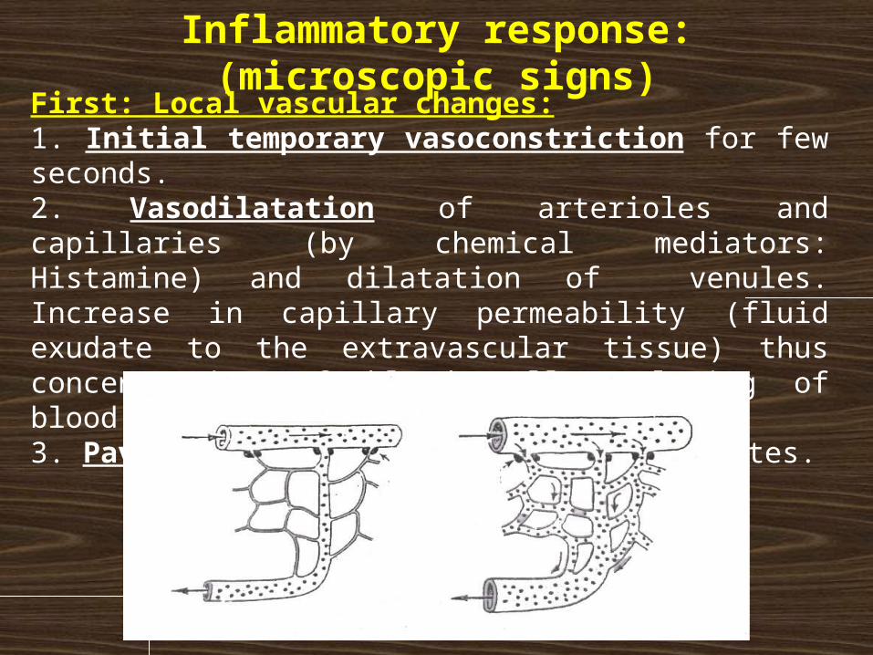
Inflammatory response: (microscopic signs)
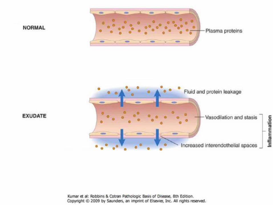
Normal Inflammation
First: Local vascular changes:1. Initial temporary vasoconstriction for few seconds.2. Vasodilatation of arterioles and capillaries (by chemical mediators: Histamine) and dilatation of venules. Increase in capillary permeability (fluid exudate to the extravascular tissue) thus concentration of blood cells, slowing of blood flow (stasis)3. Pavmentation: the migration of leukocytes.
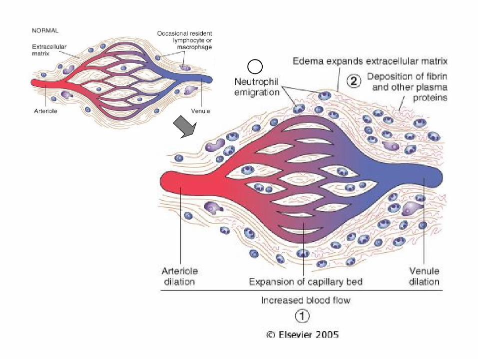
Second: Formation of inflammatory exudates:• Immigration or infiltration of the various leukocytes, fluid
and plasma proteins outside the blood vessels into the surrounding tissue without injury of the blood vessels.
• Leukocytes seem to leave the smallest blood vessels by inserting pseudopodia into the interendothelial junctions and sliding through the wall by amoeboid movement.
• This is also due to the increased capillary permeability caused by the high osmotic pressure of the surroundings.
• The early stages are marked by the predominance of polymorphs especially neutrophils migration, particularly when the inflammation is caused by pyogenic cocci, later on monocytes infiltration occurs.
****In some cases RBCs may also pass (Diapedesis)
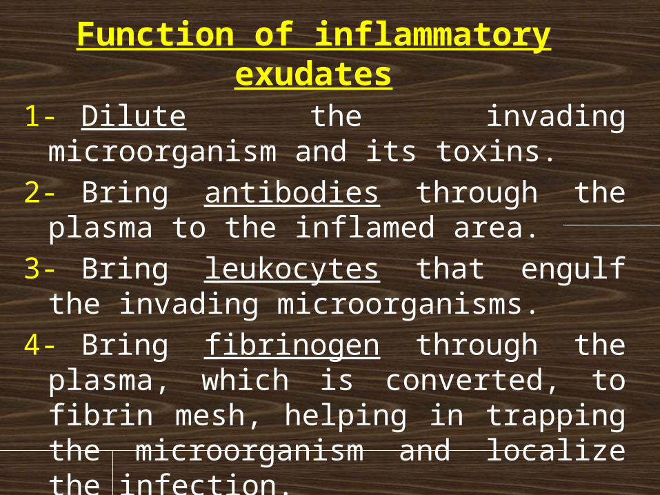
Function of inflammatory exudates
1-Dilute the invading microorganism and its toxins.
2-Bring antibodies through the plasma to the inflamed area.
3-Bring leukocytes that engulf the invading microorganisms.
4-Bring fibrinogen through the plasma, which is converted, to fibrin mesh, helping in trapping the microorganism and localize the infection.

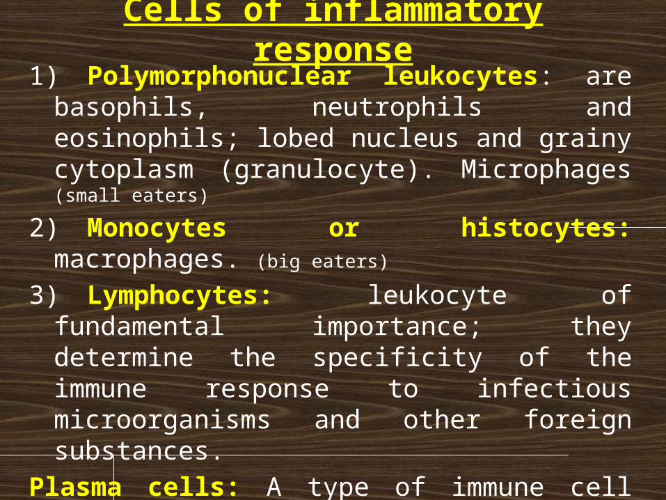
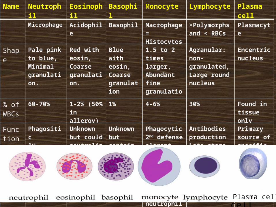
Cells of inflammatory response1) Polymorphonuclear leukocytes: are basophils,
neutrophils and eosinophils; lobed nucleus and grainy cytoplasm (granulocyte). Microphages (small eaters)
2) Monocytes or histocytes: macrophages. (big eaters)
3) Lymphocytes: leukocyte of fundamental importance; they determine the specificity of the immune response to infectious microorganisms and other foreign substances.
Plasma cells: A type of immune cell that makes large amounts of a specific antibody, developed from activated B cells (Derived from lymphocytes originate in the bone marrow). It is a type of WBCs and also called plasmacyte.
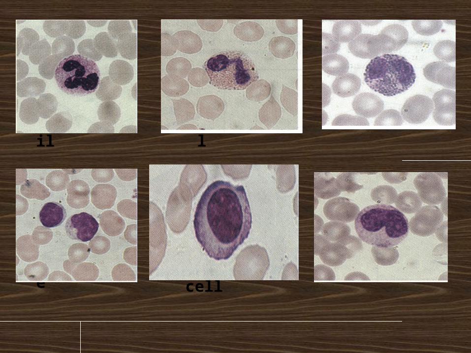
Neutrophil Eosinophil Basophil
Lymphocyte Plasma cell Monocyte
Monocytes
Name Neutrophil Eosinophil Basophil Monocyte Lymphocyte Plasma cell
Microphage Acidophile Basophil Macrophage = Histocytes
>Polymorphs and < RBCs
Plasmacyte
Shape Pale pink to blue,Minimal granulation.
Red with eosin,Coarse granulation.
Blue with eosin,Coarse granulation
1.5 to 2 times larger, Abundant fine granulation
Agranular: non-granulated, Large round nucleus
Encentric nucleus
% of WBCs
60-70% 1-2% (50% in allergy)
1% 4-6% 30% Found in tissue only
Function
Phagositic1st defense
Unknown but could neutralize histamine, serotonin and other kinins
Unknown but contain histamine &heparin
Phagocytic2nd defense elementengulf bacteria, dead cells, debris & dead neutrophils (pus cells)
Antibodies productionLate stage of the inflammation
Primary source of specific Antibodies
Plasma cell
Plasma cell
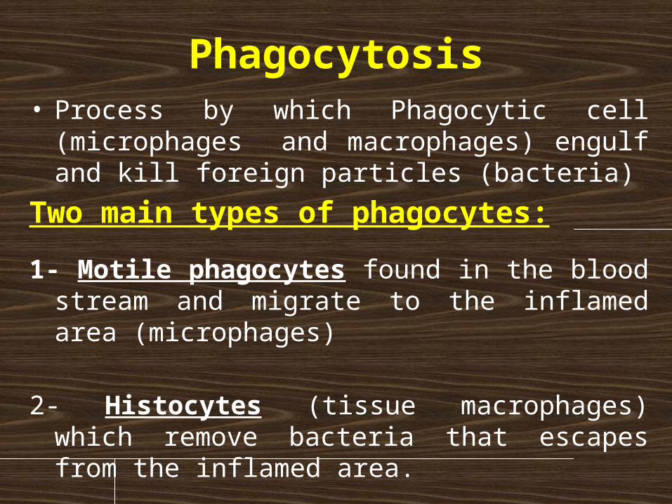
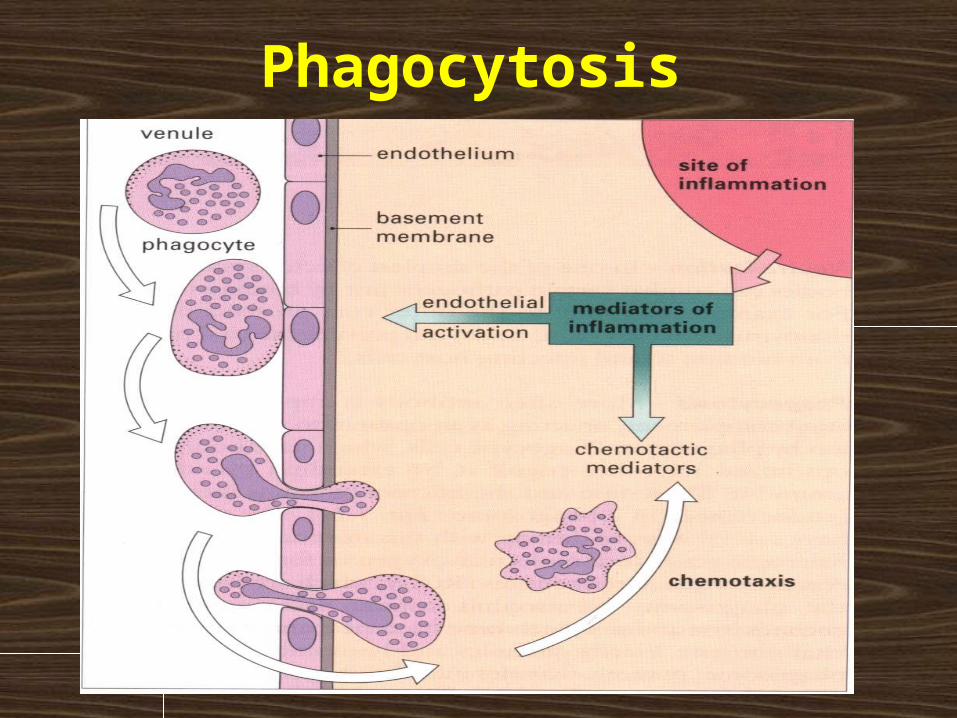
Phagocytosis• Process by which Phagocytic cell (microphages and
macrophages) engulf and kill foreign particles (bacteria)
Two main types of phagocytes:
1- Motile phagocytes found in the blood stream and migrate to the inflamed area (microphages)
2- Histocytes (tissue macrophages) which remove bacteria that escapes from the inflamed area.
Phagocytosis
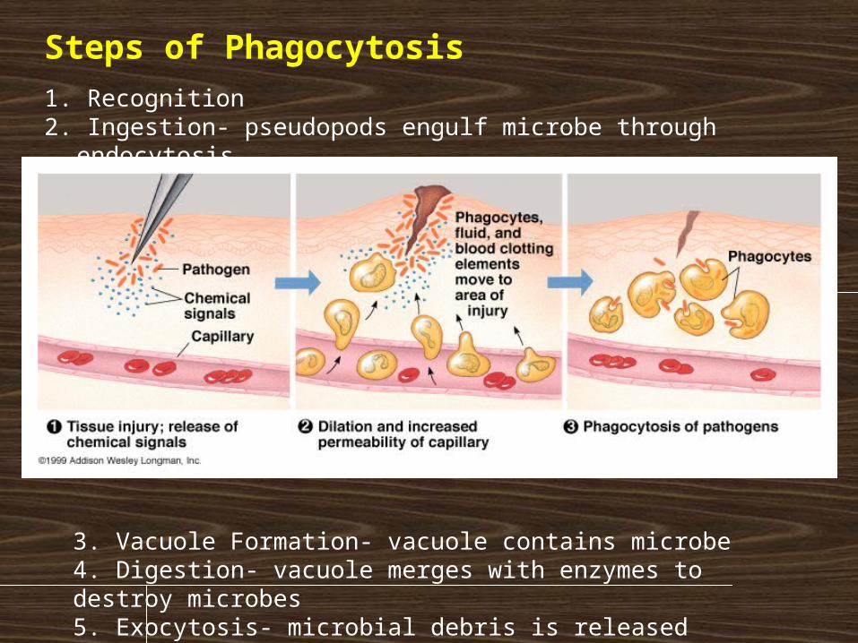
Steps of Phagocytosis
1. Recognition 2. Ingestion- pseudopods engulf microbe through endocytosis
3. Vacuole Formation- vacuole contains microbe4. Digestion- vacuole merges with enzymes to destroy microbes 5. Exocytosis- microbial debris is released
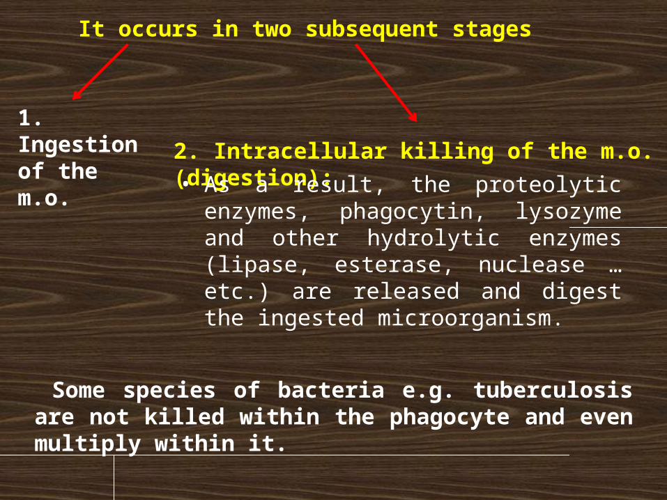
It occurs in two subsequent stages
1. Ingestion of the m.o. 2. Intracellular killing of the m.o. (digestion):
• As a result, the proteolytic enzymes, phagocytin, lysozyme and other hydrolytic enzymes (lipase, esterase, nuclease … etc.) are released and digest the ingested microorganism.
Some species of bacteria e.g. tuberculosis are not killed within the phagocyte and even multiply within it.
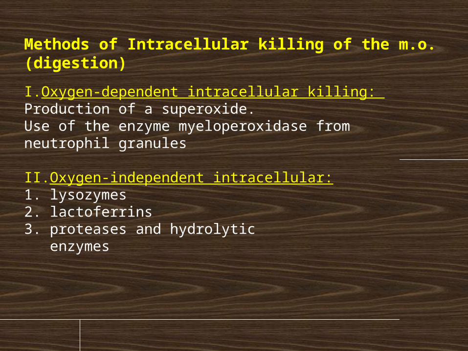
Methods of Intracellular killing of the m.o. (digestion)
I. Oxygen-dependent intracellular killing: Production of a superoxide. Use of the enzyme myeloperoxidase from neutrophil granules
II.Oxygen-independent intracellular:1. lysozymes 2. lactoferrins 3. proteases and hydrolytic enzymes
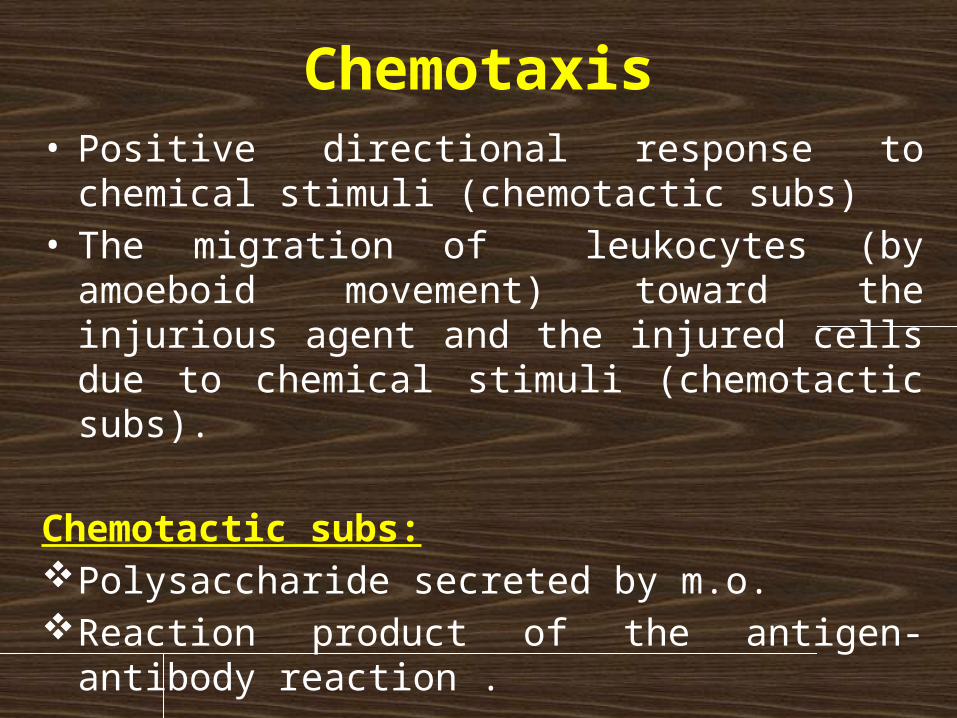
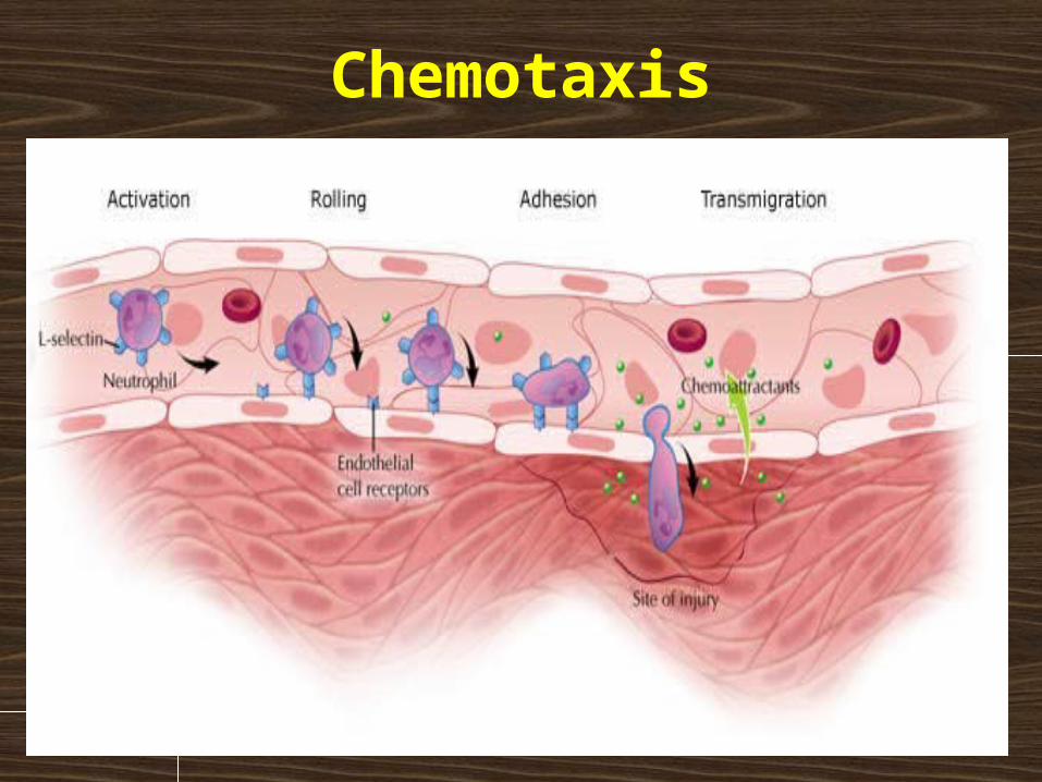
Chemotaxis• Positive directional response to chemical stimuli
(chemotactic subs)• The migration of leukocytes (by amoeboid movement)
toward the injurious agent and the injured cells due to chemical stimuli (chemotactic subs).
Chemotactic subs:Polysaccharide secreted by m.o.Reaction product of the antigen-antibody reaction .
Chemotaxis
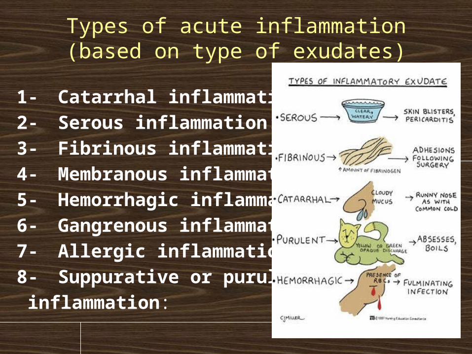
Types of acute inflammation(based on type of exudates)
1- Catarrhal inflammation: 2- Serous inflammation: 3- Fibrinous inflammation: 4- Membranous inflammation: 5- Hemorrhagic inflammation: 6- Gangrenous inflammation: 7- Allergic inflammation: 8- Suppurative or purulent inflammation:
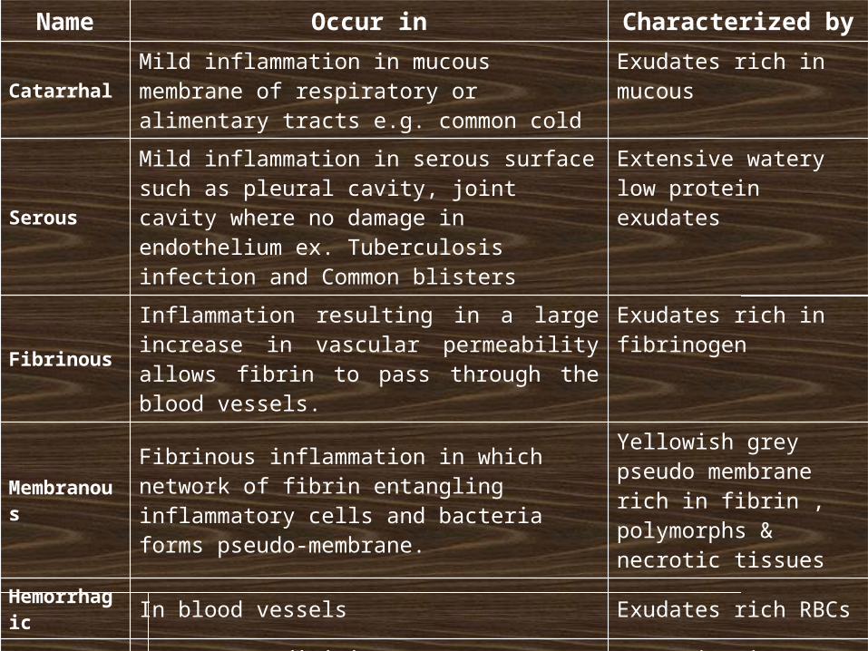
Name Occur in Characterized by
Catarrhal Mild inflammation in mucous membrane of respiratory or alimentary tracts e.g. common cold
Exudates rich in mucous
SerousMild inflammation in serous surface such as pleural cavity, joint cavity where no damage in endothelium ex. Tuberculosis infection and Common blisters
Extensive watery low protein exudates
FibrinousInflammation resulting in a large increase in vascular permeability allows fibrin to pass through the blood vessels.
Exudates rich in fibrinogen
MembranousFibrinous inflammation in which network of fibrin entangling inflammatory cells and bacteria forms pseudo-membrane.
Yellowish grey pseudo membrane rich in fibrin , polymorphs & necrotic tissues
Hemorrhagic In blood vessels Exudates rich RBCs
GangrenousAcute appendicitis Necrotic tissues resulting
from thrombi or emboli
Allergic Result to Ag – Ab reaction HypersensitivityPresence of edema & increase in vascularity.
SuppurativeCaused by pyogenic bacteria and is characterized by pus formation Example: Abscess.
Large amount of Pus & Purulent exudates produced
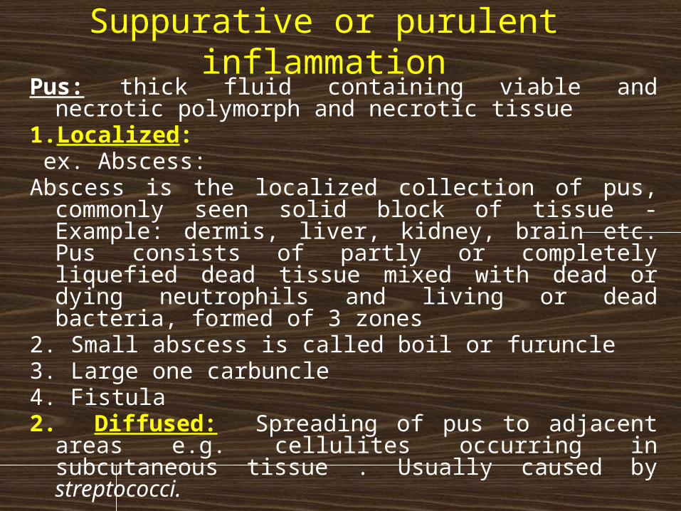
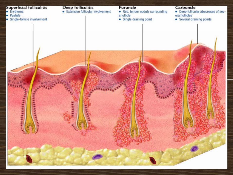
Suppurative or purulent inflammationPus: thick fluid containing viable and necrotic polymorph and
necrotic tissue 1. Localized: ex. Abscess: Abscess is the localized collection of pus, commonly seen solid
block of tissue - Example: dermis, liver, kidney, brain etc. Pus consists of partly or completely liquefied dead tissue mixed with dead or dying neutrophils and living or dead bacteria, formed of 3 zones
2. Small abscess is called boil or furuncle 3. Large one carbuncle4. Fistula2. Diffused: Spreading of pus to adjacent areas e.g. cellulites
occurring in subcutaneous tissue . Usually caused by streptococci.
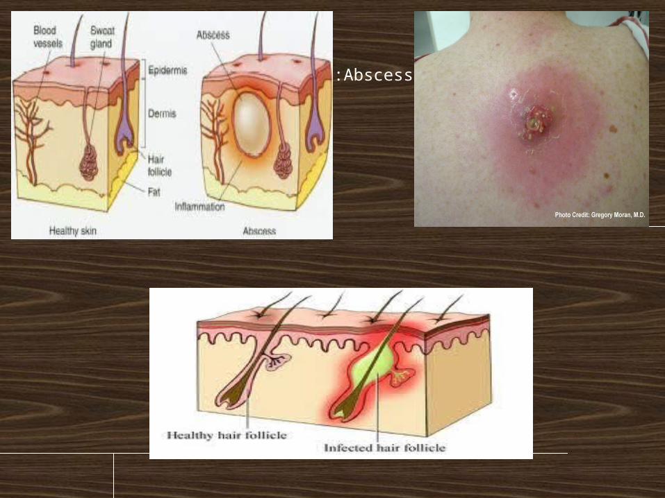
Abscess:
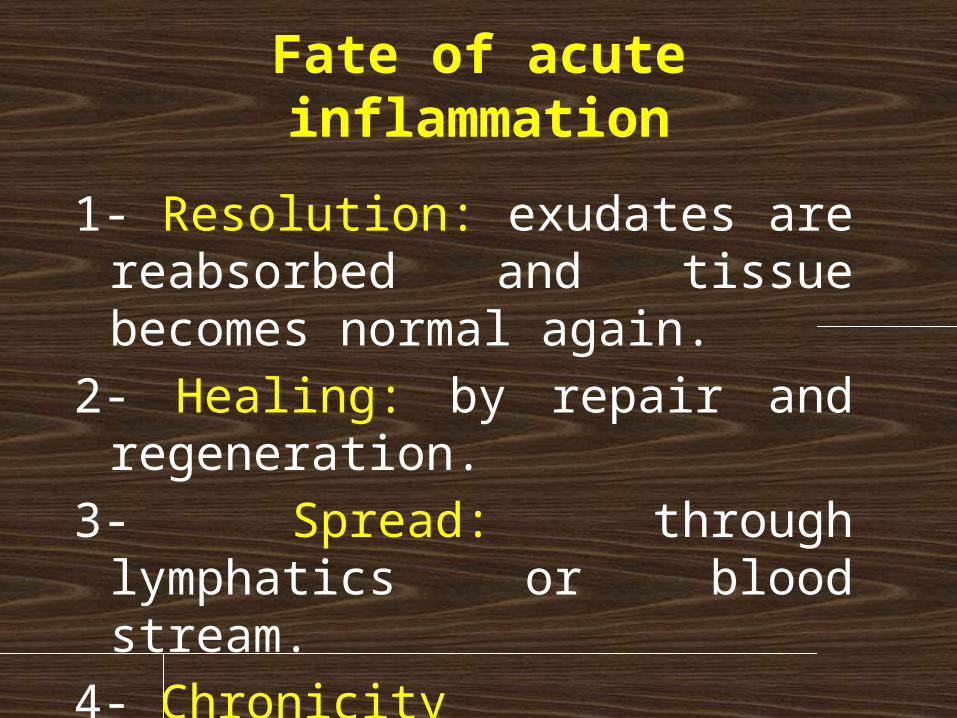
Fate of acute inflammation
1- Resolution: exudates are reabsorbed and tissue becomes normal again.
2- Healing: by repair and regeneration.
3- Spread: through lymphatics or blood stream.
4- Chronicity
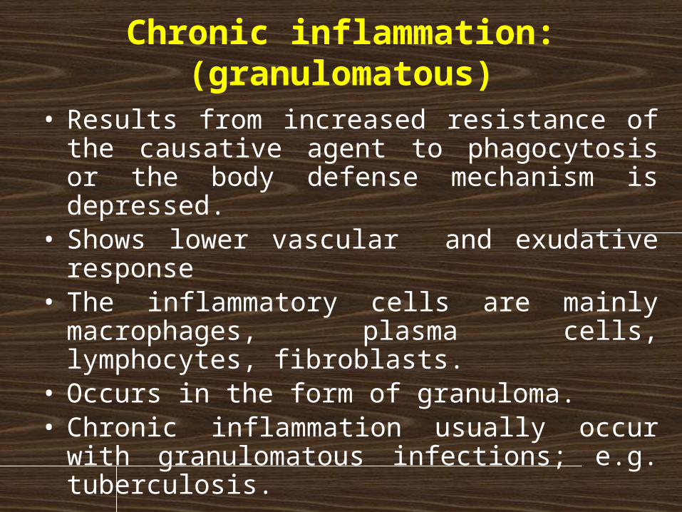
Chronic inflammation: (granulomatous)
• Results from increased resistance of the causative agent to phagocytosis or the body defense mechanism is depressed.
• Shows lower vascular and exudative response • The inflammatory cells are mainly macrophages,
plasma cells, lymphocytes, fibroblasts.• Occurs in the form of granuloma.• Chronic inflammation usually occur with
granulomatous infections; e.g. tuberculosis.
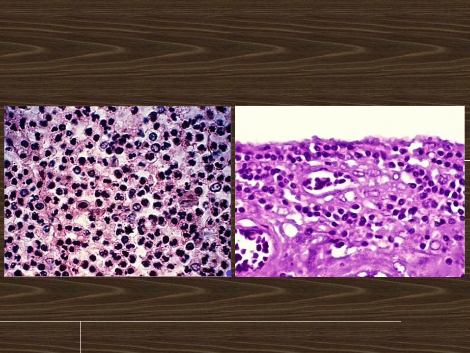
How to differentiate acute vs. chronic inflammation in sections
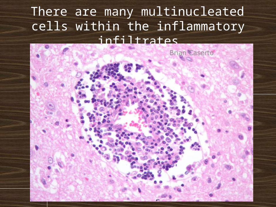
There are many multinucleated cells within the inflammatory infiltrates
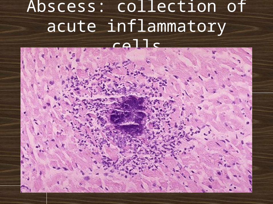
Abscess: collection of acute inflammatory cells
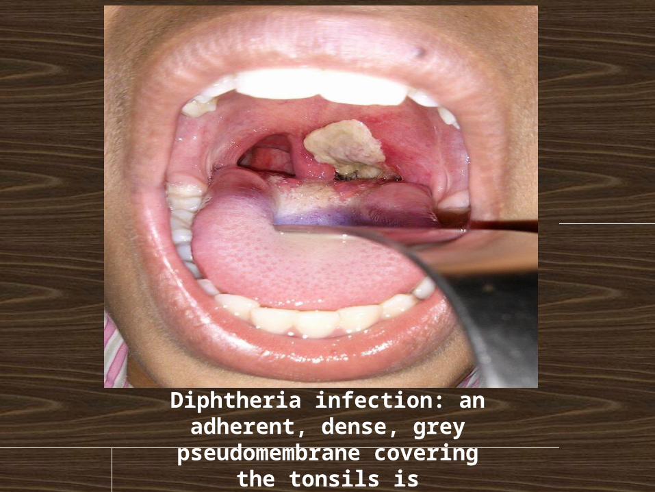
Diphtheria infection: an adherent, dense, grey
pseudomembrane covering the tonsils is classically seen
in diphtheria