pathology of immune reactivity. allergy . professor yu.i. bondarenko
DESCRIPTION
PATHOLOGY OF IMMUNE REACTIVITY. ALLERGY . Professor Yu.I. Bondarenko. Allergy is an immune response, that is accompanied damage of own tissues . - PowerPoint PPT PresentationTRANSCRIPT

PATHOLOGY OF IMMUNE REACTIVITY. ALLERGY.
Professor Yu.I. Bondarenko

• Allergy is an immune response, that is accompanied damage of own tissues.
• Allergic diseases are widely spread among people. It is considered that they occupy about 10 % of earth population. In different countries these figures change from 1 to 50 % and more.
• The cause of allergic disease is an allergen.
• Allergen – is the substance that causes development of an allergic response.
• Allergens have all properties of antigen (macromolecularity, mainly protein nature, foreign for particular organism).
• However allergic reactions can be caused by substances not only antigen nature, but also substances, not possessing these properties. To this group belong many officinal preparations, bacterial products, polysaccharides, simple chemical substances (bromine, iodine, chrome, nickel). These substances are called haptens. They become antigens (allergens) only after binding with tissues proteins.

Classification of allergens
• Exogenous allergens
• Endogenous allergens (autoallergens).
• Exogenous allergens penetrate the organism from outside
• Endoallergens are formed in the organism

Classification of allergens
a) uninfectious allergens:
Home dust Epidermal Pollen Food Industrial Officinal
b) infectious allergens:
Bacterial Fungous Viral

Food allergens
•

Domestic allergens. Main role among them domestic dust plays, which includes particles, bed-clothes, furniture, bacteria.
Epidermal allergens. To this group refer: scurf, wool, birds, fur, fish, scales. Professional sensitization by epidermal allergen is observed in sheepmen, horsemen, poultry farms workers, hairdressers.
Officinal allergens. Any officinal preparation with a little exception causes the development of officinal allergy. Medicines or their metabolites are, as usual, haptens. In case of sensitization of the organism to one preparation, allergic reactions to other medicines, having alike chemical structure can arise.

Pollen allergens. Allergic diseases are caused by shallow plants, pollen. It is called pollinosis. The diverse types of pollen can have the general allergens, therefore in people, sensitive to one type of pollen, a reaction on its other kinds is possible.
Food allergens. Many food products can be by allergens. It is usually fish, wheat, beans, tomatoes, milk, eggs. Chemical substances added to food products (dye-stuffs, antioxidants, aromatic and other substances) may also be allergens.
Industrial allergens. The industrial allergens for the most are haptens. In each industrial production a particular admission of chemical matters is used. These are: resin, glue and covering materials, plastics, dye-stuffs, metals and their salts, wood products, latex, perfumer substances, washing means, synthetic cloths and others.
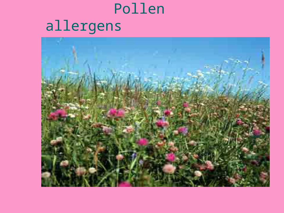
Pollen allergens

Domestic allergens

Infectious allergens Allergens of infectious origin. All the different causative agents of infectious
diseases and products of their life activity cause the development of allergic processes.
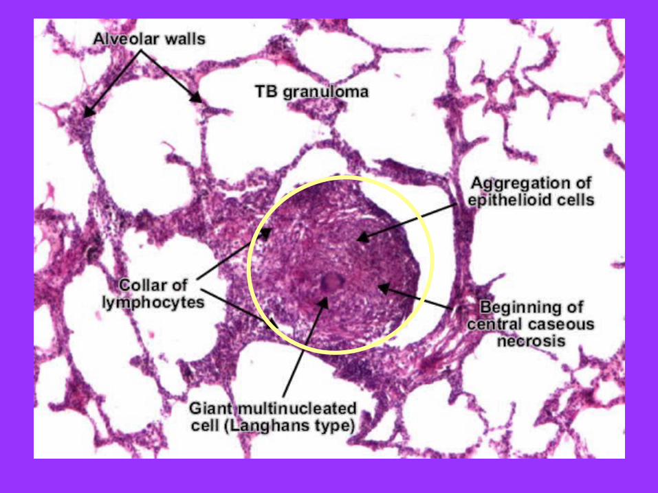
Those infectious diseases, in pathogenesis of which allergy plays leading role, were named infectious allergic. These are all the chronic infections (tuberculosis, lepra, brucellosis, syphilis, rheumatism, chronic candidosis etc.).
The widespread allergens are the fungi. Many nonpathogenic fungi while entering the organism cause sensitization and development of diverse allergic diseases (bronchial asthma).
Such fungi are contained in atmospheric air, dwellings, domestic dust, food products. With biotechnological development possibility of sensitization on enterprises on production of stern squire, vitamins, antibiotics, enzymes arises.

Classification of endogenous allergens
Natural Ecquired(brain,eye,sexuel 1.Infectious and thyroid glands) a) simplex b) complex 2.Uninfectious

Pathogenesis of allergy Pathogenesis of allergy reactionsreactions
Classification of allergic reactions by R.A.Cooke • Allergy of immediate type • Allergy of delayed-type or
hypersensitization of delayed-type
Characteristic of allergic typesCharacteristic of allergic types• The time of appearing of reaction after
contact with allergen was placed in the base of classification.
• The reactions of immediate type develop during 15-20 minutes, delayed-type – after 1-2 days.
• However it does not envelop all the variety of allergy displays. For example, some reactions develop over 4-6 or 12-18 hours.
• Therefore the different immunological mechanisms of their development was put in base of the new classification were based on pathogenic principle.
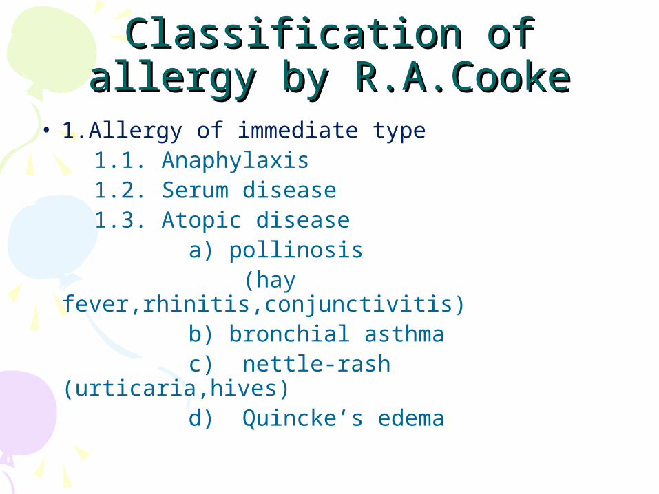
Classification of allergy by Classification of allergy by R.A.CookeR.A.Cooke
• 1.Allergy of immediate type 1.1. Anaphylaxis 1.2. Serum disease 1.3. Atopic disease a) pollinosis (hay fever,rhinitis,conjunctivitis) b) bronchial asthma c) nettle-rash (urticaria,hives) d) Quincke’s edema


Atopic RhinitesAtopic Rhinites
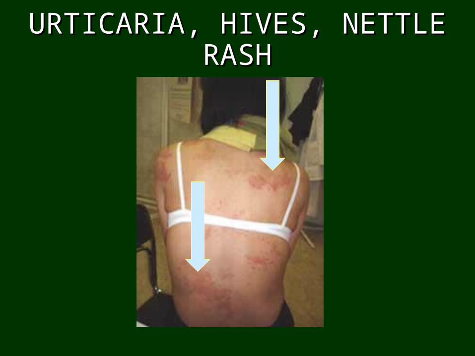
URTICARIAURTICARIA,, HIVES HIVES,, NETTLE NETTLE RASHRASH

Classification of allergy by Classification of allergy by R.A.CookeR.A.Cooke
• 2. Allergy reactions of delayed-type 2.1. Contact dermatosis 2.2. Infectious allergy 2.3. Autoallergy 2.4. Reaction of graft rejection

Classification of allergy by P.Gell, R.Coombs
• Anaphylaxic
• Cytotoxic
• Immune-complex
• Delayed hypersensitivity
• Stimulating
It is based on pathogenic principle. The peculiarities of immune mechanisms lay in its base.
Allergy development
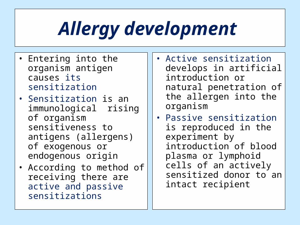
• Entering into the organism antigen causes its sensitization
• Sensitization is an immunological rising of organism sensitiveness to antigens (allergens) of exogenous or endogenous origin
• According to method of receiving there are active and passive sensitizations
• Active sensitization develops in artificial introduction or natural penetration of the allergen into the organism
• Passive sensitization is reproduced in the experiment by introduction of blood plasma or lymphoid cells of an actively sensitized donor to an intact recipient

Stages in development of allergy
• 1. Immunological stage. It covers all the changes in immune system after the penetration of an allergen into the organism, formation of antibodies or sensitized lymphocytes and their binding with the repeatedly entering allergen.
• 2. Pathochemical stage. Its sense is in formation of biological active substances. The stimulus to their formation is the binding of allergen to antibodies or sensitized lymphocytes at the end of immunological stage.
• 3. Pathophysiological stage. It is described by pathogenic action of formed mediators onto cells, organs and tissues of the organism with clinical display.

• Immunological mechanisms lie in base of allergic processes development.
• Central cell of immune system is a lymphocyte. • Lymphocytes are heterogenic according to
their functions, markers, receptors.• They develop from a stem cell. After that a lymphoid stem cell is formed, from which T-
and B-lymphocytes develop. • The T-lymphocytes acquire the specific antigen
receptors, by means of which they identify an antigen and other markers.
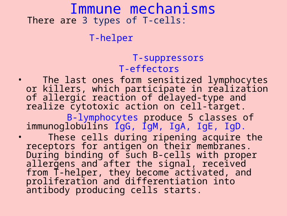
Immune mechanismsThere are 3 types of T-cells:
T-helper T-suppressors
T-effectors • The last ones form sensitized lymphocytes or
killers, which participate in realization of allergic reaction of delayed-type and realize cytotoxic action on cell-target.
B-lymphocytes produce 5 classes of immunoglobulins IgG, IgM, IgA, IgE, IgD.
• These cells during ripening acquire the receptors for antigen on their membranes. During binding of such B-cells with proper allergens and after the signal, received from T-helper, they become activated, and proliferation and differentiation into antibody producing cells starts.

Anaphilactic allergy
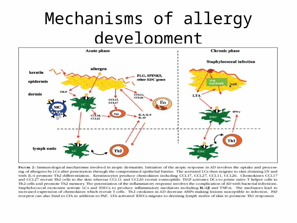
Mechanisms of allergy development

Immune mechanisms• The correlation between two groups of
subpopulations of T-helpers (Th-1 and Th-2) plays an important role in the development of immune reaction.
• They are both formed of Th-0 and differ form each other by the set of secreted lymphokines and quantity of Fc-receptors for immune globulins on their surface.
• On Th-2 there are many receptors for immune globulins A, M, E, and on Th-1 there are a few of them or they are absent.

Mechanisms of allergy development

Immune mechanisms Activation of Th-1 increases formation of IL-2, it
stimulates the secretion of immune globulins A, M and G by B-cells and switch on cellular mechanism of immunity.
• Activation of Th-2 leads though IL-4 to changing of synthesis of IgE by B-cells to proliferation of fat cells and through IL-5 to increase and proliferation of eosinophiles.
• There are antagonistic relationship between these two ways.
• The choice of way of activation depends on character of allergen.
• Besides, that the form of allergen, conditions on introduction into organism and its quantity play role.

Anaphylactic type of allergy
Anaphylactic type includes the next atopic diseases:
Atopic bronchial asthma Pollinosis Atopic dermatitis Nettle-rash (urticaria, havis) Food and officinal allergy

Immunological stage• IgE and IgG4 are formed as an answer to penetrated
allergen into the organism. • They get fixed on the mast cells and basophiles of
blood. These cells have on their surface Fc receptors for immune globulin. The state of sensitization of the organism appears.
• If the same allergen again gets into the organism or it still stays in the organism after the first penetration, binding of antigen with IgE-antibodies occurs.
• The same thing is observed with IgG4. They bind with their receptors on basophiles, macrophages, eosinophiles, thrombocytes.
• Depending on the quantity of IgE-antibodies connected to antigen, quantity of antigen can observe either inhibition of the cell activity or its activation and transfer to the pathochemical stage.

Cellular mechanisms of allergy
•

Degranulation of mast cell
Pathochemical stage
• Activation of the mast cells and basophile leads to releasing of different mediators. The process of secretion of mediators need energy, that’s why blocking of energy-formation blocks also releasing of mediators.
• A certain role in this process play cAMP and cGMP. Secretion of one of the main mediators – histamine depends on their correlation.
• Many different mediators have been excluded from the mast cells and basophiles. Some mediators are in the cell in ready form and easily are secreted (histamine, serotonin, eosinophiles chemotaxic factors).
• Some mediators are formed after stimulation of the cell (leukotriens, thrombocyte activating factors). This mediators act on vessels and target-cells, including in the development of allergic reaction eosinophiles, thrombocytes and other cells.
• As a result eosinophiles, neutrophiles, which start also to release mediators – phospholipase D, histaminase, leukotriens and others.

1. Histamin 2. Heparin 3. Serotonin 4. Eosinophiles chemotaxis factor 5. Platelet-activating factor 6. Prostaglandins (tromboxan, prostacyclin) 7. Leukotriens 8. Superoxyde anion-radical 9. Bradykinin 10. Components of complement (С3а, С5а) 11. Lyzosomal enzymes 12. Polypeptide Р
Меdiators of allergy

Histamine• Histamine is localized in ready form in granules of the
mast cells and basophile leucocytes. • In the blood of healthy people histamine almost totally
stays in basophile leucocytes. • Histamine acts on the tissues cells through the receptors
of two types – H1 and H2. • Their correlation and spreading on the cells of different
cells is different. Stimulation of H1 promotes to contraction of smooth muscles, endothelial cells and postcapillary part of microcirculation. This leads to increasing of permeability of vessels, development of edema and inflammation.
• Stimulation of H2 causes the opposite effects.
Histamine effects
• Releasing of histamine from basophile leucocytes and from the lungs is diminished through them, the function of the lymphocytes modulates, formation of migration inhibitory factor (MIF) by T-lymphocytes gets oppressed, releasing of lysosome enzymes by neutrophile leucocytes diminishes as well.
• The increasing of quantity of histamine in blood is observed in the intensive stage of bronchial asthma, nettle-rash, officinal allergy.
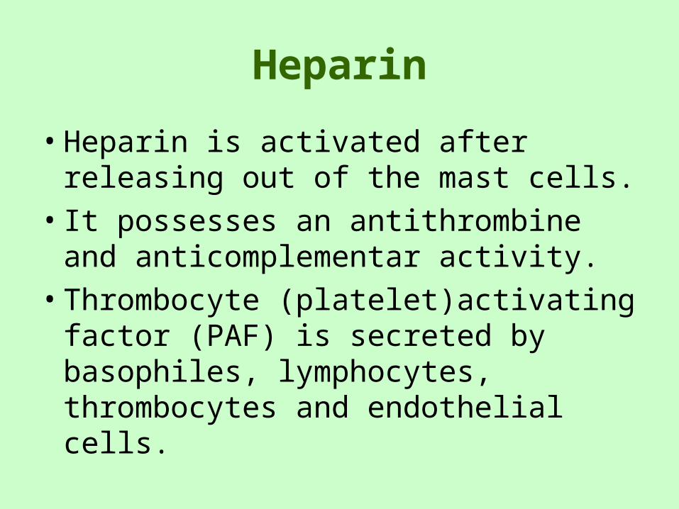
Heparin
• Heparin is activated after releasing out of the mast cells.
• It possesses an antithrombine and anticomplementar activity.
• Thrombocyte (platelet)activating factor (PAF) is secreted by basophiles, lymphocytes, thrombocytes and endothelial cells.
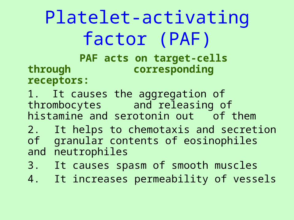
Platelet-activating factor (PAF)
PAF acts on target-cells through corresponding receptors:
1. It causes the aggregation of thrombocytes and releasing of histamine and serotonin out of them 2. It helps to chemotaxis and secretion of granular contents of eosinophiles and neutrophiles3. It causes spasm of smooth muscles4. It increases permeability of vessels
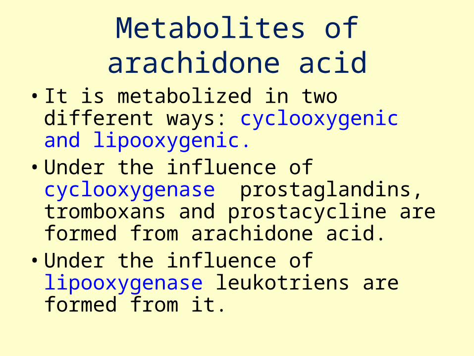
Metabolites of arachidone acid
• It is metabolized in two different ways: cyclooxygenic and lipooxygenic.
• Under the influence of cyclooxygenase prostaglandins, tromboxans and prostacycline are formed from arachidone acid.
• Under the influence of lipooxygenase leukotriens are formed from it.

Prostaglandins and leukotriens• Prostaglandins of group F possess the
ability to cause contraction of smooth muscles, including bronchi, and prostaglandins of group E provide the relaxing action.
• Leukotriens cause the spasm of smooth muscles, increase secretion of mucous, decrease coronary blood flow and power of heart contractions, increase chemotaxis of polymorphic-nuclear leukocytes, lead to development of prolonged bronchial spasm.
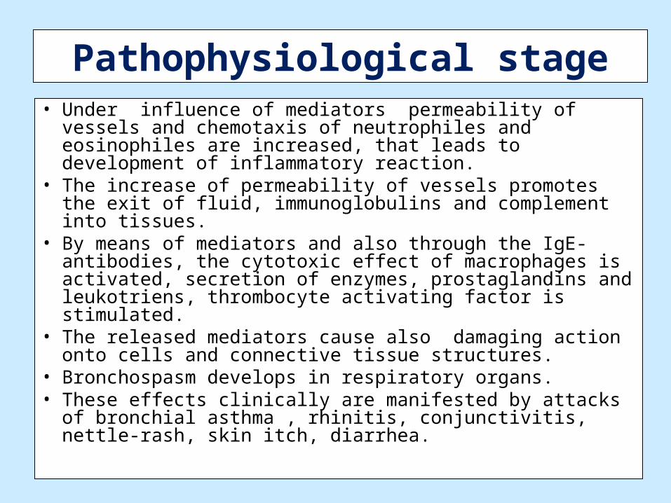
Pathophysiological stage• Under influence of mediators permeability of vessels
and chemotaxis of neutrophiles and eosinophiles are increased, that leads to development of inflammatory reaction.
• The increase of permeability of vessels promotes the exit of fluid, immunoglobulins and complement into tissues.
• By means of mediators and also through the IgE-antibodies, the cytotoxic effect of macrophages is activated, secretion of enzymes, prostaglandins and leukotriens, thrombocyte activating factor is stimulated.
• The released mediators cause also damaging action onto cells and connective tissue structures.
• Bronchospasm develops in respiratory organs. • These effects clinically are manifested by attacks of
bronchial asthma , rhinitis, conjunctivitis, nettle-rash, skin itch, diarrhea.

Medicated allergy
Anaphylactic shock
• Anaphylactic shock develops as severe complication.
• Spasm of smooth muscles of internal organs with clinical manifestation of bronchospasm (cough, expiratory dyspnea), spasm of gastro-intestinal tract muscles (spastic pain in the whole abdomen, nausea, vomiting, diarrhea), spasm of uterus in women (pain below abdomen) are observed.
• Spastic phenomena are worsened by edemas of mucous membranes of internal organs, during the edema of larynx the picture of asphyxia may develop.
• The arterial pressure sharply is decreased, the heart insufficiency, ischemia of brain, paralysis develop danger for the life of the patient appears.

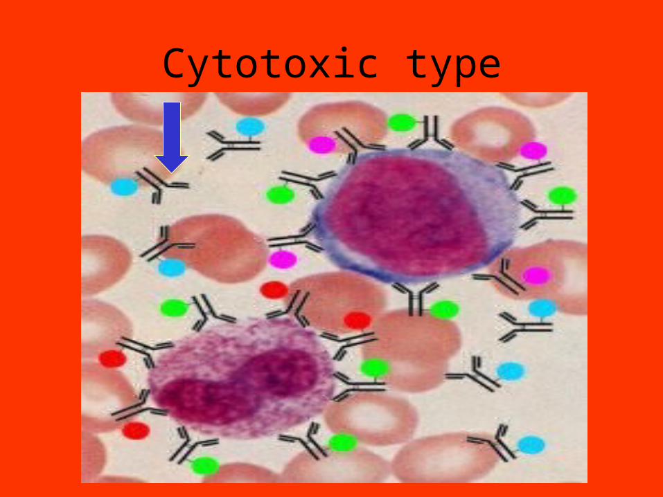
Cytotoxic type of allergic reactions
• Immunological stage. It is called cytotoxic because the antibodies that developed to antigen of the cell bind to cells and cause their damage or even lysis (cytolytic action). For swithing of this mechanism cells have to acquire autoallergen properties. Than the formation of autoantibodies is started. To development of this process further chemical substances, usually medicines, viruses, microbes. They may change the antigen structure of cellular membranes. The formed autoantibodies belong to IgG and IgM. They connect to corresponding antigens of the cells by means Fab-fragments.

Cytotoxic type

• Pathochemical stage. The main mediator of cytotoxicity is the activated enzymes of complement. Phagocytes release some lyzosomal enzymes and generate superoxide anion-radical.
• Pathophysiological stage. The damage of the cell with antigen properties may be caused by three factors:
• a) due to activation of complement, the components of which damage the cell membrane;
• b) due to activation of phagocytosis of the cells with fixed antibodies;
• c) due to activation of T-lymphocytes, natural killers, K-lymphocytes.
Immune complex type
• Immunological stage. Many exogenous and endogenous antigens participate in formation of immune complexes. Among them officinal preparations (penicillin, sulfanilamides,), antitoxic vaccines, allogen gamma-globulins, food product (milk, egg, white), inhalation allergen (home dust, fungi). In case of penetration of soluble antigen into the organism IgG and IgM antibodies are formed. These antibodies can cause the formation of precipitate and connection to antigen. Immune complex can be formed in tissues or in blood flow.
• Patochemical stage. Under the influence of immune complexes the next mediators are formed: fragments C3a, C5a, C4a of the complement, lyzosomal enzymes of phagocytes, kinines, superoxyde anion-radical.
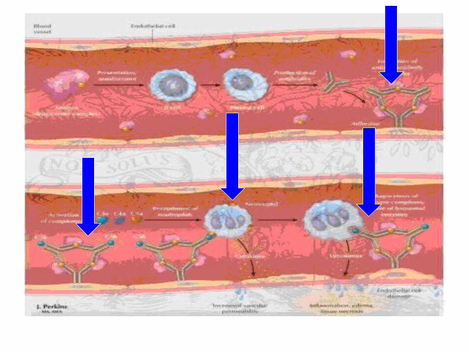
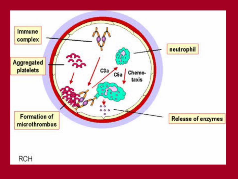

Immune complex type• Pathophysiological stage. Usually immune
complexes are placed on vessels of cannalicular apparatus of kidneys, inflammation with alteration, exudation and proliferation (glomerulonephritis) develops, in case if the complexes are placed in the lungs alveolitis appears, in skin – dermatitis. The inflammation may lead to formation of ulcers, hemorrhages, thrombosis is possible in the vessels. This type of allergic reactions is main in development of serum sick, some cases of officinal and food allergy, some autoallergic diseases (rheumatoid arthritis, systemic red lupus erythematosus). In case of activation of complement anaphylactic shock, bronchial asthma may develop.

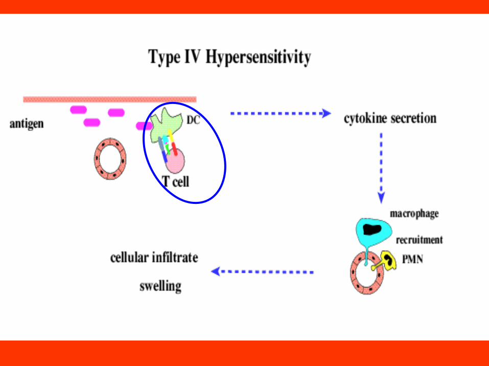
Allergic reactions of delayed type
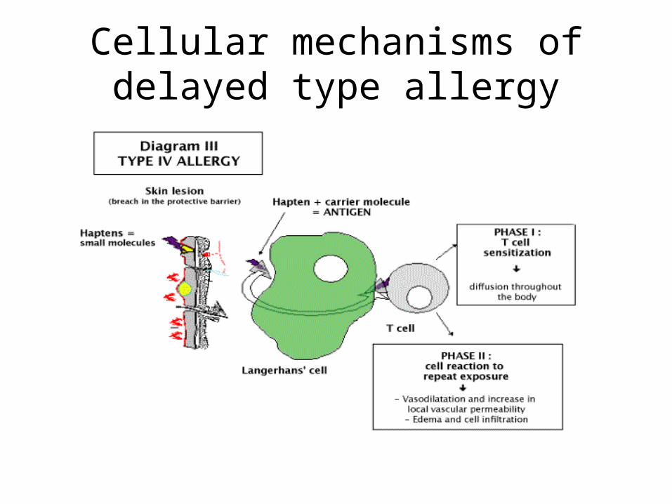
• Immunological stage. The cellular mechanism of immunity is usually activated in case of intracellular localization of the antigen (mycobacterium, brucella, histoplasma etc.) or when cells are antigen themselves. It may be microbes, fungi and their spores, which get into the organism from the outside. The cells of own tissues also may acquire the auto allergen properties. This mechanism may take place as a response to formation of complex allergens, in case of including haptens into proteins, for example, in case of contact dermatitis, which appears during the contact of the skin with different medicinal, industrious and other allergens.
Immunological mechanisms• The foreign antigen is phagocyted by
macrophages and get to T-helpers. At the same time macrophages secrete IL-1, which stimulates T-helpers. The latest excrete the growth factor pro-T-lymphocytes – IL-2, which activates and supports proliferation of T-cells. This process leads to formation of sensitized lymphocytes.They belong toT-lymphocytes and in the cell membrane they have receptors of the antibody type, which are able to connect with the antigen. In case of repeated penetration of the allergen into the organism it binds with the sensitized lymphocytes.
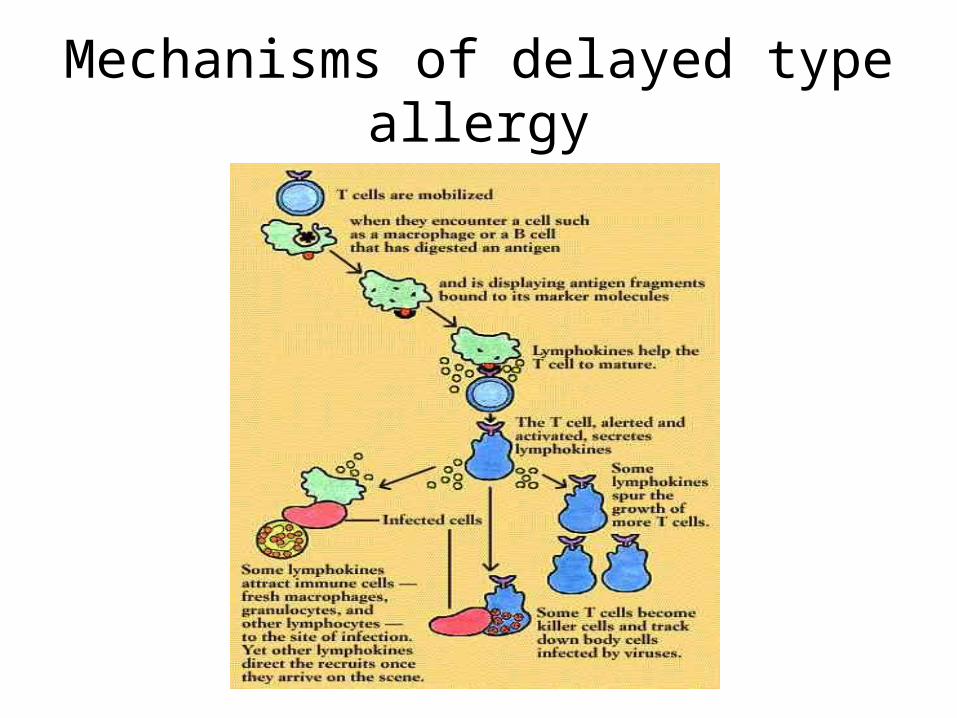
Mechanisms of delayed type allergy
Cellular mechanisms of delayed type allergy

Pathochemical stage
• It leads to morphological, biochemical and functional change in lymphocytes.
• They are presented by blast transformation and proliferation, increasing of synthesis of DNA, RNA and proteins and secretion of different mediators, which are called lymphokines. By means of lymphokines (MIF, interleukines, chemotaxic factors, factor of transfer) mobilization of different cells (macrophages, polymorph-nuclear), increasing of chemotaxic activity occur.
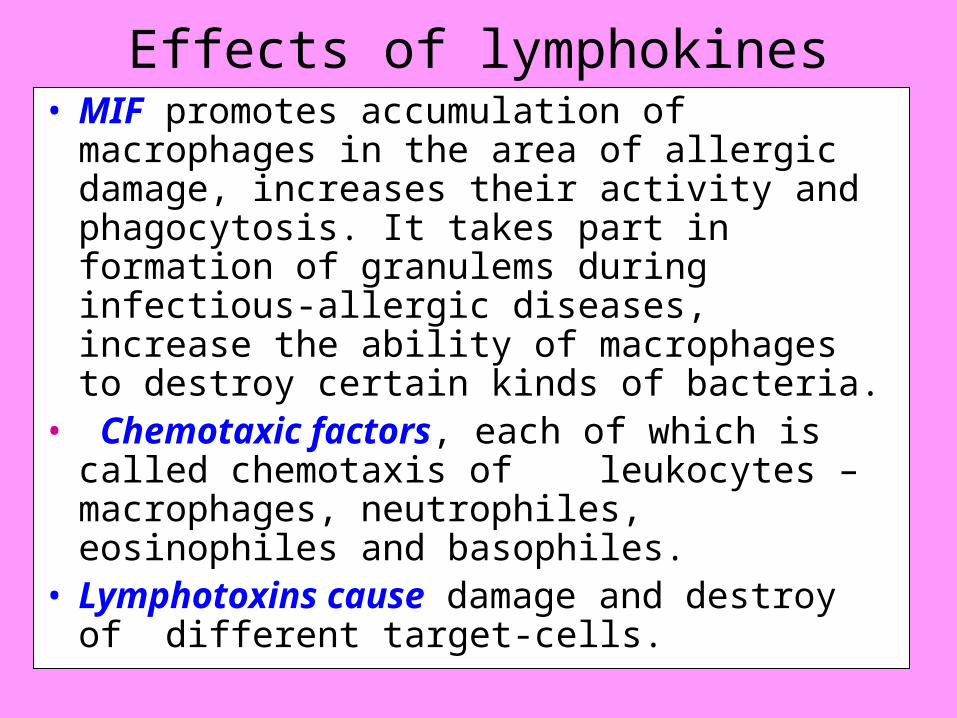
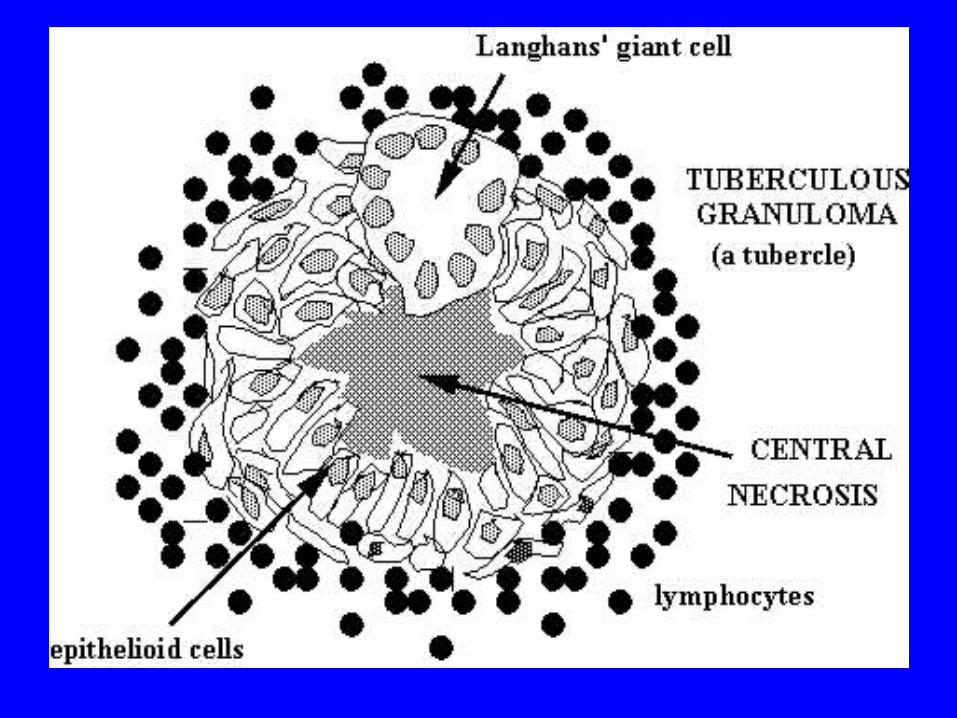
Effects of lymphokines• MIF promotes accumulation of macrophages in
the area of allergic damage, increases their activity and phagocytosis. It takes part in formation of granulems during infectious-allergic diseases, increase the ability of macrophages to destroy certain kinds of bacteria.
• Chemotaxic factors, each of which is called chemotaxis of leukocytes – macrophages, neutrophiles, eosinophiles and basophiles.
• Lymphotoxins cause damage and destroy of different target-cells.
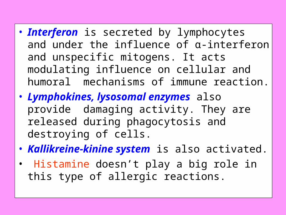
• Interferon is secreted by lymphocytes and under the influence of α-interferon and unspecific mitogens. It acts modulating influence on cellular and humoral mechanisms of immune reaction.
• Lymphokines, lysosomal enzymes also provide damaging activity. They are released during phagocytosis and destroying of cells.
• Kallikreine-kinine system is also activated.• Histamine doesn’t play a big role in this type of
allergic reactions.
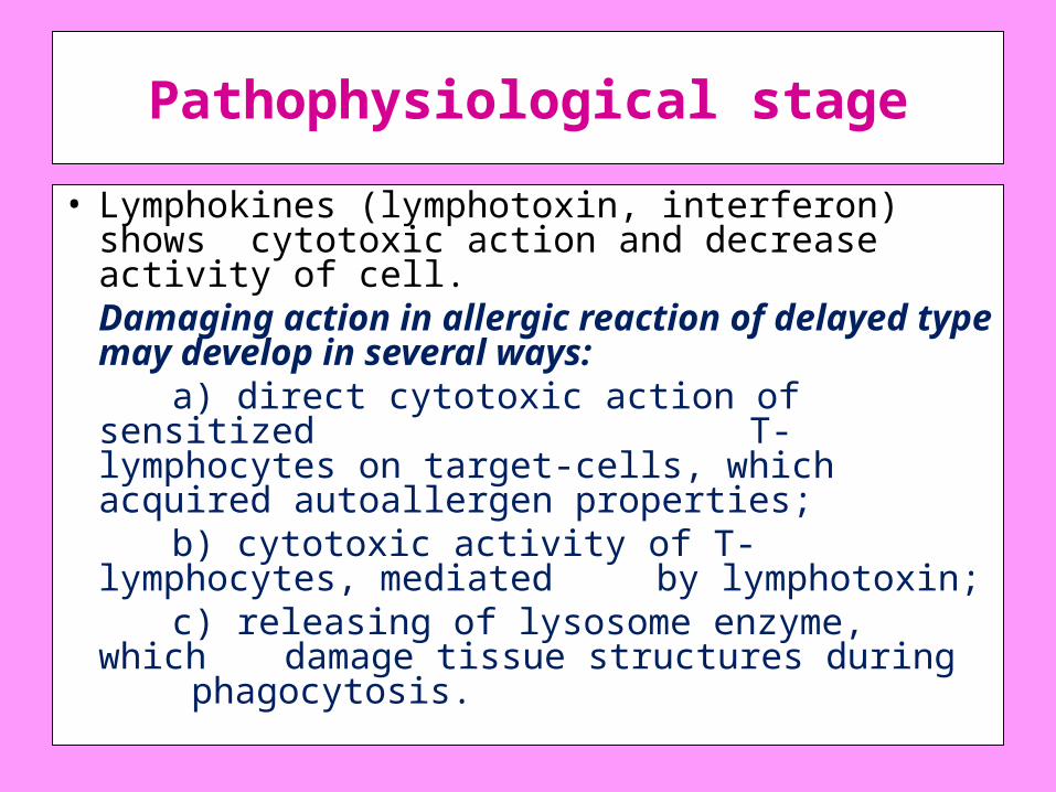
Pathophysiological stage
• Lymphokines (lymphotoxin, interferon) shows cytotoxic action and decrease activity of cell.
Damaging action in allergic reaction of delayed type may develop in several ways:
a) direct cytotoxic action of sensitized T-lymphocytes on target-cells, which acquired autoallergen properties;
b) cytotoxic activity of T-lymphocytes, mediated by lymphotoxin;
c) releasing of lysosome enzyme, which damage tissue structures during phagocytosis.
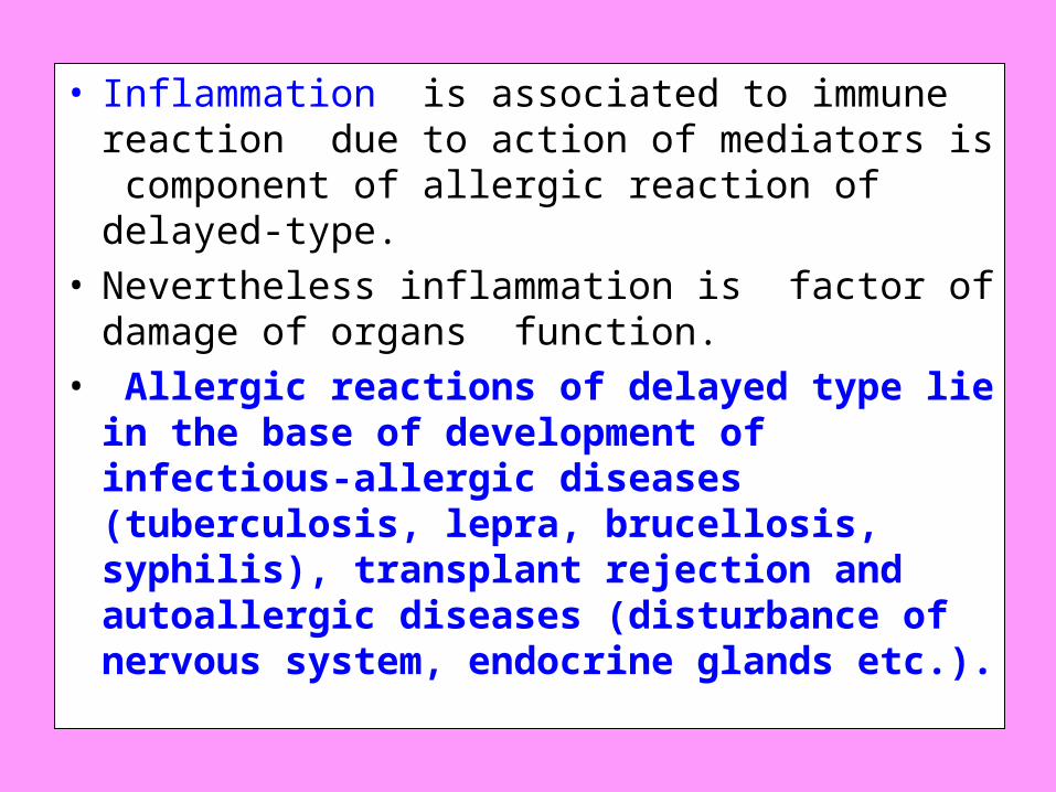
• Inflammation is associated to immune reaction due to action of mediators is component of allergic reaction of delayed-type.
• Nevertheless inflammation is factor of damage of organs function.
• Allergic reactions of delayed type lie in the base of development of infectious-allergic diseases (tuberculosis, lepra, brucellosis, syphilis), transplant rejection and autoallergic diseases (disturbance of nervous system, endocrine glands etc.).


Pseudoallergic reactions
• Pseudoallergy is pathological process, which is clinically similar to allergy but doesn’t have an immune stage of its development. The rest two stages – releasing of mediators (pathochemical) and pathophysiological (stage of clinical manifestations) take place.
• For pseudoallergic reactions are characterized only processes in the development of which the leading role play mediators, which are formed also in pathochemical stage of true allergic reactions.
Pseudoallergy• The reason of pseudoallergy is any substance
that acts directly on effector cells (fat cells, basophiles etc.) or biological fluids and cause releasing of mediators from the cells or production of them in the fluids.
• Practically most of the allergens can lead to development of both allergic and pseudoallergic reactions.
• This depends on nature of the substance, its phase, frequency of introduction into the organism and reactivity of the organism.
• Pseudoallergic reactions usually occur in officinal and food intolerance.
• Many remedies more usually lead to development of pseudoallergy than true allergy.
Pseudoallergy• Clinical picture of pseudoallergic diseases is
similar allergic diseases. • Development of such pathological processes as
increase of permeability of vessels, edema, inflammation , spasm of smooth muscles, destruction of blood cells lie in the base of this clinical picture.
• These processes may be local, organic and systemic.
• They are presented by rhinitis, nettle-rash, Quincke’s edema, periodical headaches, disturbance of gastro-intestinal tract, bronchial asthma, vaccine disease, anaphylactic shock and also damage of certain organs.

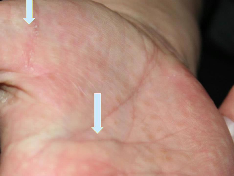
Skin reaction onto allergen

Prevention of allergy. Hyposensitization.
• Prophylaxis of an allergic disease depends on its character and group of the allergens. It directs to preventing of penetration of given allergen into the organism and influence of different irritating factors on the one. If sensitization has occurred and allergic diseases has started the next measures are appropriated.
• 1. Suppression of antibodies and sensitized lymphocytes production by means of immune depressants, ionizing radiation, cytostatics, specific lymphocyte vaccines and monoclonal antibodies.
• 2. Specific desensitization by Bezredka. Desensitization is provided by little doses of the antigen, which do not cause severe reactions. The doses are introduced repeatedly after certain intervals of time, during which produced mediators get inactivated in the organism. The main dose of the antigen is introduced after antibodies binding. This method is effective in introduction of foreign medical vaccines.
• 3. Inactivation of biological active substances. For this purpose antihistamine preparations, inhibitors of proteolytic enzymes etc.
• 4. Protection of the cells from the influence of biological active substance and also normalizing of functional disorders in organs and systems (narcotic, spasmolytic substances, receptor blockers etc.).
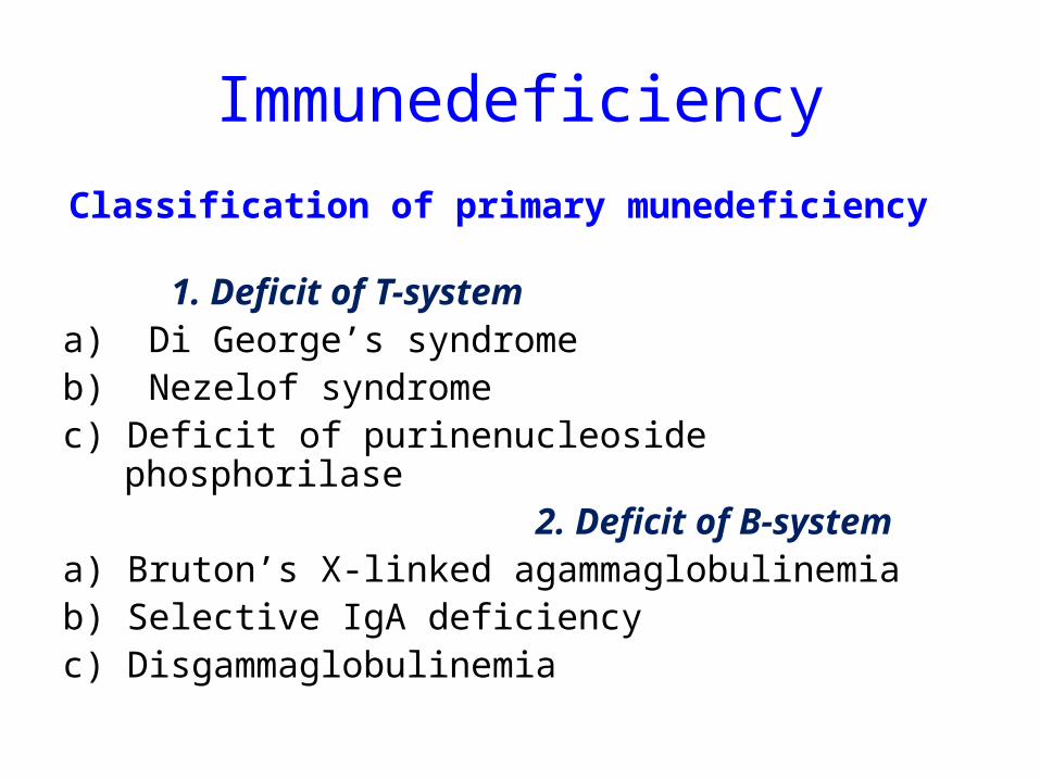
Immunedeficiency
Classification of primary munedeficiency 1. Deficit of Т-system
а) Di George’s syndromeb) Nezelof syndrome c) Deficit of purinenucleoside phosphorilase 2. Deficit of B-systemа) Bruton’s X-linked agammaglobulinemiab) Selective IgA deficiency c) Disgammaglobulinemia
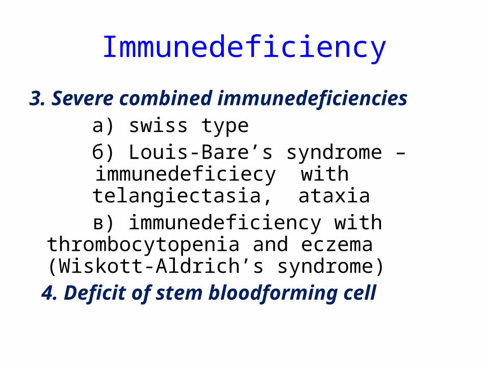
Immunedeficiency
3. Severe combined immunedeficiencies а) swiss type б) Louis-Bare’s syndrome –
immunedeficiecy with telangiectasia, ataxia
в) immunedeficiency with thrombocytopenia and eczema (Wiskott-Aldrich’s syndrome)
4. Deficit of stem bloodforming cell

TO YOU !!!THANKS A LOT !!!