pathologies guide and their orthotic indications · calcaneal spur hallux valgus foot drop club...
TRANSCRIPT

Polígono Ind. Nº 1Calle F, Nº 1528938 - Móstoles, Madrid, SPAINTel.: +34 91 334 25 32 +34 91 334 25 89
Fax: +34 91 334 25 [email protected]
Pathologies Guide and their Orthotic Indications
Path
olog
ies
Gui
de a
nd th
eir O
rtho
tic In
dica
tions
© 2014 PRIM, S.A. CATAPATO IN 01/14

INTERIOR PORTADA PAGINA 1

Since its founding in 1870 by the orthopaedic surgeon and rehabilitation specialist, Pedro Prim Fernández, PRIM’s mission has been to provide a comprehensive service to SPANISH HEALTHCARE. Initially, in the field of orthopaedics, through diagnosis, treatment and the rehabilitation of patients. Today, Prim S.A. is a Spanish privately-owned corporate group that has been listed on the Madrid Stock Exchange since 1985 and consists of three divisions:
PRIM HOSPITALESPRIM ORTOPEDIAPRIM SPA
The PRIM GROUP also includes other companies:
PRIM FISIOTERAPIA Y REHABILITACIÓN (Enraf Nonius Ibérica S.A.)PRIM CLÍNICAS ORTOPÉDICASPRIM FARMA (Luga Suministros Médicos S.L.)
PRIM ORTOPEDIA is dedicated to the DEVELOPMENT, PRODUCTION AND DISTRIBUTION of orthopaedic products and technical aids through specialist orthopaedic shops, pharmacies and cooperatives. Products include a wide range of orthoses, such as ankle, knee and wrist supports, belts, hyperextension braces, etc., as well as prostheses and prosthetic components, technical aids, incontinence products, medical equipment, etc.
The commercialisation of its own-manufactured products is carried out in Spain and abroad under registered trademarks as well known as PRIM, ORTHOPRIM, CAMP, BEBAX, SWASH, etc., exclusively manufactured by the company.
As a result of the constant technical evolution of orthopaedic products, either for conservative treatment or pre- or postoperative use, resulting from innovative designs, the availability of new materials or the latest functional concepts, as well as the existence of a wide variety of products with similar designs and indications, we have decided that it would be highly useful to provide doctors, nurses, physiotherapists, orthopaedic technicians, podiatrists and, in general, all healthcare professionals involved in the field of technical orthopaedics with a GUIDE TO MAJOR DISORDERS and their orthotic indications, which is simple and easy to use, and can help professionals to understand the biomechanical functions of orthoses in order to facilitate rapid and accurate selection of the most appropriate product, addressing issues such as the disorder to be treated, morphology, activity or other aspects relating to the patient’s needs.
With the aim of being your technological and strategic partner in the exercise of our profession, I would also like to mention that we have our own network of highly-qualified professionals with extensive knowledge and professional experience.
We hope that this guide will be of great use and technical assistance.Prim, S.A.
PREFACE
AUTHORS:Technical management and text: Vicente C. Gomar SanchoIllustrations: José Hernández GarcíaDesign and layout: J.H. Estudio Gráfico
Legal deposit:Printing:
It is prohibited, unless otherwise provided by law, to reproduce, distribute, publicly communicate or modify this work without the permission of the owners of the intellectual property. Infringement of these intellectual property rights may constitute a criminal offence (Art. 270 et seq. of the Spanish Penal Code).

CONTENTS
NECK disorders
Neck sprain, cervicalgiaSpinal disc herniationDegenerative neck disordersCervical vertebra fracture
SPINE disorders
Back painLow back painScoliosisSpinal disc herniationOsteoporosisVertebral fracture
UPPER LIMB disorders
Shoulder
Brachial palsyShoulder injuryNeck of humerus fractureClavicle fracture
Elbow
Epicondylitis, medial epicondylitisElbow fracture
HandNeurological injuryLigament and tendon injuryCarpal tunnel syndrome and Guyon’s canal syndromeQuervain’s tendinitisArthrosis, rhizarthrosisFracture and dislocation
LOWER LIMB disorders
Congenital dysplasia and dislocation of the hipPostoperative hip dislocationHip joint injury in infantile cerebral palsy
Hip
Thigh/leg
Muscle strain
Knee
Cruciate ligament and meniscus injuryLateral ligament injuryOsgood-Schlatter diseaseChondromalacia patellaePatellar dislocationPostoperative knee
Ankle
Ankle sprainAchilles tendon injuryAnkle fracture
Foot
MetatarsalgiaPlantar fasciitisCalcaneal spurHallux valgusFoot dropClub footFoot fracture
6101418
222630364044
48525660
6468
727680848892
96100104
112116120124128132
108
136140144
148152156160164168172

NECK DISORDERS
6
NECK SPRAIN, CERVICALGIA
CLINICAL
The term cervicalgia indicates pain located in the cervical area of the spine. A significant percentage of the population report having suffered cervicalgia at some point in their life, presenting pain in the posterior and lateral areas. As it is a symptom, it can have different causes with varying degrees of severity.
Postural attitudes maintained over time, poor posture during rest, work and/or sports activities with overload, minor trauma, stress, worry, etc., can cause spasms due to muscle tension. While it is usually a mild disorder without severe consequences, in some cases, it can be associated with disc disorders (discopathy) or spinal disc herniation.
SYMPTOMS
Generalised pain in the cervical area with stiffness in the neck muscles. The pain can radiate downwards, reaching the shoulders and between the shoulder blades, even the arm, and cause unilateral or bilateral headache. The musculature is tense and hard, and can result in postural problems, such as torticollis. Pain at the back of the cranium can be accompanied by weakness in the arms and hands.
NECK SPRAIN, CERVICALGIA 7

8 NECK SPRAIN, CERVICALGIA 9
ORTHOTIC TREATMENT
Rehabilitation and chiropractic techniques applied by rehabilitation specialists or physiotherapists, accompanied by medication (analgesics and muscle relaxants), complement cervical support treatment, which produces heat and, at the same time, releases tension and enables varying degrees of immobilisation. Soft polyurethane foam collars of different densities, height-adjustable rigid collars or two-piece plastezote-foam Philadelphia collars with occipital/mandibular supports provide a wide range of options depending on the severity of the cervicalgia, and are highly recommended for use on short trips or long journeys and during recovery periods of varying duration.
NECK SPRAIN, CERVICALGIA PRODUCTS
NECK DISORDERS
CC19 Straight soft collar
CC20 Contoured soft collar
CC121Contoured semi-rigid collar
E41 Height-adjustable rigid collar

NECK DISORDERS
10
SPINAL DISC HERNIATION
CLINICAL
The tissue that lies between the cervical vertebrae, known as intervertebral discs, consists of a soft gel-like central area with a hard outer covering. These intervertebral discs create joints between the vertebrae to enable mobility. When the outer covering of the disc tears, the soft central tissue can protrude from the opening created, resulting in a herniated disc.
Various factors, such as ageing, in which the intervertebral discs lose flexibility and elasticity, and the surrounding ligaments become fragile and easily torn, facilitate the appearance of a herniated disc, causing compression of the nearby spinal nerves, known as radiculopathy, and myelopathy, in cases where it affects the spinal cord.
SYMPTOMS
The presence of a cervical herniated disc can cause pain in the neck, radiating down to the shoulder and arm, and feelings of numbness or tingling in the arm and hand. The pain is usually dull and constant and difficult to locate, but on occasions, can be acute, burning and well located.
Feelings of numbness, weakness and tingling in the muscles can be indicative of a serious problem.
Other symptoms include muscle stiffness or neck cramps. These most commonly occur between the fourth, fifth and sixth cervical vertebrae.
SPINAL DISC HERNIATION 11
NECK DISORDERS
ORTHOTIC TREATMENT
Taking into account that the cervical vertebra system supports the head, which weighs around 5 kg, representing a significant mechanical demand, unloading and limiting mobility, or even immobilisation of the cervical segment, are the basic treatment objectives, along with rehabilitation and physiotherapy, accompanied by prescribed medication.
Cervical and cervicothoracic collars, with or without occipital/mandibular supports, are, depending on the severity, highly useful in conservative treatment in that they prevent compression of the intervertebral discs, nerve roots and the spinal cord. Especially useful are orthotic devices that enable adjustment of flexion and extension, and also facilitate intervertebral distraction in which separation of the vertebral bodies is sought, ensuring optimal treatment, given that 90% of herniated discs can be resolved with conservative treatment.
12 13
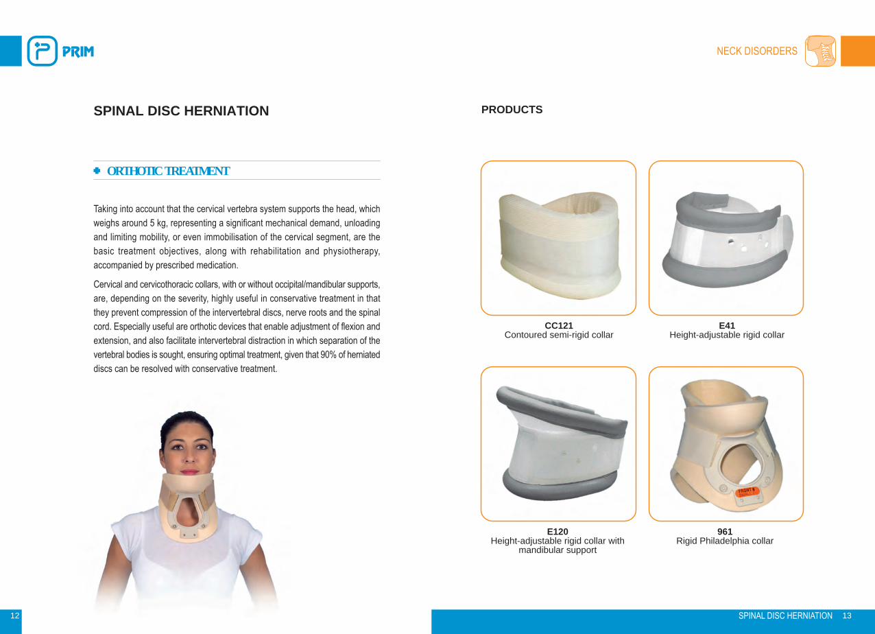
SPINAL DISC HERNIATION PRODUCTS
SPINAL DISC HERNIATION
CC121Contoured semi-rigid collar
E41 Height-adjustable rigid collar
E120Height-adjustable rigid collar with
mandibular support
961 Rigid Philadelphia collar

14
DEGENERATIVE NECK DISORDERS
CLINICAL
Degenerative neck disorders are mainly caused by deterioration of the cervical vertebrae, abnormal wear on the cartilage and bone, degeneration and mineral deposits, resulting in the loss of the physical and chemical properties of the intervertebral discs, leading to cervical osteoarthrosis, arthrosis of the neck or cervical arthrosis.
As a result of chronic degeneration of the cervical spine, including the intervertebral discs and facet joints, cervical spondylosis can occur.
Pressure on the nerve roots can produce progressive neck and shoulder pain and weakness in the arms, and even in the legs.
Abnormal vertebral growths, known as osteophytes, can appear, which, along with herniated discs, can create narrowing of the vertebral foramen, resulting in pinching of one or more nerve roots.
SYMPTOMS
Neck pain with numbness in the shoulders, arms and hands in more advanced cases.
Progressive increase of neck stiffness with loss of balance.
Posterior headache.
Loss of functional mobility of the cervical segment.
DEGENERATIVE NECK DISORDERS 15
NECK DISORDERS

NECK DISORDERS
ORTHOTIC TREATMENT
The objective is to relieve pain and prevent permanent damage to the spinal cord and nerve root. Rehabilitation and physiotherapy are effective in relieving pain, along with the use of a cervical orthosis for immobilising and unloading the head. For this, it is important to select the most appropriate cervical orthosis, depending on the required function and the degree and severity of the cervical segment degeneration.
Surgical decompression of the spinal cord may be necessary in severe cases and complemented postoperatively with stabilisation and distraction by means of an OMI-type cervical orthosis featuring an adjustable occipital/mandibular support and optional Indiana strap at the front.
16 17
DEGENERATIVE NECK DISORDERS PRODUCTS
DEGENERATIVE NECK DISORDERS
CC19 Straight soft collar
CC20 Contoured soft collar
CC121Contoured semi-rigid collar
E41 Height-adjustable rigid collar

18
VERTEBRAL FRACTURE
CLINICAL
The fracturing of one or more vertebrae, caused by sudden violent impacts, such as traffic accidents or severe sport or work injuries, is one of the most serious types of trauma that can affect the neck. Osteoporosis or cancer patients can also sustain fractures due to the weakness of the bone. In the event of injuring the spinal cord, varying degrees of paralysis can occur or even death.
SYMPTOMS
Vertebral fractures can result in severe pain, numbness, tingling in the arms and legs with loss of feeling, even paralysis, depending on the degree of severity and location of the fracture.
NECK DISORDERS
VERTEBRAL FRACTURE 19

20 VERTEBRAL FRACTURE 21
ORTHOTIC TREATMENT
The degree of severity of vertebral fracture determines the most appropriate treatment. Crush fractures can be treated with rigid cervical collars, whose function is to produce extensive immobilisation and distraction of the cervical segment to reduce intervertebral loads over a period of at least 8 weeks. In the case of major fractures of the vertebral bodies, it is necessary to use cervical traction orthoses with removable and adjustable occipital/mandibular supports, such as OMI-type cervical orthoses featuring an Indiana strap at the front, and shoulder and chest supports. In cases in which the spinal cord has been affected by the injury, a halo-type cranial traction orthosis or surgery is necessary for stabilising treatment.
VERTEBRAL FRACTURE PRODUCTS
NECK DISORDERS
961 Rigid Philadelphia collar
717247 Stabiliser for 961
E45 Cervicothoracic OMI orthosis

SPINE DISORDERS
22
BACK PAIN
CLINICAL
Any type of pain that occurs in the dorsal or thoracic area of the spine is called back pain, including that which affects joints with the ribs or the thoracic area, as they are closely linked together.
Back pain can have many causes, as there are numerous structures that are capable of producing pain.
The presence of pain can have numerous causes. It can, for example, be organic, such as infectious spondylitis, caused by an infection in one or several vertebrae; inflammatory, resulting in ankylosing spondylitis; or metabolic, as a consequence of vertebral crushing (osteoporosis).
Other sources such as tumours, trauma, Scheuerman’s disease and static or functional disorders can also be responsible for back pain.
SYMPTOMS
Pain manifests itself differently depending on its origin, ranging from fatigue and a feeling of heaviness to tingling, a sensation of burning and limited movement.
On palpation, muscle tension is evident with paravertebral muscle contractions. The source of the pain needs to be identified to establish whether it is emanating from paraspinal ligaments, facet joints or intercostal areas with trigger points or points of intense pain. The presence of herniated discs can also cause severe pain in the back.
BACK PAIN 23

SPINE DISORDERS
24 BACK PAIN 25
ORTHOTIC TREATMENT
Reducing physiological curves resulting in hyperkyphosis and the pulling forward of the shoulders (roundback) requires realignment of these curves to produce lordosis and kyphosis correction and the pulling back of the shoulders. Selection of the appropriate orthosis depends on the degree of bracing and immobilisation required, and the disorder that is causing the back pain. Selecting the most appropriate dorsolumbar orthosis for the required treatment, in terms of materials and design, is of great importance, where aspects such as the fabric, reinforcements, fastening and adjustment straps and rigid elements (stays, bands, etc.) used in its manufacture need to come into play.
BACK PAIN PRODUCTS
Camp XXI 437 Dorsolumbar belt
Elcross Gold 237 Dorsolumbar belt
Elcross Light 2137 Dorsolumbar belt

SPINE DISORDERS
26
LOW BACK PAIN
CLINICAL
Low back pain, commonly known as lumbago, is pain in the lower back that is usually produced by musculoskeletal syndrome disorders affecting the vertebrae, muscles, ligaments and nervous system.Its can have numerous causes, including degenerative disorders such as arthritis or rheumatoid arthrosis.Its most common cause is mechanical, as the lumbar segment of the spine has more mobility and needs to withstand the vertebral column’s highest mechanical demands.When the affected segment is between T12 and L4, it is called high lumbago, which is quite rare, and if the segment is between L4 and the pelvis, it is known as low lumbago.Classification is determined by duration: Acute - less than 4 weeks Sub-acute - between 4 and 12 weeks Chronic - over 12 weeksIts different characteristics enable the various lumbar processes to be distinguished. The presence of nerve root pain, acute nerve root compression, nerve root entrapment or neurogenic claudication can lead to herniated discs, spondylosis or other clinical manifestations, resulting in varying degrees of low back pain.
SYMPTOMS
Dull pain that radiates down the leg resulting in the patient having difficulty walking or even standing. In some cases, the pain can radiate to the groin, buttock or posterior thigh (sciatica). Muscle spasms and pain on palpation with burning and tingling, weakness in the legs especially after walking and tight hamstrings. The pain can be mild, severe or even disabling, and worse when bending backwards.
LOW BACK PAIN 27
SPINE DISORDERS
28 LOW BACK PAIN 29
ORTHOTIC TREATMENT
Medication and rehabilitation, along with lumbosacral orthoses, are the most common conservative treatment for low back pain. The different types of orthosis provide increased intra-abdominal pressure to unload the lumbar spine.
Limiting mobility to varying degrees depends on the orthotic device selected. Those that incorporate flexible components (fabrics, elastomers) or posterior metal and thermoplastic reinforcements and structures provide greater control and immobilisation than those that only feature elastic elements with reinforcement straps for loosening or tightening. Thus, we can distinguish between containment braces and immobilisation braces, which always act on the sagittal (flexion and extension), frontal (lateral movement) and transverse (rotational movement) planes.
LOW BACK PAIN PRODUCTS
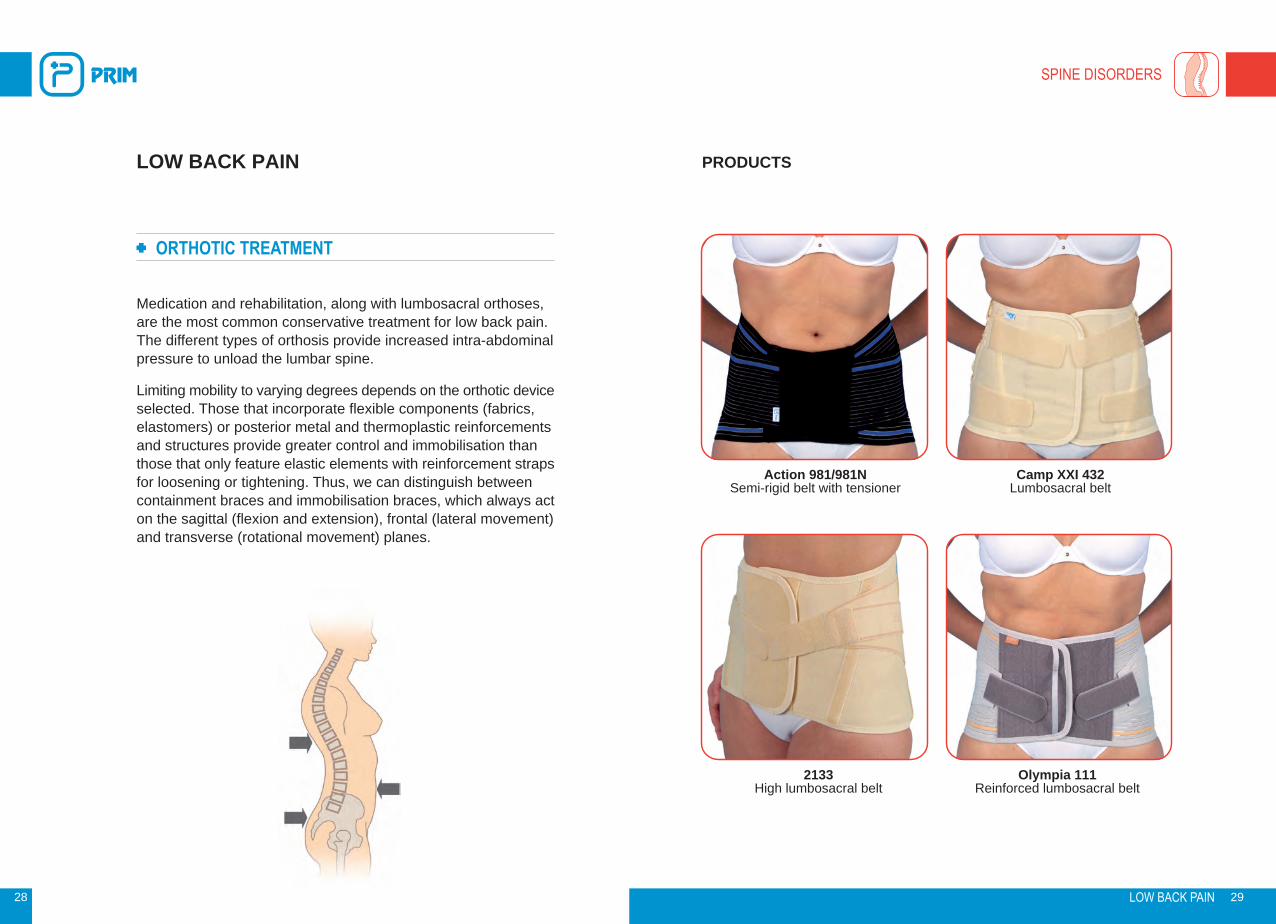
Action 981/981N Semi-rigid belt with tensioner
Camp XXI 432Lumbosacral belt
2133 High lumbosacral belt
Olympia 111Reinforced lumbosacral belt

SPINE DISORDERS
30
SCOLIOSIS
CLINICAL
Spinal column disorders can occur on two planes: the medial, where lordosis and kyphosis are observed, and the frontal, where scoliosis is evident.Scoliosis is a highly-complex spinal deformity characterised by two clear aspects: lateral curvature and vertebral rotation. The onset and progression of the deformity usually goes unnoticed, starting in puberty and affecting mainly female patients, and its origin, in most cases, is unknown, for which it is referred to as idiopathic scoliosis.The direction of the scoliosis, either right or left, is defined by the convexity of the curvature, and its location is determined by the vertebra that is most deviated laterally from the midline, known as the apical or apex vertebra. This vertebral rotation produces a thoracic deformity because, as the vertebra rotates, the ribs on the side of the convexity are pushed back, resulting in greater prominence in the back, known as gibbosity. Other disorders such as vertebral wedging - the narrowing of the vertebral body on its concave side or widening on its convex side - are visible along with variations in the thickness and elongation of the plates.Depending on their degree of flexibility, two types of scoliosis can be discerned: structural and non-structural, the latter of which has a higher degree of flexibility. Idiopathic scoliosis, which is of unknown origin, is the most common.
SYMPTOMS
In the case of infantile, juvenile and adolescent idiopathic scoliosis, onset and progression usually go unnoticed. They can be distinguished in lumbar scoliosis, which is common in girls, whose apex is found from T11 to L5, and are curves of a few degrees and accompanied by compensation curves.Thoracic curves are mostly right convexity, with no signs of discomfort, most commonly located between T5 and T12 and accompanied by compensation curves.Radiological examination, among others, provides information to determine the severity of the curve, the Risser sign, vertebral rotation and Cobb angle, along with other factors such as age or origin (neurofibromatosis, idiopathic, etc.) that enable full assessment of the scoliotic curve.This enables classification according to degree of deviation:1. 0° - 20° COBB • 2. 21° - 30° COBB • 3. 31° - 50° COBB • 4. 51° - 75° COBB • 5. 76° - 100° COBB • 6. 101° - 125° COBB • 7. 126° - > COBB.Vertebral rotation, depending on the position of the pedicles, shows if there is 0° rotation or, conversely, 1°, 2°, 3° or 4° rotations, taking as a reference the pedicle of the convexity with respect to the vertebral edge. Other factors, such as bone maturity, determine the type of treatment required.
SCOLIOSIS 31

SPINE DISORDERS
32 SCOLIOSIS 33
ORTHOTIC TREATMENT
Depending on factors such as pain, origin and etiology, degree of deviation, patient age, etc., treatment can be conservative, rehabilitational or surgical. Braces for the treatment of scoliosis can be active (corrective action) or passive (stabilisers and/or immobilisers).
Cases of juvenile and adolescent idiopathic scoliosis, whose curves are lumbar, dorsal or dorsolumbar, and in a Cobb angle range of between 20° and 50° with varying degrees of vertebral rotation, require orthotic treatment with active braces.
Throughout the history of scoliosis treatment, different types of brace have been designed, with the Boston brace being the most used today. Developed by John Hall and Billy Millar, its current design is still based on the original Boston brace for the quality of its properties, such as the materials used, components and the morphology of the module.
In the case of high dorsal or dorsolumbar curves, treatment can be carried out with the Milwaukee or Bercuoise brace or similar.
Cotrel night and/or daytime auto-traction systems are used by some inter-disciplinary teams as a way to provide more pre-intervention elasticity by reducing the Cobb angle to a certain extent and observing the curve’s degree of elasticity.
SCOLIOSIS PRODUCTS
Boston brace
Milwaukee

SPINE DISORDERS
34 SCOLIOSIS 35
ORTHOTIC TREATMENT
This orthotic device is created from a prefabricated polypropylene module with an interior Naturplast lining and has a wide range of sizes and lumbar lordosis correction options to enable the patient to select the right size according to a table of specific measurements for each patient. Some patients with peculiar morphologies may require the manufacture of a specific made-to-measure module.
The design, trim, appearance of the decompression windows and location of the corrective wedges for a custom design are determined by the curve to be treated and the relevant vertebral segment. Its correction principles are based on a set of forces applied to the convexity of the curve, seeking compression and derotation of the vertebrae, which involves correct and close positioning of the corrective wedges and proper location of the expansion windows.
SCOLIOSIS

SPINE DISORDERS
36
SPINAL DISC HERNIATION
CLINICAL
Spinal disc herniation occurs when the intervertebral disc or nucleus pulposus displaces, which can result in pressure being put on the nerve root, leading to possible neurological damage. This displacement occurs when the intervertebral disc, whose function is to absorb loads and impacts, suffers a tear in the annulus fibrosus. This occurs especially when there is prior disc degeneration with the consequent presence of arthrosis and limited mobility. Its clinical manifestations include sciatica, affecting sciatic nerve S1, and lumbago, in nerve L5, and can occur in any of the vertebrae, even in the cervical vertebrae. Disc herniation is classified according to the type of displacement of the nucleus pulposus:Disc protrusion. The annulus fibrosus suffers posterior or posterolateral deformation caused by the impact of the nucleus pulposus.Prolapse. Rupture of the nucleus pulposus through the annulus fibrosus without passing through the longitudinal ligament.Extrusion. The posterior side of the nucleus pulposus passes through the annulus fibrosus and even the longitudinal ligament.Sequestration. The extruded segment breaks free from the spinal canal due to tears in the annulus fibrosus’ collagen fibres.
SYMPTOMS
Pain in the lumbar region due to inflammation of the vertebral periosteum, dura mater, annulus fibrosus, ligaments etc., usually as a result of sudden movements or excessive overloading in the incorrect position.Tingling, loss of feeling with motor irritability and severe pain, which can radiate through the anterior, posterior and lateral thigh due to compression of the nerve roots.
SPINAL DISC HERNIATION 37
Disc protrusion Prolapse Extrusion Sequestration

SPINE DISORDERS
38 SPINAL DISC HERNIATION 39
ORTHOTIC TREATMENT
Rehabilitation techniques are, in conjunction with orthotic supports, the conservative treatment of herniated discs. Different types of orthosis produce a realignment of the lordotic curve through abdominal compression, while providing varying degrees of unloading and immobilisation. The orthoses and braces designed for this purpose provide a wide range of options.
Orthoses made from elastomers compress and reduce lordosis, as do belts and braces manufactured in fabrics of greater consistency, featuring reinforcing elements such as stays. Orthoses with posterior metal frames and thermoplastic plates, such as Knight, Taylor, MZ and Williams braces, provide greater unloading and partial immobilisation of the vertebral segment. Fully-rigid braces completely immobilise on all planes, realign the physiological curves and partially decompress with a high degree of unloading.
In surgery cases, braces are very useful during pre- and postoperative periods as a method of containment and support during the patient’s rehabilitation.
SPINAL DISC HERNIATION PRODUCTS
Elcross Gold 232 Lumbosacral belt
Camp XXI 433 Lumbosacral belt
Taylor DuoDorsolumbar spine orthosis
Knight Duo Lumbosacral spine orthosis

SPINE DISORDERS
40
OSTEOPOROSIS
CLINICAL
A disease that affects the bones due to a decrease in bone mass, in particular proteins and minerals, especially calcium. This makes the bone fragile and weaker than normal, meaning that it can fracture easily with the slightest trauma and the angle of the physiological curves can vary due to vertebral wedging, leading to hyperkyphosis and hyperlordosis.
Its onset is due to factors such as inadequate bone renewal or excessive resorption by the osteoblasts and lack of bone mass during development. Some factors, such as menopause, smoking, alcohol, sedentary lifestyle or the effect of endocrines, drugs, malnutrition, etc., promote bone mass loss and the onset of osteoporosis.
SYMPTOMS
Vertebral bone mass loss causes wedging of the vertebral bodies with the consequent increase in kyphotic and lordotic curves, resulting in hyperkyphosis and hyperlordosis, and leading to pain and decreased mobility.
Bone fragility increases the risk of compression fractures or injury from minimal trauma.
OSTEOPOROSIS 41

Camp XXI 437 Dorsolumbar belt
Elcross Gold 237 Dorsolumbar belt
SPINE DISORDERS
42 OSTEOPOROSIS 43
ORTHOTIC TREATMENT
Along with diet, vitamin D supplements and medication to restore the bone’s calcium and minerals, and physical exercise, it is necessary to realign the physiological curves to correct kyphosis and lordosis, improve load distribution and minimise the progression of the vertebral wedging, as well as providing the necessary protection for the fragile bone.
Lumbar and dorsolumbar orthoses provide systems with varying degrees of protection and unloading, with selection depending on the severity of the condition, degree of wedging or type of fracture in the spine. Hyperextension orthoses like the Jewett brace, or similar, enable realignment and prevent vertebral wedging.
The materials with which the orthosis is made, reinforcement through stays, posterior metal or thermoplastic structures, such as pelvic baskets, and the different adjustment and tightening systems determine the selection of the most appropriate orthosis based on the morphology of the patient and the state of progression of the osteoporosis.
OSTEOPOROSIS PRODUCTS
C34Hyperextension brace with adjustable pelvic support
C35 Plus Hyperextension brace with adjustable
external plate and fixed or floating pelvic band

SPINE DISORDERS
44
VERTEBRAL FRACTURE
CLINICAL
A vertebral fracture can be a fracture of the vertebral body, vertebral arch or spinous process.
In the case of a vertebral body fracture caused by crushing, the vertebral body collapses causing a burst or teardrop fracture, rupturing the anterior or posterior edge of the vertebra. The most common location is in the last dorsal or first lumbar vertebra.
In the case of the vertebra being displaced in an anterior direction, the result is spondylolisthesis or spondylolysis, the former of which can be stable or unstable and the latter stable.
Fracture-dislocation can also occur after suffering some type of trauma.
Some vertebral fractures can occur without being caused by a high-energy mechanism, for example, a bending movement can produce a wedge fracture, as is the case in patients with osteoporosis.
SYMPTOMS
Pain can sometimes go unnoticed, but is usually evident on palpation or percussion of the spinous processes. Muscle contraction is evident, resulting in limited mobility and the adoption of kyphotic postures. Any neurological signs, such as paresis, hypoesthesia or osteo-tendon reflex disturbance, are indicative that there is some degree of compression with neurological compromise, for which decompression is necessary.
VERTEBRAL FRACTURE 45

Taylor Camp XXI 637/638Spine orthosis
Knight Camp XXI 632/634Spine orthosis
SPINE DISORDERS
46 VERTEBRAL FRACTURE 47
ORTHOTIC TREATMENT
The orthotic treatment of vertebral fractures by means of braces with varying degrees of immobilisation and unloading are useful if there are no neurological signs present. If there are, decompression or alignment of the physiological curves would be necessary, and this can be carried out by using orthotic methods, as is also the case with serious CT, burns or other associated disorders in which surgery to fix and stabilise the affected vertebral segment can be delayed.
Stabilisation and immobilisation orthoses for the treatment of vertebral fractures need to be made from rigid materials (lumbar and dorsolumbar braces featuring metal or thermoplastic reinforcements) and include folding braces, Jewett hyperextension or cruciform braces, which, by means of supports located on the chest, pelvis and dorsolumbar transit area, exert compression forces that modify the physiological curve, enabling hyperkyphosis realignment and producing a hyperextension that prevents compression and vertebral wedging.
VERTEBRAL FRACTURE PRODUCTS
C35Hyperextension brace with fixed
pelvic band
C32 Hyperextension brace with adjustable external plate and floating pelvic band
UPPER LIMB DISORDERS - SHOULDER
48
BRACHIAL PALSY
CLINICAL
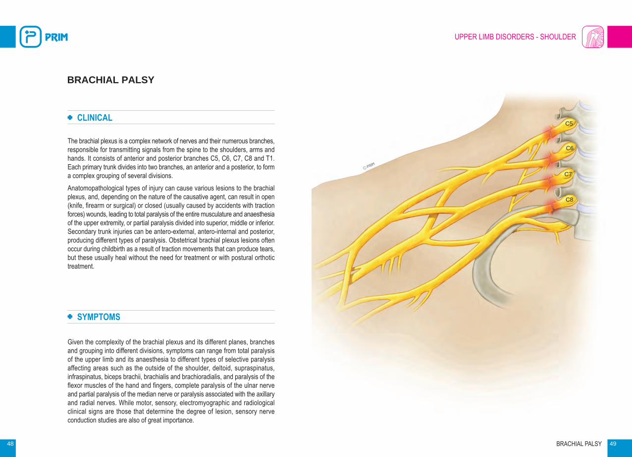
The brachial plexus is a complex network of nerves and their numerous branches, responsible for transmitting signals from the spine to the shoulders, arms and hands. It consists of anterior and posterior branches C5, C6, C7, C8 and T1. Each primary trunk divides into two branches, an anterior and a posterior, to form a complex grouping of several divisions.Anatomopathological types of injury can cause various lesions to the brachial plexus, and, depending on the nature of the causative agent, can result in open (knife, firearm or surgical) or closed (usually caused by accidents with traction forces) wounds, leading to total paralysis of the entire musculature and anaesthesia of the upper extremity, or partial paralysis divided into superior, middle or inferior. Secondary trunk injuries can be antero-external, antero-internal and posterior, producing different types of paralysis. Obstetrical brachial plexus lesions often occur during childbirth as a result of traction movements that can produce tears, but these usually heal without the need for treatment or with postural orthotic treatment.
SYMPTOMS
Given the complexity of the brachial plexus and its different planes, branches and grouping into different divisions, symptoms can range from total paralysis of the upper limb and its anaesthesia to different types of selective paralysis affecting areas such as the outside of the shoulder, deltoid, supraspinatus, infraspinatus, biceps brachii, brachialis and brachioradialis, and paralysis of the flexor muscles of the hand and fingers, complete paralysis of the ulnar nerve and partial paralysis of the median nerve or paralysis associated with the axillary and radial nerves. While motor, sensory, electromyographic and radiological clinical signs are those that determine the degree of lesion, sensory nerve conduction studies are also of great importance.
BRACHIAL PALSY 49
C5
C6
C7
C8

UPPER LIMB DISORDERS - SHOULDER
Hemisafe
50 BRACHIAL PALSY 51
ORTHOTIC TREATMENT
As well as the surgical treatment and rehabilitation techniques indicated for each case, depending on the degree of lesion and its effect on the extremity, whether it is more or less partial or complete, treatment with orthotic devices is also suitable provided that it is adapted to each particular case.
The functions of orthoses for the treatment of brachial plexus injuries include support, positioning of the limb at the right angle due its weight and preventing subluxation of the gleno-humeral joint. In some cases, unloading requires an adjustable positioning system that allows the angle of abduction and forward flexion of the shoulder to be graduated and varied, as well as the flexion of the elbow and the functional positioning of the hand, thereby enabling the orthosis to be individually adapted to the needs of each patient.
BRACHIAL PALSY PRODUCTS
TL174 Top Line shoulder support

UPPER LIMB DISORDERS - SHOULDER
52
SHOULDER INJURY
CLINICAL
A highly mobile joint consisting of two main articulations, the scapulohumeral and the acromioclavicular, and, to a lesser extent, the sternoclavicular, which enables a wide range of movements and all possible combinations on all planes, including full circumduction movements where flexion-extension is combined with abduction-adduction and internal-external rotation.It is made up of various articular and periarticular components, such as the capsular ligament, which is sleeve-shaped and consists of fibrous tissue, the coracohumeral ligament and the glenohumeral ligaments (superior, middle and inferior).A large number of injuries can occur in the shoulder joint affecting its bony components and its soft structures. Anterior or posterior dislocations, fractures, tendinitis, strains, bursitis, rotator cuff tears or adhesive capsulitis.
SYMPTOMS
Symptoms vary depending on the injury or disorder and have a common denominator of pain and varying degrees of functional disability.Day-to-day activities are affected due to the inability of the shoulder joint to perform combined movements.In particular, rotator cuff tears usually occur as a result of sudden movements or traumatic accidents with tearing of the supraspinatus, even compromising the subscapularis and infraspinatus.Dislocations can occur in various directions, the most common being anterior dislocation, which occurs as a result of an abduction and external rotation mechanism, or posterior dislocation, which occurs infrequently, due to flexion and internal rotation.
SHOULDER INJURY 53

UPPER LIMB DISORDERS - SHOULDER
54 SHOULDER INJURY 55
ORTHOTIC TREATMENT
Given the diversity of injuries that can affect the shoulder joint and taking into account that the most common are soft tissue lesions, rheumatic disorders, trauma and those that are the result of surgical treatment, different orthotic devices are required with the aim, along with rehabilitation techniques, of performing a precise orthotic function to facilitate early recovery.
Forearm supports, slings and fastening braces provide immobilisation and angled support to relieve musculoligamentous tension and, at the same time, the protection required to prevent unwanted movement.
Moreover, joint positioning orthoses, which enable adjustment of the position of the joint in order to vary, according to medical prescription, abduction and external rotation, forward flexion or a combination of all of them, allow correct articular immobilisation and unloading, while the ability to release the fastening systems enables the proposed rehabil itation programmes to be applied.
SHOULDER INJURY PRODUCTS
905Shoulder abduction orthosis
903 Shoulder immobiliser sling
UPPER LIMB DISORDERS - SHOULDER
56
NECK OF HUMERUS FRACTURE
CLINICAL
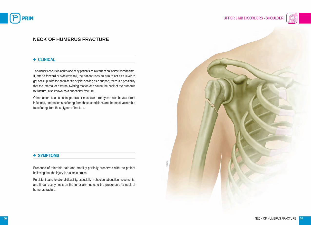
This usually occurs in adults or elderly patients as a result of an indirect mechanism. If, after a forward or sideways fall, the patient uses an arm to act as a lever to get back up, with the shoulder tip or joint serving as a support, there is a possibility that the internal or external twisting motion can cause the neck of the humerus to fracture, also known as a subcapital fracture.
Other factors such as osteoporosis or muscular atrophy can also have a direct influence, and patients suffering from these conditions are the most vulnerable to suffering from these types of fracture.
SYMPTOMS
Presence of tolerable pain and mobility partially preserved with the patient believing that the injury is a simple bruise.
Persistent pain, functional disability, especially in shoulder abduction movements, and linear ecchymosis on the inner arm indicate the presence of a neck of humerus fracture.
NECK OF HUMERUS FRACTURE 57

UPPER LIMB DISORDERS - SHOULDER
20220 09Humerus Confort
58 NECK OF HUMERUS FRACTURE 59
ORTHOTIC TREATMENT
In cases where the fracture is stable or where surgery is not indicated, orthotic devices can be used for conservative treatment in order to enable alignment, immobilisation and stabilisation of the fractured segment, including stabilisation and unloading of the glenohumeral joint, as well as compression to produce a strapping effect on the arm. These functions create optimum conditions for the consolidation of the fracture, allowing movement of the elbow and leaving the hand free to perform the necessary rehabilitation and simple household chores. These orthoses are also useful for stabilisation and protection in cases where pseudarthrosis is present in the fracture site.
NECK OF HUMERUS FRACTURE PRODUCTS
904 Shoulder abduction orthosis

UPPER LIMB DISORDERS - SHOULDER
60
CLAVICLE FRACTURE
CLINICAL
Clavicle fractures usually occur in the segment comprising the middle third of the clavicle and are caused by a violent impact to the arm, which can be extended during the impact or fall, or by direct fall onto the shoulder.
After the fall, the sternocleidomastoid muscle elevates the medial fragment of the bone, while the trapezius muscle is unable to keep the lateral fragment elevated, resulting in the shoulder drooping under the weight of the arm. The pectoralis major and the adductor muscles pull the distal fragment, making them override the proximal. Dislocation is rare, as the acromioclavicular ligament usually prevents it.
SYMPTOMS
Shoulder pain and antepulsion due to the overriding of the fractured fragments, as a result of muscle-tendon tension.
The injury often occurs during sporting activities and work or traffic accidents. In the majority of cases, it occurs in the middle third of the bone, and less frequently in the distal third.
CLAVICLE FRACTURE 61

UPPER LIMB DISORDERS - SHOULDER
ClavisanClavicle immobiliser
62 CLAVICLE FRACTURE 63
ORTHOTIC TREATMENT
In the event of being open or displaced fractures, surgery and subsequent rehabilitation is the required treatment. Immobilisation and realignment of the fracture in conservative treatments can be achieved with the use of adjustable textile orthoses or figure-of-eight devices that completely immobilise and retropulse the shoulders in order to realign the fractured segment.
CLAVICLE FRACTURE PRODUCTS

UPPER LIMB DISORDERS - ELBOW
64
EPICONDYLITIS
CLINICAL
Epicondylitis, commonly known as “tennis elbow” is an insertional disorder (enthesitis). It is caused by repetitive wrist extension movements and supination of the forearm, leading to muscle strain and degenerative processes of the tendon, where it inserts into the epicondyle region, especially affecting the short radial extensor tendon of the wrist. When this same mechanical injury occurs in the medial epicondyle, it is known as medial epicondylitis or “golfer’s elbow”. As well as occurring during sporting activities, these injuries can also be observed in work activities involving similar repetitive movements.
Its three stages show how it develops:
1. Initial imitative stage.
2. Inflammatory stage with vasomotor disorders.
3. Degenerative stage with chronic process.
SYMPTOMS
Presence of pain on the outside (epicondylitis) or inside (medial epicondylitis) of the elbow. Pain on palpation of the epicondyle or medial epicondyle, which subsides with rest. Functional disability and pain in wrist extension movements and pronosupination of the forearm, which can radiate to the arm and forearm. Inability to handle objects. It can become chronic and sometimes require surgery.
EPICONDYLITIS 65

UPPER LIMB DISORDERS - ELBOW
TL172 Top Line
TL173 Top Line elbow support
66 EPICONDYLITIS 67
ORTHOTIC TREATMENT
Along with medical and rehabilitation techniques, such as massage, shock waves, laser, etc., various types of specially-designed orthosis exist that are very important for the orthotic treatment of epicondylitis and medial epicondylitis. Devices made from neoprene provide the necessary heat for the combined application of thermotherapy and bracing-compression.
Other orthotic devices such as braces with epicondyle and medial epicondyle supports specifically compress the area below where the tendon inserts, thereby preventing mechanical stress and reducing overloads, and enabling the patient to resume work and sporting activities. Using these types of orthosis is very helpful as a method of preventing recurrent injuries.
EPICONDYLITIS PRODUCTS
P606 Top Line breathable neoprene
epicondylitis strap
P506 Elastic elbow support with gel insert
and strap

UPPER LIMB DISORDERS - ELBOW
68
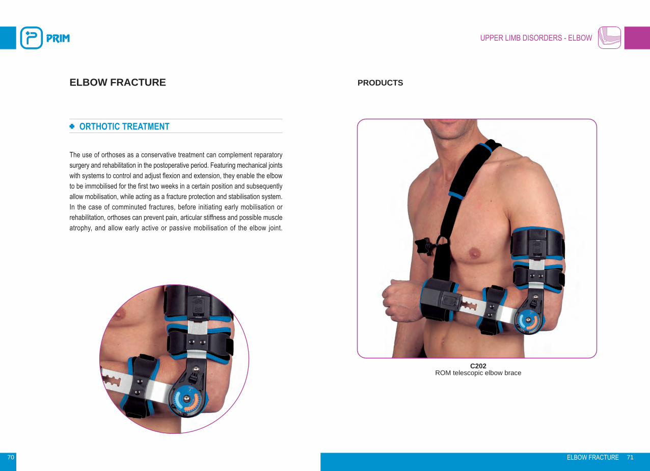
ELBOW FRACTURE
CLINICAL
Elbow fractures can equally occur in any of the elbow’s three elements - the inferior end of the humerus, the olecranon and the radial head - and can be total or partial and, in some cases, accompanied by vascular or nerve injuries with the presence of anterior linear ecchymosis, increased volume of the elbow and full functional disability. These are fractures that can therefore have serious complications, irreducibility, instability, open fractures, etc., making their evolution highly variable due to their complexity and extensive variability of fracture types.
SYMPTOMS
Symptoms vary depending on the type of fracture or dislocation-fracture, whether it has occurred by direct or indirect mechanism, whether the elbow was in flexion or extension or whether the impact was directly on the elbow. These aspects determine the type of fracture, which can be comminuted, open, etc., and the location of the action of the muscles at the time of the fracture is of great importance, due to the tension-traction exerted. Reparatory surgery is required in many cases to obtain good reduction. In any of the cases, pain, functional disability, oedema and ecchymosis, as well as other symptoms, indicate the presence of an elbow fracture.
ELBOW FRACTURE 69
UPPER LIMB DISORDERS - ELBOW
C202 ROM telescopic elbow brace
70 ELBOW FRACTURE 71
ORTHOTIC TREATMENT
The use of orthoses as a conservative treatment can complement reparatory surgery and rehabilitation in the postoperative period. Featuring mechanical joints with systems to control and adjust flexion and extension, they enable the elbow to be immobilised for the first two weeks in a certain position and subsequently allow mobilisation, while acting as a fracture protection and stabilisation system. In the case of comminuted fractures, before initiating early mobilisation or rehabilitation, orthoses can prevent pain, articular stiffness and possible muscle atrophy, and allow early active or passive mobilisation of the elbow joint.
ELBOW FRACTURE PRODUCTS

UPPER LIMB DISORDERS - HAND
72
NEUROLOGICAL INJURY
CLINICAL
The term neurological injury refers to any kind of disturbance in the functioning of the nervous system, the most frequent being spinal cord and brain injuries.Regarding the hand, causes of paralysis can be a result of injuries to the central nervous system, cerebral palsy, tumours, trauma, etc. Those caused by spinal cord injury, poliomyelitis, amyotrophic lateral sclerosis, etc., injuries to the brachial plexus and its peripheral trunks or those produced by ischemic causes.Various injuries can occur in the hand with different symptoms depending on which of the radial, median or ulnar nerves are affected, due to their muscle innervation.The result can vary from spastic to flaccid paralysis.
SYMPTOMS
Symptoms vary depending on the affected nerve. In radial nerve palsy, for example, loss of extension of the metacarpophalangeal and interphalangeal joints of the thumb can be observed and the inability to extend the wrist is evident.In the case of median nerve palsy, however, symptoms include loss of abduction of the thumb, loss of flexion of the metacarpophalangeal joint of the thumb and loss of the ability to perform thumb opposition, making it impossible to bring the thumb and index or little finger together. This can be compounded when the injury affects the forearm and elbow, causing loss of flexion in the interphalangeal joint of the thumb and distal interphalangeal flexion of the index and middle fingers.In ulnar nerve injuries, symptoms include flattening of the thenar eminence, partial ulnar claw in the 4th and 5th fingers (if the median and ulnar nerves are affected, the claw is complete), while the 5th finger is positioned in abduction and the hand shows hollowing in the interosseous spaces.
NEUROLOGICAL INJURY 73

UPPER LIMB DISORDERS - HAND
934 Functional hand orthosis
935 Positional hand orthosis
74 NEUROLOGICAL INJURY 75
ORTHOTIC TREATMENT
As well as rehabilitation techniques applied to each type of neurological injury of the hand, there is a wide variety of orthosis designs available for each particular purpose, with objectives that include protecting the skin, preventing deformities, restoring muscle balance, or as a method of stabilisation, helping functional rehabilitation, recovering function (if possible) and assisting function as a stabilising method.
Distinguishing between passive and active according to function, these first ones have the purpose of keeping the hand, or an anatomical segment of it, in a particular position, as per the prescriber’s instructions, and can be used during the day, night or both. Active or functional orthoses allow correction, alignment or rehabilitation exercises, with selection of the most suitable device depending on the prescribed function.
NEUROLOGICAL INJURY PRODUCTS
949 Short passive wrist orthosis with
thumb support
2.1Long passive wrist orthosis with
thumb support

UPPER LIMB DISORDERS - HAND
76
LIGAMENT AND TENDON INJURY
CLINICAL
The hand’s bone joints are reinforced together by ligaments, collateral ligaments situated on both sides of the joint and reinforcement anterior to the capsule.The musculature of the hand is extensive and divided into that whose origin is the hand or intrinsic musculature, which contains interosseous muscles, lumbricals, the muscle groups in the region of the little finger and thumb, and musculature whose origin is the forearm or extrinsic, whose function is flexion-extension of the fingers. This, together with the fibrous tissue that covers the musculature, makes up the soft tissue of the hand.In the event of trauma of varying degrees of severity, a number of injuries can occur, resulting in sprains or interphalangeal dislocations, which usually occur dorsally, affecting the proximal interphalangeal (PIP) and distal interphalangeal (DIP) joints or both, as well as the MCP (metacarpophalangeal), and have different degrees of classification, the most serious being that in which ligament rupture occurs with the presence of instability and functional disability.
SYMPTOMS
Shortening of the length of the finger with joint deformity. Pain and functional disability with the inability to perform any type of movement. In severe cases, it can be accompanied by a fracture.Inflammation of a tendon or group of tendons can occur, causing compression of the sheath that covers it, which is very painful and disabling. The joints can become stiff if their immobilisation exceeds 21 days.
LIGAMENT AND TENDON INJURY 77

UPPER LIMB DISORDERS - HAND
950 Short passive wrist orthosis
without thumb support
1.1Short passive thermoplastic
orthosis without thumb support
78 LIGAMENT AND TENDON INJURY 79
ORTHOTIC TREATMENT
As well as different surgical and/or therapeutic techniques, such as tendon, neuro-muscular, bone and skin repair and other aspects present in hand injuries, and rehabilitation techniques, passive and/or dynamic or active orthoses are suitable for use as a conservative treatment method. The purpose of passive orthoses is to keep the articular segments in a certain position as determined by the prescriber, and they are indicated for use as a conservative or post-surgical method. Dynamic orthoses enable functional recovery by improving mobility range and strengthening muscles and tendons. Correct selection of the most appropriate orthosis and proper fitting and monitoring by the orthopaedic technician is of paramount importance to the success of orthotic treatment.
LIGAMENT AND TENDON INJURY PRODUCTS
942 Long passive wrist orthosis with
thumb support
C180Long wrist brace with palmar bar

UPPER LIMB DISORDERS - HAND
80
CARPAL TUNNEL SYNDROME AND GRUYON'S CANAL SYNDROME
CLINICAL
Carpal tunnel syndrome.The carpal tunnel is a narrow passageway found between the bones of the base of the hand, specifically the pisiform, semi-lunate, pyramid and scaphoid, containing tendons and the median nerve in its proximal aspect, while distally it is formed by the trapezium, trapezoid, capitate and hamate. It is in this space where the median nerve can become trapped, resulting in peripheral neuropathy. This entrapment can be produced by the presence of fluid, inflammation of one of the tendons or any process that reduces space in the tunnel.Guyon’s canal syndrome. Guyon’s canal is formed by the transverse carpal ligament, the carpal ligaments and the opponens pollicis muscle of the 5th finger, with the volar carpal ligament and the palmar brevis muscle forming its roof. The ulnar nerve, which divides at this point into its superficial and deep branches, can become trapped or compressed as it passes through the canal, resulting in a neurological syndrome affecting the sensitivity of the 5th finger and part of the 4th, as well as the motor function of the hand muscles, leading to problems in bringing the fingers together and separating them.
SYMPTOMS
Carpal tunnel syndrome.A sensation of heat, numbness in the middle and index fingers, and especially the thumb. As it worsens, the patient experiences cramps and has difficulty making a fist, picking up objects and performing manual tasks. Loss of sensitivity is evident in some cases and pain is present in the upper area of the hand and wrist. Symptoms increase with activity, but can subside with massage and relaxation.Guyon’s canal syndrome.Atrophy of the hypothenar eminence and the interosseous muscles with weakness in closure movements and/or ulnar flexion of the wrist. Paresis, hypoesthesia and paresthesia in the area innervated by the ulnar affecting the flexors of the 4th and 5th fingers and the intrinsic musculature.
CARPAL TUNNEL SYNDROME AND GRUYON'S CANAL SYNDROME 81

UPPER LIMB DISORDERS - HAND
942 Long passive wrist orthosis
with thumb support
28542P Plastic orthosis for carpal tunnel
syndrome
82 CARPAL TUNNEL SYNDROME AND GRUYON'S CANAL SYNDROME 83
ORTHOTIC TREATMENT
Rehabilitation treatment, including the use of moist warm compresses, paraffin and other techniques such as laser or electrotherapy, as well as preventative treatments like wrist mobility exercises during rest periods, in conjunction with the use of unloading splints that enable immobilisation of the wrist, hand and finger joints, as in the case of Guyon’s syndrome, are very useful for keeping the joints at rest during day and nighttime use, and preventing postural attitudes during sleep like those in which the wrists are in flexion.
CARPAL TUNNEL SYNDROME AND GRUYON'S CANAL SYNDROME
PRODUCTS
950 Short passive wrist orthosis with
thumb support
1.1Short passive thermoplastic
orthosis without thumb support

UPPER LIMB DISORDERS - HAND
84
QUERVAIN’S TENDINITIS
CLINICAL
Quervain’s tendinitis, also known as Quervain’s tenosynovitis, is caused by irritation and inflammation of the tendons located at the base of the thumb, usually as a result of repetitive movements. Some traumatic injuries resulting from fractures in which an increase of tendon tension is evident can predispose the occurrence of tendinitis in the first dorsal compartment.
SYMPTOMS
Sudden or gradual pain originating in the first dorsal compartment of the wrist and thumb, radiating to the forearm.
Pain increases with mechanical effort, gripping or squeezing objects, together with pronosupination movements of the wrist.
Possible loss of sensitivity in the thumb and index finger.
QUERVAIN’S TENDINITIS 85

UPPER LIMB DISORDERS - HAND
943 Long passive wrist orthosis
with thumb support
P605 Top Line long breathable neoprene
wrist brace with thumb support
86 QUERVAIN’S TENDINITIS 87
ORTHOTIC TREATMENT
Passive wrist and thumb immobilising splints, in which the wrist, trapeziometacarpal and proximal metacarpophalangeal joints are immobilised at rest and in a functional position, enabling reduction of irritation and inflammation, are a perfect complement to rehabilitation techniques.
QUERVAIN’S TENDINITIS PRODUCTS
TL166 Top Line long wrist brace with
thumb support
949 Short passive orthosis with
thumb support

UPPER LIMB DISORDERS - HAND
88
ARTHROSIS, RHIZARTHROSIS
CLINICAL
Joint degeneration leads to arthrosis. Rheumatoid disease gives its name to the disease known as rheumatoid arthritis, in which, besides other disabling manifestations, such as pleuropulmonary and cardiac disease, articular disorders have particular clinical presence by affecting the synovial tissue of either joints or tendons, with the presence of tenosynovitis, resulting in the destruction of joint structures, causing deformity and rigidity, together with loss of cartilage and inflammation of adjacent tissue.
SYMPTOMS
The progressive articular degeneration, along with cartilage destruction, inflammation and articular deformity, mainly affecting the hand and lower limbs, causes pain and loss of mobility with articular rigidity.
ARTHROSIS, RHIZARTHROSIS 89

UPPER LIMB DISORDERS - HAND
C270Long lined thumb abduction orthosis
C170Long lined thumb abduction
orthosis
90 91
ORTHOTIC TREATMENT
It is important to combine rehabilitation techniques with different types of orthosis, which can be passive or active with the following main functions:
Pain relief, prevention of joint deformity and its progression in the hand and fingers.
Immobilisation and relaxation of the ligaments and soft tissue, thereby decreasing inflammation.
As the method of post-surgical immobilisation and alignment in endoprosthesis cases.
ARTHROSIS, RHIZARTHROSIS PRODUCTS
P609Top Line long breathable thumb orthosis with plate
TL165 Top Line thumb brace with plate
ARTHROSIS, RHIZARTHROSIS
UPPER LIMB DISORDERS - HAND
92
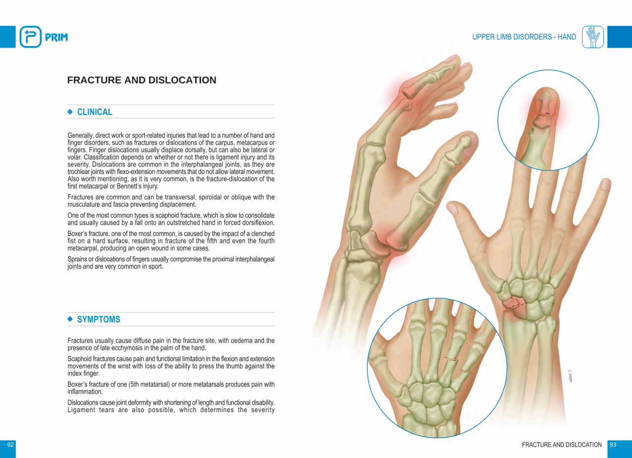
FRACTURE AND DISLOCATION
CLINICAL
Generally, direct work or sport-related injuries that lead to a number of hand and finger disorders, such as fractures or dislocations of the carpus, metacarpus or fingers. Finger dislocations usually displace dorsally, but can also be lateral or volar. Classification depends on whether or not there is ligament injury and its severity. Dislocations are common in the interphalangeal joints, as they are trochlear joints with flexo-extension movements that do not allow lateral movement. Also worth mentioning, as it is very common, is the fracture-dislocation of the first metacarpal or Bennett’s injury. Fractures are common and can be transversal, spiroidal or oblique with the musculature and fascia preventing displacement.One of the most common types is scaphoid fracture, which is slow to consolidate and usually caused by a fall onto an outstretched hand in forced dorsiflexion.Boxer’s fracture, one of the most common, is caused by the impact of a clenched fist on a hard surface, resulting in fracture of the fifth and even the fourth metacarpal, producing an open wound in some cases.Sprains or dislocations of fingers usually compromise the proximal interphalangeal joints and are very common in sport.
SYMPTOMS
Fractures usually cause diffuse pain in the fracture site, with oedema and the presence of late ecchymosis in the palm of the hand.Scaphoid fractures cause pain and functional limitation in the flexion and extension movements of the wrist with loss of the ability to press the thumb against the index finger.Boxer’s fracture of one (5th metatarsal) or more metatarsals produces pain with inflammation.Dislocations cause joint deformity with shortening of length and functional disability. Ligament tears are also possible, which determines the severity
FRACTURE AND DISLOCATION 93

943 Long passive wrist orthosis
with thumb support
UPPER LIMB DISORDERS - HAND
947 Stack distal splint
948 Stack proximal splint
94 FRACTURE AND DISLOCATION 95
ORTHOTIC TREATMENT
Immobilisation is usually used as a conservative method with finger, wrist and/or hand splints or forearm orthoses, or even a combination of both, for a maximum period of three weeks, followed by suitable rehabilitation techniques to prevent joint stiffness, especially in the fingers.Immobilisation with an orthosis requires the use of devices specifically designed for the condition being treated, taking into account aspects such as the positioning in abduction of a joint (Bennett’s dislocation) or the flexion of one or more joints (boxer’s fracture), as well as hyperextension of the distal interphalangeal joint in extensor fractures of the finger, which require correct selection of the optimum orthosis for proper orthotic treatment, given the wide range of orthoses. For wrist, hand, finger and multiple active and passive combinations, it is recommended that the specialist select the orthosis.
FRACTURE AND DISLOCATION PRODUCTS
946 RF Ortho distal splint

LOWER LIMB DISORDERS - HIP
96
CONGENITAL DYSPLASIA AND DISLOCATION OF THE HIP
CLINICAL
Dysplasia is a term that defines a broad spectrum of hip formation irregularities. Present from birth, it can be mild or involve complete dislocation of the hip. Some factors can increase the risk of developing hip dysplasia, such as family history, female gender, pregnancy history, associated malformations, etc. It is recommended that all newborns undergo exploratory tests to check for the presence of dysplasia or hip dislocation.
SYMPTOMS
Unstable hip, decrease in the normal opening of the hips, difference between the skinfolds of the lower limbs and apparent shortening of one of the limbs. Untreated dysplasia results in premature wear (arthrosis) of the joint, pain and lameness. Asymmetry is evident and may require t reatment.
CONGENITAL DYSPLASIA AND DISLOCATION OF THE HIP 97

LOWER LIMB DISORDERS - HIP
115 Pavlik harness
117 Frejka nappy
98 CONGENITAL DYSPLASIA AND DISLOCATION OF THE HIP 99
ORTHOTIC TREATMENT
Treatment of newborns with dysplasia consists of keeping the hips in abduction and maximum flexion, ensuring that the femoral head stays within the acetabulum for a period of time that depends on the infant’s age and severity of the dysplasia-dislocation. Although all orthotic devices seek equal positioning of the hips as a common denominator, their designs vary depending on the infant’s age, severity and the prescriber’s own judgement.
Frejka, Von Rosen and Ponseti nappy-type devices keeps the hips immobilised in abduction and flexion, and enable the angles to be varied and adjusted. The Pavlik harness, which features chest, shoulder and leg straps, and booties for the feet, enables abduction and flexion to be adjusted, according to medical judgement, and a varying range of mobility for the hip joints. The Pavlik device is indicated for infants between 0 and 6 months of age, but can continue to be used for a month or so more. All CHILD orthoses require special training for the parents to ensure proper fitting and complete success of this orthotic treatment.
CONGENITAL DYSPLASIA AND DISLOCATION OF THE HIP
PRODUCTS
C29 Dennis Brown splint
C30 Kindi hip abduction orthosis

LOWER LIMB DISORDERS - HIP
100
POSTOPERATIVE HIP DISLOCATION
CLINICAL
The incidence of hip dislocation in total hip replacements ranges from 1% to 15% according to various publications. Different factors can cause prosthetic hip dislocation, such as poor orientation of the components, insufficient neo-capsule, conflict between the bony structures, soft tissue tension, generalised muscle weakness with gluteal insufficiency, etc. A bad choice of implant, patient age or medial fractures are usually the most likely triggering factors. Hip arthroplasty is mainly performed due to the presence of coxarthrosis or hip fractures. Two thirds of dislocations are successfully resolved noninvasively, the rest require surgical revision. Factors such as gender (female/male ratio 2:1), age, the older the patient, the greater the risk, even patient cooperation is of paramount importance. Classified according to the time that has elapsed after surgery, dislocations can be early, less than 1 month, medium-term, between 1 month and 1 year, or late, 1 year after the procedure.
SYMPTOMS
The prosthetic femoral head is positioned outside the acetabulum or prosthetic cotyloid cavity and displaces proximally, causing considerable asymmetry of the lower limb. Pain and claudication during walking. Instability and functional disability.
POSTOPERATIVE HIP DISLOCATION 101

LOWER LIMB DISORDERS - HIP
New Camp Postoperative hip abduction orthosis
New Camp II Postoperative hip abduction orthosis
102 POSTOPERATIVE HIP DISLOCATION 103
ORTHOTIC TREATMENT
Some guidelines or recommendations in post-surgery can help to prevent hip dislocation to some extent, such as patients avoiding crossing their legs, not rotating the operated leg, limiting flexion of the hip, spreading the load, etc. In the event that the dislocation is present as a post-surgical complication, it will require an orthotic system for the multi-positional control of the hip joint. Anti-luxation hip orthoses, featuring a pelvic basket joined to a thigh support by means of a flexion and abduction-adduction control joint enables the hip joint to be kept in a certain position, allowing the patient to stand and walk, and undergo rehabilitation, which is essential for full repair. Anti-luxation hip orthoses are suitable as a preventive measure in cases where certain post-operative instability is evident during the rehabilitation period.
POSTOPERATIVE HIP DISLOCATION PRODUCTS
New Camp 3 Postoperative hip abduction orthosis

LOWER LIMB DISORDERS - HIP
104
HIP JOINT INJURY IN INFANTILE CEREBRAL PALSY
CLINICAL
Patients suffering from infantile cerebral palsy can experience impaired movement, posture and muscle tone, caused by a brain injury during its maturation period. It can acquire different clinical manifestations: in some cases, it can be spastic (tetraplegia/paresis, diplegia/paresis, hemiplegia/paresis or monoparesis) and, in others, it can be dyskinetic, ataxic, hypotonic or mixed. The many disorders that this type of patient experiences include neuromotor disorders with altered muscle tone, imbalance and persistent reflex postures, which can affect the hip joint, causing deformity in the spine and hip.
SYMPTOMS
As well as the specific symptoms of ICP patients, the hip suffers deformity in adduction, flexion and internal rotation. This is due to the hypertonicity of these muscle groups and the hypotonia of their antagonists. Due to the presence of coxa valga, subluxation or dislocation of the hip joint can occur.
HIP JOINT INJURY IN INFANTILE CEREBRAL PALSY 105

LOWER LIMB DISORDERS - HIP
Swash Variable-abduction hip orthosis
Swash LPII Variable-abduction hip
orthosis
106 HIP JOINT INJURY IN INFANTILE CEREBRAL PALSY 107
ORTHOTIC TREATMENT
Rehabilitation takes a primary role in the treatment of ICP patients and can involve physiotherapy, occupational therapy, logotherapy, etc., complemented by technical aids and specially-designed orthoses. The purpose of these orthoses is to prevent deviations and deformities, correcting them to the greatest extent possible and providing the hip joint with the necessary stability to facilitate its functioning. SWASH orthoses, which are designed to keep the hips stable and in slight abduction, enable the patient to walk and sit down during the day and prevent adduction during the night.
HIP JOINT INJURY IN INFANTILE CEREBRAL PALSY
PRODUCTS

LOWER LIMB DISORDERS - THIGH/LEG
108
MUSCLE STRAIN
CLINICAL
Muscle strain is a soft tissue injury caused by indirect trauma, usually as a result of muscle distention, or direct contusion, which can also lead to tears. The severity of the injury depends on the affected fibres, and whether the muscle has completely torn. Injuries can be classified as grade 1, 2, or 3, depending on the number of damaged or ruptured fibres. They usually occur at the muscle-tendon junction during maximum physical activity (sprinting, jumping, etc.), as a result of exceeding the elasticity of the muscle belly. Some factors such as poor fitness, overloaded muscles, lack of warm-up before exertion, muscle imbalance, age, etc., can predispose to muscle strain with varying degrees of severity.
SYMPTOMS
Sudden pain at the moment of the tear and immediate functional disability, with pain remaining after injury, even at complete rest, and loss of the muscle’s contractile function. Internal muscle bleeding with inflammation. The muscle is wel l vascularised at the t ime of injury, causing haematomas.
MUSCLE STRAIN 109

TL147 Top Line thigh support
P512 Elastic thigh support
110 MUSCLE STRAIN 111
ORTHOTIC TREATMENT
Depending on the severity of the strain and the time elapsed, different rehabilitation techniques can be applied to the injury, such as immediate cryotherapy, rest, isometric therapy etc., always accompanied by compression garments or specific orthoses that provide compression, support and heat to minimise fibre and muscle-tendon tension during the healing period. This type of orthosis is highly recommended as a method of prevention for patients returning to sporting activity.
MUSCLE STRAIN PRODUCTS
TL120 Top Line calf support
P513 Elastic calf support
LOWER LIMB DISORDERS - THIGH/LEG

LOWER LIMB DISORDERS - KNEE
112
CRUCIATE LIGAMENT AND MENISCUS INJURY
CLINICAL
The knee achieves stability through two systems consisting of stabilising ligaments - the medial-lateral and anterior cruciate ligaments (ACL) and the posterior cruciate ligament (PCL). In the event of injury, the regeneration of cruciate ligaments is uncertain. Since their function is anteroposterior stabilisation of the knee, it is advisable to determine the stability of the knee because, without it, the articular structures would progressively deteriorate. The ACL is responsible for preventing dislocation of the femur on the tibia and limiting external rotation, and can become overstretched or suffer from a partial or total tear if a rotational movement, blow or hyperextension of the knee occurs. The ACL can tear at the same time as the MLL and the medial meniscus, representing the classic knee injury. A front-on impact to the knee or a hyperextension movement can cause injury of the posterior cruciate ligament. Depending on severity, distention and whether the tear is partial or complete, they are classified as Grade 1, 2 or 3. The menisci can suffer tears due to indirect, generally violent, trauma to the knee, often associated with ligament injuries.
SYMPTOMS
A crunching sound at the moment of injury with severe pain. Inflammation of the knee after a few hours, leading to anteroposterior knee instability, and potentially resulting in arthritis in the joint. ACL injuries can be compensated by stabilising elements, and quadriceps and hamstring musculature.
CRUCIATE LIGAMENT AND MENISCUS INJURY 113

TL135 Top Line knee support
for ligament injury
114 CRUCIATE LIGAMENT AND MENISCUS INJURY 115
ORTHOTIC TREATMENT
Rehabilitation helps to recover knee mobility and improve muscle strength. Stabilising orthoses featuring flexion-extension control joints can regulate movement range and limit tension in the anterior and posterior cruciate ligaments, facilitating healing and enabling the movement range to be gradually extended. Orthoses that provide complete immobilisation are very useful for moving patients prior to surgery and as immediate treatment at the time of the injury. Other types of orthoses with control and stabilisation systems are essential as a preventative measure during sporting activities and after ligament injury recovery.
CRUCIATE LIGAMENT AND MENISCUS INJURY
PRODUCTS
R100Polycentric knee brace
for cruciate ligament injury
RR201 ROM postoperative telescopic
knee brace
LOWER LIMB DISORDERS - KNEE

LOWER LIMB DISORDERS - KNEE
116
LATERAL LIGAMENT INJURY
CLINICAL
The collateral ligaments - the internal lateral ligament (ILL) and the external lateral ligament (ELL) - provide the knee with mediolateral stability. Injury of these ligaments usually occurs when the body rotates while the foot is fixed to the ground, which is a typical movement in some sporting activities, with the medial ligament being the one that is injured most frequently. Impacts to the outer side of the knee produce a hinged movement, resulting in the stretching of the medial ligament and injury. The injury can have varying degrees of severity. Grade 1 is a ligament tear, grade 2 is a rupture of some fibres and grade 3 is complete rupture of the ligament or avulsion from its bony insertion.
SYMPTOMS
It is common for collateral ligament injury to be associated with meniscal, ILL and internal meniscus injuries, and mediolateral instability of the knee, which can be chronic, causing a feeling of insecurity and the presence of mechanical faults (sensation of the knee “moving”). Impossibility of performing any sporting activity and limitations in movement in daily life.
LATERAL LIGAMENT INJURY 117

TL136 Top Line knee support with straps
TL131 Top Line knee support with straps
118 LATERAL LIGAMENT INJURY 119
ORTHOTIC TREATMENT
In grade 1 and 2 injuries, the use of orthopaedic devices is essential to enable complete immobilisation for a period of at least 3 weeks and a prompt return to walking and isometric exercises, pain permitting. After this period, rehabilitation, along with orthotic devices, is used to gradually improve mobility. By means of mechanical flexion-extension control joints on the orthosis, it is possible to completely immobilise or set a movement range for the joint. In the case of ligament rupture, post-surgical orthotic treatment requires the use of an orthotic device for immobilisation and early rehabilitation. After surgery and subsequent rehabilitation, the use of stabilising knee supports for protection in sporting and daily activities is recommended.
LATERAL LIGAMENT INJURY PRODUCTS
TL130 Top Line articulated knee support
TL140 Top Line polycentric knee
support
LOWER LIMB DISORDERS - KNEE
LOWER LIMB DISORDERS - KNEE
120
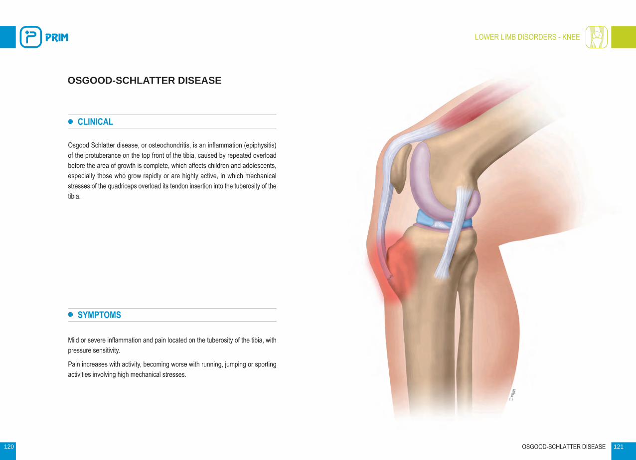
OSGOOD-SCHLATTER DISEASE
CLINICAL
Osgood Schlatter disease, or osteochondritis, is an inflammation (epiphysitis) of the protuberance on the top front of the tibia, caused by repeated overload before the area of growth is complete, which affects children and adolescents, especially those who grow rapidly or are highly active, in which mechanical stresses of the quadriceps overload its tendon insertion into the tuberosity of the tibia.
SYMPTOMS
Mild or severe inflammation and pain located on the tuberosity of the tibia, with pressure sensitivity.
Pain increases with activity, becoming worse with running, jumping or sporting activities involving high mechanical stresses.
OSGOOD-SCHLATTER DISEASE 121

TL132 Top Line patella band
P607 Top Line breathable
neoprene patella band
122 OSGOOD-SCHLATTER DISEASE 123
ORTHOTIC TREATMENT
Rest and limitation of sporting activities to reduce mechanical stresses, together with the use of patella bands and open kneecap supports with patella straps that support the patellar tendon between the kneecap and the area above the tibial tuberosity to minimise mechanical tension and overloads at the quadriceps insertion during physical activity.
As a method of preventing injury during sport, the use of patella bands is highly recommended.
OSGOOD-SCHLATTER DISEASE PRODUCTS
P618Top Line breathable neoprene
open kneecap support with straps
P616 Top Line breathable neoprene knee
support with straps
LOWER LIMB DISORDERS - KNEE

LOWER LIMB DISORDERS - KNEE
124
CHONDROMALACIA PATELLAE
CLINICAL
Chondromalacia patellae is caused by degeneration of the cartilage that forms the posterior capsule of the kneecap, and is very common in people who play sport, especially those involving considerable mechanical stresses on the knee, such as cycling, rugby, football, etc. In cases where the structure of the knee has not yet been completely damaged, it is known as femoral patella syndrome, and the symptoms are completely reversible. Some factors such as synovitis, misalignment of the joint, trauma, obesity, among others, can predispose to chondromalacia patellae.
SYMPTOMS
Symptoms vary depending on the degree of evolution, and can include oedema with softening or fibrillation of the cartilage. In severe cases, fibrillation or fissuring of the deepest layers, even ulceration, can occur.
CHONDROMALACIA PATELLAE 125

TL133 Top Line adjustable knee support
126 CHONDROMALACIA PATELLAE 127
ORTHOTIC TREATMENT
With ligament rehabilitation as a starting point in cases in which the cause is postural, the main objective should be realignment and stabilisation of the knee axis by means of a knee orthosis featuring stabilising elements that enable the patella to be centred and stabilised.
CHONDROMALACIA PATELLAE PRODUCTS
P613Top Line articulated breathable
neoprene knee support
LOWER LIMB DISORDERS - KNEE
P617 Top Line open neoprene
knee support
TL134Top Line wraparound breathable
neoprene knee support

LOWER LIMB DISORDERS - KNEE
128
PATELLAR DISLOCATION
CLINICAL
The patella is located in the front of the knee joint, in the groove formed by the femur and tibia bones, and is able to slide along the length of this groove. As a result of trauma, lateral impacts or sliding outside the groove during an abnormal movement or sudden twist or turn, the patella can become partially displaced, resulting in subluxation, or fully displaced, causing dislocation.
Some factors such as patella femoral dysplasia, patella alta or trauma can predispose to subluxation or dislocation, which can occur proximally or mediolaterally, the latter being the most common.
By relaxing the quadriceps with the leg in extension, it is possible to achieve reduction with relative ease.
SYMPTOMS
Subluxations cause pain and functional disability, with the pain reduced in cases of recurrent injury, although these episodes can damage the knee joint.
The same goes for patellar dislocations, in which the pain is greater and incapacitating, and can be associated with damage to the cartilage or other tissue in the joint itself.
The presence of oedema may be evident, in which case the patient should remain at rest with the leg elevated.
PATELLAR DISLOCATION 129

TL131 Top Line knee support with straps
TL136 Top Line knee support with straps
TL133 Top Line adjustable knee support
P508Elastic knee support with gel
patella pad
130 PATELLAR DISLOCATION 131
ORTHOTIC TREATMENT
The immediate application of cold compresses, ice packs or knee orthoses with internal pockets designed for such a purpose can provide immediate relief by reducing inflammation.
Subsequent use of an open kneecap orthosis after reducing the dislocation enables the patella to be centred and stabilised. Rehabilitation techniques to strengthen the knee muscles, together with the use of a knee orthosis, will help the patient to recover quickly, and it is advisable to continue using orthoses during sport as a method of prevention, especially in recurrent dislocations.
PATELLAR DISLOCATION PRODUCTS
LOWER LIMB DISORDERS - KNEE

LOWER LIMB DISORDERS - KNEE
132
POSTOPERATIVE KNEE
CLINICAL
The knee joint is a joint complex that is susceptible to numerous injuries, with repair requiring specific surgical techniques for each case. The different surgical techniques or arthroscopy used in the repair of ligament, meniscus or patellofemoral injuries, or post-surgical complications, such as nerve damage, infections or total replacement of the knee joint with a prosthesis due to disease or injury, lead to symptoms that require complementary treatments and rehabilitation, and the use of control and stabilisation orthoses to ensure post-surgical success, and prevent unwanted movements or mechanical situations that could jeopardise the surgical work performed.
SYMPTOMS
Inflammation or oedematisation of the joint is common in post-surgery and requires bandaging or compression orthoses to reduce the inflammation. The knee joint in post-surgery can suffer from anteroposterior instability.
POSTOPERATIVE KNEE 133

CROMS 4NROM knee brace
8626 Universal knee immobiliser
951 Knee immobiliser
134 POSTOPERATIVE KNEE 135
ORTHOTIC TREATMENT
Rehabilitation to restore the function of the knee in post-surgery requires, in some cases, the help of orthotic devices to contribute to improving the various functions indicated in each case and according to the injury treated. The indication of splints or orthoses for complete immobilisation for varying periods of time can be supplemented with orthoses featuring mechanical joints to control mobility and adjust flexion-extension and mediolateral elements to provide the necessary stability in varus-valgus, even preventing unwanted rotational movements. Compression orthoses manufactured from elastic fabric and featuring stabilisation systems prevent the occurrence of oedema, and are recommended as a method of preventing future injuries in daily life and especially during sporting activities.
POSTOPERATIVE KNEE PRODUCTS
LOWER LIMB DISORDERS - KNEE
RR201 ROM postoperative telescopic
knee brace

LOWER LIMB DISORDERS - ANKLE
136
ANKLE SPRAIN
CLINICAL
The ankle is a hinge-type joint designed to withstand high loads during work, sport and everyday activities. It is highly susceptible to injury, especially sprains, resulting from medial or lateral displacement of the joint. This displacement, which occurs laterally in the majority of cases, due to an inversion and eversion movement, can cause injury to one or more ligaments. Sprains are classified according to their severity:
- Grade 1 sprains - Ligament distention with mild inflammation occurs, complete stability is maintained, enabling loading and walking.
- Grade 2 sprains - A partial tear of the ligament occurs. Inflammation is immediate with ecchymosis. Instability while walking and loading, requiring a period of rest of about three weeks, incapacitating the patient for loading and walking.
- Grade 3 sprains - The most severe, resulting in the tearing of one or more ligaments. These sprains require at least eight weeks of healing.
SYMPTOMS
Intense pain and inflammation with the presence of ecchymosis, varying degrees of incapacitation depending on the severity of the injury. In the case of grade 1 sprains, loading and walking is possible, while in the case of a grade 3 sprain
ANKLE SPRAIN 137

TL114 Top Line ankle support with
figure-of-eight stabiliser strap
TL115Top Line ankle support with
figure-of-eight stabiliser strap
138 ANKLE SPRAIN 139
ORTHOTIC TREATMENT
Treatment should involve initial rest and immobilisation, followed by rehabilitation techniques appropriate to the severity of the injury, in conjunction with the use of orthotic devices with designs that provide the necessary functions for each case. Ranging from orthoses that provide complete immobilisation of the ankle joint on all of its planes, plantar flexion-extension, eversion-inversion, etc., to devices designed to offer compression, mediolateral stabilisation and heat, or those that feature traction straps to provide taping-type directional bandaging. The patient can vary the tension and traction of the anti-varus-valgus straps, depending on the mechanical demands required in each situation. Their use is extremely important in sport as a preventative measure after a sprain has occurred to prevent recurrent injuries.
ANKLE SPRAIN PRODUCTS
AIRFIX Mediolateral ankle stabiliser
P514 Elastic ankle stabiliser
LOWER LIMB DISORDERS - ANKLE

LOWER LIMB DISORDERS - ANKLE
140
ACHILLES TENDON INJURY
CLINICAL
The Achilles tendon is the most powerful tendon in the human body. It inserts into the posterior part of the calcaneus and is the tendon of the triceps surae muscle. Achilles tendon injuries can be classified into two groups:
- Partial or total tendon ruptures, which are acute and incapacitating.
- Tendinopathy, which can be chronic and, at the same time, insertional or non-insertional. They are sports-related (running and jumping), and particularly affect people who lack prior training and exert themselves sporadically, or those with sedentary lifestyles. In tendinopathy, the degeneration of tendon tissue (tendinosis) is evident.
SYMPTOMS
At the moment of rupture, there is an audible clicking sound, very severe pain and loss of function, with the patient only being able to continue walking with the foot in an equine position and by putting weight on the metatarsal heads. In tendinopathy, also known as tendinitis, although inflammation is not evident, the patient reports discomfort and mild pain above the heel and in the lower leg during physical activity and especially afterwards. Stiffness, inflammation and hard nodules in the tendon. Weakness and a clicking sound when moving the ankle.
ACHILLES TENDON INJURY 141

W100H Air walker
W100 Fixed walker
142 ACHILLES TENDON INJURY 143
ORTHOTIC TREATMENT
Total or partial ruptures often require surgical treatment followed by rehabilitation. To ensure that the foot remains in an equine position to ease tension in the tendon during healing, patients are recommended to use WALKER or ROM WALKER orthoses that feature internal Achilles wedges to enable walking and to keep the ankle joint in a controlled equine position. The Achilles wedges can be gradually removed and the angle reduced as the injury heals until full mobility of the ankle joint is regained. In the case of tendinopathy, compression-type ankle orthoses are available featuring stabilising systems and wedges to keep the foot in an equine position to reduce tension in the tendon.
ACHILLES TENDON INJURY PRODUCTS
W100R Articulated ROM walker
LOWER LIMB DISORDERS - ANKLE

LOWER LIMB DISORDERS - ANKLE
144
ANKLE FRACTURE
CLINICAL
The ankle is a trochlear joint formed by the distal ends of the tibia, fibula and the body of talus, and basically performs two movements: plantar flexion and dorsiflexion of the foot, with any lateral displacement likely to cause a subluxation. Along with soft tissue injuries (sprains, etc.), various types of fracture and dislocation-fracture can occur in the ankle joint. Malleolar fractures are a clear sign of subluxation, and can be unimalleolar, bimalleolar or trimalleolar, the most common being fibula fractures. The higher the fracture, the more serious the tibiofibular ligament injury. These can occur as a result of abduction, adduction and external rotation movements with foot pronation and fractures caused by internal rotation with foot supination. These types of injury are, in most cases, a result of sporting or leisure activity accidents (slipping, tripping, etc.).
SYMPTOMS
Pain and numbness in the ankle and calf with considerable limitation in ankle and foot mobility. Inability to put weight on the ankle, with the foot’s position and postural attitude deformed and irregular.
ANKLE FRACTURE 145
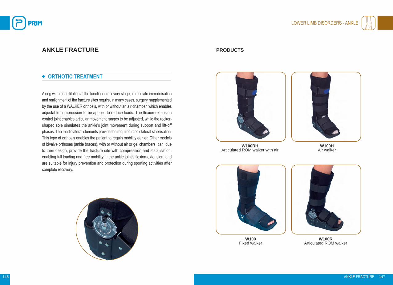
W100RH Articulated ROM walker with air
W100H Air walker
146 ANKLE FRACTURE 147
ORTHOTIC TREATMENT
Along with rehabilitation at the functional recovery stage, immediate immobilisation and realignment of the fracture sites require, in many cases, surgery, supplemented by the use of a WALKER orthosis, with or without an air chamber, which enables adjustable compression to be applied to reduce loads. The flexion-extension control joint enables articular movement ranges to be adjusted, while the rocker-shaped sole simulates the ankle’s joint movement during support and lift-off phases. The mediolateral elements provide the required mediolateral stabilisation. This type of orthosis enables the patient to regain mobility earlier. Other models of bivalve orthoses (ankle braces), with or without air or gel chambers, can, due to their design, provide the fracture site with compression and stabilisation, enabling full loading and free mobility in the ankle joint’s flexion-extension, and are suitable for injury prevention and protection during sporting activities after complete recovery.
ANKLE FRACTURE PRODUCTS
W100 Fixed walker
LOWER LIMB DISORDERS - ANKLE
W100R Articulated ROM walker
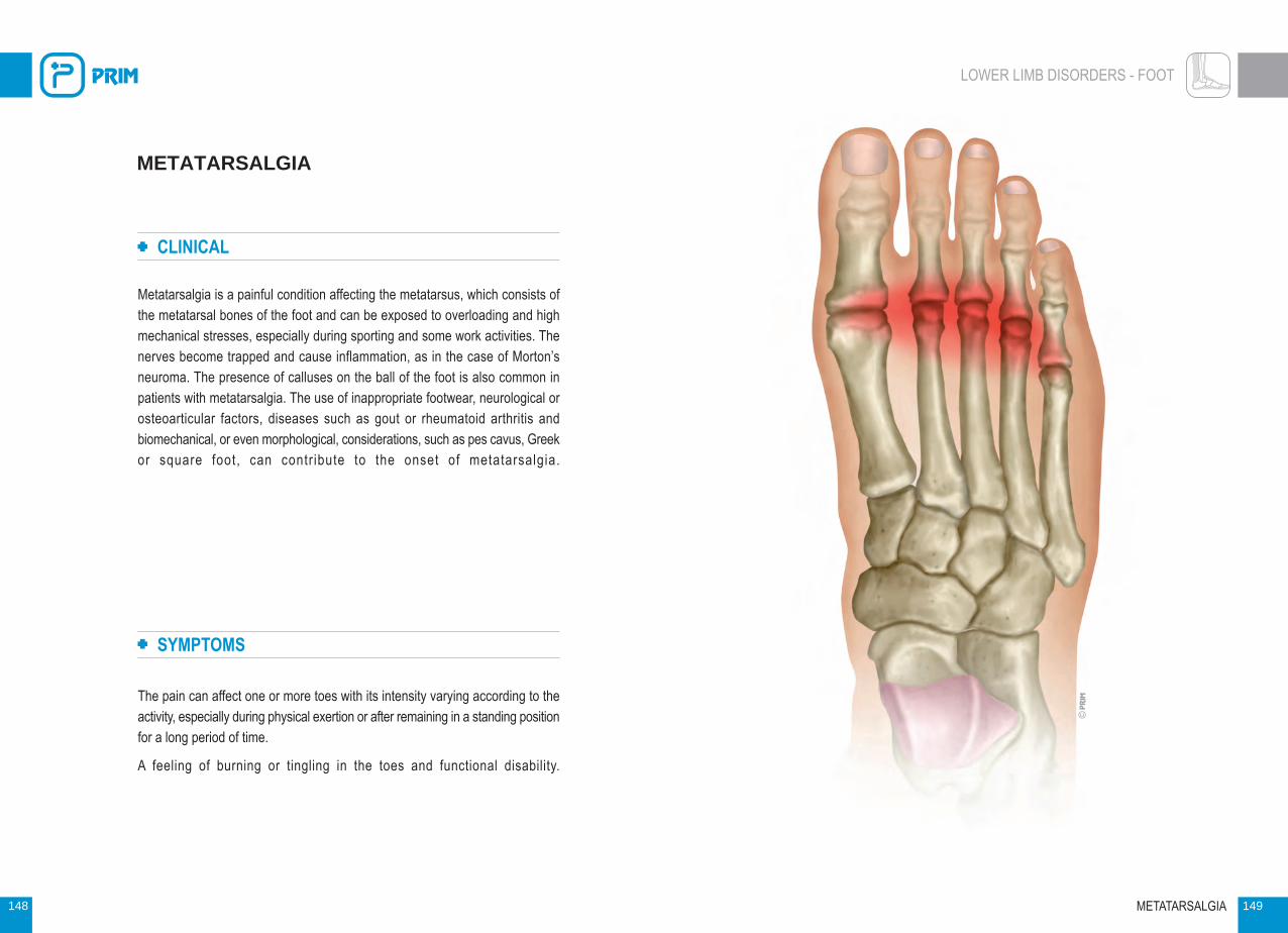
LOWER LIMB DISORDERS - FOOT
148
METATARSALGIA
CLINICAL
Metatarsalgia is a painful condition affecting the metatarsus, which consists of the metatarsal bones of the foot and can be exposed to overloading and high mechanical stresses, especially during sporting and some work activities. The nerves become trapped and cause inflammation, as in the case of Morton’s neuroma. The presence of calluses on the ball of the foot is also common in patients with metatarsalgia. The use of inappropriate footwear, neurological or osteoarticular factors, diseases such as gout or rheumatoid arthritis and biomechanical, or even morphological, considerations, such as pes cavus, Greek or square foot, can contr ibute to the onset of metatarsalgia.
SYMPTOMS
The pain can affect one or more toes with its intensity varying according to the activity, especially during physical exertion or after remaining in a standing position for a long period of time.
A feeling of burning or tingling in the toes and functional disability.
METATARSALGIA 149

LOWER LIMB DISORDERS - FOOT
P423 Plantar protector
P421 Band with metatarsal pad
150 METATARSALGIA 151
ORTHOTIC TREATMENT
Specific treatments for the disorders responsible for metatarsalgia (gout, arthritis, etc.), together with ceasing activities that overload the area, are essential to ensure that the orthotic treatment can successfully perform its function. In order to minimise overloading of the metatarsal heads, plantar orthoses featuring a bar or retrocapital support can be used to transfer loads to the metatarsal shafts and, in some cases, feature an internal and external longitudinal arch to distribute loads. These unloading supports, situated in plantar orthoses, can be made from different materials with different shores and have varying dimensions to suit the patient’s weight and activity, and provide the necessary cushioning and support for each case. Metatarsal straps fitted with retrocapital pads compress and restructure the anterior arch of the foot, thus preventing overloading of the central metatarsals.
METATARSALGIA PRODUCTS
P416 Metatarsal pad with
reinforced ring
P456 Full lined gel insole

LOWER LIMB DISORDERS - FOOT
152
PLANTAR FASCIITIS
CLINICAL
Plantar fasciitis is characterised by acute inflammation of the plantar fascia of the foot, usually caused by repetitive microtrauma, in which the thick connective tissue located in the sole of the foot at its insertion into the calcaneus becomes inflamed. Factors such as inappropriate footwear, mechanical exertion in incorrect postures and repetit ive movements can cause or aggravate it.
It is often associated with the presence of a heel spur, brought on by a periosteal reaction, because of the increase in the traction of the medial musculature of the foot.
SYMPTOMS
Pain that is usually felt in the anterior-medial base of the heel, becoming more acute when the patient gets up in the morning due to nighttime stiffness, but decreasing during the day with moderate activities. This pain, however, increases with mechanical exert ion, which requires greater overloading.
PLANTAR FASCIITIS 153

LOWER LIMB DISORDERS - FOOT
P414 Insole for rest
154 PLANTAR FASCIITIS 155
ORTHOTIC TREATMENT
Treatment is usually a combination of rehabilitation techniques, corticosteroids and the use of plantar orthoses with appropriate footwear.
Plantar orthoses facilitate the distribution of loads by providing selective distribution of plantar loads and forces, thereby enabling the plantar fascia to relax and reducing medial plantar tension. In acute cases, the use of postural orthoses at night to keep the foot at a neutral 90° angle can provide the relaxation needed to prevent the appearance of stiffness.
PLANTAR FASCIITIS PRODUCTS
P415Lined insole for rest
P441 Non-slip contoured insole
P451Double comfort insole

LOWER LIMB DISORDERS - FOOT
156
CALCANEAL SPUR
CLINICAL
A calcaneal spur is a bone spur or calcification in the base of the calcaneus at the insertion of the plantar fascia which can cause complications, such as inflammation of the plantar fascia, resulting in plantar fasciitis.
It is usually caused by excessive traction or stretching of the periosteum of the calcaneus by the plantar fascia. Flat or cavus feet and Achilles tendon contractures can also cause heel spurs as a result of increased plantar fascia tension.
SYMPTOMS
The primary symptom is pain, specifically located in the loading area of the heel. In very rare cases, redness, inflammation or other skin conditions are evident.
It is common in people who play sport or those who are overweight, and the overload can cause tears in its posterior insertion and inflammation of the tissue surrounding the bone.
CALCANEAL SPUR 157

LOWER LIMB DISORDERS - FOOT
P440 Silicone heel cup for calcaneal spur
P438 Silicone heel cup for calcaneal spur
158 CALCANEAL SPUR 159
ORTHOTIC TREATMENT
Rest and reduced everyday physical activities and sport. Rehabilitation techniques help to reduce the inflammation and, together with plantar orthoses designed for such a purpose, such as heel cups and insoles made from various materials, have the objective of selectively unloading the spur and relaxing the plantar fascia to minimise tension and overload.
Nighttime splints to keep the foot at rest at a 90° angle of flexion-extension are very useful for severe spurs associated with plantar fasci i t is.
CALCANEAL SPUR PRODUCTS
P457 Self-adhesive heel cup for
calcaneal spur
P439 Special calcaneal spur heel cup for
narrow shoes

LOWER LIMB DISORDERS - FOOT
160
HALLUX VALGUS
CLINICAL
Commonly known as a bunion, this is a complex deformity affecting the big toe, the first metatarsal and the metatarsophalangeal joint. In this deformity, the big toe is abducted with certain rotation (valgus) in the frontal plane, while the first metatarsal is adducted rotating in varus, creating an articular incongruity, resulting in the dislocation of the first metatarsophalangeal joint and affecting other structures and ligaments, the joint capsule and both the intrinsic and extrinsic musculature.
It can appear as a result of certain factors, such as abnormal development due to a pathomechanical deficit, wearing inappropriate footwear, pregnancy and menopause, and arthrosis, mainly affecting women. Today, it is considered a multicausal disorder in which genetics, footwear, hormonal and other factors may be present.
SYMPTOMS
Bone deformity with axial deviation of the first metatarsophalangeal joint. Pain due to pressure and/or friction caused by footwear.
Inflammation of soft tissue due to the bone deformity, accompanied by deformities such as hammertoe and claw toe in the adjacent toes.
HALLUX VALGUS 161

LOWER LIMB DISORDERS - FOOT
P435 Hallux valgus correction orthosis
P454 Daytime hallux valgus corrector
162 HALLUX VALGUS 163
ORTHOTIC TREATMENT
Preventative or conservative treatment by means of corrective splints for day and/or nighttime use can prevent the progression of the deformity when its onset and clinical manifestations, such as pain or signs of arthrosis, become evident. In the event of requiring surgery, splints, together with specific plantar orthoses, are very useful to ensure that the surgical correction is maintained. Plantar orthoses can improve the load distribution of the big toe and reduce pain in static and dynamic activities. Silicone or gel protectors used for exostosis can relieve pain when wearing shoes. In addition, post-surgery shoes can help patients to walk and stand while wearing a postoperative dressing during recovery.
HALLUX VALGUS PRODUCTS
P407 Daytime hallux valgus protector
P419 Hallux valgus protector with
reinforced ring

LOWER LIMB DISORDERS - FOOT
164
DROP FOOT
CLINICAL
Foot drop is characterised by a deformity in which the foot permanently remains in a position of plantar flexion, with functional limitation in performing dorsiflexion.
It can be flaccid or spastic, depending on the cause of the paralysis. The foot drop can be reducible or, conversely, the deformity is structured, making realignment impossible.
Foot drop is usually accompanied by other disorders, such as equinovarus (club foot) and equinovalgus, and, in some cases, is the result of paralysis of the tibialis anterior or an after-effect of poliomyelitis, stroke or neurological disease, such as multiple sclerosis or Duchenne muscular dystrophy.
SYMPTOMS
The foot is in permanent plantar flexion, which can be reducible, as in flaccid paralysis, or irreducible, as in the case of spastic paralysis or structured deformities.
Possible loss of feeling and steppage gait in flaccid paralysis. In irreducible foot drop, hip and knee flexion is evident to compensate for the asymmetry caused by the structured or irreducible plantar flexion.
DROP FOOT 165

LOWER LIMB DISORDERS - FOOT
NA410 Drop-foot orthosis
D81 Rancho de los Amigos
drop-foot orthosis
166 DROP FOOT 167
ORTHOTIC TREATMENT
Rehabilitation and surgical treatment, in some cases, also require postural or corrective orthoses to prevent foot drop, especially in patients that are bedridden for long periods (post-surgery, ICU, etc.).
Functional orthoses that enable dorsiflexion of the foot during the lift-off phase and plantar flexion in the heel support phase facilitate a more harmonious walking style with lower energy consumption and greater stability. Rancho de los Amigos-type polypropylene orthoses are suitable for flaccid paralysis without severe deformities. In the case of foot drop due to spastic paralysis, Klenzak-type orthoses with spring-loaded ankle joints offer great control, combined with anti-varus-valgus straps.
DROP FOOT PRODUCTS
ATX01Drop-foot orthosis
NA400Drop-foot orthosis

LOWER LIMB DISORDERS - FOOT
168
CLUB FOOT
CLINICAL
Club foot, or congenital talipes equinovarus, is clinically an equinus deformity, in which the hindfoot is inwardly angulated and supinated, while the forefoot is adducted, which can, in some cases, be associated with internal rotation of the tibia. Approximately 50% of cases are bilateral. Its etiology is diverse, and many different theories exist about its causes, such as mechanical disturbance due to poor uterine position, disorders during embryonic development, genetic causes due to malformation in chromosome maps, or those that consider it to be a result of an imbalance in internal and external peroneal musculature.
In club foot, osteoarticular and musculoligamentous disorders are evident.
Other congenital foot deformities include those that affect the forefoot, such as metatarsus varus and metatarsus varus-adductus, distinguished by their clinical presentation and evolution, which can involve supination of the forefoot at the Lisfranc joint.
SYMPTOMS
An equinus deformity with hindfoot varus-supination and forefoot adduction.
Metatarsus varus is characterised by a deviation in forefoot adduction, while metatarsus varus-adductus shows adduction and supination in the Lisfranc joint, which can be combined in some cases with adduction-supination in Chopart’s joint.
CLUB FOOT 169
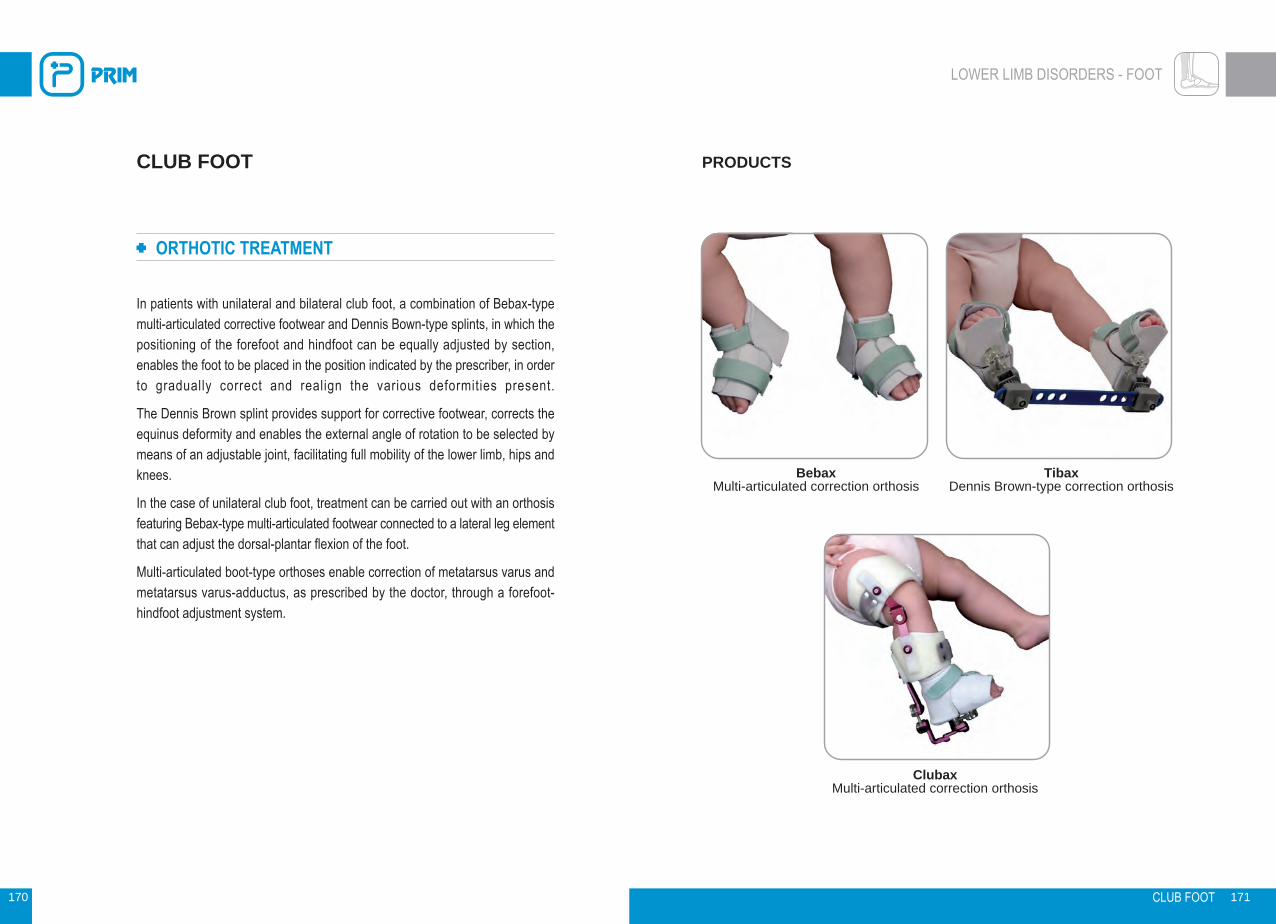
LOWER LIMB DISORDERS - FOOT
Bebax Multi-articulated correction orthosis
Tibax Dennis Brown-type correction orthosis
170 CLUB FOOT 171
ORTHOTIC TREATMENT
In patients with unilateral and bilateral club foot, a combination of Bebax-type multi-articulated corrective footwear and Dennis Bown-type splints, in which the positioning of the forefoot and hindfoot can be equally adjusted by section, enables the foot to be placed in the position indicated by the prescriber, in order to gradually correct and realign the various deformities present.
The Dennis Brown splint provides support for corrective footwear, corrects the equinus deformity and enables the external angle of rotation to be selected by means of an adjustable joint, facilitating full mobility of the lower limb, hips and knees.
In the case of unilateral club foot, treatment can be carried out with an orthosis featuring Bebax-type multi-articulated footwear connected to a lateral leg element that can adjust the dorsal-plantar flexion of the foot.
Multi-articulated boot-type orthoses enable correction of metatarsus varus and metatarsus varus-adductus, as prescribed by the doctor, through a forefoot-hindfoot adjustment system.
CLUB FOOT PRODUCTS
Clubax Multi-articulated correction orthosis

LOWER LIMB DISORDERS - FOOT
172
FOOT FRACTURE
CLINICAL
Various types of fractures can occur in the foot, given its complex osteoarticular system and its high work rate. Some fractures are more common, such as metatarsal fractures, which can occur in the diaphysis or neck.
Toe fractures are usually caused by crushing or direct blows, the first and fifth toes being most commonly affected, usually due to trauma.
Subtalar dislocation is usually associated with fractures of the posterior process of the talus.
Calcaneal fractures can occur as a result of falls from a certain height, in which the calcaneus and the antero-external and postero-external subtalar joint absorb all of the energy, affecting, in some cases, the scaphoid and cuboid due to their articular interrelationship, which can be very complex.
SYMPTOMS
In toe fractures, inflammation and ecchymosis can occur and an interphalangeal sprain of the 5th toe can appear.
Metatarsal fractures cause pain in the forefoot, with oedema and dorsal and plantar ecchymosis, and prevent the patient from walking.
Calcaneus and subtalar joint fracture can produce severe pain in the heel, tarsal region and ankle, oedema and plantar ecchymosis. Intense pain on heel percussion and eversion-inversion movements. The tibia-tarsus joint retains movement, but it is limited by pain.
FOOT FRACTURE 173
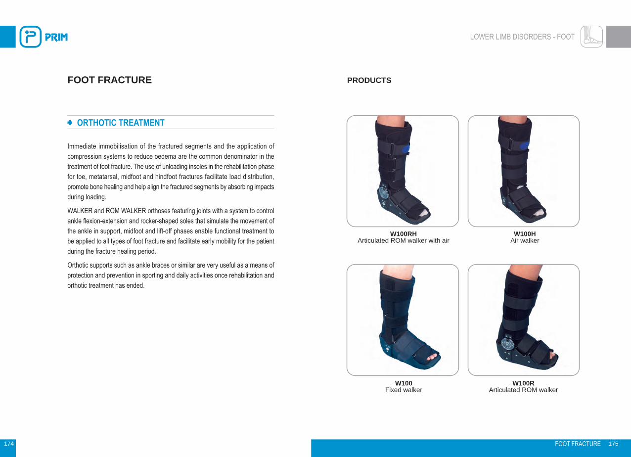
LOWER LIMB DISORDERS - FOOT
W100RH Articulated ROM walker with air
W100H Air walker
174 FOOT FRACTURE 175
ORTHOTIC TREATMENT
Immediate immobilisation of the fractured segments and the application of compression systems to reduce oedema are the common denominator in the treatment of foot fracture. The use of unloading insoles in the rehabilitation phase for toe, metatarsal, midfoot and hindfoot fractures facilitate load distribution, promote bone healing and help align the fractured segments by absorbing impacts during loading.
WALKER and ROM WALKER orthoses featuring joints with a system to control ankle flexion-extension and rocker-shaped soles that simulate the movement of the ankle in support, midfoot and lift-off phases enable functional treatment to be applied to all types of foot fracture and facilitate early mobility for the patient during the fracture healing period.
Orthotic supports such as ankle braces or similar are very useful as a means of protection and prevention in sporting and daily activities once rehabilitation and orthotic treatment has ended.
FOOT FRACTURE PRODUCTS
W100 Fixed walker
W100R Articulated ROM walker

NOTES NOTES