pathogen screening and gill na /k atpase … k. 2014. pathogen...pathogen screening and gill na+/k+-...
TRANSCRIPT

Pathogen Screening and Gill Na+/K+- ATPase Assessment of South Delta Chinook and Steelhead 2014 Release Groups
Ken Nichols
September 2014
U.S. Fish & Wildlife Service
US Fish and Wildlife Service California-Nevada Fish Health Center 24411 Coleman Fish Hatchery Rd Anderson, CA 96007 (530) 365-4271 Fax: (530) 365-7150 http://www.fws.gov/canvfhc/

SUMMARY
The health and physiological condition of the study fish can help explain their performance and survival during the studies. Juvenile Chinook salmon and steelhead trout were surveyed for specific fish pathogens and smolt development using gill Na+/K+-ATPase (gill ATPase) activity levels. In both steelhead and Chinook release groups, survival over the 24 holding period was high. No significant pathogen infections were detected in Chinook or steelhead release groups. Gill ATPase levels were stable or increasing over the study period suggesting levels of smolt development would not be a factor in fish performance.
Recommended citation for this report is:
Nichols, K. 2014. Pathogen Screening and Gill Na+/K+- ATPase Assessment of South Delta Chinook and Steelhead 2014 Release Groups. U.S. Fish & Wildlife Service, California-Nevada Fish Health Center, Anderson, CA. Available: http://www.fws.gov/canvfhc/reports.asp.
Notice:
The mention of trade names or commercial products in this report does not constitute endorsement or recommendation for use by the Federal government. The findings and conclusions in this report are those of the author and do not necessarily represent the views of the US Fish and Wildlife Service.
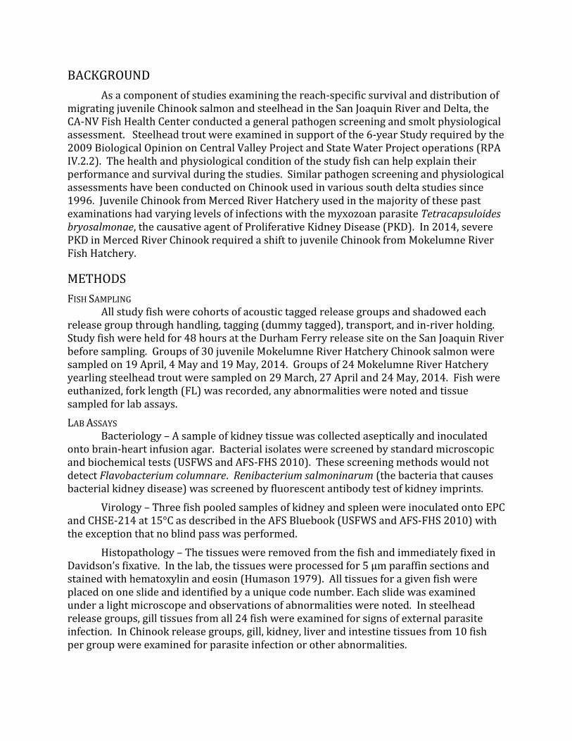
BACKGROUND
As a component of studies examining the reach-specific survival and distribution of migrating juvenile Chinook salmon and steelhead in the San Joaquin River and Delta, the CA-NV Fish Health Center conducted a general pathogen screening and smolt physiological assessment. Steelhead trout were examined in support of the 6-year Study required by the 2009 Biological Opinion on Central Valley Project and State Water Project operations (RPA IV.2.2). The health and physiological condition of the study fish can help explain their performance and survival during the studies. Similar pathogen screening and physiological assessments have been conducted on Chinook used in various south delta studies since 1996. Juvenile Chinook from Merced River Hatchery used in the majority of these past examinations had varying levels of infections with the myxozoan parasite Tetracapsuloides bryosalmonae, the causative agent of Proliferative Kidney Disease (PKD). In 2014, severe PKD in Merced River Chinook required a shift to juvenile Chinook from Mokelumne River Fish Hatchery.
METHODS
FISH SAMPLING All study fish were cohorts of acoustic tagged release groups and shadowed each
release group through handling, tagging (dummy tagged), transport, and in-river holding. Study fish were held for 48 hours at the Durham Ferry release site on the San Joaquin River before sampling. Groups of 30 juvenile Mokelumne River Hatchery Chinook salmon were sampled on 19 April, 4 May and 19 May, 2014. Groups of 24 Mokelumne River Hatchery yearling steelhead trout were sampled on 29 March, 27 April and 24 May, 2014. Fish were euthanized, fork length (FL) was recorded, any abnormalities were noted and tissue sampled for lab assays.
LAB ASSAYS Bacteriology – A sample of kidney tissue was collected aseptically and inoculated
onto brain-heart infusion agar. Bacterial isolates were screened by standard microscopic and biochemical tests (USFWS and AFS-FHS 2010). These screening methods would not detect Flavobacterium columnare. Renibacterium salmoninarum (the bacteria that causes bacterial kidney disease) was screened by fluorescent antibody test of kidney imprints.
Virology – Three fish pooled samples of kidney and spleen were inoculated onto EPC and CHSE-214 at 15°C as described in the AFS Bluebook (USFWS and AFS-FHS 2010) with the exception that no blind pass was performed.
Histopathology – The tissues were removed from the fish and immediately fixed in Davidson’s fixative. In the lab, the tissues were processed for 5 μm paraffin sections and stained with hematoxylin and eosin (Humason 1979). All tissues for a given fish were placed on one slide and identified by a unique code number. Each slide was examined under a light microscope and observations of abnormalities were noted. In steelhead release groups, gill tissues from all 24 fish were examined for signs of external parasite infection. In Chinook release groups, gill, kidney, liver and intestine tissues from 10 fish per group were examined for parasite infection or other abnormalities.

Gill ATPase – Gill Na+/K+-Adenosine Triphosphatase (gill ATPase) activity was assayed by the method of McCormick (1993). Gill ATPase activity is correlated with osmoregulatory ability in saltwater, and high concentrations are found in the chloride cells of the lamellae.
RESULTS
FISH CONDITION Chinook – Prior to the health assessment, one fish died in 19 April group and no
mortality occurred in the 4 May or 19 May release groups (Table 1). A penetrating abdominal wound (external abnormality) and degenerated intestine (internal abnormality) were noted on this single mortality. No significant scale loss or pale gills were noted in any of the Chinook health sample groups. Overall, sutures from tagging surgery were in good conditions with minor inflammation noted in 3% (1/30) of fish sampled 19 April; a loose suture noted in 3% of (1/30) fish sampled 4 May; and minor hemorrhaging noted in 13% (4/30) of fish sampled 19 May.
Table 1. Chinook release group mean (± sd) fork length (FL), mortality over the 48 hr. holding period, fish with external abnormalities (Ext Abn), fish with internal abnormalities (Int Abn) and number of fish sampled for lab assays (N).
Group FL (mm) Mortality Ext Abn Int Abn N
19 April 96.2 ±5.1 1/30 (3%) 0/30 (0%) 0/30 (0%) 29
4 May 101.2 ±4.4 0/30 (0%) 0/30 (0%) 0/30 (3%) 30
19 May 99.5 ±5.2 0/30 (0%) 0/30 (0%) 0/30 (3%) 30
Steelhead – One fish died prior to the health assessment in the 29 March release group, and no mortality occurred in the 27 April or 24 May groups (Table 2). No wounds or clinical signs of infection were observed on the single mortality. In the 29 March health assessment group, no significant external or internal abnormalities were noted, and minor hemorrhaging or inflammation (Figure 1) at the suture site was observed in 21% (5/24) of fish. In the 27 April health assessment group, cloudy eyes were noted in 4% (1/24) of fish, and partly open or bleeding sutures were observed in 8% (2/24) of fish. In the 24 May group, significant scale loss (>50% of body) was noted in 13% (3/24) of fish; 4% (1/24) of fish had eye abnormalities; and minor hemorrhaging or partly open sutures were noted in 21% (5/24) of fish.

Table 2. Steelhead release group mean (± sd) fork length (FL), mortality over the 48 hr. holding period, fish with external abnormalities (Ext Abn), fish with internal abnormalities (Int Abn) and number of fish sampled for lab assays (N).
Group FL (mm) Mortality Ext Abn Int Abn N
29 March 240 (±14) 1/24 (4%) 0/24 (0%) 0/24 (0%) 23
27 April 250 (±13) 0/24 (0%) 1/24 (4%) 0/24 (0%) 24
24 May 249 (±17) 0/24 (0%) 4/24 (17%) 0/24 (0%) 24
BACTERIOLOGY AND VIROLOGY In both Chinook and steelhead sample groups, no virus or other cytopathic effects
were observed by cell culture over the 21 day incubation period. No obligate bacterial pathogens were detected, and other isolates were isolated in 3-24% of sample groups (Table 3). These other isolates were common fauna in the environment and fishes GI tract (Aoki 1999) and were likely contaminates due to field sampling conditions.
Figure 1. Examples of normal sutures and minor hemorrhaging at suture site in fish assessed after holding for 48 hours.

Table 3. Summary of bacteria isolated from the kidneys of dummy tagged fish.
Species Aeromonas /Pseudomonas various Gram positive bacteria
Chinook 3% (3/87) 17% (15/87)
Steelhead 11% (8/71) 24% (17/71)
HISTOPATHOLOGY Chinook – No significant abnormalities or signs of infection were detected in tissues
from the 30 fish examined.
Steelhead – No significant abnormalities were observed on the gills of 69 fish examined; however, subclinical parasite infections were observed. Light infections with Capriniana piscium (Figure 2A, formerly known as Trichophrya, presumptive identification) were observed in 75% (52/69) of gills. Cyst-like xenoma (Figure 2B) caused by an unidentified microsporidian were observed in 3% (2/69) of gill samples. There was no associated lesion or other sign of impairment associated with these infections.
Figure 2. Histology sections (H&E stained) of steelhead gills with (A) Capriniana piscium (formerly Trichophrya) infections, and (B) Cyst-like xenoma. Note the absence of significant inflammation or lesion in both infections.
GILL ATPASE ACTIVITY Chinook – Gill ATPase activity levels (µmol ADP*mg protein-1*hr-1) ranged from 0.6
to 14.3. Two fish from the 19 April sample group were excluded from the analysis due extremely high activity levels which were likely errors in the protein measurement. The activity levels in the 4 May release group were lower than the 19 April and 19 May groups (Figure 3, P<0.001, ANOVA).

Figure 3. Boxplot of median gill ATPase activity (µmol ADP·mg protein-1·hr-1) in juvenile Chinook salmon sampled from the 19 April, 4 May and 19 May release groups. Groups with letter subscripts in common were not significantly different (P<0.001, ANOVA).
Steelhead – Gill ATPase activity levels (µmol ADP*mg protein-1*hr-1) ranged from 0.2 to 5.7. Activity levels tended to increase with the highest levels observed in the May release group (Figure 4, P=0.008, ANOVA).
DISCUSSION
No significant health issues were observed in either the Chinook or steelhead release groups in 2014. The Chinook salmon from Mokelumne River Hatchery used in the study this year did not have any signs of T. bryosalmonae infections common in the Merced River Hatchery Chinook during past years. The minor suture issues observed in both Chinook and steelhead release groups were observed in only a few individuals and did not impact overall health of the fish. Several steelhead from the 24 May release group were observed to have significant scale loss which may have been an indication of higher smolt development.
Gill ATPase activity levels were stable or increasing over the study period suggesting smolt development would not be a significant factor in fish performance. Gill ATPase activity in salmonids typically increases and peaks near the time of most active

migratory behavior (Duston, Saunders and Knox 1991; Ewing, Ewing and Satterthwaite 2001; Wedemeyer 1996). In Chinook sample groups, gill ATPase levels were similar in the first (19 April) and last (19 May) release groups suggesting these fish were not yet past time peak smolt development. The cause of the lower median gill ATPase levels observed in the second (4 May) Chinook release group was not apparent. While in steelhead sample groups, gill ATPase levels increased over time the relationship with migration behavior may not be consistent. In unpublished CA-NV Fish Health Center data, steelhead have demonstrated the ability to significantly increase activity levels in only a few days following hatchery release.
Figure 4. Boxplot of median gill ATPase activity (µmol ADP·mg protein-1·hr-1) in juvenile steelhead from the March, April and May release groups. Groups with letter subscripts in common were not significantly different (P=0.008, ANOVA).
ACKNOWLEDGMENTS
Biologists with the USFWS Stockton FWO provided help with logistics and fish handling. Scott with at the CA-NV Fish Health Center provided valuable assistance with field sampling and laboratory assays.

REFERENCES
Aoki T. 1999. Motile Aeromonads. Chapter 11 In: Fish Diseases and Disorders, Vol. 3: Viral, Bacterial and Fungal Infections, Woo P T K and Bruno D W, editors, CABI Pub. New York.
Duston J, R L Saunders and D E Knox. 1991. Effects of increases in freshwater temperature on loss of smolt characteristics in Atlantic salmon (Salmo salar). Canadian Journal of Aquatic Animal Sciences 48: 164-169.
Ewing R D, G S Ewing and T D Satterthwaite. 2001. Changes in gill Na+, K+-ATPase specific activity during seaward migration of wild juvenile Chinook salmon. Journal of Fish Biology 58: 1414-1426.
Humason G L. 1979. Animal Tissue Techniques, 4th edition. W H Freeman and Co., San Francisco.
McCormick S D. 1993. Methods for Nonlethal Gill Biopsy and Measurement of Na+, K+-ATPase Activity. Canadian Journal of Fisheries and Aquatic Sciences. 50: 656-658.
USFWS and AFS-FHS (U.S. Fish and Wildlife Service and American Fisheries Society-Fish Health Section). 2010. Standard procedures for aquatic animal health inspections. In AFS-FHS. FHS blue book: suggested procedures for the detection and identification of certain finfish and shellfish pathogens, 2010 edition. AFS-FHS, Bethesda, Maryland.
Wedemeyer G A. 1996. Physiology of Fish in Intensive Culture Systems. Chapman & Hall, New York.