patellar instability: looking outside the mpfl · pdf filepatellar instability: looking...
TRANSCRIPT

8/10/2016
1
Patellar Instability: Looking Outside the MPFL
Jack Farr, MD
OrthoIndy Cartilage Restoration Center
Indianapolis, IN
Financial Disclosures
Royalties• Arthrex T3 AMZ jig• Depuy/Synthes PFAConsultant• Aesculap• Arthrex• ArthoCare• BioMet• DePuy• Exactech• MedShape• NuTech• Mitek• Moximed• RTI• Joint Preservation• Vericel (formerly Sanofi/Genzyme)• Schwartz Biomedical• Skeletal Kinetics• ISTO• Zimmer• SBMFellowship Grants• Arthrex• Ossur• Biomet
Table 4. Concomitant Procedures
Procedure Indication
Femoral rotational osteotomyIncreased femoral anteversion
Rotational Tibial Osteotomy Increased external tibial torsion
Varus Producing Femoral Osteotomy Genu valgum
TTO Abnormal TT‐PCL, Abnormal Caton‐Deschamps Ratio
MPFL repair/reconstruction Incompetence of the MPFL
Lateral release/lengthening Fixed patella tilt with lateral retinacular tightness
Trochleoplasty Not currently indicated as adjunct to cartilage restoration
AAOS Let’s Discuss: Joint Preservation of the Knee Chapter 13: Issues Specific to Cartilage Restoration in the Patellofemoral Joint Sherman SL, Nuelle C, Farr J

8/10/2016
2
Outside the MPFL
• Proximal Medial‐‐Yanke
• Axial and Coronal Alignment
• Proximal Lateral
• Distal (tibial tuberosity)
• Trochlear Dysplasia
• Patellar Alta
Excessive Femoral Anteversion
Derotation of Femur

8/10/2016
3
Valgus Malalignment
Lateral Lengthening
Superficial oblique layer of lateral retinaculum incised adjacent to patella
Exposure for concomitant patellar chondral restoration with AMZ
Deep Layer of Lateral Retinaculum
Lateral Lengthening
A second step‐cut is 2 cm posterior through the deep transverse/capsule/synovium: Repair allows 1.5‐2.0 cm lengthening
Deep Transverse Layer
Superficial Oblique Layer

8/10/2016
4
Tibial Tuberosity Osteotomy
• TT‐TG distance > 20mm—BUT DON’T STOP THERE It’s just a number…………that is not infallible
• Assess Femoral/Tibial rotational abnormalities
e.g., Increased femoral anteversion: CT (Teitge); MRI (Noyes) to assess hip/knee/ankle rotation
If Marked Dysplasia,There is no TG
Courtesy of David Dejour MD
TT‐Posterior Cruciate Ligament
Seitlinger et al AJSM 2012
• Control 18.4 mm
95% < 24 mm
• Instability Patients
38% > 24 mm
57% with TT‐TG > 20 had TT‐PCL > 24 mm
43% with TT‐TG > 20 had TT‐PCL < 24 mm (could these have femoral and/or TF rotation?)
Seitlinger AJSM 2012
8/10/2016
5
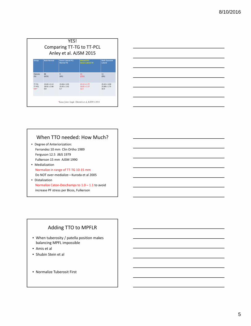
YES!Comparing TT‐TG to TT‐PCLAnley et al. AJSM 2015
Group Both Normal Excess Lateral PCLNormal TG
Normal PCLExcess Lateral TG
Both ExcessiveLateral
Patients(%)
88(62%)
9(6%)
33(23%)
11(8%)
TT‐TGTT‐PCLKJA*
15.04 ± 3.1218.92 ± 2.666.8
15.06 ± 3.5525.92 ± 2.426.7
23.22 ± 2.7720.82 ± 2.2712.5
25.42 ± 3.9925.88 ± 1.7510.3
*Knee Joint Angle Dietrich et al, KSSTA 2014
When TTO needed: How Much?• Degree of Anteriorization:
Ferrandez 10 mm Clin Ortho 1989
Ferguson 12.5 JBJS 1979
Fulkerson 15 mm AJSM 1990
• Medialization
Normalize in range of TT‐TG 10‐15 mm
Do NOT over‐medialize—Kuroda et al 2005
• Distalization
Normalize Caton‐Deschamps to 1.0 – 1.1 to avoid
increase PF stress per Bicos, Fulkerson
Adding TTO to MPFLR
• When tuberosity / patella position makes balancing MPFL impossible
• Amis et al
• Shubin Stein et al
• Normalize Tuberosit First

8/10/2016
6
RPI with Chronic Patellar SubluxationDO NOT PULL Patella into Position
Creating Sloped Osteotomy
Saw through cutting block and tibia exiting on retractor
AMZ cutting block with slope selector
Conflict of Interest Disclosure
Farr on Design team for the Tracker AMZ Jig System (DePuy Mitek) and T3 (Arthrex)
Alternative AMZ Jig System
Conflict of Interest Disclosure: Design Surgeons Farr, Cole, Nawab; Arthrex

8/10/2016
7
Completing the Osteotomy
Osteotomes connect posterior cut proximally
Saw using first cut as captured guide
Steep osteotomy completed with free tuberosity pedicle
Tuberosity fixed with 2 interfragmentary screwsAnterized 15mm; medialized 7mm
8/10/2016
8
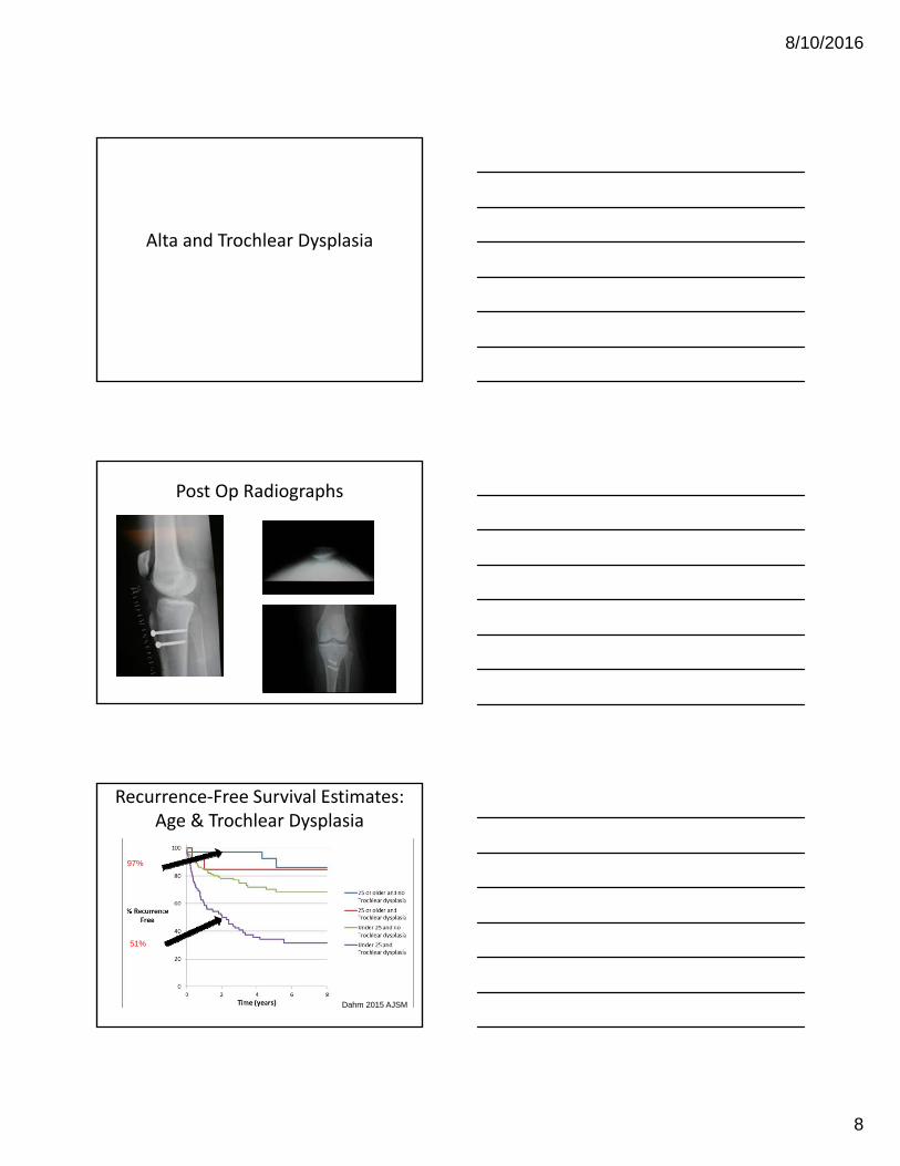
Alta and Trochlear Dysplasia
Post Op Radiographs
Recurrence‐Free Survival Estimates: Age & Trochlear Dysplasia
97%
51%
Dahm 2015 AJSM

8/10/2016
9
Recurrence‐Free Survival Estimates: Trochlear Dysplasia & Patella Alta
48%
83%
Dahm 2015 AJSM
Conclusions
• Trochlear dysplasia is the strongest risk factor for recurrent patellar instability
• Patients of any age with trochlear dysplasia: > 3.2x increased risk of recurrence
• Trochlear dysplasia & patella alta: 4.2x
Dahm 2015 AJSM
RPI
• The 2nd strongest risk factor was age
• Age (yrs) (%) w/ recurrent PF instability
≥ 40 0%
≥ 25 16%
< 25 47%

8/10/2016
10
Proposed algorithm
Patellar AltaCaton‐Deschamps > 1.2
36.4mm
26mm
36.5mm /26.3mm
= 1.4
42 mm
26.3mm
Distalization to treat Patellar AltaMayer et al. AJSM 2012
“V” shape cutsMeasure bone to be resectedallowing for saw blade kerf
8/10/2016
11
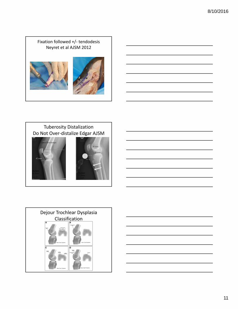
Fixation followed +/‐ tendodesisNeyret et al AJSM 2012
Tuberosity DistalizationDo Not Over‐distalize Edgar AJSM
36.4mm
26mm
36.5mm26.3mm
= 1.39
Caton-Deschamps
28.4mm27.2mm= 1.03
28 mm
28 mm
42 mm
Dejour Trochlear Dysplasia Classification

8/10/2016
12
Trochlear Dysplasia
Deepening Trochleoplasty: Caution
Formation Gentle Groove

8/10/2016
13
Thank You