patellar groove replacement in patellar luxation with ... · patellar groove replacement in...
TRANSCRIPT
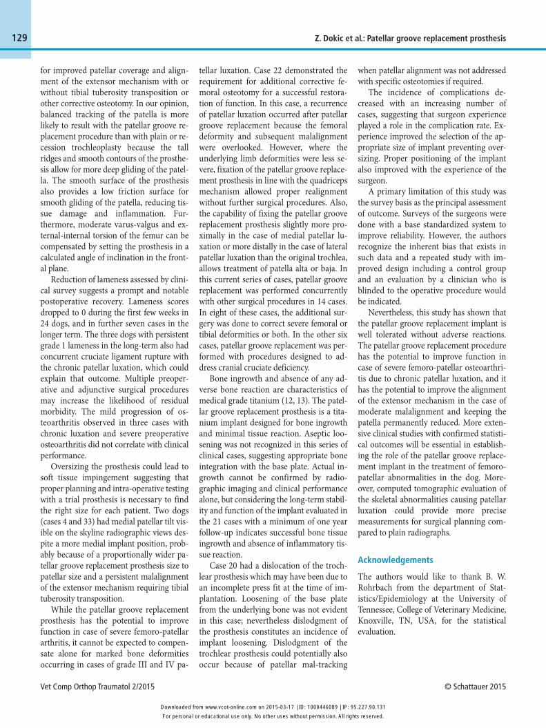
© Schattauer 2015 Vet Comp Orthop Traumatol 2/2015
124Clinical Communication
Patellar groove replacement in patellar luxation with severe femoro-patellar osteoarthritisZ. Dokic1; D. Lorinson1; J. P. Weigel2; A. Vezzoni31Chirurgische Überweisungspraxis, Vösendorf, Austria; 2College of Veterinary Medicine, Department of Small Animal Clinical Sciences, University of Tennessee, Knoxville, TN, USA; 3Clinica Veterinaria Vezzoni srl, Cremona, Italy
KeywordsDog, patellar luxation, partial stifle joint prosthesis
SummaryObjective: To report a novel method of treat-ing femoro-patellar instability in association with severe femoro-patellar osteoarthritis, by substituting the femoral trochlear with a pa-tellar groove replacement prosthesis. Study design: Retrospective case series.Methods: Preoperative lameness was scored from 0–4, and radiographic studies including standard positions for patellar luxation were obtained for evidence of malalignment and femoro-patellar osteoarthritis. Cases with or without previous surgeries were included. The size of trochlear implant was determined by transparent templates and confirmed intra-operatively with trials. Radiographic im-ages, together with clinical examinations, were reviewed immediately and at three
months postoperatively and at longer term when available. Results: Thirty-five cases of patellar luxation ranging from grades II to IV were included. Eleven of these cases had prior surgical inter-ventions which failed to stabilize the patella. Fourteen dogs required additional surgical procedures in conjunction with patellar groove replacement. Complications occurred in six patients, of which three required revi-sion. Complete resolution of subjectively- assessed lameness was evident in 24/35 cases by the third month and in another seven of 35 patients on the longer term re-evaluations. Clinical significance: Use of a patellar groove replacement prosthesis has the po-tential to decrease the lameness associated with severe femoro-patellar arthritis, to im-prove patellar stability, and to correct the alignment of the extensor mechanism.
Correspondence to:Aldo VezzoniClinica Veterinaria Vezzoni srlV. Massarotti, 24Cremona 261000ItalyPhone: +39 0372 23451E-mail: [email protected]
Vet Comp Orthop Traumatol 2015; 28: 124–130http://dx.doi.org/10.3415/VCOT-14-07-0106Received: July 13, 2014Accepted: December 10, 2014Epub ahead of print: February 4, 2015
IntroductionChronic instability and erosion of the femoro-patellar joint surfaces result in lameness and progressive osteoarthritis (1, 2). Osteochondral trochleoplasties have been used to deepen the trochlear groove to provide patellar stability, with or without additional surgical procedures (3, 4). How-
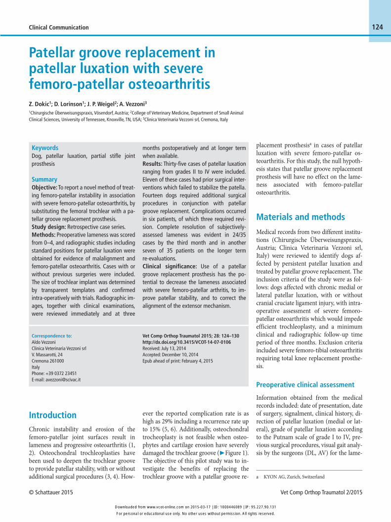
ever the reported complication rate is as high as 29% including a recurrence rate up to 15% (5, 6). Additionally, osteochondral trocheoplasty is not feasible when osteo-phytes and cartilage erosion have severely damaged the trochlear groove (▶ Figure 1). The objective of this pilot study was to in-vestigate the benefits of replacing the trochlear groove with a patellar groove re-
placement prosthesisa in cases of patellar luxation with severe femoro-patellar os-teoarthritis. For this study, the null hypoth-esis states that patellar groove replacement prosthesis will have no effect on the lame-ness associated with femoro-patellar osteoarthritis.
Materials and methods
Medical records from two different institu-tions (Chirurgische Überweisungspraxis, Austria; Clinica Veterinaria Vezzoni srl, Italy) were reviewed to identify dogs af-fected by persistent patellar luxation and treated by patellar groove replacement. The inclusion criteria of the study were as fol-lows: dogs affected with chronic medial or lateral patellar luxation, with or without cranial cruciate ligament injury, with intra-operative assessment of severe femoro- patellar osteoarthritis which would impede efficient trochleoplasty, and a minimum clinical and radiographic follow-up time period of three months. Exclusion criteria included severe femoro-tibial osteoarthritis requiring total knee replacement prosthe-sis.
Preoperative clinical assessment
Information obtained from the medical records included: date of presentation, date of surgery, signalment, clinical history, di-rection of patellar luxation (medial or lat-eral), grade of patellar luxation according to the Putnam scale of grade I to IV, pre-vious surgical procedures, visual gait analy-sis by the surgeons (DL, AV) for the lame-
a KYON AG, Zurich, Switzerland
For personal or educational use only. No other uses without permission. All rights reserved.Downloaded from www.vcot-online.com on 2015-03-17 | ID: 1000446089 | IP: 95.227.90.131
Vet Comp Orthop Traumatol 2/2015 © Schattauer 2015
125 Z. Dokic et al.: Patellar groove replacement prosthesis
ness score (7). Lameness was assessed sub-jectively at the walk and trot and graded from 0 to 4 using the following scale: 0 = no lameness; 1 = mild, intermittent weight-bearing lameness; 2 = persistent moderate weight-bearing lameness; 3 = persistent se-vere weight-bearing lameness, with or without intermittent non-weight bearing; and 4 = persistent non-weight-bearing lameness (▶ Appendix Table 1: Available online at www.vcot-online.com).
Implant design
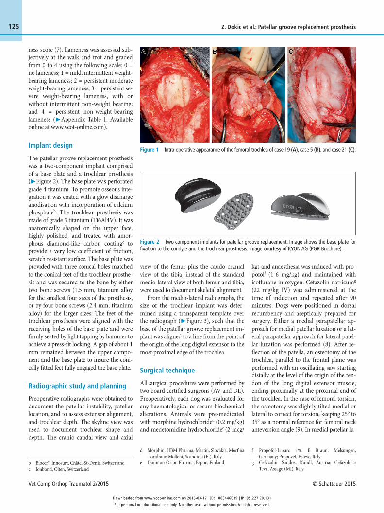
The patellar groove replacement prosthesis was a two-component implant comprised of a base plate and a trochlear prosthesis (▶ Figure 2). The base plate was perforated grade 4 titanium. To promote osseous inte-gration it was coated with a glow discharge anodisation with incorporation of calcium phosphateb. The trochlear prosthesis was made of grade 5 titanium (Ti6Al4V). It was anatomically shaped on the upper face, highly polished, and treated with amor-phous diamond-like carbon coatingc to provide a very low coefficient of friction, scratch resistant surface. The base plate was provided with three conical holes matched to the conical feet of the trochlear prosthe-sis and was secured to the bone by either two bone screws (1.5 mm, titanium alloy for the smallest four sizes of the prosthesis, or by four bone screws (2.4 mm, titanium alloy) for the larger sizes. The feet of the trochlear prosthesis were aligned with the receiving holes of the base plate and were firmly seated by light tapping by hammer to achieve a press-fit locking. A gap of about 1 mm remained between the upper compo-nent and the base plate to insure the coni-cally fitted feet fully engaged the base plate.
Radiographic study and planning
Preoperative radiographs were obtained to document the patellar instability, patellar location, and to assess extensor alignment, and trochlear depth. The skyline view was used to document trochlear shape and depth. The cranio-caudal view and axial
view of the femur plus the caudo-cranial view of the tibia, instead of the standard medio-lateral view of both femur and tibia, were used to document skeletal alignment.
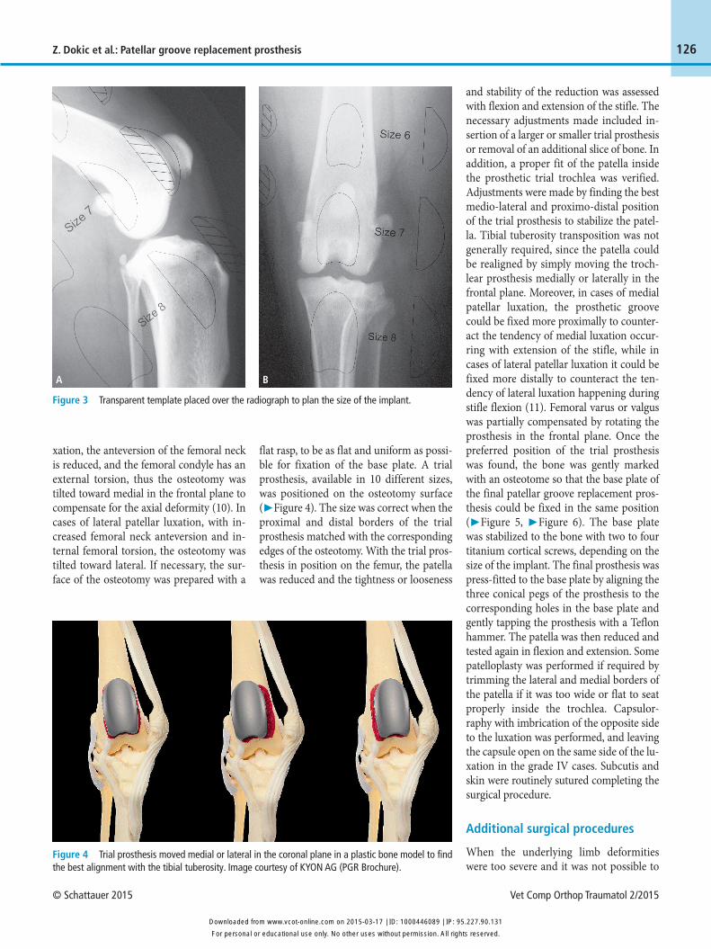
From the medio-lateral radiographs, the size of the trochlear implant was deter-mined using a transparent template over the radiograph (▶ Figure 3), such that the base of the patellar groove replacement im-plant was aligned to a line from the point of the origin of the long digital extensor to the most proximal edge of the trochlea.
Surgical technique
All surgical procedures were performed by two board certified surgeons (AV and DL). Preoperatively, each dog was evaluated for any haematological or serum biochemical alterations. Animals were pre-medicated with morphine hydrochlorided (0.2 mg/kg) and medetomidine hydrochloridee (2 mcg/
kg) and anaesthesia was induced with pro-pofolf (1-6 mg/kg) and maintained with isoflurane in oxygen. Cefazolin natricumg (22 mg/kg IV) was administered at the time of induction and repeated after 90 minutes. Dogs were positioned in dorsal recumbency and aseptically prepared for surgery. Either a medial parapatellar ap-proach for medial patellar luxation or a lat-eral parapatellar approach for lateral patel-lar luxation was performed (8). After re-flection of the patella, an osteotomy of the trochlea, parallel to the frontal plane was performed with an oscillating saw starting distally at the level of the origin of the ten-don of the long digital extensor muscle, ending proximally at the proximal end of the trochlea. In the case of femoral torsion, the osteotomy was slightly tilted medial or lateral to correct for torsion, keeping 25° to 35° as a normal reference for femoral neck anteversion angle (9). In medial patellar lu-
Figure 1 Intra-operative appearance of the femoral trochlea of case 19 (A), case 5 (B), and case 21 (C).
Figure 2 Two component implants for patellar groove replacement. Image shows the base plate for fixation to the condyle and the trochlear prosthesis. Image courtesy of KYON AG (PGR Brochure).
b Biocer®: Innosurf, Châtel-St-Denis, Switzerlandc Ionbond, Olten, Switzerland
f Propofol-Lipuro 1%: B Braun, Melsungen, Germany; Propovet, Esteve, Italy
g Cefazolin: Sandos, Kundl, Austria; Cefazolina: Teva, Assago (MI), Italy
d Morphin: HBM Pharma, Martin, Slovakia; Morfina cloridrato: Molteni, Scandicci (FI), Italy
e Domitor: Orion Pharma, Espoo, Finland
For personal or educational use only. No other uses without permission. All rights reserved.Downloaded from www.vcot-online.com on 2015-03-17 | ID: 1000446089 | IP: 95.227.90.131
© Schattauer 2015 Vet Comp Orthop Traumatol 2/2015
126
and stability of the reduction was assessed with flexion and extension of the stifle. The necessary adjustments made included in-sertion of a larger or smaller trial prosthesis or removal of an additional slice of bone. In addition, a proper fit of the patella inside the prosthetic trial trochlea was verified. Adjustments were made by finding the best medio-lateral and proximo-distal position of the trial prosthesis to stabilize the patel-la. Tibial tuberosity transposition was not generally required, since the patella could be realigned by simply moving the troch-lear prosthesis medially or laterally in the frontal plane. Moreover, in cases of medial patellar luxation, the prosthetic groove could be fixed more proximally to counter-act the tendency of medial luxation occur-ring with extension of the stifle, while in cases of lateral patellar luxation it could be fixed more distally to counteract the ten-dency of lateral luxation happening during stifle flexion (11). Femoral varus or valgus was partially compensated by rotating the prosthesis in the frontal plane. Once the preferred position of the trial prosthesis was found, the bone was gently marked with an osteotome so that the base plate of the final patellar groove replacement pros-thesis could be fixed in the same position (▶ Figure 5, ▶ Figure 6). The base plate was stabilized to the bone with two to four titanium cortical screws, depending on the size of the implant. The final prosthesis was press-fitted to the base plate by aligning the three conical pegs of the prosthesis to the corresponding holes in the base plate and gently tapping the prosthesis with a Teflon hammer. The patella was then reduced and tested again in flexion and extension. Some patelloplasty was performed if required by trimming the lateral and medial borders of the patella if it was too wide or flat to seat properly inside the trochlea. Capsulor-raphy with imbrication of the opposite side to the luxation was performed, and leaving the capsule open on the same side of the lu-xation in the grade IV cases. Subcutis and skin were routinely sutured completing the surgical procedure.
Additional surgical procedures
When the underlying limb deformities were too severe and it was not possible to
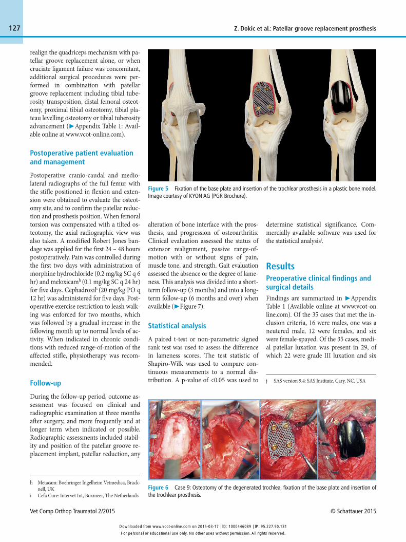
flat rasp, to be as flat and uniform as possi-ble for fixation of the base plate. A trial prosthesis, available in 10 different sizes, was positioned on the osteotomy surface (▶ Figure 4). The size was correct when the proximal and distal borders of the trial prosthesis matched with the corresponding edges of the osteotomy. With the trial pros-thesis in position on the femur, the patella was reduced and the tightness or looseness
Z. Dokic et al.: Patellar groove replacement prosthesis
xation, the anteversion of the femoral neck is reduced, and the femoral condyle has an external torsion, thus the osteotomy was tilted toward medial in the frontal plane to compensate for the axial deformity (10). In cases of lateral patellar luxation, with in-creased femoral neck anteversion and in-ternal femoral torsion, the osteotomy was tilted toward lateral. If necessary, the sur-face of the osteotomy was prepared with a
Figure 3 Transparent template placed over the radiograph to plan the size of the implant.
A B
Figure 4 Trial prosthesis moved medial or lateral in the coronal plane in a plastic bone model to find the best alignment with the tibial tuberosity. Image courtesy of KYON AG (PGR Brochure).
For personal or educational use only. No other uses without permission. All rights reserved.Downloaded from www.vcot-online.com on 2015-03-17 | ID: 1000446089 | IP: 95.227.90.131
Vet Comp Orthop Traumatol 2/2015 © Schattauer 2015
127 Z. Dokic et al.: Patellar groove replacement prosthesis
realign the quadriceps mechanism with pa-tellar groove replacement alone, or when cruciate ligament failure was concomitant, additional surgical procedures were per-formed in combination with patellar groove replacement including tibial tube-rosity transposition, distal femoral osteot-omy, proximal tibial osteotomy, tibial pla-teau levelling osteotomy or tibial tuberosity advancement (▶ Appendix Table 1: Avail-able online at www.vcot-online.com).
Postoperative patient evaluation and management
Postoperative cranio-caudal and medio-lateral radiographs of the full femur with the stifle positioned in flexion and exten-sion were obtained to evaluate the osteot-omy site, and to confirm the patellar reduc-tion and prosthesis position. When femoral torsion was compensated with a tilted os-teotomy, the axial radiographic view was also taken. A modified Robert Jones ban-dage was applied for the first 24 – 48 hours postoperatively. Pain was controlled during the first two days with administration of morphine hydrochloride (0.2 mg/kg SC q 6 hr) and meloxicamh (0.1 mg/kg SC q 24 hr) for five days. Cephadroxili (20 mg/kg PO q 12 hr) was administered for five days. Post-operative exercise restriction to leash walk-ing was enforced for two months, which was followed by a gradual increase in the following month up to normal levels of ac-tivity. When indicated in chronic condi-tions with reduced range-of-motion of the affected stifle, physiotherapy was recom-mended.
Follow-up
During the follow-up period, outcome as-sessment was focused on clinical and radiographic examination at three months after surgery, and more frequently and at longer term when indicated or possible. Radiographic assessments included stabil-ity and position of the patellar groove re-placement implant, patellar reduction, any
alteration of bone interface with the pros-thesis, and progression of osteoarthritis. Clinical evaluation assessed the status of extensor realignment, passive range-of-motion with or without signs of pain, muscle tone, and strength. Gait evaluation assessed the absence or the degree of lame-ness. This analysis was divided into a short-term follow-up (3 months) and into a long-term follow-up (6 months and over) when available (▶ Figure 7).
Statistical analysis
A paired t-test or non-parametric signed rank test was used to assess the difference in lameness scores. The test statistic of Shapiro-Wilk was used to compare con-tinuous measurements to a normal dis-tribution. A p-value of <0.05 was used to
determine statistical significance. Com-mercially available software was used for the statistical analysisj.
Results Preoperative clinical findings and surgical details Findings are summarized in ▶ Appendix Table 1 (Available online at www.vcot-online.com). Of the 35 cases that met the in-clusion criteria, 16 were males, one was a neutered male, 12 were females, and six were female-spayed. Of the 35 cases, medi-al patellar luxation was present in 29, of which 22 were grade III luxation and six
Figure 5 Fixation of the base plate and insertion of the trochlear prosthesis in a plastic bone model. Image courtesy of KYON AG (PGR Brochure).
Figure 6 Case 9: Osteotomy of the degenerated trochlea, fixation of the base plate and insertion of the trochlear prosthesis.
h Metacam: Boehringer Ingelheim Vetmedica, Brack-nell, UK
i Cefa Cure: Intervet Int, Boxmeer, The Netherlands
j SAS version 9.4: SAS Institute, Cary, NC, USA
For personal or educational use only. No other uses without permission. All rights reserved.Downloaded from www.vcot-online.com on 2015-03-17 | ID: 1000446089 | IP: 95.227.90.131

© Schattauer 2015 Vet Comp Orthop Traumatol 2/2015
128
Figure 7 Case 6: Preoperative (Pre-op), postoperative (Post-op) and three years follow-up (FU) radiographs.
Z. Dokic et al.: Patellar groove replacement prosthesis
were a grade IV. Lateral patellar luxation was present in six cases, of which one was a grade II luxation and five were a grade IV. The age ranged from eight to 125 months with a mean of 54.5 months. Body weight ranged from 1.5 – 97.5 kg with a mean of 21.9 kg. All 35 cases had short-term (3 months) follow-up, whereas 33/35 had a long-term follow-up (6 to 56 months). The median time for long-term follow-up was 12 months and the mean 19 months.
The sizes of prosthesis used in these cases were as follows: 1 (n = 3), 2 (n = 3), 3 (n = 4), 4 (n = 5), 5 (n = 1), 6 (n = 2), 7 (n = 8), 8 (n = 2), 9 (n = 5), 10 (n = 2).
Outcome
Findings are summarized in ▶ Appendix Table 1 (Available online at www.vcot- online.com). All 35 cases were lame prior to surgery with lameness scores ranging from 2 – 4 (median score of 3). Three months following surgery, all cases im-proved with lameness scores ranging from 0 to 1 (mean score of 0.3). Specifically 24/35 cases were not lame at three months while 11/35 cases were still lame but with a lesser score compared to the preoperative lameness. The long-term evaluation deter-mined that 23 of the 33 with a lameness score 0 at three months remained un-changed in longer term evaluation, seven improved from a lameness score of 1 to 0 and three remained with a score of 1. Dif-ferences in lameness scores between pre-surgery and short-term follow-up and long-term follow-up were significant (p <0.0001) indicating an improvement in function following patellar groove replace-ment surgery. Differences in lameness
scores between the short-term and long-term follow-up periods were also signifi-cant (p <0.0156) indicating a continued improvement beyond three months follow-ing patellar groove replacement. The null hypothesis was rejected with these results, confirming a positive clinical effect with the patellar groove replacement procedure.
Not all cases had patellar groove re-placement surgery alone as 14 patients required additional surgical treatment in-cluding tibial tuberosity transposition (n = 5), tibial plateau levelling osteotomy (n = 4), tibial tuberosity advancement (n = 1), distal femoral osteotomy (n = 3), and proximal tibial osteotomy (n = 3), and a lateral fabellar-tibial suture in one case. However in 21 cases where the underlying limb deformities were less severe, patellar groove replacement was the only procedure done.
Complications
Complications were observed in six cases, of which three required surgical revision. In one unrevised case (case 2), an oversized implant resulted in capsule tension and thickening. Two others were a Chihuahua (case 4) and a Yorkshire Terrier (case 33) with medial tilting of the patella inside the patellar groove replacement prosthesis which nevertheless did not cause lameness or discomfort on stifle manipulation. Of the three cases requiring surgical revision, a mongrel (case 7) demonstrated painful impingement during range-of-motion in the third and fourth week after surgery, where the patella slipped over the distal end of the prosthesis creating a “clunk” sensation. Revision performed one month
after surgery included excision of 3–4 mm of excess bone over the extensor fossa and of redundant fibrous tissue from the proxi-mal border of the patella, which led to complete improvement at three months. A large breed dog (case 20) experienced sudden lameness four months after surgery due to dislodgement of the prosthesis. The case was successfully revised with re-fitting of a replacement prosthetic trochlea in-cluding the base which had been altered by friction with the loose implant. The dog had regained normal function by eight months following revision. A medium size dog (case 22) had a recurrence of medial patellar luxation one month after surgery requiring limb alignment because of ex-cessive distal femoral varus (anatomical lateral distal femoral angle 107°) and exter-nal femoral torsion (femoral anterversion angle 15°). Distal femoral osteotomy with a final anatomic lateral distal femoral angle of 95° and femoral anteversion angle of 30° was performed. Patellar reduction was con-firmed 11 months following the additional osteotomies. At three months following the patellar groove replacement procedure, three dogs (cases 3, 5, and 19) that had chronic patellar luxation and severe preop-erative osteoarthritis had some progression of the arthritis evident on the follow-up radiographs. In the remaining 32 cases pro-gression of osteoarthritis was not observed.
Discussion
The patellar groove replacement procedure has the potential to restore function to dogs with severe femoro-patellar arthritis. The patellar groove replacement also provides
For personal or educational use only. No other uses without permission. All rights reserved.Downloaded from www.vcot-online.com on 2015-03-17 | ID: 1000446089 | IP: 95.227.90.131
Vet Comp Orthop Traumatol 2/2015 © Schattauer 2015
129 Z. Dokic et al.: Patellar groove replacement prosthesis
for improved patellar coverage and align-ment of the extensor mechanism with or without tibial tuberosity transposition or other corrective osteotomy. In our opinion, balanced tracking of the patella is more likely to result with the patellar groove re-placement procedure than with plain or re-cession trochleoplasty because the tall ridges and smooth contours of the prosthe-sis allow for more deep gliding of the patel-la. The smooth surface of the prosthesis also provides a low friction surface for smooth gliding of the patella, reducing tis-sue damage and inflammation. Fur-thermore, moderate varus-valgus and ex-ternal-internal torsion of the femur can be compensated by setting the prosthesis in a calculated angle of inclination in the front-al plane.
Reduction of lameness assessed by clini-cal survey suggests a prompt and notable postoperative recovery. Lameness scores dropped to 0 during the first few weeks in 24 dogs, and in further seven cases in the longer term. The three dogs with persistent grade 1 lameness in the long-term also had concurrent cruciate ligament rupture with the chronic patellar luxation, which could explain that outcome. Multiple preoper-ative and adjunctive surgical procedures may increase the likelihood of residual morbidity. The mild progression of os-teoarthritis observed in three cases with chronic luxation and severe preoperative osteoarthritis did not correlate with clinical performance.
Oversizing the prosthesis could lead to soft tissue impingement suggesting that proper planning and intra-operative testing with a trial prosthesis is necessary to find the right size for each patient. Two dogs (cases 4 and 33) had medial patellar tilt vis-ible on the skyline radiographic views des-pite a more medial implant position, prob-ably because of a proportionally wider pa-tellar groove replacement prosthesis size to patellar size and a persistent malalignment of the extensor mechanism requiring tibial tuberosity transposition.
While the patellar groove replacement prosthesis has the potential to improve function in case of severe femoro-patellar arthritis, it cannot be expected to compen-sate alone for marked bone deformities occurring in cases of grade III and IV pa-
tellar luxation. Case 22 demonstrated the requirement for additional corrective fe-moral osteotomy for a successful restora-tion of function. In this case, a recurrence of patellar luxation occurred after patellar groove replacement because the femoral deformity and subsequent malalignment were overlooked. However, where the underlying limb deformities were less se-vere, fixation of the patellar groove replace-ment prosthesis in line with the quadriceps mechanism allowed proper realignment without further surgical procedures. Also, the capability of fixing the patellar groove replacement prosthesis slightly more pro-ximally in the case of medial patellar lu-xation or more distally in the case of lateral patellar luxation than the original trochlea, allows treatment of patella alta or baja. In this current series of cases, patellar groove replacement was performed concurrently with other surgical procedures in 14 cases. In eight of these cases, the additional sur-gery was done to correct severe femoral or tibial deformities or both. In the other six cases, patellar groove replacement was per-formed with procedures designed to ad-dress cranial cruciate deficiency.
Bone ingrowth and absence of any ad-verse bone reaction are characteristics of medical grade titanium (12, 13). The patel-lar groove replacement prosthesis is a tita-nium implant designed for bone ingrowth and minimal tissue reaction. Aseptic loo-sening was not recognized in this series of clinical cases, suggesting appropriate bone integration with the base plate. Actual in-growth cannot be confirmed by radio-graphic imaging and clinical performance alone, but considering the long-term stabil-ity and function of the implant evaluated in the 21 cases with a minimum of one year follow-up indicates successful bone tissue ingrowth and absence of inflammatory tis-sue reaction.
Case 20 had a dislocation of the troch-lear prosthesis which may have been due to an incomplete press fit at the time of im-plantation. Loosening of the base plate from the underlying bone was not evident in this case; nevertheless dislodgment of the prosthesis constitutes an incidence of implant loosening. Dislodgment of the trochlear prosthesis could potentially also occur because of patellar mal-tracking
when patellar alignment was not addressed with specific osteotomies if required.
The incidence of complications de-creased with an increasing number of cases, suggesting that surgeon experience played a role in the complication rate. Ex-perience improved the selection of the ap-propriate size of implant preventing over-sizing. Proper positioning of the implant also improved with the experience of the surgeon.
A primary limitation of this study was the survey basis as the principal assessment of outcome. Surveys of the surgeons were done with a base standardized system to improve reliability. However, the authors recognize the inherent bias that exists in such data and a repeated study with im-proved design including a control group and an evaluation by a clinician who is blinded to the operative procedure would be indicated.
Nevertheless, this study has shown that the patellar groove replacement implant is well tolerated without adverse reactions. The patellar groove replacement procedure has the potential to improve function in case of severe femoro-patellar osteoarthri-tis due to chronic patellar luxation, and it has the potential to improve the alignment of the extensor mechanism in the case of moderate malalignment and keeping the patella permanently reduced. More exten-sive clinical studies with confirmed statisti-cal outcomes will be essential in establish-ing the role of the patellar groove replace-ment implant in the treatment of femoro-patellar abnormalities in the dog. More-over, computed tomographic evaluation of the skeletal abnormalities causing patellar luxation could provide more precise measurements for surgical planning com-pared to plain radiographs.
Acknowledgements
The authors would like to thank B. W. Rohrbach from the department of Stat-istics/Epidemiology at the University of Tennessee, College of Veterinary Medicine, Knoxville, TN, USA, for the statistical evaluation.
For personal or educational use only. No other uses without permission. All rights reserved.Downloaded from www.vcot-online.com on 2015-03-17 | ID: 1000446089 | IP: 95.227.90.131

© Schattauer 2015 Vet Comp Orthop Traumatol 2/2015
130Z. Dokic et al.: Patellar groove replacement prosthesis
Conflict of interest
A. Vezzoni teaches the PGR technique at KYON courses; no payments however are received from KYON for using this tech-nique in clinical cases. No other conflicts of interest to declare.
References1. Roy RG, Wallace LJ, Johnston GR, et al. A retro-
spective evaluation of stifle osteoarthritis in dogs with bilateral medial patellar luxation and unilat-eral surgical repair. Vet Surg 1992; 21: 475–479.
2. Alam MR, Lee HB, Kim MS, et al. Surgical model of osteoarthritis secondary to medial patellar lu-xation in dogs. Veterinarni Medicina 2011; 3: 123–130.
3. Slocum B, Devine T. Trochlear recession for cor-rection of luxating patella in the dog. J Am Vet Med Assoc 1985; 186: 365–369.
4. Talcott K, Goring R, Haan J. Rectangular recession trochleoplasty for treatment of patella luxation in dogs and cats. Vet Comp Orthop Traumatol 2000; 13: 39–43.
5. DeAngelis M, Hohn RB. Evaluation of surgical correction of canine patella luxation in 142 cases. J Am Vet Med Assoc 1970; 156: 587–594.
6. Arthurs GI, Langley-Hobbs SJ. Complications as-sociated with corrective surgery for patella lu-xation in 109 dogs. Vet Surg 2006; 35: 559–566.
7. Putnam RW. Patellar luxation in the dog. Masters [thesis]. Guelph, Canada: University of Guelph; 1968.
8. Piermattei DL, Flo GL, Decamp CE. The stifle joint. In: Brinker, Piermattei and Flo’s Handbook of Small Animal Orthopedics and Fracture Repair. 4th ed. St. Louis, Missouri: Saunders; 2006. pg. 562–632.
9. Dudley RM, Kowaleski MP, Drost WT, et al. Radiographic and computed tomographic deter-mination of femoral varus and torsion in the dog. Vet Rad Ultrasound 2006; 47: 546–552.
10. Kowaleski MP. Femoral corrective osteotomy for medial patellar luxation. Proceedings of the American College of Veterinary Surgeons Sympo-sium; 2006 October 5–7; Washington DC, USA. pg. 473–476.
11. Mostafa A, Griffon D, Thomas M, et al. Proximod-istal alignment of the canine patella: Radiographic evaluation and association with medial and lateral patella luxation. Vet Surg 2008; 31: 201–211.
12. Leventhal GS. Titanium, a metal for surgery. J Bone Joint Surg Am 1951; 33: 473-474.
13. Brånemark PI. Osseointegration and its experi-mental background. J Prosthet Dent 1983; 50: 399-410.
For personal or educational use only. No other uses without permission. All rights reserved.Downloaded from www.vcot-online.com on 2015-03-17 | ID: 1000446089 | IP: 95.227.90.131