part i lecture 11 applications to biophysics and ... · 1 1.021, 3.021, 10.333, 22.00 introduction...
TRANSCRIPT

1
1.021, 3.021, 10.333, 22.00 Introduction to Modeling and Simulation
Part I – Continuum and particle methods
Markus J. BuehlerLaboratory for Atomistic and Molecular MechanicsDepartment of Civil and Environmental EngineeringMassachusetts Institute of Technology
Applications to biophysics and bionanomechanics (cont’d)Lecture 11

2
Content overview
I. Particle and continuum methods1. Atoms, molecules, chemistry2. Continuum modeling approaches and solution approaches 3. Statistical mechanics4. Molecular dynamics, Monte Carlo5. Visualization and data analysis 6. Mechanical properties – application: how things fail (and
how to prevent it)7. Multi-scale modeling paradigm8. Biological systems (simulation in biophysics) – how
proteins work and how to model them
II. Quantum mechanical methods1. It’s A Quantum World: The Theory of Quantum Mechanics2. Quantum Mechanics: Practice Makes Perfect3. The Many-Body Problem: From Many-Body to Single-
Particle4. Quantum modeling of materials5. From Atoms to Solids6. Basic properties of materials7. Advanced properties of materials8. What else can we do?
Lectures 2-13
Lectures 14-26

3
Overview: Material covered so far…Lecture 1: Broad introduction to IM/S
Lecture 2: Introduction to atomistic and continuum modeling (multi-scale modeling paradigm, difference between continuum and atomistic approach, case study: diffusion)
Lecture 3: Basic statistical mechanics – property calculation I (property calculation: microscopic states vs. macroscopic properties, ensembles, probability density and partition function)
Lecture 4: Property calculation II (Monte Carlo, advanced property calculation, introduction to chemical interactions)
Lecture 5: How to model chemical interactions I (example: movie of copper deformation/dislocations, etc.)
Lecture 6: How to model chemical interactions II (EAM, a bit of ReaxFF—chemical reactions)
Lecture 7: Application to modeling brittle materials I
Lecture 8: Application to modeling brittle materials II
Lecture 9: Application – Applications to materials failure
Lecture 10: Applications to biophysics and bionanomechanics
Lecture 11: Applications to biophysics and bionanomechanics (cont’d)

4
Lecture 11: Applications to biophysics and bionanomechanics (cont’d)
Outline:1. Force fields for proteins: (brief) review2. Fracture of protein domains – Bell model 3. Examples – materials and applications
Goal of today’s lecture: Fracture model for protein domains: “Bell model”Method to apply loading in molecular dynamics simulation (nanomechanics of single molecules)Applications to disease and other aspects
1. Force fields for proteins: (brief) review
5

6
Chemistry, structure and properties are linked
Cartoon
Chemical structure
• Covalent bonds (C-C, C-O, C-H, C-N..)• Electrostatic interactions (charged amino acid side chains)• H-bonds (e.g. between H and O)• vdW interactions (uncharged parts of molecules)
Presence of various chemical bonds:

7http://www.ch.embnet.org/MD_tutorial/pages/MD.Part2.html
Model for covalent bonds
20stretchstretch )(
21 rrk −=φ
20bendbend )(
21 θθφ −= k
))cos(1(21
rotrot ϑφ −= kCourtesy of the EMBnet Education & Training Committee. Used with permission. Images created for the CHARMM tutorial by Dr. Dmitry Kuznetsov (Swiss Institute of Bioinformatics) for the EMBnet Education & Training committee (http://www.embnet.org)

8
Summary: CHARMM potential (pset #3)
bondHvdWMetallicCovalentElec −++++= UUUUUUtotal
=0 for proteins
rotbendstretchCovalent UUUU ++=
20stretchstretch )(
21 rrk −=φ
20bendbend )(
21 θθφ −= k
:ElecU Coulomb potentialij
jiij r
qqr
1
)(ε
φ =
:vdWU LJ potential⎥⎥
⎦
⎤
⎢⎢
⎣
⎡
⎟⎟⎠
⎞⎜⎜⎝
⎛−⎟
⎟⎠
⎞⎜⎜⎝
⎛=
612
4)(ijij
ij rrr σσεφ
:bondH−U )(cos65)( DHA4
10
bondH
12
bondHbondH θφ
⎥⎥
⎦
⎤
⎢⎢
⎣
⎡
⎟⎟⎠
⎞⎜⎜⎝
⎛−⎟
⎟⎠
⎞⎜⎜⎝
⎛= −−
−ijij
ij rR
rRDr
))cos(1(21
rotrot ϑφ −= k
2. Fracture of protein domains –Bell model
9

10
Experimental techniques
Courtesy of Elsevier, Inc., http://www.sciencedirect.com. Used with permission.

11
How to apply load to a molecule
(in molecular dynamics simulations)

12
Virtual atommoves w/ velocity
Steered molecular dynamics used to apply forces to protein structures
Steered molecular dynamics (SMD)
kvx
end point of molecule
v

13
)( xtvkf −⋅=
Virtual atommoves w/ velocity
Steered molecular dynamics used to apply forces to protein structures
)( xtvkf −⋅=
SMD deformation speed vector
time
Distance between end point of molecule and virtual atom
Steered molecular dynamics (SMD)
kvx
fv
end point of molecule
xtv −⋅SMD spring constant

14
f
x
SMD mimics AFM single molecule experiments
xk
vAtomic force microscope
k xv

15
SMD is a useful approach to probe the nanomechanics of proteins (elastic deformation,
“plastic” – permanent deformation, etc.)
Example: titin unfolding (CHARMM force field)
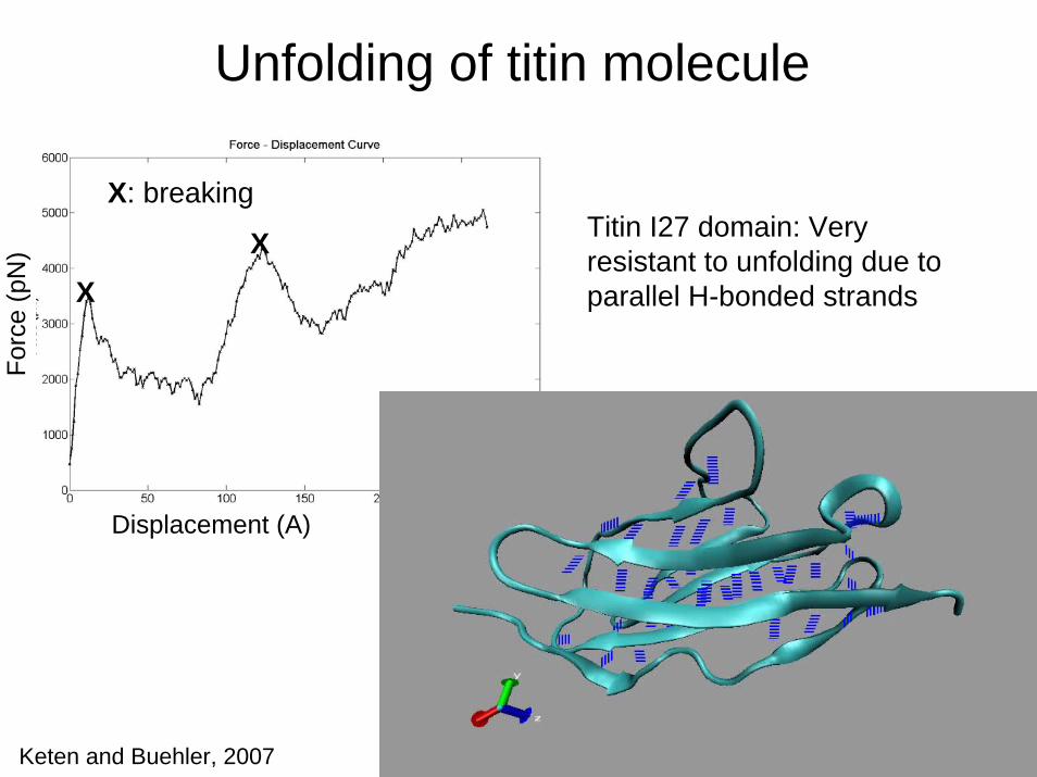
16
Displacement (A)
Forc
e (p
N)
Unfolding of titin molecule
Titin I27 domain: Very resistant to unfolding due to parallel H-bonded strands
X: breaking
XX
Keten and Buehler, 2007

17
Protein unfolding - ReaxFF
PnIB 1AKG
ReaxFF modelingM. Buehler, JoMMS, 2007
F
F
AHs

18
Protein unfolding - CHARMM
CHARMM modeling
Covalent bonds don’t break
M. Buehler, JoMMS, 2007

19
Comparison – CHARMM vs. ReaxFF
M. Buehler, JoMMS, 2007

Application to alpha-helical proteins
20

Vimentin intermediate filaments
Image courtesy of Greenmonster on Flickr.
Image courtesy of Bluebie Pixie on Flickr.
License: CC-BY.
Image of neuron and cell nucleus © sources unknown. All rights reserved. This content is excluded from our Creative Commons license. For more information, see http://ocw.mit.edu/fairuse.
Sourc
e: Q
in,
Z.,
L.
Kre
pla
k, a
nd M
. Bueh
ler.
"H
iera
rchic
al S
truct
ure
Contr
ols
Nan
om
echanic
al P
roper
ties
of Vim
entin I
nte
rmed
iate
Fila
men
ts."
PL
oSO
NE 4
, no.
10 (
2009).
doi:
10.1
371/j
ourn
al.pone.
0007294.
Lice
nse
CC B
Y.

22
Alpha-helical protein: stretching
M. Buehler, JoMMS, 2007
ReaxFF modeling of AHstretching
A: First H-bonds break (turns open)B: Stretch covalent backboneC: Backbone breaks

Coarse-graining approach
23
Describe interaction between “beads” and not “atoms”
Same concept as force fields for atoms
See also: http://dx.doi.org/10.1371/journal.pone.0006015

Case study: From nanoscale filaments to micrometer meshworks
24

Movie: MD simulation of AH coiled coil
25
Image removed due to copyright restrictions. Please see http://dx.doi.org/10.1103/PhysRevLett.104.198304.
See also: Z. Qin, ACS Nano, 2011, and Z. Qin BioNanoScience, 2010.

26
What about varying pulling speeds?
Changing the time-scale of observation of fracture

27
Variation of pulling speed
Image by MIT OCW. After Ackbarow and Buehler, 2007.
00
4,000
8,000 00 0.2 0.4
500
1,000
1,500
Forc
e (p
N)
12,000
50 100
Strain (%)
150 200
v = 65 m/sv = 45 m/sv = 25 m/sv = 7.5 m/sv = 1 m/smodelmodel 0.1 nm/s

28
Force at angular point fAP=fracture force
vf ln~AP
See also Ackbarow and Buehler, J. Mat. Sci., 2007
Pulling speed (m/s)
Forc
e at
AP
(pN
)

29
General results…

30
Rupture force vs. pulling speed
Buehler et al., Nature Materials, 2009
APf
Reprinted by permission from Macmillan Publishers Ltd: Nature Materials. Source: Buehler, M. ,and Yung, Y. "Chemomechanical Behaviour of Protein Constituents." Nature Materials 8, no. 3 (2009): 175-88. © 2009.

31
How to make sense of these results?

A few fundamental properties of bonds
Bonds have a “bond energy” (energy barrier to break)
Arrhenius relationship gives probability for energy barrier to be overcome, given a temperature
All bonds vibrate at frequency ω
32
⎟⎟⎠
⎞⎜⎜⎝
⎛−=
TkEpB
bexp

33
⎟⎟⎠
⎞⎜⎜⎝
⎛−=
TkEpB
bexp
Probability for bond rupture (Arrhenius relation)
Bell model
temperatureBoltzmann constant
heightof energy
barrier
distance to energybarrier
“bond”

34
⎟⎟⎠
⎞⎜⎜⎝
⎛ ⋅−−=
TkxfEp
B
Bbexp
Probability for bond rupture (Arrhenius relation)
Bell model
temperatureBoltzmann constant
heightof energy
barrier
distance to energybarrier
force applied(lower energybarrier)
“bond”
APff =

35
⎟⎟⎠
⎞⎜⎜⎝
⎛ ⋅−−=
TkxfEp
B
Bbexp
Probability for bond rupture (Arrhenius relation)
Bell model
Off-rate = probability times vibrational frequency
sec/1101 130 ×=ω
τωωχ 1)(exp00 =⎟⎟
⎠
⎞⎜⎜⎝
⎛⋅
⋅−−⋅=⋅=
TkxfEp
b
bb
bond vibrations

36
⎟⎟⎠
⎞⎜⎜⎝
⎛ ⋅−−=
TkxfEp
B
Bbexp
Probability for bond rupture (Arrhenius relation)
Bell model
Off-rate = probability times vibrational frequency
sec/1101 130 ×=ω
τωωχ 1)(exp00 =⎟⎟
⎠
⎞⎜⎜⎝
⎛⋅
⋅−−⋅=⋅=
TkxfEp
b
bb
“How often bond breaks per unit time”bond vibrations

37
⎟⎟⎠
⎞⎜⎜⎝
⎛ ⋅−−=
TkxfEp
B
Bbexp
Probability for bond rupture (Arrhenius relation)
Bell model
Off-rate = probability times vibrational frequency
sec/1101 130 ×=ω
τωωχ 1)(exp00 =⎟⎟
⎠
⎞⎜⎜⎝
⎛⋅
⋅−−⋅=⋅=
TkxfEp
b
bb
=τ bond lifetime(inverse of off-rate)

38
Bell model
tΔ↓
pulling speed (at end of molecule)vtx =ΔΔ /
???
tΔ
vtx =ΔΔ /
xΔxΔ→

39
Bell model
broken turntΔ↓
tΔ
xΔ
vtx =ΔΔ /
pulling speed (at end of molecule)vtx =ΔΔ /
xΔ→
xΔ→
xΔ→
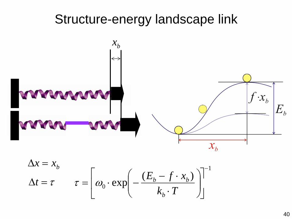
Structure-energy landscape link
40
bx
bxx =Δ
τ=Δt1
0)(exp
−
⎥⎦
⎤⎢⎣
⎡⎟⎟⎠
⎞⎜⎜⎝
⎛⋅
⋅−−⋅=
TkxfE
b
bbωτ

41
Bell model
vtxxTk
xfEx bb
bbb =ΔΔ=⋅⎟⎟
⎠
⎞⎜⎜⎝
⎛⋅
⋅−−⋅=⋅ /)(exp0ωχ
Bond breaking at (lateral applied displacement):bx
pulling speed
bxx =Δ
tΔ↓
tΔ
xΔ
vtx =ΔΔ /
τ/1=
broken turn

42
Bell model
vxTk
xfEb
b
bb =⋅⎟⎟⎠
⎞⎜⎜⎝
⎛⋅
⋅−−⋅
)(exp0ω
Solve this expression for f :

43
Bell model
vxTk
xfEb
b
bb =⋅⎟⎟⎠
⎞⎜⎜⎝
⎛⋅
⋅−−⋅
)(exp0ω
Solve this expression for f :
( )( )
⎟⎟⎠
⎞⎜⎜⎝
⎛⎟⎟⎠
⎞⎜⎜⎝
⎛⋅
−⋅⋅⋅
−⋅
=
⎟⎟⎠
⎞⎜⎜⎝
⎛⋅
−⋅⋅
−⋅
=
⎟⎟⎠
⎞⎜⎜⎝
⎛⋅−
⋅⋅
+⋅
=⋅−⋅+
=
⋅−⋅=⋅+−
=⋅+⋅
⋅−−
TkEx
xTkv
xTkf
TkEx
xTkv
xTkf
xTk
Ex
Tkvx
Tkx
xvTkEf
xvTkxfE
vxTk
xfE
b
bb
b
b
b
b
b
bb
b
b
b
b
bb
b
b
b
b
b
b
bbb
bbbb
bb
bb
explnln
)ln(ln
)ln(ln)ln(ln
)ln(ln
ln)ln()(
0
0
00
0
0
ω
ω
ωω
ω
ω ln(..)

Simplification and grouping of variables
44
⎟⎟⎠
⎞⎜⎜⎝
⎛⎟⎟⎠
⎞⎜⎜⎝
⎛⋅
−⋅⋅⋅⋅
−⋅⋅
=Tk
Exx
Tkvx
TkExvfb
bb
b
b
b
bbb explnln),;( 0ω
⎟⎟⎠
⎞⎜⎜⎝
⎛⋅
−⋅⋅==Tk
Exvb
bb exp: 00 ω
Only system parameters,[distance/length]

45
Bell model
vxTk
xfEb
b
bb =⋅⎟⎟⎠
⎞⎜⎜⎝
⎛⋅
⋅−−⋅
)(exp0ω
Results in:
bvavx
Tkvx
TkExvfb
b
b
bbb +⋅=⋅
⋅−⋅
⋅= lnlnln),;( 0
0ln vx
Tkb
xTka
b
B
b
B
⋅⋅
−=
⋅=

46
behavior of strengthvf ln~
bvaExvf bb +⋅= ln),;(
Pulling speed (m/s)
Eb= 5.6 kcal/mol and xb= 0.17 Ǻ (results obtained from fitting to the simulation data)
Forc
e at
AP
(pN
)

Pulling speed (m/s)
bvaExvf bb
Forc
e at
AP
(pN
)
47
Scaling with Eb : shifts curve
+⋅= ln),;(
↑bE
0ln vx
Tkbx
Tkab
B
b
B ⋅⋅
−=⋅
= ⎟⎟⎠
⎞⎜⎜⎝
⎛⋅
−⋅⋅=Tk
Exvb
bb exp00 ω

Pulling speed (m/s)
bvaExvf bb +Fo
rce
at A
P (p
N)
48
⋅= ln),;(
↓bx
0ln vx
Tkbx
Tkab
B
b
B ⋅⋅
−=⋅
= ⎟⎟⎠
⎞⎜⎜⎝
⎛⋅
−⋅⋅=Tk
Exvb
bb exp00 ω
Scaling with xb: changes slope

49
Simulation results
Bertaud, Hester, Jimenez, and Buehler, J. Phys. Cond. Matt., 2010
Courtesy of IOP Publishing, Inc. Used with permission. Source: Fig. 3 from Bertaud, J., Hester, J. et al. "Energy Landscape, Structure andRate Effects on Strength Properties of Alpha-helical Proteins." J Phys.: Condens. Matter 22 (2010): 035102. doi:10.1088/0953-8984/22/3/035102.

50
Mechanisms associated with protein fracture

51
Change in fracture mechanism
Single AH structure
Simulation span: 250 nsReaches deformation speed O(cm/sec)
FDM: Sequential HB breaking
SDM: Concurrent HB breaking (3..5 HBs)
Courtesy of National Academy of Sciences, U. S. A. Used with permission. Source: Ackbarow, Theodor, et al. "Hierarchies, Multiple Energy Barriers, and Robustness Govern the Fracture Mechanics of Alpha-helical and Beta-sheet Protein Domains." PNAS 104 (October 16, 2007): 16410-5. Copyright 2007 National Academy of Sciences, U.S.A.

52
Analysis of energy landscape parameters
Energy single H-bond: ≈3-4 kcal/mol
What does this mean???
Courtesy of National Academy of Sciences, U. S. A. Used with permission. Source: Ackbarow, Theodor, et al. "Hierarchies, Multiple Energy Barriers, and Robustness Govern the Fracture Mechanics of Alpha-helical and Beta-sheet Protein Domains." PNAS 104 (October 16, 2007): 16410-5. Copyright 2007 National Academy of Sciences, U.S.A.

53
H-bond rupture dynamics: mechanism
Courtesy of National Academy of Sciences, U. S. A. Used with permission. Source: Ackbarow, Theodor, et al. "Hierarchies, Multiple Energy Barriers, and Robustness Govern the Fracture Mechanics of Alpha-helical and Beta-sheet Protein Domains." PNAS 104 (October 16, 2007): 16410-5. Copyright 2007 National Academy of Sciences, U.S.A.

54
I: All HBs are intact
II: Rupture of 3 HBs – simultaneously; within τ ≈ 20 ps
III: Rest of the AH relaxes – slower deformation…
H-bond rupture dynamics: mechanism
Courtesy of National Academy of Sciences, U. S. A. Used with permission. Source: Ackbarow, Theodor, et al. "Hierarchies, Multiple Energy Barriers, and Robustness Govern the Fracture Mechanics of Alpha-helical and Beta-sheet Protein Domains." PNAS 104 (October 16, 2007): 16410-5. Copyright 2007 National Academy of Sciences, U.S.A.

3. Examples – materials and applications
E.g. disease diagnosis, mechanisms, etc.
55

56
Genetic diseases – defects in protein materials
Defect at DNA level causes structure modification
Question: how does such a structure modification influence material behavior / material properties?
Four letter code “DNA”
Sequence of amino acids“polypeptide”(1D structure)
CHANGED
Folding (3D structure)STRUCTURAL
DEFECT
.. - Proline - Serine –Proline - Alanine - ..ACGT
DEFECT IN SEQUENCE

57
Structural change in protein molecules can lead to fatal diseases
Single point mutations in IF structure causes severe diseases such as rapid aging disease progeria – HGPS (Nature, 2003; Nature, 2006, PNAS, 2006)Cell nucleus loses stability under mechanical (e.g. cyclic) loading, failure occurs at heart (fatigue)
Genetic defect:
substitution of a single DNA base: Amino acid guanine is switched to adenine
Image of patient removed due to copyright restrictions.

58
Structural change in protein molecules can lead to fatal diseases
Single point mutations in IF structure causes severe diseases such as rapid aging disease progeria – HGPS (Nature, 2003; Nature, 2006, PNAS, 2006)Cell nucleus loses stability under cyclic loadingFailure occurs at heart (fatigue)
Experiment suggests that mechanical properties of nucleus change
Fractures
Image of patient removed due to copyright restrictions.
Courtesy of National Academy of Sciences, U. S. A. Used with permission. Source: Dahl, et al. "Distinct Structural and Mechanical Properties of the Nuclear Lamina in Hutchinson–Gilford Progeria Syndrome." PNAS 103 (2006): 10271-6. Copyright 2006 National Academy of Sciences, U.S.A.

59
Mechanisms of progeria
Images courtesy of National Academy of Sciences, U. S. A. Used with permission. Source: Dahl, et al. "Distinct Structural and Mechanical Properties of the Nuclear Lamina in Hutchinson–Gilford Progeria Syndrome." PNAS 103 (2006): 10271-6. Copyright 2006 National Academy of Sciences, U.S.A.

60
Deformation of red blood cells
Courtesy of Elsevier, Inc., http://www.sciencedirect.com. Used with permission.

61
Stages of malaria and effect on cell stiffness
Disease stagesH-RBC (healthy)Pf-U-RBC (exposed but not infected)Pf-R-pRBC (ring stage)Pf-T-pRBC(trophozoite stage)Pf-S-pRBC (schizont stage)Consequence: Due to rigidity, RBCs can not move easily through capillaries in the lung
Courtesy of Elsevier, Inc., http://www.sciencedirect.com. Used with permission.

62
Cell deformation
Courtesy of Elsevier, Inc., http://www.sciencedirect.com. Used with permission.

63
Deformation of red blood cells
Courtesy of Elsevier, Inc., http://www.sciencedirect.com. Used with permission.

64
Mechanical signature of cancer cells (AFM)
Cancer cells=soft
Healthy cells=stiff
Reprinted by permission from Macmillan Publishers Ltd: Nature Nanotechnology. Source: Cross, S., Y. Jin, et al. "Nanomechanical Analysis of Cells from Cancer Patients." Nature Nanotechnology 2, no. 12 (2007): 780-3. © 2007.

MIT OpenCourseWarehttp://ocw.mit.edu
3.021J / 1.021J / 10.333J / 18.361J / 22.00J Introduction to Modeling and SimulationSpring 2012
For information about citing these materials or our Terms of use, visit: http://ocw.mit.edu/terms.