paromomycin. ii. paromobiosamine, a diaminohexosyl-d-ribose
TRANSCRIPT
July 5, 1959 COXMUNICATIONS TO THE EDITOR 3481
be identical with D-glucosamine hydrochloride by infrared spectrum, X-ray diffraction, optical rota- tion and comparison of the salicylidene derivatives.
Since the crystalline N,N', N"-triacetylparomam- ine [Anal. Calcd. for C18H31N3010 (449.5) : C, 48.10; H, 6.95; N, 9.35. Found: C, 48.04; H, 7.03;
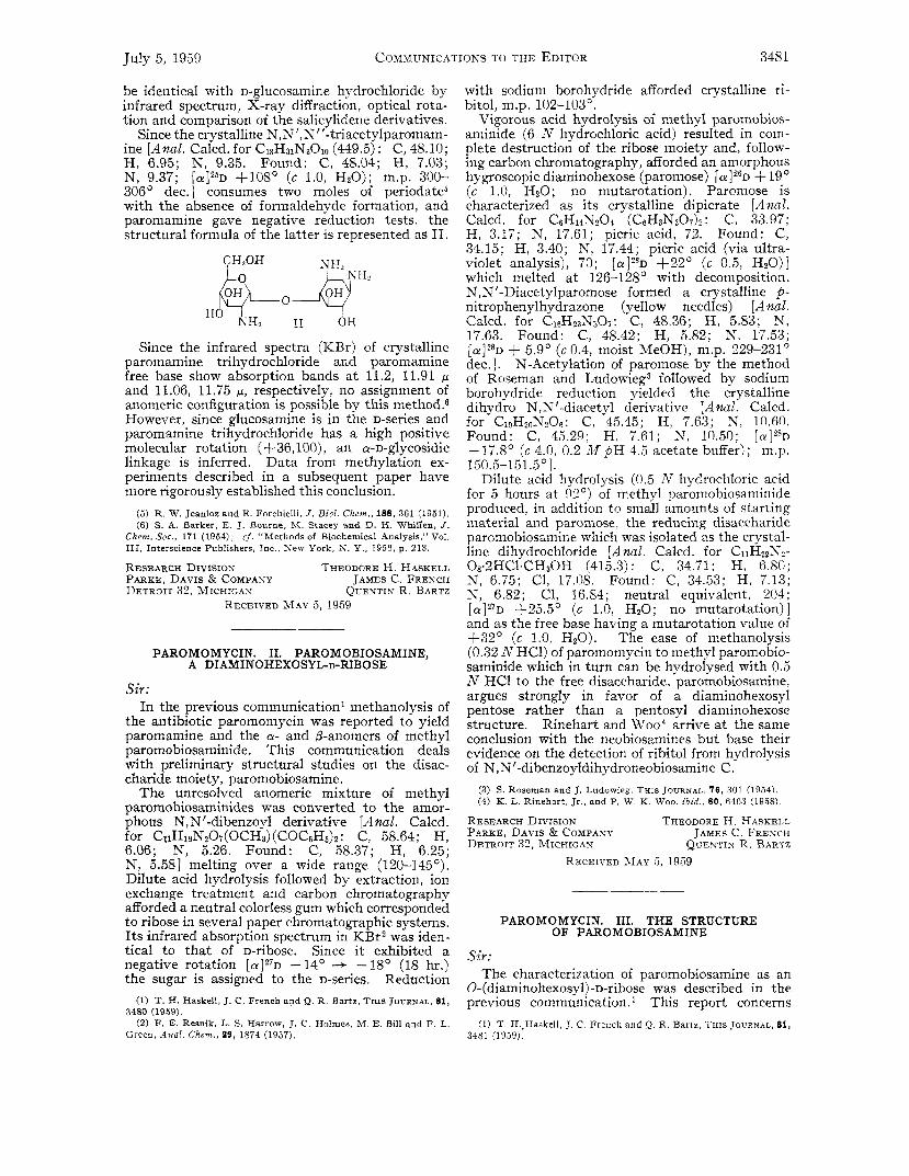
306' dec.] consumes two moles of periodate5 with the absence of formaldehyde formation] and paromamine gave negative reduction tests, the structural formula of the latter is represented as IT.
N, 9.37; [aIz5D +108' (c 1.0, HzO); m.p. 300-
CHlOH @ @H>
HO "7 II OH
Since the infrared spectra (KBr) of crystalline paromamine trihydrochloride and paromamine free base show absorption bands a t 11.2, 11.91 p and 11.06, 11.75 g, respectively, no assignment of anomeric configuration is possible by this method.6 However, since glucosamine is in the D-series and paromamine trihydrochloride has a high positive molecular rotation (+36, loo), an a-D-glycosidic linkage is inferred. Data from methylation ex- periments described in a subsequent paper have more rigorously established this conclusion.
( 5 ) R . W. Jeanloz and E. Forchielli, J. Bid . Chem.. 188, 361 (1951). (6 ) S. A. Barker, E. J. Bourne, M. Stacey and D. H. Whiffen, J .
rf. "Methods of Biochemical Analysis," Vol. Chem. Soc., 171 (1954); 111, Interscience Publishers, Inc. , S e w York, S. Y., 1956, p. 213.
RESEARCH DIVISIOT THEODORE H. HASKELL PARKE, DAVIS & COMPANY JAMES C. FRENCH DETROIT 32, MICHIGAX QVENTIN R. BARTZ
RECEIVED MAY 5 , 1959
PAROMOMYCIN. 11. PAROMOBIOSAMINE, A DIAMINOHEXOSYL-D-RIBOSE
Sir : In the previous communication' methanolysis of
the antibiotic paromomycin was reported to yield paromamine and the cy- and /3-anomers of methyl paromobiosaminide. This communication deals with preliminary structural studies on the disac- charide moiety, paromobiosamine.
The unresolved anomeric mixture of methyl paromobiosaminides was converted to the amor- phous NIN'-dibenzoyl derivative [A nul. Calcd. for CltH1eN207(0CH3) (COCsH&: C, 58.64; H, 6.06; N, 5.26. Found: C, 58.37; H I 6.25; N, 5.581 melting over a wide range (120-145'). Dilute acid hydrolysis followed by extraction, ion exchange treatment and carbon chromatography afforded a neutral colorless gum which corresponded to ribose in several paper chromatographic systems. Its infrared absorption spectrum in KBr2 was iden- tical to that of D-ribose. Since it exhibited a negative rotation [ a ] 2 7 ~ -14' 4 -118' (18 hr.) the sugar is assigned to the D-series. Reduction
(1) T. H. Haskell, J. C. French and Q . R. Bartz, THIS JOURNAL, 81,
(2) F. E. Resnik, L. S. Harrow, J. C . Holmes, M. E. Bill and F. L. 3480 (1959).
Green, Anal. Chem,, 29, 1874 (1057).
with sodium borohydride afforded crystalline ri- bitol, m.p. 102-103".
Vigorous acid hydrolysis of methyl paromobios- aminide (6 N hydrochloric acid) resulted in com- plete destruction of the ribose moiety and, follow- ing carbon chromatography, afforded an amorphous hygroscopic diaminohexose (paromose) [ a ] 2 6 D + 19 ' (c 1.0, HzO; no mutarotation). Paromose is characterized as its crystalline dipicrate [ A nul. Calcd. for c6H14N20.1 (CeH3N307)2: C, 33.97; H, 3.17; N, 17.61; picric acid, 72. Found: C, 34.15; H, 3.40; N, 17.44; picric acid (via ultra- violet analysis), 70; [ a ] ? * ~ 4-22' (c 0.5, HzO)] which melted at 126-128' with decomposition. N,N'-Diacetylparomose formed a crystalline fi- nitrophenylhydrazone (yellow needles) [A nal. Calcd. for C&23Nj07: C, 48.36; H, 5.83; N, 17.63. Found: C, 48.42; H, 5.82; N, 17.53; [cy]"D + 5.9' (c 0.4, moist MeOH), m.p. 229-231' dec.]. N-Acetylation of paromose by the method of Roseman and Ludowieg3 followed by sodium borohydride reduction yielded the crystalline dihydro N,N '-diacetyl derivative [Anal. Calcd. for CloHznN206: C, 45.45; H, 7.63; W, 10.60. Found: C, 45.29; H, 7.61; N, 10.50; [cy]'*D - 17.8' (c 4.0, 0.2 Af pH 4.5 acetate buffer); m.p. 150.5-1 5 1.5 O 1.
Dilute acid hydrolysis (0.5 AT hydrochloric acid for 5 hours a t 92') of methyl paromobiosaniinide produced, in addition to small amounts of starting material and paromose, the reducing disaccharide paromobiosamine which was isolated as the crystal- line dihydrochloride [A nul. Calcd. for CllH22N2-
N, 6.75; C1, 17.08. Found: C, 34.53; H, 7.13; N, 6.82; C1, 16.84; neutral equivalent, 204; [ a I z 7 ~ 1-25.5' (c 1.0, HzO; no mutarotation)] and as the free base having a mutarotation value of +32' (c 1.0, HzO). The ease of methanolysis (0.32 N HCI) of paromomycin to methyl paromobio- saminide which in turn can be hydrolysed with 0.5 N HCI to the free disaccharide, paromobiosamine, argues strongly in favor of a diaminohexosyl pentose rather than a pentosyl diaminohexose structure. Rinehart and Woo4 arrive at the same conclusion with the neobiosamines but base their evidence on the detection of ribitol from hydrolysis of N,N'-dibenzoyldihydroneobiosamine C.
08.2HCl.CH30H (415.3) : C. 34.71; H, 6.80;
(3) S. Roseman and J. Ludowieg, THIS JOVRNAL, 76, 301 (1954). (4) K. L. Rinehart , Jr., and P. W. K. Woo, i b i d . , 80, 6403 (1958).
RESEARCH DIVISION THEODORE H. HASKELL PARKE, DAVIS & COMPANY JAMES C. FRENCH DETROIT 32, MICHIGAY QUENTIN R , RARTZ
RECEIVED MAY 5 , 1959
PAROMOMYCIN. 111. THE STRUCTURE OF PAROMOBIOSAMINE
Sir : The characterization of paromobiosamine as an
0-(diaminohexosy1)-D-ribose was described in the previous communication.1 This report concerns
( 1 ) T. H..Haskell, J . C. French and Q. R. Bartz, THIS J O U R N A L , 81, 3481 (1059).