palm vein database and experimental framework for...
TRANSCRIPT
Palm Vein Database and Experimental Framework forReproducible Research
Pedro Tome and Sebastien MarcelIdiap Research Institute
Centre du Parc, Rue Marconi 19, CH-1920 Martigny, Switzerland{pedro.tome, sebastien.marcel}@idiap.ch
Abstract:A palm vein database acquired by a contactless sensor together with an experimen-
tal framework freely available for fair reproducible research purposes are described.The palm vein recognition system uses automatic palm region segmentation and circu-lar Gabor filter approach to enhance the veins in the preprocessing, LBP features andhistogram intersection as matching. Results are presented comparing two automaticsegmentation using the ROI-1 region proportioned by the acquisition sensor and theROI-2 region generated by the recognition software developed. Complete benchmarkresults using popular methods and the source code are attached to the database as areference for other researchers.
1 Introduction
Automatic palm vein recognition has emerged as a reliable technology to provide greaterlevel of security to personal authentication system [WESS05]. Among the various humanhand biometric characteristics that can be used to recognize a person, such as geome-try, fingerprint, palm print or knuckle print, the palm veins are perhaps the most suc-cessful form with highest recognition rates achieved between the different characteris-tics [MCT12] as palm vein patterns are considered stable and reliable. This means thatonce a person has reached adulthood, the hand structure, veins and configuration remainrelatively stable throughout the person’s life [YDS06]. In addition, they can be acquiredwithout contact and require the presence of blood in the veins to be registered, whichmakes more robust these systems against the liveness problem and the spoofing attacks.The palm vein imaging acquisition requires infrared (IR) illumination (generally, NearIR)and standard cameras with a simple CCD or CMOS sensor. Therefore, palm vein imagesare grayscale images in which dark grey to black veins appear on the grey background.
Because the scarce number of palm vein databases and the different unclear and complexprotocols provided by the databases in the literature, no fair reproducible and comparableresearch can be carried out. For these reasons, the VERA Palm vein database and theexperimental framework are introduced and described in this paper freely available for re-search purposes at www.idiap.ch/dataset and www.idiap.ch/scientific-research/resources. Baseline experimental results obtained by the authors using popularly usedapproaches are also presented.
Camera
Ultrasound
sensor
Diffuser
LED on/off
LED control
distance
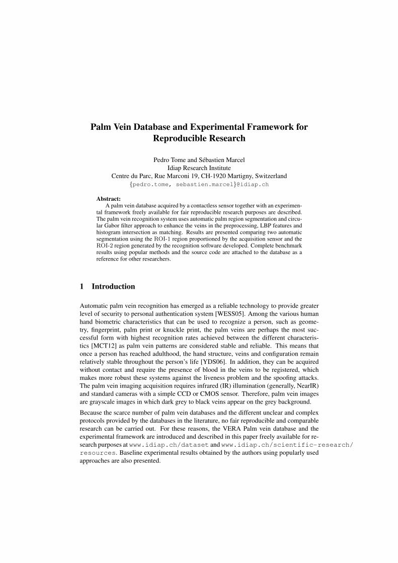
Figure 1: Palm vein prototype sensor description and palm vein image acquisition example.
2 State-of-the-art
The most complete research in palm vein pattern recognition was conducted by Fujitsuin Japan, supported by a patent and described in [Wat08]. The database is comprised of150, 000 palm vein images from 75, 000 subjects on a different rank of ages. This databasewas collected for commercial purposes, therefore no details are available and reproductionof the study is impossible. On the other hand, from a non-commercial point of view,there are a scarce number of free available palm vein databases in the literature [HSTR08,KK11]. The most relevant one is the CASIA Multi-spectral [HSTR08], a contactless ac-quisition from 100 subjects using six different wavelengths (visible, 460, 630, 700, 850and 940 nm) of the illumination. On the other hand, the PUT database [KK11] is a smalldatabase comprises of 50 subjects acquired on a contact sensor using just one wavelengthof 880 nm for illumination. It is also important to highlight other database collectionsmentioned in the literature that are not publicly available such as [MCT12, Lee12]. In thiscontext, researchers working on palm vein recognition built their own acquisition devicesto acquire vein pattern images. This resulted in many different proposals for the choice ofregion of interest (ROI), different positioning equipment, various image parameters suchas resolution, and different image collection processes. For those reasons, all these workspresent different protocols and performance results, which in such different conditions arethus difficult to compare. To the best of our knowledge, there are no works in the literatureproviding any kind of experimental framework which allows the fair comparison of theperformance results similar to the new one that we present here.
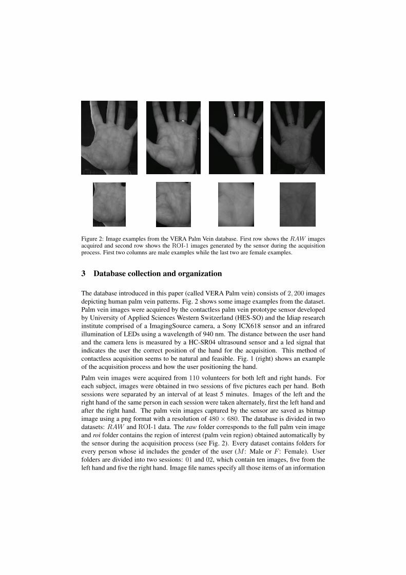
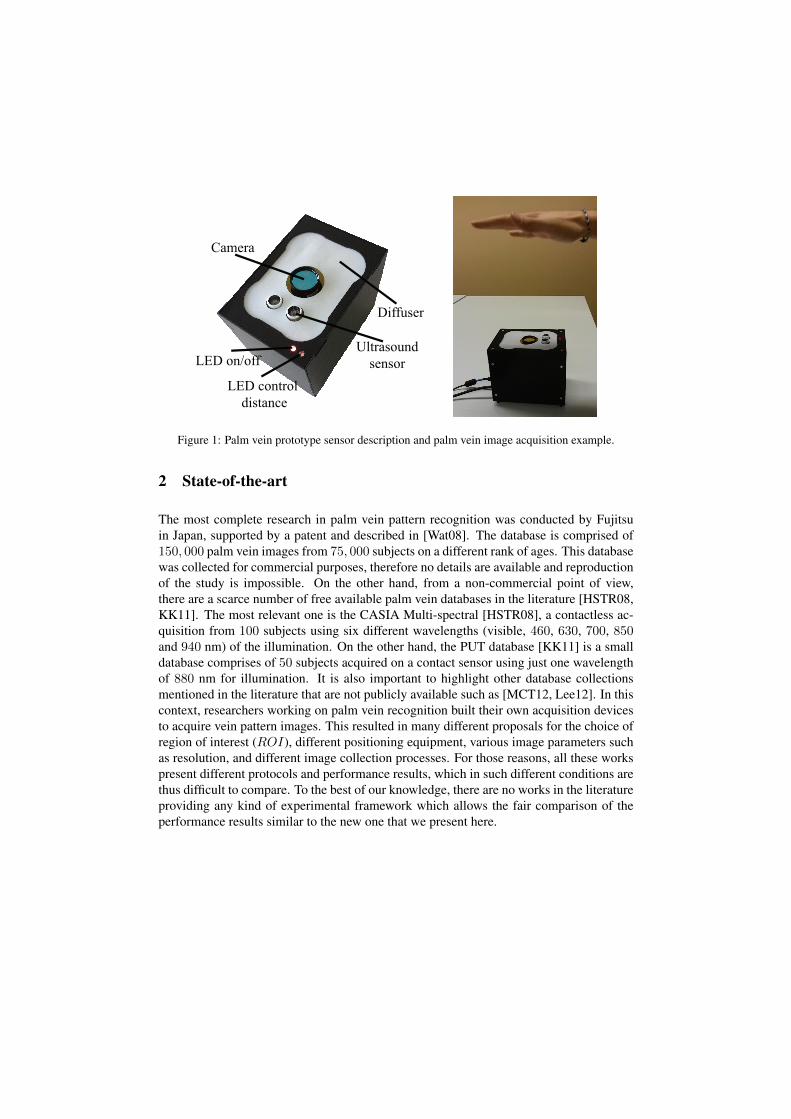
Figure 2: Image examples from the VERA Palm Vein database. First row shows the RAW imagesacquired and second row shows the ROI-1 images generated by the sensor during the acquisitionprocess. First two columns are male examples while the last two are female examples.
3 Database collection and organization
The database introduced in this paper (called VERA Palm vein) consists of 2, 200 imagesdepicting human palm vein patterns. Fig. 2 shows some image examples from the dataset.Palm vein images were acquired by the contactless palm vein prototype sensor developedby University of Applied Sciences Western Switzerland (HES-SO) and the Idiap researchinstitute comprised of a ImagingSource camera, a Sony ICX618 sensor and an infraredillumination of LEDs using a wavelength of 940 nm. The distance between the user handand the camera lens is measured by a HC-SR04 ultrasound sensor and a led signal thatindicates the user the correct position of the hand for the acquisition. This method ofcontactless acquisition seems to be natural and feasible. Fig. 1 (right) shows an exampleof the acquisition process and how the user positioning the hand.
Palm vein images were acquired from 110 volunteers for both left and right hands. Foreach subject, images were obtained in two sessions of five pictures each per hand. Bothsessions were separated by an interval of at least 5 minutes. Images of the left and theright hand of the same person in each session were taken alternately, first the left hand andafter the right hand. The palm vein images captured by the sensor are saved as bitmapimage using a png format with a resolution of 480 × 680. The database is divided in twodatasets: RAW and ROI-1 data. The raw folder corresponds to the full palm vein imageand roi folder contains the region of interest (palm vein region) obtained automatically bythe sensor during the acquisition process (see Fig. 2). Every dataset contains folders forevery person whose id includes the gender of the user (M : Male or F : Female). Userfolders are divided into two sessions: 01 and 02, which contain ten images, five from theleft hand and five the right hand. Image file names specify all those items of an information
ProtocolWorld set Development set Evaluation set
Clients # Files Clients Enrolment Probe Client Enrolment Probenom L&R 20 400 30 120 480 60 240 960nom L 20 200 30 60 240 60 120 480nom R 20 200 30 60 240 60 120 480
Table 1: Database detailed description based on number of images for for the three protocols definedand the different sets.
exactly using the next format: “UUU H X Y .png”, where UUU defines the user id, Hthe hand (L: left or R: right), X the session, and finally, Y the number of the acquisition.For example, the image named “021 L 1 2.png” is the second image in the first session ofthe left palm of the 21th user and has the path: “.../021-M /01/021 L 1 2.png”.
4 Experimental framework
This work presents an open source and extensible experimental palm vein frameworkcalled PalmveinRecLib: bob.palmvein1, which allows fair and reproducible benchmarkson palm vein recognition. This framework includes a complete module for scores analysisand allows to run a complete palm vein recognition experiment, from the preprocessing ofRAW images (including segmentation) to the computation of biometric scores and theirevaluation. This framework is totally open source and modular, which means that all al-gorithm parameters are fixed, available and each block can be replaced or improved bynew algorithms and approaches. The system implements several baseline methods fromthe state-of-the-art and is divided on three stages: i) segmentation and normalization, ii)feature extraction, and iii) matching.
In the segmentation process the hand contour is localised by a binarization from grayscalepalm vein images. Then the hand landmarks (peaks and valleys) are extracted using theradial distance function (RDF) between the reference point (generally the starting of thewrist) and the contour points extracted [KW14]. The palm region is extracted as a squareregion based on the located hand landmarks and a scaling and rotation normalization onthe extracted palm vein region is performed. Finally, the palm veins are enhanced by usingthe Circular Gabor Filter (CGF) approach [ZY09]. Once the palm vein region is extractedand normalised, local binary patterns (LBP) are computed to serve as features [MD14] andthe histogram intersection metric [SB91] is adopted as a similarity measure to compute thescores.
5 Experimental protocol and baseline results
The VERA Palm vein database is presented with three different protocols: i) nom L&R- normal operation mode, where left and right hand of the same subject are considered
1Freely available at https://pypi.python.org/pypi/bob.palmvein
0.01 0.1 1 10 100FAR (%)
50
60
70
80
90
100
CA
R(%
)
ROC curve for ROI-2 system
Dev. nom L&R
Eval. nom L&R
Dev. nom L
Eval. nom L
Dev. nom R
Eval. nom R
0.01 0.1 1 10 100FAR (%)
50
60
70
80
90
100
CA
R(%
)
ROC curve for ROI-1 system
Dev. nom L&R
Eval. nom L&R
Dev. nom L
Eval. nom L
Dev. nom R
Eval. nom R
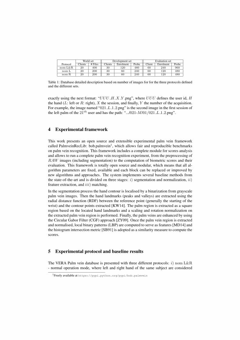
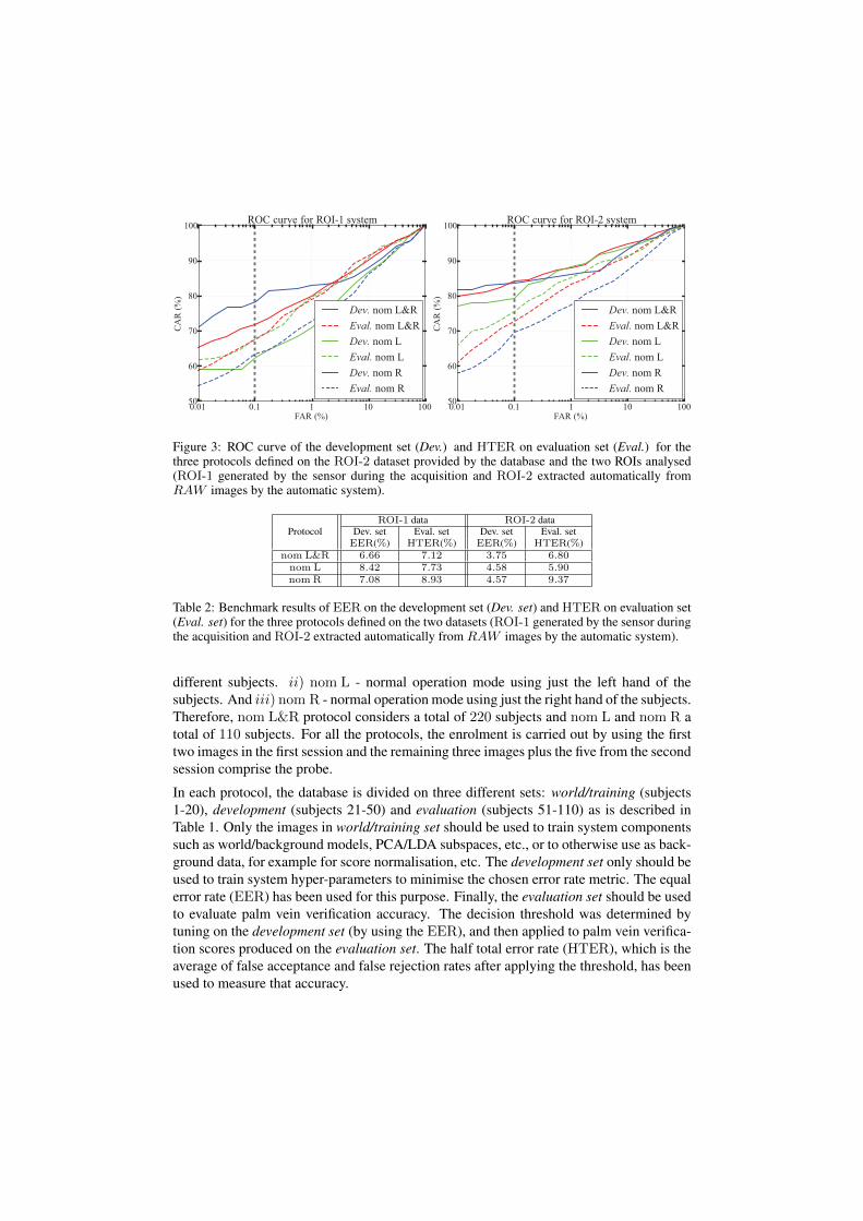
Figure 3: ROC curve of the development set (Dev.) and HTER on evaluation set (Eval.) for thethree protocols defined on the ROI-2 dataset provided by the database and the two ROIs analysed(ROI-1 generated by the sensor during the acquisition and ROI-2 extracted automatically fromRAW images by the automatic system).
ProtocolROI-1 data ROI-2 data
Dev. set Eval. set Dev. set Eval. setEER(%) HTER(%) EER(%) HTER(%)
nom L&R 6.66 7.12 3.75 6.80nom L 8.42 7.73 4.58 5.90nom R 7.08 8.93 4.57 9.37
Table 2: Benchmark results of EER on the development set (Dev. set) and HTER on evaluation set(Eval. set) for the three protocols defined on the two datasets (ROI-1 generated by the sensor duringthe acquisition and ROI-2 extracted automatically from RAW images by the automatic system).
different subjects. ii) nom L - normal operation mode using just the left hand of thesubjects. And iii) nom R - normal operation mode using just the right hand of the subjects.Therefore, nom L&R protocol considers a total of 220 subjects and nom L and nom R atotal of 110 subjects. For all the protocols, the enrolment is carried out by using the firsttwo images in the first session and the remaining three images plus the five from the secondsession comprise the probe.
In each protocol, the database is divided on three different sets: world/training (subjects1-20), development (subjects 21-50) and evaluation (subjects 51-110) as is described inTable 1. Only the images in world/training set should be used to train system componentssuch as world/background models, PCA/LDA subspaces, etc., or to otherwise use as back-ground data, for example for score normalisation, etc. The development set only should beused to train system hyper-parameters to minimise the chosen error rate metric. The equalerror rate (EER) has been used for this purpose. Finally, the evaluation set should be usedto evaluate palm vein verification accuracy. The decision threshold was determined bytuning on the development set (by using the EER), and then applied to palm vein verifica-tion scores produced on the evaluation set. The half total error rate (HTER), which is theaverage of false acceptance and false rejection rates after applying the threshold, has beenused to measure that accuracy.
Table 2 and Fig. 3 show the benchmark results for the three protocols on the different setsdefined. As we can see the ROI-2 images produce better results than the ROI-1 regions,this means that the automatic segmentation implemented align better the palm vein region.Focusing our attention of ROI-2 results, the system achieved a rate of 3.75% of EER onthe development set and 6.80% of HTER on the evaluation set on the nom L&R protocol.Results on both hands achieved similar recognition rates of EER on the developmentset, but however, left hand obtained a rate of 5.90% of HTER on the evaluation set incomparison to the 9.37% of HTER of the right hand. This difference can be explainedbased on the enrolment images. On the evaluation set of the left hand there are no effectsof blurring images, while on the right hand, there are several subjects that experiment thisproblem on their enrolment images, and therefore, the HTER rate increases.
6 Conclusion
This paper presents a new palm vein database acquired by a contactless sensor togetherwith an open source experimental framework freely available for reproducible researchpurposes. The scarce number of databases and the unclear protocols proposed so far inthe literature of this field make this database a valuable reference for the improvementof palm vein recognition systems. The results obtained so far demonstrate the utility ofthe database and open the opportunity to research on new approaches in the palm veinpattern recognition field. Therefore, the collected database will be useful for the researchcommunity as a reference database that provides replicable and clear analysis protocolsand a free experimental framework for the fair reproducible research on the palm veinrecognition field.
Acknowledgements
This work has been partially supported by the EU FP7 BEAT (284989) project and theSwiss Centre for Biometrics Research and Testing for support. The authors would like tothank the University of Applied Sciences Western Switzerland (HES-SO) for developingthe palm vein sensor.
References
[HSTR08] Ying Hao, Zhenan Sun, Tieniu Tan, and Chao Ren. Multispectral palm image fusion foraccurate contact-free palmprint recognition. In Proc. on IEEE International Conferenceon Image Processing (ICIP), pages 281–284, 2008.
[KK11] R. Kabaciski and M. Kowalski. Vein pattern database and benchmark results. Electron-ics Letters, 47:1127–1128(1), September 2011.
[KW14] W. Kang and Q. Wu. Contactless Palm Vein Recognition Using a Mutual Foreground-Based Local Binary Pattern. IEEE Transactions on Information Forensics and Security,9(11):1974–1985, Nov 2014.
[Lee12] Jen-Chun Lee. A novel biometric system based on palm vein image. Pattern RecognitionLetters, 33(12):1520 – 1528, 2012.
[MCT12] Goh Kah Ong Michael, Tee Connie, and Andrew Beng Jin Teoh. A contactless biomet-ric system using multiple hand features. Journal of Visual Communication and ImageRepresentation, 23(7):1068 – 1084, 2012.
[MD14] Leila Mirmohamadsadeghi and Andrzej Drygajlo. Palm vein recognition with localtexture patterns. IET Biometrics, pages 1–9, January 2014.
[SB91] MichaelJ. Swain and DanaH. Ballard. Color indexing. International Journal of Com-puter Vision, 7(1):11–32, 1991.
[Wat08] Masaki Watanabe. Palm Vein Authentication. In NaliniK. Ratha and Venu Govindaraju,editors, Advances in Biometrics, pages 75–88. Springer London, 2008.
[WESS05] M. Watanabe, T. Endoh, M. Shiohara, and S. Sasaki. Palm vein authentication technol-ogy and its applications. In Proc. on Biometrics Symposium, pages 37–38, 2005.
[YDS06] Erdem Yoruk, Helin Dutagaci, and Bulent Sankur. Hand Biometrics. Image VisionComputing, 24(5):483–497, May 2006.
[ZY09] Jing Zhang and Jinfeng Yang. Finger-Vein Image Enhancement Based on Combinationof Gray-Level Grouping and Circular Gabor Filter. In International Conference onInformation Engineering and Computer Science (ICIECS), pages 1–4, Dec 2009.