palate hyperpigmentation caused by prolonged use of the anti-malarial chloroquine
TRANSCRIPT
SINE QUA NON CLINICOPATHOLOGIC CORRELAT
Palate Hyperpigmentation Caused by Prolonged Useof the Anti-Malarial Chloroquine
Mario Rodrigues de Melo Filho • Celsia Adriane Dias da Silva •
Maurıcio da Rocha Dourado • Maria Betania de Oliveira Pires •
Sabina Pena Borges Pego • Edmilson Martins de Freitas
Received: 26 April 2011 / Accepted: 10 August 2011 / Published online: 30 August 2011
� Springer Science+Business Media, LLC 2011
Abstract The side-effects of many drugs manifest in the
oral mucosa. The anti-malarial agent chloroquine diphos-
phate, which is also used to treat immunological, derma-
tological, and rheumatological disorders, usually causes
pigmentary changes in the oral mucosa. This report pre-
sents a case of palate pigmentation related to the prolonged
use of chloroquine diphosphate caused by the deposition of
drug metabolites in the mucosa. Healthcare professionals
must be aware of these drugs and their adverse effects in
order to make the correct diagnosis, decide on the optimal
treatment for the condition, or refer the patient to an
appropriate specialist.
Keywords Pigmentary changes � Oral mucosa �Anti-malarials
History
A 64-year-old woman with dark skin tone was referred to
us for evaluation of a dark stain on the roof of mouth that
had appeared around 3 months before presentation without
any painful symptoms. The patient had arterial hyperten-
sion and rheumatoid arthritis among other systemic dis-
eases, and she was using a pacemaker. She reported the use
of the following medicines: prednisone, methotrexate,
meloxicam, aspirin (100 mg) and chloroquine phosphate
(200 mg/day, started approximately 15 years prior).
Clinical Examination
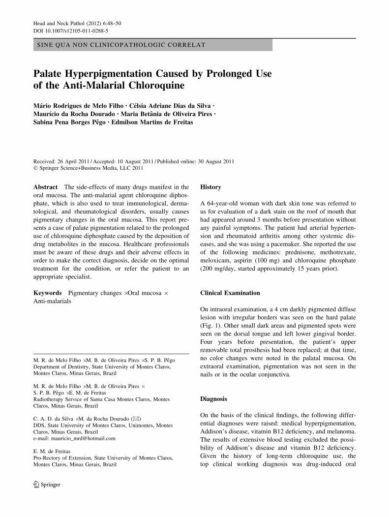
On intraoral examination, a 4 cm darkly pigmented diffuse
lesion with irregular borders was seen on the hard palate
(Fig. 1). Other small dark areas and pigmented spots were
seen on the dorsal tongue and left lower gingival border.
Four years before presentation, the patient’s upper
removable total prosthesis had been replaced; at that time,
no color changes were noted in the palatal mucosa. On
extraoral examination, pigmentation was not seen in the
nails or in the ocular conjunctiva.
Diagnosis
On the basis of the clinical findings, the following differ-
ential diagnoses were raised: medical hyperpigmentation,
Addison’s disease, vitamin B12 deficiency, and melanoma.
The results of extensive blood testing excluded the possi-
bility of Addison’s disease and vitamin B12 deficiency.
Given the history of long-term chloroquine use, the
top clinical working diagnosis was drug-induced oral
M. R. de Melo Filho � M. B. de Oliveira Pires � S. P. B. Pego
Department of Dentistry, State University of Montes Claros,
Montes Claros, Minas Gerais, Brazil
M. R. de Melo Filho � M. B. de Oliveira Pires �S. P. B. Pego � E. M. de Freitas
Radiotherapy Service of Santa Casa Montes Claros, Montes
Claros, Minas Gerais, Brazil
C. A. D. da Silva � M. da Rocha Dourado (&)
DDS, State University of Montes Claros, Unimontes, Montes
Claros, Minas Gerais, Brazil
e-mail: [email protected]
E. M. de Freitas
Pro-Rectory of Extension, State University of Montes Claros,
Montes Claros, Minas Gerais, Brazil
123
Head and Neck Pathol (2012) 6:48–50
DOI 10.1007/s12105-011-0288-5
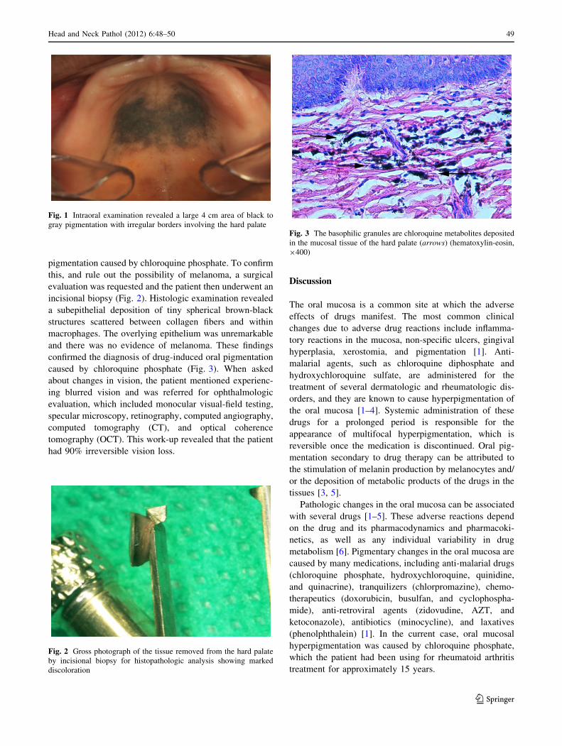
pigmentation caused by chloroquine phosphate. To confirm
this, and rule out the possibility of melanoma, a surgical
evaluation was requested and the patient then underwent an
incisional biopsy (Fig. 2). Histologic examination revealed
a subepithelial deposition of tiny spherical brown-black
structures scattered between collagen fibers and within
macrophages. The overlying epithelium was unremarkable
and there was no evidence of melanoma. These findings
confirmed the diagnosis of drug-induced oral pigmentation
caused by chloroquine phosphate (Fig. 3). When asked
about changes in vision, the patient mentioned experienc-
ing blurred vision and was referred for ophthalmologic
evaluation, which included monocular visual-field testing,
specular microscopy, retinography, computed angiography,
computed tomography (CT), and optical coherence
tomography (OCT). This work-up revealed that the patient
had 90% irreversible vision loss.
Discussion
The oral mucosa is a common site at which the adverse
effects of drugs manifest. The most common clinical
changes due to adverse drug reactions include inflamma-
tory reactions in the mucosa, non-specific ulcers, gingival
hyperplasia, xerostomia, and pigmentation [1]. Anti-
malarial agents, such as chloroquine diphosphate and
hydroxychloroquine sulfate, are administered for the
treatment of several dermatologic and rheumatologic dis-
orders, and they are known to cause hyperpigmentation of
the oral mucosa [1–4]. Systemic administration of these
drugs for a prolonged period is responsible for the
appearance of multifocal hyperpigmentation, which is
reversible once the medication is discontinued. Oral pig-
mentation secondary to drug therapy can be attributed to
the stimulation of melanin production by melanocytes and/
or the deposition of metabolic products of the drugs in the
tissues [3, 5].
Pathologic changes in the oral mucosa can be associated
with several drugs [1–5]. These adverse reactions depend
on the drug and its pharmacodynamics and pharmacoki-
netics, as well as any individual variability in drug
metabolism [6]. Pigmentary changes in the oral mucosa are
caused by many medications, including anti-malarial drugs
(chloroquine phosphate, hydroxychloroquine, quinidine,
and quinacrine), tranquilizers (chlorpromazine), chemo-
therapeutics (doxorubicin, busulfan, and cyclophospha-
mide), anti-retroviral agents (zidovudine, AZT, and
ketoconazole), antibiotics (minocycline), and laxatives
(phenolphthalein) [1]. In the current case, oral mucosal
hyperpigmentation was caused by chloroquine phosphate,
which the patient had been using for rheumatoid arthritis
treatment for approximately 15 years.
Fig. 1 Intraoral examination revealed a large 4 cm area of black to
gray pigmentation with irregular borders involving the hard palate
Fig. 2 Gross photograph of the tissue removed from the hard palate
by incisional biopsy for histopathologic analysis showing marked
discoloration
Fig. 3 The basophilic granules are chloroquine metabolites deposited
in the mucosal tissue of the hard palate (arrows) (hematoxylin-eosin,
9400)
Head and Neck Pathol (2012) 6:48–50 49
123

Chloroquine phosphate and hydroxychloroquine sulfate
have been used since the Second World War as anti-
malarial agents. Furthermore, because of their immuno-
suppressive and anti-inflammatory actions, they came into
widespread use for treating systemic and discoid lupus
erythematosus, rheumatoid arthritis, cutaneous porphyria,
solar hives, and other dermatological conditions [3, 5].
Although these drugs yield good therapeutic results, there
are significant concerns regarding the ocular toxicity of
these drugs, particularly chloroquine. Chronic use of these
drugs can result in retinal degeneration [2, 7, 8] that can
persist even after the medication has been discontinued
[5, 9]. The two most important irreversible ocular side
effects of chloroquine are retinotoxicity and corneal
deposits [8, 9]. The patient in the current case had 90% loss
of vision due to the deposition of chloroquine in the pig-
mented retinal tissues. The daily dose of the drug seems to
determine the development of ocular disease, and it should
be established depending on the patient’s weight; in the case
of chloroquine, it should not exceed 4 mg kg-1 day-1 [5].
Unlike the ocular effects, the pigmentary changes
caused by chloroquine are reversible once the dose is
reduced or the drug is discontinued. These changes can
appear in the upper lip, the oral mucosa, and the hard
palate, the latter being the site at which the prevalence is
highest [9]. The hyperpigmented areas can be of several
different sizes, and are normally diffuse, macular lesions
with well-defined edges. In terms of color variations, they
can be brown, black, or gray [3]. The characteristics of the
lesion found in our patient are consistent with those
described above. Although the pigmentary lesions caused
by drugs can sometimes be esthetically unpleasant, they do
not require treatment, since they usually disappear once the
drug is discontinued [1].
A dental surgeon must be aware of the many adverse
reactions and side effects from medications that can com-
promise the oral tissues in order to identify them and treat
them, as well as prevent them whenever possible [6].
Important factors to consider in the differential diagnosis
include the site and color of the lesion, changes in pig-
mentation over time, family medical history, and
medication use including duration [3]. Patients who
develop intraoral signs and symptoms attributable to
adverse drug reactions should seek assistance from
healthcare professionals, who must be able to recognize
such signs and symptoms, be aware of the commonly
involved medicines, and establish a clear relationship
between the drug and the oral side effects [3]. Even though
the patient in this case sought assistance relatively late, her
condition was correctly diagnosed because of good anam-
nesis. She was quickly referred for specialized evaluation,
thereby preventing worse damage, which would have been
total vision loss. In summary, pigmentary alterations in the
oral mucosa caused by the use of medications, particularly
anti-malarial drugs, should be included as part of the
clinical differential diagnosis of hyperpigmentation of the
oral mucosa.
References
1. Porter SR, Scully C. Adverse drug reactions in the mouth. Clin
Dermatol. 2000;18:525–32.
2. Arana LA, Arana J, Hasimoto AR, et al. Tomografia de coerencia
optica na avaliacao da camada de fibras nervosas peripapilar nos
usuarios de cloroquina. Arq Bras Oftalmol. 2010;73:28–32.
3. Kleinegger CL, Hammond HL, Finkelstein MW. Oral mucosal
hyperpigmentation secondary to antimalarial drug therapy. Oral
Surg Oral Med Oral Pathol Oral Radiol Endod. 2000;90:189–94.
4. Martin TJM, Sharp I. Oral mucosal pigmentation secondary to
treatment with mepacrine, with sparing of the denture bearing area.
Br J Oral Maxillofac Surg. 2004;42:351–3.
5. Gouveia EB, Morales MAS, Gouveia GB, et al. Toxicidade ocular
por derivados da 4-aminoquinolona. Arq Bras Oftalmol. 2007;70:
1046–51. [online].
6. Seymour RA, Rudralingham M. Oral and dental adverse drug
reactions. Periodontology. 2000;2008(46):9–26.
7. Lacava AC. Complicacoes oculares da terapeutica com a cloroqu-
ina e derivados. Arq Bras Oftalmol. 2010;73:384–9.
8. Rodrigues LD, Shinjo SK, Oyamada MK, et al. Metodos
diagnosticos para retinopatia induzida pelo difosfato de cloroquina
nos portadores de lupus eritematoso sistemico. Arq Bras Oftalmol.
2009;72:313–20. (online).
9. Lerman MA, Karimbux N, Guze KA, et al. Pigmentation of the
hard palate. Oral Surg Oral Med Oral Pathol Oral Radiol Endod.
2009;107:8–12.
50 Head and Neck Pathol (2012) 6:48–50
123