pain processing in the human brain - helsinki
TRANSCRIPT

Department of Psychiatry
University of Helsinki
Pain processing in the human brain–
views from magnetoencephalography andfunctional magnetic resonance imaging
Tuukka Raij
Brain Research Unit of
Low Temperature Laboratory, and
Advanced Magnetic Imaging Centre
Helsinki University of Technology
2005
Finnish Graduate School of Neuroscience

Department of Psychiatry
University of Helsinki
Pain processing in the human brain–views from magnetoencephalography and
functional magnetic resonance imaging
Tuukka Raij
Brain Research Unit of
Low Temperature Laboratory, and Advanced Magnetic Imaging Centre
Helsinki University of Technology
ACADEMIC DISSERTATION
To be publicly discussed by permission of the Faculty of Medicine of the
University of Helsinki in the Lecture Hall F1, Helsinki University of Technology,
Otakaari 3 A, on May 27, 2005 at 12 noon.

ISBN 952-91-8736-X (printed version)
ISBN 952-10-2477-1 (PDF)
Picaset Oy
Helsinki 2005

Supervisors
Professor Riitta Hari, M.D., Ph.D.
Docent Nina Forss, M.D., Ph.D.
Brain Research Unit
Low Temperature Laboratory
Helsinki University of Technology
Espoo, Finland
Reviewers
Professor Eija Kalso, M.D., Ph.D.
Pain Clinic
Department of Anesthesia and Intensive Care
Helsinki University Central Hospital
Helsinki, Finland
Docent Juha Huttunen, M.D., Ph.D.
Department of Clinical Neurophysiology
Helsinki University Central Hospital
Helsinki, Finland
Opponent
Professor Alfons Schnitzler, M.D., Ph.D.
Department of Neurology
Heinrich-Heine University
Düsseldorf, Germany

CONTENTS
ABSTRACT ....................................................................................................................................................I
ABBREVIATIONS...................................................................................................................................... II
LIST OF PUBLICATIONS .......................................................................................................................III
1 INTRODUCTION...................................................................................................................................... 1
2 BACKGROUND ........................................................................................................................................ 2
2.1 PAIN SYSTEM........................................................................................................................................ 2
2.1.1 Nociceptors and peripheral pathways........................................................................................ 2
2.1.2 Spinal transmission...................................................................................................................... 3
2.1.3 Central projections of the spinal nociceptive pathways............................................................. 4
2.1.4 Descending regulation............................................................................................................... 13
2.3 PAIN–MOTOR-SYSTEM INTERACTION................................................................................................. 142.2.1 Spontaneous oscillatory activity of the motor cortex and oscillatory corticomuscular
communication.................................................................................................................................... 14
2.3 HYPNOSIS AND SUBJECTIVE REALITY................................................................................................ 152.3.1 Hypnosis..................................................................................................................................... 15
2.3.2 Subjective reality........................................................................................................................ 16
2.4 BRAIN IMAGING .................................................................................................................................. 16
2.4.1 Image of pain............................................................................................................................. 16
2.4.2 Magnetoencephalography (MEG) and electroencephalography (EEG)................................. 17
2.4.3 Functional magnetic resonance imaging (fMRI)...................................................................... 21
2.5 PAINFUL STIMULATION ....................................................................................................................... 24
3 AIMS OF THE STUDY .......................................................................................................................... 25
4 MATERIALS AND METHODS............................................................................................................ 26
4.1 SUBJECTS............................................................................................................................................ 264.2 STIMULATION , PSYCHOPHYSICAL MEASUREMENTS, QUESTIONNAIRES, AND SCREENING............... 26
4.3 MEG AND EEG RECORDINGS............................................................................................................ 274.4 ANALYSIS OF MEG AND EEG DATA ................................................................................................. 28
4.5 FMRI MEASUREMENTS....................................................................................................................... 28
4.6 PREPROCESSING AND ANALYSIS OF THE FMRI DATA ........................................................................ 29
5 EXPERIMENTS ...................................................................................................................................... 30
5.1 OPTIMUM INTER-STIMULUS INTERVAL FOR MEASUREMENT OF CORTICAL ELECTROMAGNETIC
RESPONSES TO PAINFUL LASER STIMULI IS 4–5 S (STUDY I) .................................................................... 30
5.1.1 Stimuli......................................................................................................................................... 30
5.1.2 Results........................................................................................................................................ 31
5.1.3 Discussion.................................................................................................................................. 32
5.2 FIRST AND SECOND PAIN SHARE A COMMON CORTICAL NETWORK (STUDY II) ................................ 32
5.2.1 Methods...................................................................................................................................... 33
5.2.2 Results........................................................................................................................................ 33
5.2.3 Discussion.................................................................................................................................. 34
5.3 PAINFUL Aδ- AND C-FIBER STIMULI SUPPRESS THE MOTOR-CORTEX OSCILLATORY ACTIVITY
(STUDY III) ............................................................................................................................................... 35
5.3.1 Methods...................................................................................................................................... 36

5.3.2 Results........................................................................................................................................ 36
5.3.3 Discussion.................................................................................................................................. 37
5.4 PAINFUL LASER-STIMULI ENHANCE OSCILLATORY CORTEX–MUSCLE COUPLING............................. 385.4.1 Methods...................................................................................................................................... 38
5.4.2 Results........................................................................................................................................ 38
5.4.3 Discussion.................................................................................................................................. 39
5.5 SUBJECTIVE REALITY OF PAIN IS ASSOCIATED WITH ACTIVATION OF THE SENSORY PAIN CIRCUITRY
AND OF THE MEDIAL PREFRONTAL CORTEX............................................................................................. 405.5.1 Methods...................................................................................................................................... 40
5.5.2 Results........................................................................................................................................ 41
5.5.3 Discussion.................................................................................................................................. 42
6 GENERAL DISCUSSION...................................................................................................................... 44
6.1 TIME-LOCKED ELECTROMAGNETIC SIGNATURE OF PAIN IN THE BRAIN............................................ 44
6.2 EFFECTS OF PAIN ON THE MOTOR CORTEX......................................................................................... 446.3 SUBJECTIVE REALITY OF PAIN, AND BRAIN CORRELATES OF PSYCHOLOGICALLY VS. PHYSICALLY
INDUCED PAIN........................................................................................................................................... 456.3.1 Real vs. unreal pain................................................................................................................... 45
6.3.2 Subjective reality and psychotic disorders...............................................................................46
6.4 SUGGESTION-INDUCED PERCEPTION.................................................................................................. 466.5 FROM ANSWERS TO QUESTIONS.......................................................................................................... 47
7 CONCLUSIONS ...................................................................................................................................... 48
ACKNOWLEGMENTS............................................................................................................................. 50
REFERENCES............................................................................................................................................ 52
PUBLICATIONS ........................................................................................................................................ 62

Abstract
Pain remains a major cause of suffering. Economical losses due to pain-related
disability are huge, and many chronic pain disorders remain resistant to treatment.
A part of these problems arise from inadequate understanding of pain. Applying
advanced brain imaging technology and pain-selective laser stimulation this thesis
aims to increase knowledge about processing of pain in the human brain. In Study
I, we characterized pain-evoked magnetic fields, recorded by whole-scalp
magnetoencephalography (MEG), and defined optimum interstimulus interval for
these recordings. This knowledge was applied in Study II, where we characterized
cortical responses to painful C-fiber stimulation and showed that MEG responses to
Aδ- and C-fiber-mediated pain have origins in a common cortical network,
including the secondary somatosensory cortices and the posterior parietal cortex.
Studies III and IV added to clinically interesting evidence of pain–motor-cortex
interaction by showing pain-related modulation of the motor cortex function (Study
III), and pain-related modulation of the cortex–muscle oscillatory communication
(Study IV). In Study V, we applied functional magnetic resonance imaging to
characterize similarities and differences between brain correlates of psychologically
and physically induced pain. In addition, we found correlates of subjective reality
of pain in the pain-processing circuitry and in the medial prefrontal cortex. Our
findings build basis for studies on brain function in pain disorders and may be
helpful for studies on reality distortions in psychiatric disorders.

Abbreviations
ACC Anterior cingulate cortex
EEG Electroencephalography
ECD Equivalent current dipole
EMG Electromyography
fMRI Functional magnetic resonance imaging
ISI Inter-stimulus interval
MEG Magnetoencephalography
MI Primary motor cortex
mPFC Medial prefrontal cortex
MRI Magnetic resonance imaging
PPC Posterior parietal cortex
SI Primary somatosensory cortex
SII Secondary somatosensory cortex
SQUID Superconducting quantum interference device
VAS Visual analogue scale
List of Publications
This thesis is based on the following five publications, which will be referred to by
the roman numerals.
I Raij TT, Vartiainen NV, Jousmäki V, Hari R. Effects of interstimulus interval on
cortical responses to painful laser stimulation. J Clin Neurophysiol 2003, 20:
73–79.
II Forss N, Raij TT, Seppä M, Hari R. Common cortical network for first and
second pain. Neuroimage 2005, 24:132–42.
III Raij TT, Forss N, Stancak A, Hari R. Modulation of motor-cortex oscillatory
activity by painful Aδ- and C-fiber stimuli. Neuroimage 2004, 23:569–73.
IV Stancak A, Raij TT, Pohja M, Forss N, Stancak A, Hari R. Oscillatory motor
cortex–muscle coupling during painful laser and nonpainful tactile stimulation.
Neuroimage 2005, in press.
V Raij TT, Numminen J, Närvänen S, Hiltunen J, Hari R. Brain correlates of
subjective reality of physically and psychologically induced pain. Proc Natl Acad
Sci USA 2005, 102: 2147–2151.

1
1 Introduction
Pain is a major cause of disability and suffering. More than one third of population
suffer from chronic pain in some part of their life, and up to 50% of these disorders
restrict daily life. The amount of individual suffering is difficult to measure, but
economical losses due to pain are estimated to be about 100 billion (100 x 109)
dollars per year only in the United States (NIH 1998).
Failures of current treatment become understandable in the light of
complexity of the human pain. Already the peripheral and spinal transmission of
noxious signals involves a complex system, where numerous dysfunctions may lead
to chronic pain. Several pathways—working under descending control—transmit
the noxious signals to various brain regions, and finally these signals interact with
signals from the higher-order brain structures. Such a top-down regulation reflects
also various psychological phenomena that are influenced by socio-cultural factors.
Unraveling mechanisms at all levels of this complexity—in health and in
disease—benefits development of more effective prevention and treatment οf
pathological pain.
The present thesis focuses on pain-related brain mechanisms in healthy subjects.
Such a research has been enabled during the last decades by development of non-
invasive brain research tools, including whole-scalp magnetoencephalography
(MEG) and functional magnetic resonance imaging (fMRI). These tools were
applied in five separate studies to increase understanding of experimental pain in
healthy subjects and thereby to build grounds for later studies on pathological pain.
2
2 Background
Specific issues addressed by this thesis include recovery cycles of the pain-evoked
cortical responses, brain responses to pain-transmitting Aδ- and C-fiber activation,
effect of pain on the motor cortex, and brain correlates of suggestion-induced pain
and of subjective reality of pain. For a general background for these studies, I will
next describe structural and functional anatomy of the pain system, spontaneous
oscillatory activity of the motor cortex, as well as the coherence between this
oscillatory activity and peripheral motor-unit firing. I will then introduce briefly
hypnotic suggestions and subjective reality to finish the introduction with
description of the neuroimaging methods applied in this thesis.
2.1 Pain system
Pain system has developed to provide information about present or potential tissue
damage for protective purposes. Here I aim to give a general view about the
structure and function of the most important parts of this system, as presented in
recent reviews (Jessel and Kelly 1991; Willis and Westlund 1997; Peyron et al.
1999; Treede et al. 1999; Schnitzler and Ploner 2000; Craig 2003). It is to be noted
that current knowledge about the pain system is largely based on animal studies,
and many details remain debated.
2.1.1 Nociceptors and peripheral pathways
Free nerve endings that register noxious events are widespread in the superficial
skin, periosteum, peritoneum, vascular walls, and meninges. These nerve endings
can be classified—according to their reactivity—to thermal, mechanical, and
polymodal nociceptors. The nociceptors activated either by thermal or mechanical
stimuli belong to thinly myelinated Aδ-fibers that conduct impulses at 5–30 m/s.

3
The polymodal nociceptors activated by mechanical, thermal and chemical stimuli
belong to unmyelinated C-fibers that conduct at 0.5–2 m/s. Both fiber types project
to the dorsal horn of the spinal cord or—from the facial region—to the trigeminal
ganglia.
2.1.2 Spinal transmission
Peripheral nociceptive neurons synapse in the dorsal horn of the spinal cord with i)
projection neurons that send ascending axons towards higher centers, ii ) inhibitory
interneurons that are involved in regulation of transmission of the nociceptive
information, and iii ) excitatory interneurons that relay input to the projection
neurons. Projection neurons of the most superficial lamina (lamina I) of the dorsal
horn transmit information that is important for the maintenance of general
homeostasis, and include two major classes of nociceptive neurons. Spinal
nociceptive-specific neurons receive input predominantly from the nociceptive C-
fibers, whereas so called HPC cells (HPC for reactivity to heat, pinch, and cold)
receive input predominantly from Aδ-fibers.
In addition to these lamina I connections, peripheral nociceptive fibers
synapse in various deeper laminae. In lamina V, they connect to wide-dynamic-
range neurons that have large receptive fields and receive input from
mechanoreceptors in addition to nociceptors. Ascending axons of the projection
neurons of both laminae I and V cross the spinal cord and conduct nociceptive
information in the anterolateral system. Conduction velocities correspond to those
of the peripheral neurons that give them the input (Tran et al. 2001). These
ascending pathways include the spinothalamic tract, where anterior parts contain
predominantly lamina V neurons and lateral parts predominantly lamina I neurons.
Lesions of the anterior parts of the spinothalamic tract affect mainly tactile
perception and movements, whereas lesions of the lateral parts reduce peripheral
pain. Furthermore, stimulation of the lateral spinothalamic tract elicits pain.

4
Therefore spinothalamic projections of the lamina I nociceptive neurons seem to
have a predominant role in pain perception.
2.1.3 Central projections of the spinal nociceptive pathways
Projection areas of the spinal pathways can be roughly divided to brainstem nuclei
that exert autonomic responses to pain, and to higher-level circuitries that processes
sensory, emotional, and cognitive dimensions of pain.
2.1.3.1 Brainstem and midbrain—autonomic regulation
Projection sites of nociceptive pathways in the brainstem and midbrain include the
reticular activating system, the catecholaminergic nuclei, the parabrachial nucleus,
the periaqueductal grey, the superior colliculus, the pretectal nuclei, the red
nucleus, and several other nuclei (presented in detail in Willis and Westlund
(1997).
The catecholaminergic nuclei of the brainstem receive input at least from
lamina I (pain-specific) pathways and regulate vigilance and attention through
cortical projections. In addition, they regulate bodily functions via the autonomic
nervous system and are involved in descending modulation of the spinal
nociceptive afferents.
Parabrachial nucleus is involved in cardiovascular regulation, and periaqueductal
grey in endogenous analgesia. In addition, stimulation of the projection areas of the
nociceptive neurons in the periaqueductal grey exerts automatic coping behavior,
such as fight or flight reaction. All these nuclei are interconnected with
hypothalamus and amygdala, and the parabrachial nucleus may provide input to the
insula via ventrobasal thalamus. The superior colliculus is suggested to play a role
in pain-related visuomotor orienting, the pretectal nuclei in endogenous analgesia,
and the red nucleus in pain-related motor functions.

5
Only a few imaging studies have reported pain-related activation of these
structures in humans, possibly because autonomic responses tend to habituate
during prolonged stimulation (Petrovic et al. 2004). Single-trial fMRI studies may
prove to be a powerful tool to study responses of the brainstem and midbrain to
pain in humans (Bingel et al. 2002).
2.1.3.2 Additional extra-thalamic projections
Direct pathways arising from the spinal cord project to the amygdala that is
involved in emotional responses, and to the hypothalamus that regulates bodily
functions by hormone secretion (Willis and Westlund 1997).
2.1.3.3 Thalamus—more than a relay station
Thalamus is involved in the transfer and modulation of both sensory and motor
information. In addition, it is a part of neuronal circuitries related to cognitive
functions, such as memory and language. Thalamus involves 50–60 nuclei that
project to one or a few cortical areas and receive feedback projections from the
cortex. In addition, it contains intralaminar and reticular nuclei that project to wide-
spread cortical areas and regulate general arousal (Herrero et al. 2002).
In addition to the intralaminar and reticular nuclei, the main thalamic
projection sites of the pain pathways are the ventral posterior (lateral, medial, and
inferior) nuclei, posterior part of the ventromedial nucleus, and ventrocaudal part of
the medial dorsal nucleus. The ventral posterior nuclei receive input from both
lamina V pathways and lamina I (nociceptive-specific) pathways, whereas the
posterior part of the ventromedial nucleus, ventrocaudal part of the medial dorsal
nucleus, and the parafascicular nucleus receive input exclusively from the lamina I
(nociception-specific) tracts.
The ventral posterior lateral and medial nuclei are parts of the same system, but
whereas the medial nucleus receives innervation from the trigeminal region, the
lateral nucleus receives its input from the rest of the body. These nuclei project

6
further to the contralateral primary somatosensory (SI) cortex and send minor
projections to the bilateral secondary somatosensory (SII) cortices. The ventral
posterior inferior nucleus sends axons mainly to the SII cortex. In addition to
nociceptive spinothalamic pathways, all ventral posterior nuclei receive input from
tactile pathways.
Fig. 1. Schematic summary of the main thalamocortical projections of the pain pathways. The
cortical projection areas are illustrated in the human brain on right. Dashed arrows present minor
pathways. Thalamic nuclei in the bold-lined boxes receive input from the spinal lamina V pathways
in addition to the lamina I (pain-specific) pathways. Whereas the projections to SI and cACC are
predominantly contralateral, projections to insula and SII are bilateral. VPL, VPM, and VPI =
ventral posterior lateral, medial, and inferior thalamic nuclei respectively, VMpo = posterior part of
the ventromedial nucleus, MDvc = ventrocaudal part of the medial dorsal nucleus, SI = primary
somatosensory cortex, SII = secondary somatosensory cortex, cACC = caudal anterior cingulate
cortex. Brain image segmented by Mika Seppä at BRU, LTL.
Of the nociceptive-specific thalamic nuclei, the posterior part of the
ventromedial nucleus projects to the insula, and the ventrocaudal part of the medial

7
dorsal nucleus to the anterior cingulate cortex (ACC). In addition, insular
projections of the posterior part of the ventromedial nucleus may send collaterals to
the SI cortex. Fig. 1 illustrates the most important thalamocortical projections. For
this thesis, it is of special interest that some pain-related thalamic nuclei
(parafascicular nucleus and the ventral and central lateral nuclei) project to the
motor areas, including the motor cortex and the basal ganglia.
Whereas mainly thalamic nuclei contralateral to the stimulus relay
nociceptive information to the cortex, imaging studies during painful stimulation
often show bilateral thalamic activation The thalamic activation may therefore
reflect other functions than relay of nociceptive input, such as regulation of arousal
(Peyron et al. 2000). In principle, spatial resolution of functional magnetic
resonance imaging (fMRI) allows identification of single thalamic nuclei, but this
requires special measurement techniques. Therefore, characterization of the pain-
related thalamic nuclei in living human brain has not yet been completed. Thalamus
is, however, of great interest in pain research, because thalamic lesions frequently
result in pain of the contralateral side of the body, and because neurosurgical
interventions to thalamus may relieve chronic pain (Duncan et al. 1998).
2.1.3.4 Somatosensory cortices, posterior insula, and the sensory component of
pain
The sensory component of pain, including location, intensity, and quality of pain
has been suggested to be associated with activity of the contralateral SI cortex, the
bilateral SII cortices, and the bilateral posterior insula (Treede et al. 1999;
Schnitzler and Ploner 2000; Craig 2003). Amplitudes of evoked MEG responses
from the SI region are linearly correlated with the intensity of painful stimuli
(Timmermann et al. 2001), and the pain-elicited increase of the cerebral blood flow
in the SI region is somatotopically organized (Andersson et al. 1997; Bingel et al.
2004). Based on these findings SI cortex has been suggested to be involved in

8
encoding of the stimulus intensity and location (Schnitzler and Ploner 2000). It is,
however, to be noted that the SI cortex has been activated only in about a half of
the pain imaging studies (Bushnell et al. 1999; Peyron et al. 2000), and many of
these studies have applied methods unable to differentiate activation within the SI
cortex from activation in the adjacent motor and posterior parietal cortices.
Furthermore, some pain stimuli may activate tactile system in addition to the pain
system.
In contrast to the SI region, the intensity-response function of the SII cortex
is S-shaped, showing a major increase in amplitude when stimuli become clearly
painful (Timmermann et al. 2001). Lesion of the SII cortex may impair pain
thresholds (Schnitzler and Ploner 2000) and lead into an inability to recognize the
quality of the painful stimulus, even when the patient is asked to pick up pain-
related terms, such as “hot, burning, and pain” from a list (Ploner et al. 1999a).
Together with the known role of the SII cortex in tactile feature analysis and object
recognition, these finding suggest that SII contributes to recognizing stimuli as
painful (Schnitzler and Ploner 2000). In addition, SII has connections to memory-
related temporal-lobe structures, and to the motor system (Jones and Powell 1969),
suggesting that the SII cortex has a contribution to learning and memory of pain, as
well as to pain–motor integration. In macaque monkeys, SII region involves two
somatotopical body representations (Krubitzer et al. 1995), and in humans, the SII
cortex seems to comprise four histologically separate regions (Eickhoff et al. 2002).
Functional specialization of these subregions in pain processing remains to be
discovered.
Electric stimulation of the human posterior insula results in painful
sensations that differ in quality (e.g. burning vs. tingling) depending on the
stimulation site (Ostrowsky et al. 2002). Furthermore, pain-related activation of the
posterior insula is predominantly contralateral and shows only little modulation by

9
attentional manipulation, suggesting a role for this region in processing of sensory-
discriminative dimension of pain (Brooks et al. 2002).
2.1.3.5 Cingulate cortex and the emotional component of pain
Most of the pain imaging studies have reported activation of the anterior cingulate
cortex (Peyron et al. 2000). Cingulate gyrus is a part of the limbic system and could
be therefore related to the emotional component of pain. This view is supported by
the finding of correlation between activity of the dorsal ACC and subjective
unpleasantness of pain in a positron-emission-tomography study where the
unpleasantness was manipulated by hypnotic suggestion without affecting sensory
component of pain (Rainville et al. 1997). In addition to this unpleasantness-related
activation, several pain-related activation sites have been reported in ACC (Büchel
et al. 2002). Although ACC may have an important role in pain processing, it is not
a region specific to pain. Caudal ACC near the “unpleasantness region” is
associated at least with motor planning, conflict monitoring (Eisenberger and
Lieberman 2004), response selection (Fitzgerald and Folan-Curran 2002), and
attention (Davis et al. 1997). Middle ACC may be related to cognitive control
(Ridderinkhof et al. 2004) and rostral ACC to emotional processing (Phan et al.
2002), anticipation of pain (Eisenberger and Lieberman 2004), and endogenous
analgesia (Petrovic et al. 2002).
Role of ACC in cognitive processing has been recently emphasized
(Gallagher and Frith 2003; Ridderinkhof et al. 2004). An alternative, or at least
complementary, explanation to the observed associations may be, however, that
ACC regulates autonomic bodily arousal according to internally or externally
generated demands (Critchley 2004). Pain-related activation of the cingulate cortex
is not restricted to ACC, but has been observed also in the posterior cingulate
cortex (Baciu et al. 1999; Becerra et al. 2001; Brooks et al. 2002; Niddam et al.
2002; Strigo et al. 2003).

10
2.1.3.6 Insula and its projections to amygdala—feeling about internal body state
and gating information for the limbic system
Pain-related activation occurs in the posterior, middle, and anterior insula. The
posterior insula receives somatosensory, visual, and auditory input, whereas input
to the anterior insula is mainly olfactory, gustatory, and visceral (Augustine 1996).
Insular activity is associated with many emotional, sensory, and motor functions,
but only pain-related findings are discussed here.
The posterior insula may encode sensory aspects of pain (see 2.1.3.4) and
integrate pain-related and contextual information before triggering the limbic areas
of the medial temporal lobe (Schnitzler and Ploner 2000). This view is in line with
the finding that patients with insular damage have adequate sensory-discriminative
capacity but inadequate emotional response to pain (Berthier et al. 1988).
Activation of the anterior insula is associated with changes in the internal
body state, such as temperature change, and tissue damage. These changes need not
be physical, but also different emotions are related to activation of similar insular
regions. The anterior insula has been suggested to be a part of a neuronal system
that monitors internal bodily state to maintain homeostasis (Craig 2002).
Furthermore, neural projections from insula may be involved in endogenous
analgesia (Jasmin et al. 2003).
2.1.3.7 Prefrontal and parietal association cortices
Prefrontal and parietal association cortices, activated during numerous study
procedures—including painful stimulation—are related to higher-order mental
functions. In pain, these cortices are assumed to be involved in modulation of pain
by regulating attention and endogenous analgesia (Petrovic et al. 2002; Wager et al.
2004). They may apply information from memory and sensory systems to assign
meaning to pain, and subserve planning and execution of coping strategies.

11
2.1.3.8 Central motor system
Pain-related activation of the central motor system, including the primary,
premotor, and supplementary motor cortices, basal ganglia, and the cerebellum, is
frequently reported in brain-imaging studies (Davis 2000; Peyron et al. 2000).
These activations are, however, difficult to interpret, because contamination may
arise if the subject moves more during painful stimulation than during control
period, or if the subject suppresses a reflex elicited by the painful stimulation.
Alternative explanation for these activations could be that motor programs are
automatically activated by pain or that the functional state of the motor system
changes, reflecting preparation for the voluntary motor movements. Recent findings
suggest that in addition to preparing and executing motor functions, motor system
is involved in perception, such as understanding motor actions of others (Rizzolatti
et al. 2001), and ownership of body parts (Ehrsson et al. 2004). Therefore the motor
system coud be somehow involved in pain perception. Interestingly, stimulation of
the primary motor cortex relieves chronic pain (Tsubokawa et al. 1991a, b).
In addition to its motor functions, cerebellum may contribute to various
non-motor brain circuits, including those that subserve emotional associative
learning (McIntosh and Gonzalez-Lima 1998). Such learning is likely to be
involved in pain processing and could be related to the pain-related cerebellar
activations.
Basal ganglia include a group of deep nuclei that comprise globus pallidus,
subthalamic nucleus, and substantia nigra, as well as nucleus caudatus and
putamen, which constitute the striatum together with the nucleus accumbens. Basal
ganglia are a major part of the extrapyramidal motor system and are involved in
larger-scale neuronal circuitries related to cognitive and emotional-motivational
functions (Herrero et al. 2002). In addition, basal ganglia process sensory
information, and some of their neurons respond differentially to painful and

12
nonpainful somatosensory stimulation (Chudler and Dong 1995). Diseases of the
basal ganglia typically produce involuntary stereotypical movements, resting
tremor, and apathy with difficulties of initiative and spontaneous movements,
thoughts and emotional responses (Herrero et al. 2002). Sometimes these disorders
are associated with intermittent, poorly localized pain (Chudler and Dong 1995).
Basal ganglia have been suggested to be involved in processing of all dimensions of
pain, and in integration between pain and motor functions. Particularly, basal
ganglia may gate or modulate nociceptive information to higher motor areas.
Moreover, stimulation studies suggest that basal ganglia are involved in pain
modulation via connection to the medial thalamus (Chudler and Dong 1995).
2.1.3.9 Reward system and encoding of punishment
Human reward system includes the ventral striatum, the sublenticular extended
amygdala, the ventral tegmentum, and the orbital gyrus. This network has been
recently shown to be activated both during pain and anticipation of pain (Becerra et
al. 2001; Jensen et al. 2003). Most likely the reward system is involved, in addition
to reward, in processing of punishment that can be seen as the other end of the
continuum.
2.1.3.10 Network level—towards synthesis
All the above-mentioned pain-related areas are connected with each other, either
directly or indirectly. Timing of different activations is an important aspect for
understanding information processing in these networks. Temporal resolution of
fMRI and positron emission tomography is too poor to separate serial from parallel
activations and to follow proceeding of serial activation. Instead, MEG and EEG
can record such processes within millisecond scale. Together with a few
intracranial recordings performed during surgery, MEG and EEG studies have
shown that noxious input from hand receives the bilateral SII cortices, ACC, and

13
superior parietal cortex (the SI cortex or the posterior parietal cortex; see 6.1) at
about the same time, around 150 ms after the onset of stimulus (Kakigi et al. 1995;
Lenz et al. 1998; Ploner et al. 1999b). Then, at about 200 ms, bilateral insula
becomes activated, at latencies similar to those of the later response of ACC (Lenz
et al. 1998; Frot and Mauguiere 2003).
Little is known about interaction between different brain areas during pain
processing. New methods for studying such an interaction, however, promise
interesting views into interregional communication (Gross et al. 2001).
2.1.4 Descending regulation
All the levels of the pain pathways are under control of descending modulating
system. Animal studies have identified several structures where stimulation or
pharmacological manipulation produces analgesia: the periaqueductal grey, nucleus
raphe magnus, locus coeruleus, subcoeruleus, parabrachial area, parts of the
reticular formation, the pretectal nucleus, thalamus, and insula (Willis and
Westlund 1997; Jasmin et al. 2003). The periaqueductal grey sends projectios via
nucleus raphe magnus, reticular formation, catecholamine cell group of the brain
stem, and parabrachial area to the spinal cord. These projections regulate
transmission of the pain signal in the dorsal horn. Stimulation of the periaqueductal
grey results in analgesia, but also in aversion. Interestingly, such aversion is not
associated with the analgesic effect of stimulation of the anterior pretectal nucleus.
The endogenous analgesia system is under regulation of hierarchically
higher brain regions. Activity of the prefrontal cortex is associated with placebo
analgesia and covariates with activity of the brain stem, suggesting that signals
from the prefrontal cortex may activate endogenous analgesia system (Petrovic et
al. 2002). Furthermore, prefrontal cortex may regulate directly activity of the
cortical pain-processing areas (Miller 2000; Wager et al. 2004).
14
2.3 Pain–motor-system interaction
Interaction between pain and the motor system is essential for protective behavior.
Effects of pain on the spinal motor system are well understood, but little is known
about cortical interaction. A sensitive method to follow functional state of the
primary motor cortex is by means of its oscillatory activity.
2.2.1 Spontaneous oscillatory activity of the motor cortex and oscillatory
corticomuscular communication
Neuronal circuitries express continuous spontaneous oscillatory activity. Perhaps
the best known of such an activity is the 10 Hz “alpha” activity arising from the
occipital lobe and from the parieto-occipital sulcus. The level of these oscillations
changes when the neuronal circuits are recruited in a task; for example the posterior
alpha activity is suppressed during visual processing.
Spontaneous oscillatory activity arising around the central sulcus is called
mu rhythm. It has predominant frequency bands around 10 and 20 Hz (Hari and
Salmelin 1997). Several intracranial and magnetoencephalographic recordings have
shown that the ~20 Hz component arises predominantly from the MI cortex (Jasper
and Penfield 1949; Papakostopoulos et al. 1980; Salmelin and Hari 1994). The ~20
Hz component is suppressed when the MI cortex is activated (Jasper and Penfield
1949; Salenius et al. 1997b; Schnitzler et al. 1997). Such a suppression is followed
by an increase of the oscillatory level during which excitability of the MI cortex is
decreased (Chen et al. 1999). Based on these findings, ~20 Hz suppression has been
used as indicator of the MI cortex activation (Schnitzler et al. 1997; Hari et al.
1998).
These ~20-Hz oscillations of the MI cortex establish coherence with
peripheral muscle firing during isometric muscle contraction (Conway et al. 1995;
Grosse et al. 2002; Salenius and Hari 2003). Although functional role of the

15
coherence remains under debate, it offers a unique view into dynamics of
corticomuscular communication (Grosse et al. 2002; Salenius and Hari 2003).
2.3 Hypnosis and subjective reality
According to definition of the International Association for the Study of Pain, pain
is “an unpleasant sensory and emotional experience associated with actual or
potential tissue damage, or described in terms of such damage". Although purely
psychogenic pain is rare, pain is influenced strongly by psychological factors.
Hypnosis was applied in Study V to produce an experience of pain as vivid as
possible, without any physical stimulation.
2.3.1 Hypnosis
Hypnosis is characterized by increased responsiviness to suggestions, and
hypnotizability is typically measured by subject’s responsiviness to suggestions
under hypnosis (Weitzenhoffer and Hilgard 1962). This “state” is largely dependent
on personal characteristics, such as openness to suggestions in general, and abilities
of imagery and attention. The hypnotist can increase suggestibility by stepwise
relaxation and by creating expectations, beliefs, and confident atmosphere (Barber
2000; Kallio and Revonsuo 2003). Hypnotic experiences can be explained by
everyday psychological phenomena, such as attention, imagery, expectations,
beliefs, and social interaction, including strong expectations and role play (Barber
2000; Kallio and Revonsuo 2003). Discussion whether hypnosis is an altered state
of consciousness or not is, however, still continuing. This discussion is largely
motivated by occasional reports of amazingly vivid subjective experiences that
occur under hypnosis. Neuroimaging studies have supported these findings by
showing that hypnosis can modulate more efficiently than does imagery alone brain
processing of visual and pain stimuli (Kosslyn et al. 2000; Derbyshire et al. 2004).
Furthermore, when the signal-to-noise ratio of external stimuli is low and mental
images of similar stimulus strong, imaginal and real stimuli can be mistaken for

16
each other (Bryant and Mallard 2003). It remains, however, elusive, whether such a
mixing can occur if stimuli are presented with intensities that can be clearly
perceived.
2.3.2 Subjective reality
Although neuroimaging can not answer the fundamental philosophical question
about objective reality, raised by e.g. Plato and Descartes, it may be useful to study
how the subjective experience of reality is constructed in the human brain.
Studies on healthy and diseased subjects have shown several psychological
factors that are connected to the subjective reality: memory, expectations,
orientation and attention, sensory processes, and the cognition about the origins of
the experience (Bentall 1990; David 1999; Brebion et al. 2000; Aleman et al. 2003;
Barnes et al. 2003). Brain basis of construction of the experience of reality remains
largely unknown, but it has been shown that activity in the perigenual anterior
cingulate cortex correlates to the subjective reality of imaginal hearing (Szechtman
et al. 1998). Furthermore, studies on hallucinations suggest involvement of sensory
cortices in these internally generated experiences that appear to the subject more or
less real (Tiihonen et al. 1992; Weiss and Heckers 1999).
2.4 Brain imaging
2.4.1 Image of pain
Until 1990’s, pain processing was widely believed to occur without significant
contribution from the cerebral cortex. This view was based on evidence that lesions
of the somatosensory cortex only rarely affected pain perception, and that direct
stimulation of the somatosensory cortex during neurosurgery only rarely elicited
pain (Schnitzler and Ploner 2000). These findings were, however, contradicted by
later lesion studies and neurophysiological findings.

17
Compared with electroencephalography (EEG), more reliable localization
of the brain activity by magnetoencephalography (MEG) advanced understanding
of the cortical pain processing in 1980’s, and researchers at our Brain Research
Unit localized cortical responses to painful dental and nasal stimuli in the bilateral
SII cortices (Hari et al. 1983; Huttunen et al. 1986). Later, positron emission
tomography studies (Jones et al. 1991; Talbot et al. 1991) showed multiple
activation sites in the cerebral cortex during painful thermal stimulation in
comparison with otherwise similar but nonpainful heat. Thereafter, hundreds of
neuroimaging studies have confirmed these findings and characterized wide-spread
cortical and subcortical activation during painful stimulation in healthy subjects
(Peyron et al. 2000). Together with increasing evidence from lesion and stimulation
studies these findings have greatly advanced understanding of the pain-related brain
function.
I will next describe brain-imaging methods applied in this thesis: MEG,
EEG, and fMRI.
2.4.2 Magnetoencephalography (MEG) and electroencephalography (EEG)
Magnetoencephalography (MEG) and electroencephalography (EEG) measure
noninvasively electric signalling in the brain. Compared with neuroimaging
methods such as functional magnetic resonance imaging and positron emission
tomography, MEG and EEG have excellent time resolution but limitations in the
localization of neuronal activity. The following discussion is largely based on the
reviews of Hämäläinen et al. (1993), Hari and Forss (1999), and Hari (2005).
2.4.2.1 Basics of MEG (and EEG)
When a neuron receives chemical or electrical impulses from other neurons via
synapses or gap junctions, its activity changes. These impulses open ion channels
and more ions can flux across the cell membrane. These ions result in an electric

18
current along the interior of the postsynaptic dendrite. In addition to this primary
current, external volume currents to the opposite direction complete current loop.
Passive dendritic currents are longer lasting than action potentials, and magnetic
fields associated with these currents summate. In addition, the magnetic fields
associated with currents of opposite direction that occur during action potentials
cancel each other when viewed from distance. Therefore, MEG (and EEG) signal
are thought to reflect mainly postsynaptic, dendritic, currents.
Only currents tangential to the skull (or tangential components of tilted
currents) can be detected by a magnetometer. This is because in a spherical
conductor, fields associated with radial primary currents and their volume currents
cancel each other. Magnetic field associated with a tangential current is detected
outside the skull because of asymmetry of the volume currents. Fortunately, the
pyramidal cells—assumed to be the main source of the MEG signal—are oriented
perpendicularly toward the cortical surface. Because about two thirds of the surface
of the human brain is fissural cortex, currents in the pyramidal cells are mostly
tangential to the skull, and their fields detectable by MEG. Deep sources are poorly
detected because of the symmetry of conductor, and because the signal decays
rapidly as a function of distance (signal strength = 1/r2, where r = distance from the
source).
Because neuromagnetic signals are very weak, typically 10–8–10–9of the
earths magnetic field, MEG measurements are performed in a magnetically
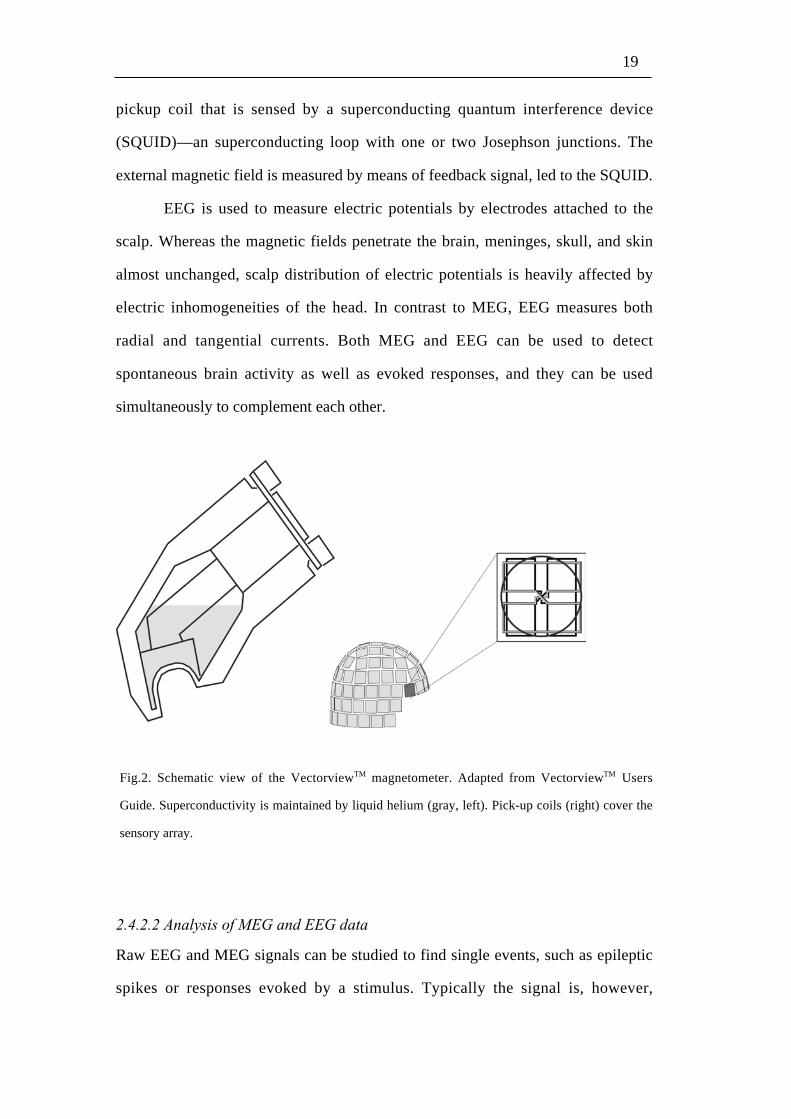
shielded room to lower the magnetic noise. The modern helmet-shaped
neuromagnetometers (Fig. 2) house hundreds of signal detectors. These detectors,
magnetometers and gradiometers, are merged in liquid helium to maintain
superconductivity necessary to detect weak magnetic fields. Magnetometers are
loop-form pick-up coils that give maximum signal on both sides of a dipolar
current. Planar gradiometers are figure-of-eight shaped coils that give maximum
signal above the current dipole. Changing magnetic field induces a current in a
19
pickup coil that is sensed by a superconducting quantum interference device
(SQUID)—an superconducting loop with one or two Josephson junctions. The
external magnetic field is measured by means of feedback signal, led to the SQUID.
EEG is used to measure electric potentials by electrodes attached to the
scalp. Whereas the magnetic fields penetrate the brain, meninges, skull, and skin
almost unchanged, scalp distribution of electric potentials is heavily affected by
electric inhomogeneities of the head. In contrast to MEG, EEG measures both
radial and tangential currents. Both MEG and EEG can be used to detect
spontaneous brain activity as well as evoked responses, and they can be used
simultaneously to complement each other.
Fig.2. Schematic view of the VectorviewTM magnetometer. Adapted from VectorviewTM Users
Guide. Superconductivity is maintained by liquid helium (gray, left). Pick-up coils (right) cover the
sensory array.
2.4.2.2 Analysis of MEG and EEG data
Raw EEG and MEG signals can be studied to find single events, such as epileptic
spikes or responses evoked by a stimulus. Typically the signal is, however,

20
averaged with respect to stimulus onset or to same task event to enhance signal-to-
noise ratio. The signal distribution of the averaged responses is then searched
visually to obtain the first guess for the activated areas and to select time windows
for further analysis. There is no unique solution to the inverse problem, i.e. which
current distribution in the brain produces the measured magnetic field pattern, but
anatomical and physiological knowledge can be utilized to constrain the possible
solutions.
For source modelling, the head is typically modelled as a spherically
symmetric volume conductor. Although a brain-shaped “realistic” conductor model
is superior in source localization in some brain regions, the sphere model is
computationally less demanding, and it offers an adequate model for most of the
cortical regions, including the primary visual, auditory, and sensorimotor cortices
(Tarkiainen et al. 2003).
A widely applied method is to model neuronal activity as current dipoles.
Optimum dipole model, the equivalent current dipole (ECD), is searched for by a
least-squares fit. Multi-dipole model, combining several single dipoles, can then be
introduced. Validity of the dipole model can be evaluated by comparing the
measured signals with the responses predicted by the model. If the signals are
inadequately explained by the model, the data are re-evaluated. Finally, a model
with the smallest possible amount of dipoles that best describes the measured
fields—and agrees with known anatomy—is accepted. Localization error in ECD
analysis of MEG data is typically only 2–4 mm, but several limitations have to be
kept in mind. For example, a distributed source can be interpreted as stronger and
deeper than the actual one, and confidence limits of the ECD location are relatively
high in the direction of depth.
An alternative method to model MEG data is to use minimum norm
estimates that apply less restrictions to the source configurations (Uutela et al.
21
1999). These methods are less user-dependent, but in practice, a priori knowledge
has to be applied to avoid false positive results (Stenbacka et al. 2002).
Computation of sources is more complicated for EEG than MEG signals,
because tissues of different electric conductivities distort the distribution of electric
potentials, and because in EEG both radially and tangentially oriented currents
must be considered.
2.4.3 Functional magnetic resonance imaging (fMRI)
Functional magnetic resonance imaging (fMRI) measures neuronal activity
indirectly, typically by detecting changes in blood oxygenation. Because coupling
between neuronal activation and blood-oxygenation change is slow (response peaks
4–6 s after the neural activation), fMRI is inferior to MEG and EEG in temporal
resolution. fMRI is, however, superior to MEG and EEG in localization accuracy,
and detects both superficial and deep activations. The following discussion is
mainly based on the textbooks of Brown and Semelka (1995), and of Jessard et al.
(2001).
2.4.3.1 Basics of MRI
Signal detected by MRI arises mainly from the protons (hydrogen nuclei of tissue,
mainly water). Rotation of protons around their axis is called spin. Because of
interaction between spins and the external magnetic field, protons precess.
Frequency of this precession (Larmor frequency) depends on magnetic field. In
MRI, a strong external magnetic field (B0) is used to align the spins. This results in
longitudinal magnetization of tissue (M0) in the direction of B0. Radiofrequency
pulse at the Larmor frequency can be applied to tilt the magnetization out of
equilibrium. When the pulse is then turned off, protons start to return to original
orientation and emit radiofrequency signal. Returning of the magnetization depends
on properties of tissue, and can be measured indirectly (T1-weighted images).

22
In the end of radio-frequency pulse, protons precess in coherence, i.e
precession movement of the protons is in phase. This results in summation and
therefore in a strong signal. This signal decays rapidly because of i) interaction at
the atomic and molecular level (T2-effect), and because of ii ) inhomogeneities in
the external magnetic field (T2*-effect). As well as the return of longitudinal
magnetization, the dephasing of the precession depends on tissue properties. This is
why anatomical structures can be viewed both by T1- and T2-weighted MR images.
Most of the functional MRI studies apply blood-oxygenation level
dependent (BOLD) signal. This method is based on different magnetic properties of
oxygenated and deoxygenated hemoglobin. When neurons are activated, relative
proportion of oxygenated blood increases locally, and associated signal change can
be detected in T2*- (and T2- to less extent) weighted images (Ogawa et al. 1990).
Slice selection can be accomplished in MR imaging by inducing
longitudinal magnetic gradient and applying radiofrequency pulses that excite only
the nuclei in certain field strength. Structural T1-images are then typically collected
row by row in a selected slice so that after each excitation pulse, magnetic gradients
are manipulated to result in unique combination of phase and frequency of signal
from each point of this row. In functional imaging, high speed is required to detect
changes that occur in the blood oxygenation in a time scale of seconds. Such a
high-speed-image collection is enabled in echo-planar imaging by changing
gradients so that the whole slice can be collected after one excitation pulse. This
results in loss of spatial accuracy and decrease of image quality. Whereas spatial
resolution of structural images is typically about 1 mm, it is typically 3–4
millimeters in echo planar images. Because data are collected in echo planar
imaging for a relatively long period after the excitation pulse, field
inhomogeneities, susceptibility effects and chemical shifts have more time to distort
spin phasing and spatial encoding decreasing the image quality. Compared with
other available methods, however, high-speed collection of the BOLD signal from

23
the whole brain with echo planar imaging is a powerful tool to study human brain
function.
2.4.3.2 Preprocessing and analysis of fMRI data
To achieve optimal results, functional volumes need to be preprocessed before
analysis. Commonly applied preprocessing includes movement correction and
spatial smoothing. In addition, volumes of all subjects are normalized to a common
template, whenever a group-level analysis is included. For movement correction,
translation and rotation parameters are defined in each dimension for all the other
volumes with respect to the first. Using these parameters, each volume is then
aligned to match the first volume. Volumes are smoothed by a gaussian filter to
increase signal-to-noise ratio, to compensate for inter-individual variance in
functional anatomy, and to make data to conform more closely to statistical models
(Friston et al. 1994).
Normalization applies both linear and nonlinear transformation to fit
volumes to a common template volume (Friston et al. 1995a).
For data analysis, a general linear model, based on the study protocol, is
first created (Friston et al. 1995b). This model is then convolved with
hemodynamic response function to take into account the time lag between neuronal
activation and hemodynamic response. Additional functions can be included as
regressors to compensate for slow signal drifts; this procedure corresponds to high-
pass filtering. As fMRI signal is temporally autocorrelated, an autoregressive model
is also included (Bullmore et al. 1996). The time course of the signal is then fitted,
voxel by voxel, to the model by a least-squares fit, resulting in multipliers of the
general linear model (parameter estimates) and their variance for each condition.
Statistical parametric maps (SPMs) are formed by comparing these parameter
estimates between conditions (e.g. task vs. rest). An alternative method is to
correlate the model function with the time behavior of the signal (Bandettini et al.

24
1993). This method can be applied also to find out brain areas where time behaviors
of the signals are similar to each other (functional connectivity; Friston 1994).
For a group analysis, the individual contrast or correlation images are fed
voxel by voxel into statistical tests (Holmes and Friston 1998). Statistical decision
making in fMRI studies has to take into account the problem of multiple
comparisons; testing hundreds of thousands of voxels results in numerous false
positive findings if 95% confidence level is applied. Therefore one needs to use
conservative statistical thresholds and/or knowledge about functional neuroanatomy
and extent of the activation to restrict the amount of false positive results.
2.5 Painful stimulation
In pain imaging studies, stimuli are most frequently delivered to the skin for
practical reasons. Electric or mechanical stimuli cause clear pain, and tactile
sensation as well. Because tactile and pain systems are overlapping, it is difficult to
recognize pain-related brain activity by brain imaging if the tactile component is
present. Thulium-laser pulses, applied in this thesis, are absorbed in the water of the
most superficial skin, where the nociceptors are located. These pulses heat the skin
up to > 45 °C, activating nociceptors without significant activation of tactile
receptors (Bromm and Lorenz 1998).

25
3 Aims of the study
This thesis aimed to increase understanding about brain functions related to pain.
Specific aims of Studies I–V were the following.
To define recovery cycles of pain-evoked MEG and EEG responses for
optimization of evoked-response measurements for clinical diagnostics and basic
research (Study I)
To compare sites and time courses of brain responses to pain mediated via slowly
conducting C- and faster conducting Aδ-fibers (Study II). Such information would
benefit studies on C-fiber function in pain disorders.
To study effects of noxious input on the spontaneous oscillatory activity of the
primary motor cortex (Study III) and on the oscillatory corticomuscular
communication (Study IV) to better understand the effect of pain on the motor
cortex.
To study brain correlates of suggestion-induced pain and of subjective reality of
pain (Study V). Such information could be useful for understanding effects of
psychological factors in pain, the nature of hypnotically induced experiences, and
construction of the subjective reality in the brain.

26
4 Materials and methods
4.1 Subjects
Altogether 33 subjects (14 females, 19 males; mean age 28 years, range 19–44
years) were studied by MEG or fMRI, some of them several times. For Study V,
subjects were prescreened from among 103 volunteers by suggestibility. All
subjects were, by self report, healthy and without any medication. They were
mainly students from the Helsinki University of Technology and University of
Helsinki. All measurements had prior approval by the local ethics committee and
subjects gave written informed consent before participation.
Study N of subjects Stimuli Recording
I 8 Painful laser pulses MEG and EEG
II 10 Painful laser pulses MEG
III 9 Painful laser pulses MEG
IV 7 Painful laser and non-painful tactile pulses MEG and EMG
V 14 Painful laser pulses and hypnotic suggestions fMRI
Table 1. Number of subjects, stimulation, and recording of brain activity in the five studies.
4.2 Stimulation, psychophysical measurements, questionnaires, and
screening
Painful stimuli were delivered with a thulium-YAG stimulator (1 ms pulse
duration, 2000 nm wavelength, Baasel Lasertech, Starnberg, Germany). The stimuli
were conducted to the measurement room via an optic fiber and directed to the left
hand dorsum. To avoid skin burns and adaptation, stimulation site was manually
moved in an area of about 10 cm2. In Study IV, tactile stimuli were produced by a
pneumatic stimulator, where air pressure pulse of 300 kPa bulges out a thin
diaphragm for about 170 ms (Mertens and Lütkenhoner 2000; Simoes et al. 2001).
In the psychophysical part of Study III, reaction times were collected by applying a
key pad in which finger lift releases a light beam. Hypnotic suggestions were given
by an experienced hypnotist, Dr. Sakari Närvänen.

27
During each experiment, subjects estimated mean pain intensity right after
stimulation sessions. In Study V, subject filled in a detailed questionnaire,
including type, location and temporal behavior of pain, as well as mean intensity,
unpleasantness, and reality of pain applying visual analog scales (VAS). In VAS,
one end represents the minimum and the other end the maximum of a measure.
Subjects draw a vertical line in between these end points according to their
evaluation.
I prescreened suggestible subjects to Study V by Stanford Hypnotic
Susceptibility Scale Form C (Weitzenhoffer and Hilgard 1962). This test includes
induction of hypnosis by sequential relaxation and by suggestions for focused
attention. Subjects’ responses to 12 suggestions for different perceptions and
experiences are then observed and/or interviewed.
4.3 MEG and EEG recordings
During MEG and EEG recordings (Studies I–IV), activity was recorded with a 306-
channel helmet-shaped neuromagnetometer (Vectorview™, Neuromag Ltd,
Helsinki, Finland) at the Brain Research Unit of the Low Temperature Laboratory
(Fig. 2.). The device contains 102 identical units of two gradiometers and one
magnetometer in each.
Four head-position-indicator coils were placed to the scalp and the locations
of the coils with respect to anatomical landmarks of the head were determined with
a 3-D digitizer. When the subject was then seated under the MEG helmet, weak
electrical currents were led to the coils and the exact head position was found by
measuring the resulting magnetic signals. During the MEG (Studies I–IV) and EEG
(Study I) recordings, the subject was sitting comfortably in a magnetically shielded
room, with the head supported against the helmet-shaped sensor array of the
neuromagnetometer.
The MEG signals were bandpass filtered through 0.03–172 Hz and digitized
at 600 Hz. Traces coinciding with amplitudes exceeding 150 µV in the
simultaneously recorded vertical electro-oculogram were automatically rejected
from the analysis.

28
Scalp EEG was recorded in Study I from the midline locations Fz, Cz, and
Pz of the international 10-20 system, referred to the left mastoid. The filter settings,
analysis period, and prestimulus baseline were the same as for the MEG recordings.
In Study IV, surface electromyograms were recorded from the first m.
interosseus digitorum, and the m. opponens pollicis with the same filter settings as
MEG. For the MEG studies, magnetic resonance images were acquired with a 1.5-T
Siemens Magneton system of the Department of Radiology, University of Helsinki.
4.4 Analysis of MEG and EEG data
In Studies I–IV, source modeling was based on laser-evoked fields recorded by the
204 gradiometers, and the procedure followed MEG analysis methods generally
applied in our laboratory (see 2.2.2). Only ECDs accounting for more than 80% of
the signal variance in 10–20 channels around the local signal maximum were
selected for a multidipole model. Source strengths and response latencies were
calculated from the multidipole models (or from the evoked potentials recorded
from Cz electrode of EEG in Study I).
Level of the motor-cortex ~20-Hz oscillations (see 2.4) was quantified in
Study III by first filtering the MEG signals through 15–25 Hz, then rectifying them,
and finally averaging with respect to stimulus onset (Salmelin and Hari 1994).
Signal strength was then calculated from one channel at signal maximum over the
M1 cortex of each hemispheres.
In Study IV, cortex–muscle coherence was calculated as cross correlation
between original MEG signal from the contralateral MI cortex and the rectified
EMG from hand muscles. Cortical sources of coherent signal were modelled as
ECDs. Coherence between these MI sources and EMGs was then computed with
respect to the stimulus onset applying fast-fourier transform.
4.5 fMRI measurements
Functional MR images were acquired by a Signa VH/i 3.0T MRI scanner (General
Electrics, Milwaukee, WI, USA) at the Advanced Magnetic Imaging Centre of the
Helsinki University of Technology. A gradient-echo echo-planar imaging sequence

29
(time to repetition = 3.0 s, TE = 32 ms, flip angle = 90°, field of view = 20 cm, 96 x
96 matrix, slice thickness 3 mm, no spacing) was applied to obtain BOLD signal.
The whole brain was covered by 37 oblique axial slices. Structural images were
collected for each subject by T1-weighted 3D spoiled gradient-echo sequence
(SPGR; time to repetition = 8.4 ms, time to echo = 1.8 ms, time to inversion = 300
ms, flip angle 15°, number of excitations = 2).
4.6 Preprocessing and analysis of the fMRI data
Functional data were preprocessed and analyzed by Statistical Parametric Mapping
software (SPM2, http://www.fil.ion.ucl.ac.uk/spm). Volumes for each subject were
realigned, spatially normalized to the average brain of the Montreal Neurological
Institute (MNI, resulting in cubic voxels of 8 mm3), and smoothed with an 8-mm
(full width at half maximum) Gaussian kernel. The analysis applied the general-
linear-model approach (see 2.3.2).

30
5 Experiments
5.1 Optimum inter-stimulus interval for measurement of cortical
electromagnetic responses to painful laser stimuli is 4–5 s (Study I)
Cortical pain-evoked responses offer a well-established and well-replicable
measure of pain-related cortical processing (Bromm and Lorenz 1998). Therefore
these responses are increasingly measured both in clinical diagnostics of
neurological diseases and in pain research (Spiegel et al. 2000).
Assuming stationary noise, the signal-to-noise ratio (SNR) is proportional to
the square root of the number of averaged responses. SNR can not be enhanced,
however, simply by increasing number of stimuli during a fixed measurement time
because the cortical responses to single stimuli decrease in amplitude with
shortening of the inter-stimulus interval (ISI). SNR of cortical evoked responses
during a fixed measurement time can be mathematically optimized if one knows
how amplitude of the these responses behaves as a function of inter-stimulus
interval (ISI; Ahlfors et al. 1993). So far, effect of ISI on cortical pain-evoked fields
had remained, however, unknown.
5.1.1 Stimuli
Each subject received altogether 10 blocks of laser stimuli in two experimental
sessions. Stimuli were delivered at ISIs of 2, 4, 8, and 16 s in the first session and
of 0.5, 1, 2, and 4 s in the second. Each ISI varied randomly by 20% around the
mean to minimize effects of stimulus anticipation. Order of stimulation blocks
(each with certain ISI) was otherwise randomized, but to study replicability of the
responses, the 2-s ISI always started and ended the measurement session.

31
5.1.2 Results
Subjects reported the laser stimuli to elicit pricking pain, followed by a weaker
sensation of burning pain. Perceived intensity of pain decreased as a function of
ISI, being 4.4 ± 0.6 (mean ± SEM on 0–10 scale) for the 0.5-s ISI and 3.1 ± 0.5 for
the 16-s ISI.
In line with earlier studies (Hari et al. 1983; Huttunen et al. 1986; Kakigi et
al. 1995; Frot et al. 1999; Ploner et al. 1999b; Kanda et al. 2000), laser stimuli
elicited bilateral activation of the SII cortex, with peak at about 155 ms in the
contralateral and at 160 ms in the ipsilateral SII cortex. In addition, weak activation
of the superior parietal cortex was observed in 7 out of the 8 subjects.
Laser-evoked potentials (LEPs), recorded from the position Cz, consisted of
a surface-negative peak at 190–230 ms, followed by a surface-positive peak at
310–330 ms. Both of these peak latencies were longer than those of laser-evoked
fields (LEFs; p < 0.05).
Fig. 3. Normalized amplitudes (mean of 8 subjects) of LEFs and LEPs as a function of ISI. An
exponential model function with time constant of 3.5 s is presented with black line. SIIc =
contralateral secondary somatosensory cortex, SIIi = ipsilateral secondary somatosensory cortex, SIc
= contralateral region of the primary somatosensory cortex, ISI = inter-stimulus interval. LEF =
laser-evoked field, LEP = laser-evoked potential.
Both LEFs and LEPs increased in amplitude as a function of ISI until about
4 s. Thereafter prolongation of the ISI had only little effect on the response

32
strengths. Exponential curve with a time constant of 3.5 s fitted best to the
measured signal strengths as a function of ISI (Fig. 3). As the optimum ISI for
obtaining the best SNR during a fixed measurement time is about 1.26 x this time
constant (Ahlfors et al. 1993), the optimum ISI for both magnetic and electric pain-
evoked responses is about 4–5 s.
5.1.3 Discussion
Our findings show that both LEFs and LEPs increase strongly in amplitude up to
ISIs of 4 seconds and start to saturate thereafter. Based on this recovery cycle, ISIs
of 4–5 s are optimal for recording of LEFs and LEFs. These findings add to prior
and later findings of increase of LEP amplitudes with increasing ISI (Jacobson et
al. 1985; Dowman 1996; Truini et al. 2004).
The lack of quickly recovering cortical response, such as the SI response
during tactile stimulation (Wikström et al. 1996), adds to differences between
processing of tactile and noxious input in the superior parietal region.
5.2 First and second pain share a common cortical network (Study II)
Microneurographic recordings have implied altered function of pain-mediating C-
fibers in a specific chronic lower limb pain syndrome (Orstavik et al. 2003). Lack
of practical methods for selective C-fiber stimulation has, however, restricted
MEG/EEG studies in this field. Bragard et al (1996) managed to selectively activate
C-fibers when laser stimuli were restricted to tiny skin areas, based on higher
density and lower activation threshold of C-nociceptors than Aδ-nociceptors. After
that, several studies have reported C-fiber-mediated evoked responses. Stimuli in
these studies have elicited, however, mainly other percepts than pain, such as
warmth or itch (Opsommer et al. 2001; Tran et al. 2002; Kakigi et al. 2003) that
may be subserved by different neural systems than pain (Andrew and Craig 2001).
Furthermore, cortical responses to separate C-fiber and Aδ-fiber stimulation have

33
not been compared in same subjects. We therefore aimed to study cortical
responses elicited by clearly painful C-nociceptor stimuli and to compare these
responses with those elicited by painful Aδ-stimuli.
5.2.1 Methods
C-nociceptors were activated by laser stimulation of tiny skin areas. The method of
Bragard et al. (1996)—that applied an aluminium plate perforated by small holes
and attached to skin—was modified and further developed to allow flexible moving
of the stimulation site, to avoid mechanical contact to skin, and to elicit clear pain.
These aims were achieved by connecting a plastic plate with a single small hole to
the hand piece of the thulium-laser stimulator. This restrictor allowed delivery of
laser pulses (about 50 mJ) to a skin area of 0.2–0.3 mm2. Plastic, instead of
aluminium, was used to reduce acoustic noise resulting from laser-beam absorption.
Responses to “large-area” (about 500 mJ/10 mm2) laser stimulation were
measured in a separate session, and about 100 responses were averaged for both
types of stimuli. Both Aδ- and C-fiber stimuli were delivered to the dorsum of the
left hand at ISIs that varied randomly between 4.5 s and 5.5 s.
5.2.2 Results
Subjects rated both stimuli as clearly painful (mean ± SEM intensity 5 ± 1 for Aδ-
stimuli and 4 ± 1 for C-stimuli on 0–10 scale). Aδ-stimuli produced immediately
sharp pain, followed by a weaker pain and/or warmth sensation, whereas C-stimuli
were associated with a delayed onset of pain.
To Aδ-stimuli, responses in the SII region peaked at 167 ± 7 ms
(contralateral hemisphere) and 179 ± 7 ms (ipsilateral hemisphere; Fig. 4). The
corresponding SII responses to C-stimuli peaked at 811 ± 14 ms and 823 ± 21 ms,
respectively. In addition, we observed a response arising from the right posterior

34
parietal cortex (PPC), peaking at 183 ± 22 ms during Aδ-stimulation and at 833 ±
22 ms during C-stimulation (Fig. 4). The PPC responses were posterior (15–16
mm), medial (14–20 mm), and superior (10–13 mm) to the 20-ms SI response to
electric median nerve stimulation in the same subjects (P < 0.05).
Fig. 4. Mean source locations and time courses during painful Aδ- and C-fiber stimulation.
Confidence intervals of sources of the pain-evoked fields are superimposed on the average image of
elastic transformations of all subjects’ MR images. SIIc = contralateral secondary somatosensory
cortex, SIIi = ipsilateral secondary somatosensory cortex, PPC = posterior parietal cortex.
5.2.3 Discussion
These results demonstrate that cortical responses to painful C-fiber stimulation can
be reliably recorded. Comparison of the responses mediated via C- and Aδ-fibers
revealed that both stimuli activate the cortical network including the SII cortices
and superior parietal region. In contrast to earlier MEG studies suggesting pain-
related activation of the SI cortex (Ploner et al. 1999b; Kanda et al. 2000; Ploner et
al. 2000; Tran et al. 2002), we localized superior parietal responses consistently to
the posterior parietal cortex (PPC), as well during Aδ-fiber- as during C-fiber-
35
mediated pain. Possible reasons for such a discrepancy include at least differences
in stimulation paradigms (Peyron et al. 2000) and different constraints in the
analysis (Stenbacka et al. 2002). Whereas SI may be related to encoding of
intensity and location of pain (see 2.1.3.4), PPC is a multimodal area that has been
linked at least with spatial attention (Davis et al. 2002).
Ploner et al. (Ploner et al. 2002) recently compared magnetic fields during
early (Aδ-fiber-mediated) and late (C-fiber-mediated) phase of painful “large-area”
laser-evoked responses. Activation of the superior parietal cortex was relatively
stronger during early than late phase of activation. By stimulating Aδ- and C-
nociceptors separately we found ratios of superior parietal and SII activation
strengths during both Aδ- and C-fiber-mediated activation to be similar. Assuming
the same source for the superior parietal activation in both studies, these findings
suggest that the relatively weaker activation of the superior parietal cortex (Ploner
et al. 2002) may be specific to second pain (i.e. the late phase of biphasic pain
following a single stimulus) but independent of the pain-mediating fiber type.
By characterizing cortical responses to painful C-fiber stimulation, our
findings build basis for studies on involvement of different fiber systems in pain
disorders.
5.3 Painful A δ- and C-fiber stimuli suppress the motor-cortex
oscillatory activity (Study III)
In addition to brain structures related to sensory, emotional and cognitive
components of pain, painful stimulation is associated with activation of the motor
system (Melzack and Wall 1965). In contrast to the well-known pain-related spinal
reflexes, effect of pain on the brain-level motor system remains poorly understood.
These mechanisms may have clinical relevance, because pain is sometimes
associated with motor dysfunction (Juottonen 2002), and on the other hand, motor
cortex stimulation relieves chronic pain (Tsubokawa et al. 1991a, b). We aimed to

36
study effect of selectively noxious input on the primary motor (MI) cortex in
healthy subjects.
5.3.1 Methods
Measurement was similar and partly overlapping with Study II. Instead of evoked
responses, level of the spontaneous ~20-Hz activity, arising from the MI region,
was analyzed (see 4.4). In a separate session, reaction times were measured i) to
sound presented at varying time points with respect to Aδ-fiber stimuli and ii) to
Aδ-fiber stimuli.
5.3.2 Results
Aδ-stimulation elicited pricking pain at short latencies (intensity 5 ± 1, mean ±
SEM on the 0–10 scale) that was followed by burning pain. C-stimuli resulted in
delayed onset of pain (intensity 4 ± 1).
Level of the ~20-Hz oscillations started to decrease 180 ± 10 ms (mean ± SEM)
after Aδ-fiber stimuli and 820 ± 30 ms after C-fiber stimuli, and the suppression
peaked 160–170 ms later for both stimuli (at 340 ± 30 and 990 ± 40; Fig. 5). A
similar, but about 50% weaker, suppression of the ~20-Hz activity was observed in
7 out of 9 subjects in the ipsilateral MI region.
During Aδ-stimulation, onsets of the ~20-Hz suppressions coincided roughly
with the peaks of the simultaneously recorded evoked responses from the
contralateral SII cortex.
Reaction times to Aδ-stimuli coincided with the peaks of the motor-cortex ~20-
Hz suppression, and reaction times to tones were 10 ± 2 ms shorter (p < 0.01) when
the motor responses to these tones occurred after, rather than before, the onset of
laser-stimulus-induced ~20-Hz suppression. Reaction times to Aδ-fiber stimuli
were 335 ± 10 ms.

37
Fig. 5. Sources of the ~20-Hz oscillations in one subject (left), and grand average of temporal
behavior of these oscillations with respect to painful Aδ- and C-fiber stimuli.
5.3.3 Discussion
As suppression of the ~20-Hz oscillations is associated with activation of the MI
cortex (Jasper and Penfield 1949; Salmelin and Hari 1994; Salenius et al. 1997b;
Schnitzler et al. 1997; Hari et al. 1998), our results could reflect MI excitation by
noxious input. Such an excitation would be unlikely to be explained by general
arousal only, because much stronger suppression was observed in the right than in
the left hemisphere. We did not stimulate right hand to test whether the effect
would be contralateral rather than right-hemisphere specific, but the effect of
electric median-nerve stimulation on the motor cortex 20-Hz oscillations has been
demonstrated to be clearly contralateral (Salenius et al. 1997b).
Although many factors may contribute to the observed fastening of reaction
times after painful stimuli, this finding suggests that noxious stimuli are not
associated with a reduced ability to perform voluntary movements, as has been
suggested by a previous study (Valeriani et al. 1999). Interestingly, peak latencies
of the ~20-Hz suppression and reaction times coincided at the time window during
which also ability to perform voluntary motor acts seems to be enhanced. Aδ-
stimuli suppressed the 20-Hz level at very short latency, suggesting automatic
priming of the MI cortex, rather than a voluntary response to pain. Such a priming
could be related to preparation for protective voluntary movements.

38
5.4 Painful laser-stimuli enhance oscillatory cortex–muscle coupling
Findings of Study III supported involvement of the MI cortex in response to pain,
arising the question whether noxious input affects communication between the MI
cortex and peripheral muscles. Such a communication can be studied by means of
coherence between MI oscillations and electromyographic signals from the muscles
(Conway et al. 1995; Salenius et al. 1997a); for reviews, see: (Grosse et al. 2002;
Salenius and Hari 2003). Somatotopy of the motor-cortex sources of the coherent
MEG signal and increase of time lag from the cortical to the peripheral signal with
increasing distance from the cortex suggest that the cortex–muscle coherence offers
an unique view into dynamics of the motor system (Salenius and Hari 2003).
5.4.1 Methods
During recordings, subjects pressed a rubber tube with their left index finger and
thumb. MEG and EMG signals were recorded from each subject during three
different conditions (1 min each) that were repeated five times in an order balanced
across subjects. These conditions included: isometric muscle contraction, isometric
muscle contraction plus noxious laser stimulation, and isometric muscle contraction
plus innocuous tactile stimulation.
5.4.2 Results
Laser stimuli with intensities of 440 ± 20 mJ were perceived as weak, but clear
pain, and the tactile stimuli as mild touch. Handgrips were performed at about one
third of the maximum voluntary contraction, and they were associated with a
MEG–EMG coherence in the ~20-Hz band in all three conditions in 6 out of 7
subjects. The coherence increased phasically after both types of stimuli but
significantly later after laser than tactile stimuli (mean ± SEM peak latencies 1.1 ±
0.1 s vs. 0.6 ± 0.1 s; P < 0.05; Fig. 6). This coherence increase lasted longer after
laser than tactile stimuli (0.9 ± 0.1 s vs. 0.5 ± 0.1 s, P < 0.05).

39
Fig. 6. Normalized grand average of coherence between the motor cortex and peripheral hand
muscles during isometric muscle contraction without stimuli (left), with respect to laser stimuli
(middle), and with respect to tactile stimuli (right).
5.4.3 Discussion
These findings show that, in addition to innocuous tactile stimuli, selectively
noxious laser stimuli are followed by an increase of the 20-Hz MEG–EMG
coherence. Although vibratory muscle-tendon stimuli may not increase
cortex–muscle coherence (Mima et al. 2000), our finding of the coherence increase
after tactile stimuli agrees with prior findings on electric median-nerve stimulation
(Hari and Salenius 1999). Coherence increase after selectively noxious laser stimuli
offer another piece of evidence for involvement of the MI cortex in response to
pain.
Probably because of the muscle contraction and small number of subjects,
significant suppression of the motor-cortex ~20-Hz oscillations was not present in
this study. The latency of the coherence increase was, however, clearly longer than
the latency of the ~20-Hz suppression observed in Study III. According to Killner
et al. (2003), increase of the cortex–muscle coherence may be related to resetting of
the descending motor commands. Such a resetting could be necessary after
modulation of the MI activity by tactile or noxious input.

40
5.5 Subjective reality of pain is associated with activation of the
sensory pain circuitry and of the medial prefrontal cortex
Ability to differentiate events arising from one's mind from those arising from the
external world may be especially demanding in experience of pain that has great
contribution from psychological factors (Rainville et al. 1997; Petrovic and Ingvar
2002; Derbyshire et al. 2004; Wager et al. 2004). Top-down activation of the pain
circuitry was recently demonstrated after hypnotic suggestion for pain, and such an
activation was proposed to form a possible basis for pain disorders without
appropriate physical origin (Derbyshire et al. 2004). It is, however, unknown,
whether this activation was associated with actual experience of pain or with
anticipation of pain; anticipation of pain may be associated with a similar activation
pattern than actual pain (Koyama et al. 1998; Ploghaus et al. 1999; Porro et al.
2002). In addition, it remains elusive, how real such a suggestion-induced pain is
experienced, how it differs from pain of physical origin, and how the subjective
reality of pain is constructed in the human brain. To address these questions, we
induced pain to the left hand of healthy, suggestible volunteers by either laser
pulses or hypnotic suggestion during fMRI.
5.5.1 Methods
Subjects were hypnotized before entering the scanner for the first sessions and
instructed to signal with a small foot movement when the maximum tolerable pain
was achieved and when the pain was totally relieved. Suggestions were given via
head phones to induce and relieve pain several times during the first scanning
session. This session was followed by another session where laser-induced pain
alternated with rest, while subject remained under hypnosis but did not receive any
suggestions. Laser stimulation was repeated in a separate session, during which the
subject was alert, and half of the laser-pulse series were delivered with non-painful
intensities for control purposes. Subjects filled in a detailed questionnaire about
their experiences right after the brain scanning. This questionnaire included
estimates of the reality of pain in a range from "imaginal pain" to "real physical
pain associated with injury or painful stimulation of the hand".

41
5.5.2 Results
Subjects described the suggestion-induced pain most frequently as burning or
aching in the left hand, and the laser-induced pain as continuous burning and
fluctuating pricking pain. The location, intensity, and unpleasantness were similar
for laser- and suggestion-induced pain (50–60/100 on VAS for intensity and
unpleasantness). Although the subjective reality of pain varied between subjects
also during laser stimulation (without any suggestion, and independently of whether
the subject was under hypnosis or not), each subject estimated the reality of pain to
be higher for laser- than suggestion-induced pain (87 ± 3 vs. 62 ± 5; P < 0.001).
Similarly to laser-induced pain, stable phase of suggestion-induced pain
(from the subject’s sign of maximum pain to beginning of the pain relief) was
associated with activation of the well known pain circuitry, including bilateral
insulas and SII cortices, and the contralateral caudal ACC. Furthermore, activation
strengths of the SII cortex correlated with the subjective estimates of pain intensity
during both laser- and suggestion-induced pain.
Despite similarities between the activation patterns, the contralateral (right)
posterior insula, the posterior superior SII cortex bilaterally, and the ipsilateral (left)
cerebellum were more strongly activated during laser-induced than suggestion-
induced pain. Of these areas, activation strengths of the posterior insula and of the
SII cortices correlated positively with the subjective reality of laser-induced pain.
During suggestion-induced pain, subjective reality estimates correlated
positively with activation strengths of two areas in the medial prefrontal cortex
(mPFC; Fig. 7): the perigenual anterior cingulate cortex (pACC) and an area
extending from the rostral anterior cingulate cortex (rACC) to the pericingulate
cortex. Similar trend was evident during laser-induced pain. Although intensity and
reality estimates were mutually correlated (P < 0.05), the correlations between the
reality estimates and activation strengths were significant even if the intensity and
unpleasantness estimates were included in the correlation analysis as confounding
factors.
During both laser- and suggestion-induced pain, signals from rACC
covaried with signals from the pain circuitry, whereas signals from pACC covaried
with signals from bilateral medial temporal lobes, inferior parietal cortices, and
posterior cingulate cortices.

42
Fig. 7. Correlation between activation strengths in the medial prefrontal cortex and the subjective
reality of pain. rACC = rostral anterior cingulate cortex (Talairach coordinates (x, y, z): 8, 36, 20;
–8, 32, 19), pACC = perigenual anterior cingulate cortex (Talairach coordinates: 8, 43, 0).
5.5.3 Discussion
These findings suggest that suggestion-induced pain is associated with activation
pattern very similar to that observed during physically induced pain. The activation
of the sensory pain circuitry was, however, stronger, and the reality estimates were
higher during laser-induced than suggestion-induced pain.
Subjects reported the suggestion-induced pain to be most frequently burning or
aching even if they were allowed to imagine whatever type of pain. Reports of
burning pain could have resulted from that subjects knew about the forthcoming
laser stimulation and had tried laser stimuli. Only 2 out of 14 subjects reported,
however, pricking, although laser stimuli cause clear pricking, in addition to
burning sensation. In addition, only a few test pulses were given to the subjects
prior to scanning, and subjects were told that the suggested pain could be of any
kind. It is therefore likely that burning and aching sensations are for some reason
more prone than pricking to imaginal pain.

43
Subjective reality of pain was associated with activation of the sensory pain
circuitry and of the medial prefrontal cortex. Stronger activation of the sensory pain
circuitry could be related to more clear, and therefore to subjectively more real
perception. Pericingulate part of the observed medial prefontal network is related to
self monitoring and to monitoring of intentions of self and others (Gallagher and
Frith 2003), phenomena very close to source monitoring, i.e. monitoring whether
percept is of external or internal origin. Interestingly, activity of pACC covaried
with areas associated with memory (thalamus and medial temporal lobe), attention
(inferior parietal cortices), and imagery (inferior parietal cortex; Cabeza and
Nyberg 2000)—i.e. factors connected to the experience of reality (Bentall 1990;
David 1999; Brebion et al. 2000; Aleman et al. 2003; Barnes et al. 2003). Although
fMRI studies can not reveal causal relationship between subjective experience and
brain activations nor between spatially separate activations, these findings open
intriguing views into study of subjective reality.

44
6 General discussion
Dysfunction can be hardly understood without understanding the normal function.
This thesis aimed to increase understanding of pain-related brain function in
healthy subjects, building thereby basis for studies on mechanisms and treatment of
pain disorders.
6.1 Time-locked electromagnetic signature of pain in the brain
Pain-evoked MEG and EEG responses are clinically useful (Spiegel et al. 2000)
and offer a valuable tool for pain research. Because good signal-to-noise ratio is
essential for reliable results in any measurement, our finding of optimum ISI (Study
I) is likely to be useful for both clinical diagnostics and research. Because cortical
disinhibition has been suggested in chronic regional pain syndrome (Schwenkreis et
al. 2003), it is of interest whether recovery cycles of pain-evoked responses would
differ in these pain patients.
Characterization of cortical responses to C-fiber-mediated pain, and
comparing these responses with those mediated via Aδ-fibers, builds basis for
studying involvement of these different pain-mediating systems in clinical disorders
(Orstavik et al. 2003). Our finding that both Aδ− and C-fiber-mediated pain are
associated consistently with PPC activation arises question about the role of this
area in pain perception.
6.2 Effects of pain on the motor cortex
Our findings add to the evidence of motor-cortex involvement in pain by showing
suppression of the motor-cortex spontaneous oscillatory activity and increase of the
corticomuscular oscillatory communication after selectively noxious stimuli.
Suppression of the MI oscillations may indicate excitation of the MI cortex (Jasper

45
and Penfield 1949; Salmelin and Hari 1994; Salenius et al. 1997b; Schnitzler et al.
1997; Hari et al. 1998). In pain such an excitation could facilitate protective
voluntary movements. If continued during chronic tension-type pain, however, such
an excitation could contribute to the vicious circle between pain and the excitatory
input to the painful muscles. In comparison with adaptation of tactile reseptors,
adaptation of nociceptors is minimal. Therefore, chronic pain could have major
influence on the motor cortex.
6.3 Subjective reality of pain, and brain correlates of psychologically
vs. physically induced pain
Although pain as a hallucination is rare, physical pain is strongly influenced by
psychological factors. Therefore, our findings on brain correlates of suggestion-
induced pain and subjective reality of pain are of clinical interest. In addition to
understanding of pain, these findings may benefit research of reality disorders,
observed in psychiatric diseases.
6.3.1 Real vs. unreal pain
Sometimes chronic pain disorders can not be explained by physical findings. These
disorders are frustrating for both patients and health-care professionals, and may
arise suspicion about the reality of pain, especially when the complaint of pain
disagrees with the observed behavior. Differentiating “unreal and real pain” would
be of great interest for guiding treatments and reimbursements. In this context
“unreal pain” is often intermixed with “pain of psychological origin”. Our findings
support the evidence that the subjective reality of pain is affected besides physically
induced activity in the pain circuitries, also by psychological factors. In this
framework, functional state of the sensory pain circuitry could be informative about
46
the origins of pain, but the medial prefrontal cortex should be considered in
assessment of subjective reality of pain as well.
6.3.2 Subjective reality and psychotic disorders
Although psychological factors strongly modulate pain, painful hallucinations are
very rare. In fact, schizophrenic patients and their close relatives are less sensitive
to pain than non-schizophrenic controls; for a recent discussion, see Murthy et al.
(2004). Our results on brain mechanisms of subjective reality of pain could have,
however, validity for subjective reality of other hallucinations (and perhaps of
delusions). Although mechanisms of psychotic disorders most likely differ from
those of hypnotic experiences, it is evident that the clinical research benefits from
understanding of how the experience of reality is constructed in the healthy brain.
6.4 Suggestion-induced perception
Use of term hypnotic hallucination in classical hypnosis literature (Weitzenhoffer
and Hilgard 1962) gives an impression that suggestible subjects would be unable to
differentiate suggestion-induced and real percepts. To the best of my knowledge,
there have not been prior studies comparing reality of physically and hypnotically
induced percepts that would have been of equal subjective intensity and intense
enough to be clearly perceived. Our results suggest that although hypnotic
experiences can be very intense, even the most suggestible subjects are able to
differentiate suggestion-induced percepts from the percepts of external origin by
the experience of reality; This finding is further supported by differences in brain
activation.
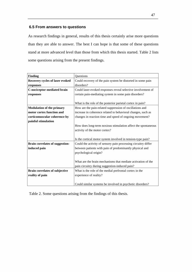
47
6.5 From answers to questions
As research findings in general, results of this thesis certainly arise more questions
than they are able to answer. The best I can hope is that some of these questions
stand at more advanced level than those from which this thesis started. Table 2 lists
some questions arising from the present findings.
Finding Questions
Recovery cycles of laser evokedresponses
Could recovery of the pain system be distorted in some pain
disorders?
C-nociceptor-mediated brainresponses
Could laser-evoked responses reveal selective involvement of
certain pain-mediating system in some pain disorders?
What is the role of the posterior parietal cortex in pain?
Modulation of the primarymotor cortex function andcorticomuscular coherence bypainful stimulation
How are the pain-related suppression of oscillations andincrease in coherence related to behavioral changes, such as
changes in reaction time and speed of ongoing movement?
How does long-term noxious stimulation affect the spontaneous
activity of the motor cortex?
Is the cortical motor system involved in tension-type pain?
Brain correlates of suggestion-induced pain
Could the activity of sensory pain processing circuitry differ
between patients with pain of predominantly physical and
psychological origin?
What are the brain mechanisms that mediate activation of thepain circuitry during suggestion-induced pain?
Brain correlates of subjectivereality of pain
What is the role of the medial prefrontal cortex in the
experience of reality?
Could similar systems be involved in psychotic disorders?
Table 2. Some questions arising from the findings of this thesis.
48
7 Conclusions
Whereas acute pain has an important protective function, chronic pain does not
have physiological meaning. Knowledge about pain is needed especially to help
those numerous people suffering from chronic pain. This thesis aimed to increase
understanding about brain function related to acute pain, and to build thereby basis
for studies on chronic pain. We found that both laser-evoked fields and potentials
increase in amplitude with increasing ISI up to about 4 s and saturate thereafter.
Based on these findings, optimum signal-to-noise ratio in a fixed measurement time
is achieved by inter-stimulus intervals of 4–5 s (Study I). Characterization of
cortical responses to painful C-fiber stimuli revealed that the C-fiber-mediated pain
activates similar cortical network than Aδ−mediated pain, including the bilateral
somatosensory cortices and the posterior parietal cortex (Study II).
Selectively painful laser stimuli suppressed spontaneous oscillatory activity
of the MI cortex (Study III) and, during isometric muscle contraction, increased
oscillatory cortex–muscle coupling (Study IV).
Suggestion-induced and laser-induced pain were associated with similar
activation of the emotional parts of the brains’s pain circuitry, whereas the laser-
induced pain was related to stronger activations of sensory parts of this circuitry
(Study V). Subjective reality of laser-induced pain correlated with activation
strengths in the sensory pain-processing areas. During suggestion-induced pain, the
subjective reality of pain correlated with activation strengths in the medial
prefrontal cortex, and similar trend was evident during laser-induced pain.
Our findings help to apply cortical laser-evoked responses more effectively
(Study I), and to address involvement of Aδ- vs. C-fiber system in chronic pain
disorders (Study II). The findings of pain-related PPC (Study II) and MI (Studies
III and IV) activation arise question about role of these regions in acute and chronic

49
pain. Results on brain correlates of suggestion-induced pain and subjective reality
of pain (Study V) benefit understanding of psychological modulation of pain, and
may be helpful for studies on mechanisms of reality distortions in psychiatric
disorders.

50
Acknowledgments
Work for this thesis was conducted at the Brain Research Unit of the Low
Temperature Laboratory (LTL) and at the Advanced Magnetic Imaging Centre of
the Helsinki University of Technology, and it was financially supported by the
Finnish Graduate School for Neuroscience, the Signe and Ane Gyllenberg
foundation, and the Maud Kuistila foundation, and via LTL by the Academy of
Finland.
It has been honor to work in such a high-quality unit as LTL is. I am
thankful for this possibility to the founder of LTL, late Academician Olli V.
Lounasmaa, to current director of LTL, Professor Mikko Paalanen, and to the head
of the Brain Research Unit, Professor Riitta Hari. I have had a privilege to conduct
my graduate studies under supervision of Prof. Hari, whos enthusiasm and
knowledge about human mind and brain I can’t stop admiring. To my other
supervisor, Doc. Nina Forss, I owe greatest thanks for her skilful guidance,
inspiring scientific discussions, and for her great impact on the atmosphere of LTL.
I want to thank Prof. Eija Kalso and Doc. Juha Huttunen for constructive
suggestions on the manuscript of this thesis.
I am grateful to my co-authors Phil. Lic. Jaana Hiltunen, and Drs. Veikko
Jousmäki, Jussi Numminen, Sakari Närvänen, Andrej Stancak, and Nuutti
Vartiainen for collaboration.
Great thanks to the “AIVO social group” and the “graduate students’ floor
ball team” for the great time spent together, and to Dr. Tommi Raij for his inspiring
example.
I’m thankful to my parents, sister, brother, captains of the Venekerho, and
all my friends for their support and relaxing company.
I owe my greatest thanks to Mr. Samuel Aulanko, Mrs. Mia Ilman, Dr. Ole
Jensen, Dr. Raimo Joensuu, Mrs. Marita Kattelus, Dr. Hanna Renvall, Mr. Ville
Renvall, Prof. Riitta Salmelin, Dr. Martin Schürmann, Mr. Mika Seppä, Dr.
Cristina Simões, Mr. Topi Tanskanen, and Dr. Antti Tarkiainen for their expert
help and advice.
The great equipment and support provided by Neuromag Ltd. and General
Electrics have been essential for my work. Thank you!

51
Great thanks to the computer support team—Samuli Hakala, Jan Kujala,
Mika Seppä, Antti Tarkiainen, and Kimmo Uutela; without their help this thesis
would have taken much longer time.
I would also like to thank Sari Avikainen, Peter Berglund, Gina Caetano,
Katri Cornelissen, Nobuya Fujiki, Teija Halme, Päivi Helenius, Liisa Helle, Linda
Henriksson, Kati Hirvonen, Yevhen Hlushchuk, Marja Holmström, Annika Hulden,
Kaisa Hytönen, Marianne Inkinen, Antti Jalava, Helge Kainulainen, Erika
Kirveskari, Tuire Koivisto, Hannu Laaksonen, Martin Lehécka, Sari Levänen, Mia
Liljeström, Sasu Liuhanen, Sanna Malinen, Mika Martikainen, Seppo Mattila, Pirjo
Muukkonen, Jyrki Mäkelä, Satu Pakarinen, Lauri Parkkonen, Tiina Parviainen,
Liisi Pasanen, Marjatta Pohja, Antti Puurula, Miiamaaria Saarela, Timo Saarinen,
Stephan Salenius, Ronny Schreiber, Teija Silén, Linda Stenbacka, Oguz Tanzer,
Johanna Uusvuori, Simo Vanni, Minna Vihla, and all the other former and present
members of LTL personnel; without them there would not be this Center of
Excellence. I also wish to thank Flamine Alary, Ken-Ichi Kaneko, Marieke
Longcamp, Takeshi Morita, Nobuyuki Nishitani and all the other visitors who have
created lively international atmosphere in our laboratory. Special thanks to Juha
Järveläinen for friendship and inspiring conversations.
I highly appreciate participation of the subjects in my painful studies.
Most of all I want to thank my wife Niina for her patience and support, my
daughter Pinja for her energy and creativity, and my son Oiva for smiles in the
mornings. This thesis I dedicate to them.
May 2005
Tuukka Raij

52
References
Ahlfors SP, Ilmoniemi RJ, and Portin K. The effect of stimulation rate on thesignal-to-noise ratio of evoked responses. Electroencephalogr Clin
Neurophysiol 1993, 88: 339–342.Aleman A, Bocker KB, Hijman R, de Haan EH, and Kahn RS. Cognitive basis of
hallucinations in schizophrenia: role of top-down information processing.
Schizophr Res 2003, 64: 175–185.Andersson JL, Lilja A, Hartvig P, Langstrom B, Gordh T, Handwerker H, and
Torebjork E. Somatotopic organization along the central sulcus, for painlocalization in humans, as revealed by positron emission tomography. Exp
Brain Res 1997, 117: 192–199.Andrew D, and Craig AD. Spinothalamic lamina I neurons selectively sensitive to
histamine: a central neural pathway for itch. Nat Neurosci 2001, 4: 72-77.Augustine JR. Circuitry and functional aspects of the insular lobe in primates
including humans. Brain Res Brain Res Rev 1996, 22: 229–244.Baciu MV, Bonaz BL, Papillon E, Bost RA, Le Bas JF, Fournet J, and Segebarth
CM. Central processing of rectal pain: a functional MR imaging study. Am
J Neuroradiol 1999, 20: 1920–1924.Bandettini PA, Jesmanowicz A, Wong EC, and Hyde JS. Processing strategies for
time-course data sets in functional MRI of the human brain. Magn Reson
Med 1993, 30: 161–173.Barber TX. A deeper understanding of hypnosis: its secrets, its nature, its essence.
Am J Clin Hypn 2000, 42: 208–272.Barnes J, Boubert L, Harris J, Lee A, and David AS. Reality monitoring and visual
hallucinations in Parkinson's disease. Neuropsychologia 2003, 41: 565-574.Becerra L, Breiter HC, Wise R, Gonzalez RG, and Borsook D. Reward circuitry
activation by noxious thermal stimuli. Neuron 2001, 32: 927–946.
Bentall RP. The illusion of reality: a review and integration of psychologicalresearch on hallucinations. Psychol Bull 1990, 107: 82–95.
Berthier M, Starkstein S, and Leiguarda R. Asymbolia for pain: a sensory-limbicdisconnection syndrome. Ann Neurol 1988, 24: 41–49.
Bingel U, Lorenz J, Glauche V, Knab R, Glascher J, Weiller C, and Büchel C.Somatotopic organization of human somatosensory cortices for pain: asingle trial fMRI study. Neuroimage 2004, 23: 224–232.
Bingel U, Quante M, Knab R, Bromm B, Weiller C, and Büchel C. Subcorticalstructures involved in pain processing: evidence from single-trial fMRI.Pain 2002, 99: 313–321.

53
Bragard D, Chen AC, and Plaghki L. Direct isolation of ultra-late (C-fibre) evokedbrain potentials by CO2 laser stimulation of tiny cutaneous surface areas inman. Neurosci Lett 1996, 209: 81–84.
Brebion G, Amador X, David A, Malaspina D, Sharif Z, and Gorman JM. Positivesymptomatology and source-monitoring failure in schizophrenia—an
analysis of symptom-specific effects. Psychiatry Res 2000, 95: 119–131.Bromm B, and Lorenz J. Neurophysiological evaluation of pain.
Electroencephalogr Clin Neurophysiol 1998, 107: 227–253.Brooks JC, Nurmikko TJ, Bimson WE, Singh KD, and Roberts N. fMRI of
thermal pain: effects of stimulus laterality and attention. Neuroimage 2002,15: 293–301.
Brown MA, and Semelka RC. MRI: Basic Principles & Applications, Wiley, NewYork, 1995.
Bryant RA, and Mallard D. Seeing is believing: the reality of hypnotichallucinations. Conscious Cogn 2003, 12: 219–230.
Büchel C, Bornhovd K, Quante M, Glauche V, Bromm B, and Weiller C.
Dissociable neural responses related to pain intensity, stimulus intensity,and stimulus awareness within the anterior cingulate cortex: a parametricsingle-trial laser functional magnetic resonance imaging study. J Neurosci
2002, 22: 970–976.Bullmore E, Brammer M, Williams SC, Rabe-Hesketh S, Janot N, David A,
Mellers J, Howard R, and Sham P. Statistical methods of estimation andinference for functional MR image analysis. Magn Reson Med 1996, 35:261–277.
Bushnell MC, Duncan GH, Hofbauer RK, Ha B, Chen JI, and Carrier B. Painperception: is there a role for primary somatosensory cortex? Proc Natl
Acad Sci U S A 1999, 96: 7705–7709.
Cabeza R, and Nyberg L. Imaging cognition II: An empirical review of 275 PETand fMRI studies. J Cogn Neurosci 2000, 12: 1–47.
Chen R, Corwell B, and Hallett M. Modulation of motor cortex excitability bymedian nerve and digit stimulation. Exp Brain Res 1999, 129: 77–86.
Chudler EH, and Dong WK. The role of the basal ganglia in nociception and pain.Pain 1995, 60: 3-38.
Conway BA, Halliday DM, Farmer SF, Shahani U, Maas P, Weir AI, andRosenberg JR. Synchronization between motor cortex and spinalmotoneuronal pool during the performance of a maintained motor task inman. J Physiol (London) 1995, 489: 917–924.
Craig AD. How do you feel? Interoception: the sense of the physiological
condition of the body. Nat Rev Neurosci 2002, 3: 655–666.Craig AD. Pain mechanisms: labeled lines versus convergence in central
processing. Ann Rev Neurosci 2003, 26: 1–30.

54
Critchley HD. The human cortex responds to an interoceptive challenge. Proc Natl
Acad Sci U S A 2004, 101: 6333–6334.David AS. Auditory hallucinations: phenomenology, neuropsychology and
neuroimaging update. Acta Psychiatr Scand Suppl 1999, 395: 95–104.Davis KD. Studies of pain using functional magnetic resonance imaging. In: Pain
Imaging, Casey KL, and Bushnell MC (Eds.), IASP PRESS, Seattle, 2000,pp. 195210.
Davis KD, Pope GE, Crawley AP, and Mikulis DJ. Neural correlates of pricklesensation: a percept-related fMRI study. Nat Neurosci 2002, 5: 1121-1122.
Davis KD, Taylor SJ, Crawley AP, Wood ML, and Mikulis DJ. Functional MRI ofpain- and attention-related activations in the human cingulate cortex. J
Neurophysiol 1997, 77: 33703380.Derbyshire SW, Whalley MG, Stenger VA, and Oakley DA. Cerebral activation
during hypnotically induced and imagined pain. Neuroimage 2004, 23:392401.
Dowman R. Effects of interstimulus interval on scalp topographies evoked by
noxious sural nerve stimulation. Psychophysiol 1996, 33: 398–408.Duncan GH, Kupers RC, Marchand S, Villemure JG, Gybels JM, and Bushnell
MC. Stimulation of human thalamus for pain relief: possible modulatorycircuits revealed by positron emission tomography. J Neurophysiol 1998,80: 3326–3330.
Ehrsson HH, Spence C, and Passingham RE. That's my hand! Activity in premotorcortex reflects feeling of ownership of a limb. Science 2004, 305: 875–877.
Eickhoff S, Geyer S, Amunts K, Mohlberg H, and Zilles K. Cytoarchitectonicanalysis and stereotaxic map of the human secondary somatosensorycortex. Abstract in Hum Brain Mapp Meeting, New York, 2002.
Eisenberger NI, and Lieberman MD. Why rejection hurts: a common neural alarm
system for physical and social pain. Trends Cogn Sci 2004, 8: 294–300.Fitzgerald MJT, and Folan-Curran J. Olfactory and limbic systems. In: Clinical
Neuroanatomy and Related Neuroscience, Saunders, Edinburgh, 2002, pp.275–292.
Friston K, Worsley K, Frackowiak R, Mazziotta J, and Evans A. Assessing thesignificance of focal activations using their spatial extent. Hum Brain
Mapp 1994, 1: 214–220.Friston KJ. Functional and effective connectivity in neuroimaging: A synthesis.
Hum Brain Mapp 1994, 2: 56–78.Friston KJ, Ashburner J, Frith CD, Poline J-P, Heather JD, and Frackowiak RSJ.
Spatial registration and normalization of images. Hum Brain Mapp 1995a,
2: 165–189.Friston KJ, Holmes AP, Worsley KJ, Poline J-P, Frith CD, and Frackowiak RSJ.
Statistical parametric maps in functional imaging: a general linearapproach. Hum Brain Mapp 1995b, 2: 189–210.

55
Frot M, and Mauguiere F. Dual representation of pain in the operculo-insularcortex in humans. Brain 2003, 126: 438–450.
Frot M, Rambaud L, Guenot M, and Mauguiere F. Intracortical recordings of earlypain-related CO2-laser evoked potentials in the human secondsomatosensory (SII) area. Clin Neurophysiol 1999, 110: 133–145.
Gallagher HL, and Frith CD. Functional imaging of 'theory of mind'. Trends Cogn
Sci 2003, 7: 77–83.Gross J, Kujala J, Hämäläinen M, Timmermann L, Schnitzler A, and Salmelin R.
Dynamic imaging of coherent sources: Studying neural interactions in thehuman brain. Proc Natl Acad Sci U S A 2001, 98: 694–699.
Grosse P, Cassidy MJ, and Brown P. EEG-EMG, MEG-EMG and EMG-EMGfrequency analysis: physiological principles and clinical applications. Clin
Neurophysiol 2002, 113: 1523–1531.Hämäläinen M, Hari R, Ilmoniemi RJ, Knuutila J, and Lounasmaa OV.
Magnetoencephalography—theory, instrumentation, and applications tononinvasive studies of the working human brain. Rev Mod Phys 1993, 65:
413–497.Hari R. Magnetoencephalography in clinical neurophysiological assesment of
human cortical functions. In: Electroencephalography, Niedermeyer E, andLopes da Silva F (Eds.), Lippincott Williams & Wilkins, Philadelphia,2005, pp. 1165–1197.
Hari R, and Forss N. Magnetoencephalography in the study of humansomatosensory cortical processing. Phil Trans R Soc Lond B Biol Sci 1999,354: 1145–1154.
Hari R, Forss N, Avikainen S, Kirveskari E, Salenius S, and Rizzolatti G.Activation of human primary motor cortex during action observation: aneuromagnetic study. Proc Natl Acad Sci USA 1998, 95: 15061–15065.
Hari R, Kaukoranta E, Reinikainen K, Huopaniemi T, and Mauno J.Neuromagnetic localization of cortical activity evoked by painful dentalstimulation in man. Neurosci Lett 1983, 42: 77–82.
Hari R, and Salenius S. Rhythmical corticomotor communication. NeuroReport
1999, 10: R1–10.Hari R, and Salmelin R. Human cortical oscillations: a neuromagnetic view
through the skull. Trends Neurosci 1997, 20: 44–49.Herrero MT, Barcia C, and Navarro JM. Functional anatomy of thalamus and basal
ganglia. Childs Nerv Syst 2002, 18: 386–404.Holmes AP, and Friston KJ. Generalisability, random effects & population
inference. Neuroimage 1998,S754.
Huttunen J, Kobal G, Kaukoranta E, and Hari R. Cortical responses to painful CO2
stimulation of nasal mucosa; a magnetoencephalographic study in man.Electroencephalogr Clin Neurophysiol 1986, 64: 347–349.

56
Jacobson RC, Chapman CR, and Gerlach R. Stimulus intensity and inter-stimulusinterval effects on pain-related cerebral potentials. Electroencephalogr Clin
Neurophysiol 1985, 62: 352–363.Jasmin L, Rabkin SD, Granato A, Boudah A, and Ohara PT. Analgesia and
hyperalgesia from GABA-mediated modulation of the cerebral cortex.
Nature 2003, 424: 316–320.Jasper H, and Penfield W. Electrocorticograms in man: Effect of voluntary
movement upon the electrical activity of the precentral gyrus. Archiv für
Psychiatrie und Zeitschrift Neurologie 1949, 183: 163–174.Jensen J, McIntosh AR, Crawley AP, Mikulis DJ, Remington G, and Kapur S.
Direct activation of the ventral striatum in anticipation of aversive stimuli.Neuron 2003, 40: 12511257.
Jessard P, Matthews PM, and Smith SM. Functional MRI an introduction to
methods, Oxford University Press, New York, 2001.Jessel TM, and Kelly DD. Pain and analgesia. In: Principles of Neural Science,
Kandel ER, Schwartz JH, and Jessel TM (Eds.), Appleton & Lange,
Connecticut, 1991, pp. 385399.Jones AK, Brown WD, Friston KJ, Qi LY, and Frackowiak RS. Cortical and
subcortical localization of response to pain in man using positron emissiontomography. Proc R Soc Lond B Biol Sci 1991, 244: 3944.
Jones EG, and Powell TP. Connexions of the somatic sensory cortex of the rhesusmonkey. I. Ipsilateral cortical connexions. Brain 1969, 92: 477502.
Kakigi R, Koyama S, Hoshiyama M, Kitamura Y, Shimojo M, and Watanabe S.Pain-related magnetic fields following painful CO2 laser stimulation inman. Neurosci Lett 1995, 192: 45–48.
Kakigi R, Tran TD, Qiu Y, Wang X, Nguyen TB, Inui K, Watanabe S, andHoshiyama M. Cerebral responses following stimulation of unmyelinated
C-fibers in humans: electro- and magnetoencephalographic study. Neurosci
Res 2003, 45: 255–275.Kallio S, and Revonsuo A. Hypnotic phenomena and altered states of
consciousness: a multilevel framework of description and explanation.Contemporary Hypnosis 2003, 20: 111–164.
Kanda M, Nagamine T, Ikeda A, Ohara S, Kunieda T, Fujiwara N, Yazawa S,Sawamoto N, Matsumoto R, Taki W, and Shibasaki H. Primarysomatosensory cortex is actively involved in pain processing in human.Brain Res 2000, 853: 282–289.
Kilner JM, Salenius S, Baker SN, Jackson A, Hari R, and Lemon RN. Task-dependent modulations of cortical oscillatory activity in human subjects
during a bimanual precision grip task. NeuroImage 2003, 18: 67–73.Kosslyn SM, Thompson WL, Costantini-Ferrando MF, Alpert NM, and Spiegel D.
Hypnotic visual illusion alters color processing in the brain. Am J
Psychiatry 2000, 157: 1279–1284.
57
Koyama T, Tanaka YZ, and Mikami A. Nociceptive neurons in the macaqueanterior cingulate activate during anticipation of pain. Neuroreport 1998, 9:2663–2667.
Krubitzer L, Clarey J, Tweedale R, Elston G, and Calford M. A redefinition ofsomatosensory areas in the lateral sulcus of macaque monkeys. J Neurosci
1995, 15: 3821–3839.Lenz FA, Rios M, Zirh A, Chau D, Krauss G, and Lesser RP. Painful stimuli
evoke potentials recorded over the human anterior cingulate gyrus. J
Neurophysiol 1998, 79: 2231–2234.McIntosh AR, and Gonzalez-Lima F. Large-scale functional connectivity in
associative learning: interrelations of the rat auditory, visual, and limbicsystems. J Neurophysiol 1998, 80: 3148–3162.
Melzack R, and Wall PD. Pain mechanisms: a new theory. Science 1965, 150:971–979.
Mertens M, and Lütkenhoner B. Efficient neuromagnetic determination oflandmarks in the somatosensory cortex. Clin Neurophysiol 2000, 111:
1478–1487.Miller EK. The prefrontal cortex and cognitive control. Nat Rev Neurosci 2000, 1:
59–65.Mima T, Steger J, Schulman AE, Gerloff C, and Hallett M.
Electroencephalographic measurement of motor cortex control of muscleactivity in humans. Clin Neurophysiol 2000, 111: 326–237.
Murthy BV, Narayan B, and Nayagam S. Reduced perception of pain inschizophrenia: its relevance to the clinical diagnosis of compartmentsyndrome. Injury 2004, 35: 1192–1193.
Niddam DM, Yeh TC, Wu YT, Lee PL, Ho LT, Arendt-Nielsen L, Chen AC, andHsieh JC. Event-related functional MRI study on central representation of
acute muscle pain induced by electrical stimulation. Neuroimage 2002, 17:1437–1450.
NIH. The NIH Guide: New Directions in Pain Research I, Washington, DC, 1998.Ogawa S, Lee TM, Kay AR, and Tank DW. Brain magnetic resonance imaging
with contrast dependent on blood oxygenation. Proc Natl Acad Sci USA
1990, 87: 9868–9872.Opsommer E, Weiss T, Miltner WH, and Plaghki L. Scalp topography of ultralate
(C-fibres) evoked potentials following thulium YAG laser stimuli to tinyskin surface areas in humans. Clin Neurophysiol 2001, 112: 1868–1874.
Orstavik K, Weidner C, Schmidt R, Schmelz M, Hilliges M, Jorum E, HandwerkerH, and Torebjork E. Pathological C-fibres in patients with a chronic painful
condition. Brain 2003, 126: 567–578.Ostrowsky K, Magnin M, Ryvlin P, Isnard J, Guenot M, and Mauguiere F.
Representation of pain and somatic sensation in the human insula: a study

58
of responses to direct electrical cortical stimulation. Cerebral Cortex 2002,12: 376–385.
Papakostopoulos D, Crow H, and Newton P. Spatiotemporal characteristics ofintrinsic evoked and event related potentials in the human cortex. In:Rhytmic EEG Activities and Cortical Functioning, Pfurtscheller G, Buser
P, and Lopez da Silva F (Eds.), Elsevier, 1980, pp. 179–200.Petrovic P, and Ingvar M. Imaging cognitive modulation of pain processing. Pain
2002, 95: 1–5.Petrovic P, Kalso E, Petersson KM, and Ingvar M. Placebo and opioid
analgesia—imaging a shared neuronal network. Science 2002, 295:1737–1740.
Petrovic P, Petersson KM, Hansson P, and Ingvar M. Brainstem involvement inthe initial response to pain. Neuroimage 2004, 22: 995–1005.
Peyron R, Garcia-Larrea L, Gregoire MC, Costes N, Convers P, Lavenne F,Mauguiere F, Michel D, and Laurent B. Haemodynamic brain responses toacute pain in humans: sensory and attentional networks. Brain 1999, 122:
1765–1780.Peyron R, Laurent B, and Garcia-Larrea L. Functional imaging of brain responses
to pain. A review and meta-analysis (2000). Neurophysiol Clin 2000, 30:263–288.
Phan KL, Wager T, Taylor SF, and Liberzon I. Functional neuroanatomy ofemotion: a meta-analysis of emotion activation studies in PET and fMRI.Neuroimage 2002, 16: 331–348.
Ploghaus A, Tracey I, Gati JS, Clare S, Menon RS, Matthews PM, and RawlinsJN. Dissociating pain from its anticipation in the human brain. Science
1999, 284: 1979–1981.Ploner M, Freund HJ, and Schnitzler A. Pain affect without pain sensation in a
patient with a postcentral lesion. Pain 1999a, 81: 211–214.Ploner M, Gross J, Timmermann L, and Schnitzler A. Cortical representation of
first and second pain sensation in humans. Proc Natl Acad Sci USA 2002,99: 12444–12448.
Ploner M, Schmitz F, Freund HJ, and Schnitzler A. Parallel activation of primaryand secondary somatosensory cortices in human pain processing. J
Neurophysiol 1999b, 81: 3100–3104.Ploner M, Schmitz F, Freund HJ, and Schnitzler A. Differential organization of
touch and pain in human primary somatosensory cortex. J Neurophysiol
2000, 83: 1770–1776.Porro CA, Baraldi P, Pagnoni G, Serafini M, Facchin P, Maieron M, and Nichelli
P. Does anticipation of pain affect cortical nociceptive systems? J Neurosci
2002, 22: 3206–3214.

59
Rainville P, Duncan GH, Price DD, Carrier B, and Bushnell MC. Pain affectencoded in human anterior cingulate but not somatosensory cortex. Science
1997, 277: 968–971.Ridderinkhof KR, Ullsperger M, Crone EA, and Nieuwenhuis S. The role of the
medial frontal cortex in cognitive control. Science 2004, 306: 443–447.
Rizzolatti G, Fogassi L, and Gallese V. Neurophysiological mechanismsunderlying the understanding and imitation of action. Nat Rev Neurosci
2001, 2: 661–670.Salenius S, and Hari R. Synchronous cortical oscillatory activity during motor
action. Curr Opin Neurobiol 2003, 13: 678–684.Salenius S, Portin K, Kajola M, Salmelin R, and Hari R. Cortical control of human
motoneuron firing during isometric contraction. J Neurophysiol 1997a, 77:3401–3405.
Salenius S, Schnitzler A, Salmelin R, Jousmäki V, and Hari R. Modulation ofhuman cortical rolandic rhythms during natural sensorimotor tasks.Neuroimage 1997b, 5: 221–228.
Salmelin R, and Hari R. Spatiotemporal characteristics of sensorimotorneuromagnetic rhythms related to thumb movement. Neuroscience 1994,60: 537–550.
Schnitzler A, and Ploner M. Neurophysiology and functional neuroanatomy ofpain perception. J Clin Neurophysiol 2000, 17: 592–603.
Schnitzler A, Salenius S, Salmelin R, Jousmäki V, and Hari R. Involvement ofprimary motor cortex in motor imagery: a neuromagnetic study.Neuroimage 1997, 6: 201–208.
Schwenkreis P, Janssen F, Rommel O, Pleger B, Volker B, Hosbach I, DertwinkelR, Maier C, and Tegenthoff M. Bilateral motor cortex disinhibition incomplex regional pain syndrome (CRPS) type I of the hand. Neurology
2003, 61: 515–519.Simoes C, Mertens M, Forss N, Jousmäki V, Lutkenhoner B, and Hari R.
Functional overlap of finger representations in human SI and SII cortices. J
Neurophysiol 2001, 86: 1661–1665.Spiegel J, Hansen C, and Treede RD. Clinical evaluation criteria for the
assessment of impaired pain sensitivity by thulium-laser evoked potentials.Clin Neurophysiol 2000, 111: 725–735.
Stenbacka L, Vanni S, Uutela K, and Hari R. Comparison of minimum currentestimate and dipole modeling in the analysis of simulated activity in thehuman visual cortices. Neuroimage 2002, 16: 936–943.
Strigo IA, Duncan GH, Boivin M, and Bushnell MC. Differentiation of visceral
and cutaneous pain in the human brain. J Neurophysiol 2003, 89:3294–3303.

60
Szechtman H, Woody E, Bowers KS, and Nahmias C. Where the imaginal appearsreal: a positron emission tomography study of auditory hallucinations. Proc
Natl Acad Sci USA 1998, 95: 1956–1960.Talbot JD, Marrett S, Evans AC, Meyer E, Bushnell MC, and Duncan GH.
Multiple representations of pain in human cerebral cortex. Science 1991,
251: 1355–1358.Tarkiainen A, Liljeström M, Seppä M, and Salmelin R. The 3D topography of
MEG source localization accuracy: effects of conductor model and noise.Clin Neurophysiol 2003, 114: 1977–1992.
Tiihonen J, Hari R, Naukkarinen H, Rimon R, Jousmäki V, and Kajola M.Auditory hallucinations may modify activity of the human auditory cortex.American Journal of Psychiatry 1992, 149: 255–257.
Timmermann L, Ploner M, Haucke K, Schmitz F, Baltissen R, and Schnitzler A.Differential coding of pain intensity in the human primary and secondarysomatosensory cortex. J Neurophysiol 2001, 86: 1499–1503.
Tran TD, Inui K, Hoshiyama M, Lam K, Qiu Y, and Kakigi R. Cerebral activation
by the signals ascending through unmyelinated C-fibers in humans: amagnetoencephalographic study. Neuroscience 2002, 113: 375–386.
Tran TD, Lam K, Hoshiyama M, and Kakigi R. A new method for measuring theconduction velocities of Abeta-, Adelta- and C-fibers following electric andCO2 laser stimulation in humans. Neurosci Lett 2001, 301: 187–190.
Treede RD, Kenshalo DR, Gracely RH, and Jones AK. The cortical representationof pain. Pain 1999, 79: 105–111.
Truini A, Rossi P, Galeotti F, Romaniello A, Virtuoso M, De Lena C, Leandri M,and Cruccu G. Excitability of the Adelta nociceptive pathways as assessedby the recovery cycle of laser evoked potentials in humans. Exp Brain Res
2004, 155: 120–123.
Tsubokawa T, Katayama Y, Yamamoto T, Hirayama T, and Koyama S. Treatmentof thalamic pain by chronic motor cortex stimulation. Pacing Clin
Electrophysiol 1991a, 14: 131–134.Tsubokawa T, Katayama Y, Yamamoto T, Hirayama T, and Koyama S. Chronic
motor cortex stimulation for the treatment of central pain. Acta Neurochir
Suppl 1991b, 52: 137–139.Uutela K, Hämäläinen M, and Somersalo E. Visualization of
magnetoencephalographic data using minimum current estimates.Neuroimage 1999, 10: 173–180.
Valeriani M, Restuccia D, Di Lazzaro V, Oliviero A, Profice P, Le Pera D, SaturnoE, and Tonali P. Inhibition of the human primary motor area by painful
heat stimulation of the skin. Clin Neurophysiol 1999, 110: 1475–1480.Wager TD, Rilling JK, Smith EE, Sokolik A, Casey KL, Davidson RJ, Kosslyn
SM, Rose RM, and Cohen JD. Placebo-induced changes in fMRI in theanticipation and experience of pain. Science 2004, 303: 1162–1167.

61
Weiss AP, and Heckers S. Neuroimaging of hallucinations: a review of theliterature. Psychiatry Research 1999, 92: 61–74.
Weitzenhoffer AM, and Hilgard ER Stanford Hypnotic Susceptibility Scale: Form
C, Consulting Psychologists Press, Palo Alto, 1962.Wikström H, Huttunen J, Korvenoja A, Virtanen J, Salonen O, Aronen H, and
Ilmoniemi RJ. Effects of interstimulus interval on somatosensory evokedmagnetic fields (SEFs): a hypothesis concerning SEF generation at theprimary sensorimotor cortex. Electroencephalogr Clin Neurophysiol 1996,100: 479–487.
Willis WD, and Westlund KN. Neuroanatomy of the pain system and of thepathways that modulate pain. J Clin Neurophysiol 1997, 14: 2–31.