paediatrics hiv
TRANSCRIPT
NUR HANISAH BINTI ZAINOREN
PAEDIATRIC

CONTENT
• Introduction
• Clinical manifestations
• Diagnosis
• Management

INTRODUCTION

• HIV is the virus which attacks the T-cells in the immune system
• AIDS is the syndrome which appears in advanced stages of HIV infection
• HIV is a virus
• AIDS is a medical condition

CLINICAL MANIFESTATIONS
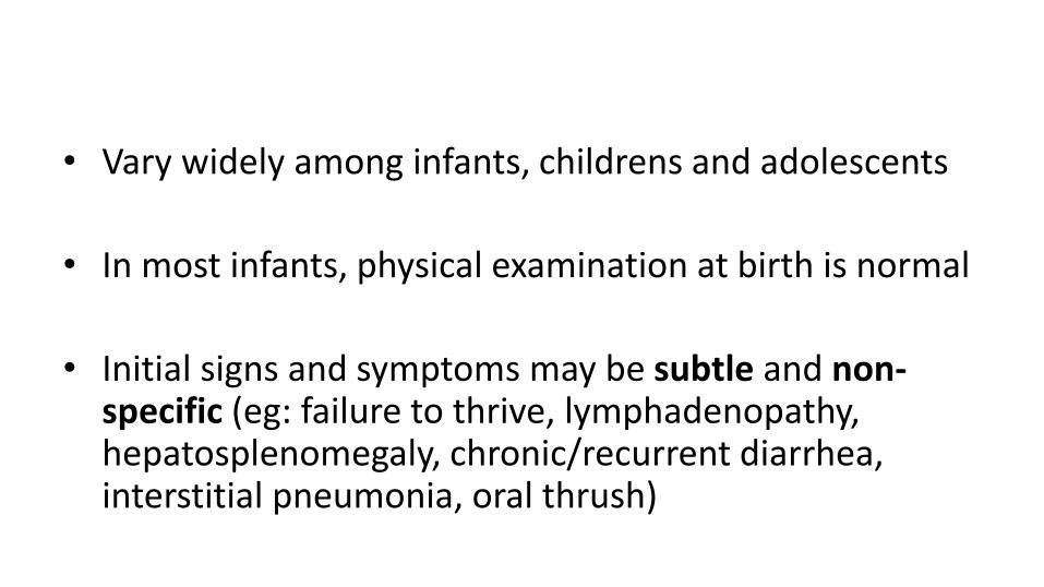
• Vary widely among infants, childrens and adolescents
• In most infants, physical examination at birth is normal
• Initial signs and symptoms may be subtle and non-specific (eg: failure to thrive, lymphadenopathy, hepatosplenomegaly, chronic/recurrent diarrhea, interstitial pneumonia, oral thrush)

• In United States and Europe: systemic and pulmonaryfindings are common
• In Africa: chronic diarrhea, wasting, malnutrition are common

• Symptoms commonly found MORE in CHILDREN than adults are:
– Recurrent bacterial infection
– Chronic parotid swelling
– Lymphocytic interstitial pneumonitis
– Early onset neurologic deterioration

• Paediatic HIV staged by two parameters
– Clinical status
– Degree of immunologic impairment

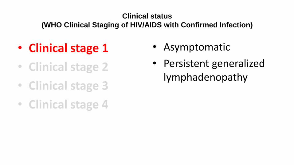
Clinical status
(WHO Clinical Staging of HIV/AIDS with Confirmed Infection)
• Clinical stage 1
• Clinical stage 2
• Clinical stage 3
• Clinical stage 4
Clinical status
(WHO Clinical Staging of HIV/AIDS with Confirmed Infection)
• Clinical stage 1
• Clinical stage 2
• Clinical stage 3
• Clinical stage 4
• Asymptomatic
• Persistent generalized lymphadenopathy

Clinical status
(WHO Clinical Staging of HIV/AIDS with Confirmed Infection)
• Clinical stage 1
• Clinical stage 2
• Clinical stage 3
• Clinical stage 4
• Unexplained persistent hepatosplenomegaly• Papular pruritic eruptions• Fungal nail infection• Angular cheilitis• Lineal gingival erythema• Extensive wart virus infection• Extensive molluscum contagiosum• Recurrent oral ulceration• Unexplained persistent parotid enlargement• Herpes zoster• Recurrent/chronic URTI (otitis media,
otorrhea, sinusitis, tonsillitis)

Clinical status
(WHO Clinical Staging of HIV/AIDS with Confirmed Infection)
• Clinical stage 1
• Clinical stage 2
• Clinical stage 3
• Clinical stage 4
• Unexplained moderate malnutrition or wasting not adequately responding to standard therapy
• Unexplained persistent diarrhea (14 days or more)• Unexplained persistent fever• Persistent oral candidiasis• Oral hairy leukoplakia• Acute necrotizing ulcerative gingivitis/periodontitis• Lymph node TB• Pulmonary TB• Severe recurrent bacteria pneumonia• Symptomatic lymphoid interstitial pneumonitis• Chronic HIV-associated lung disease including
bronchiectasis• Unexplained anemia, neutropenia or chronic
thrombocytopenia
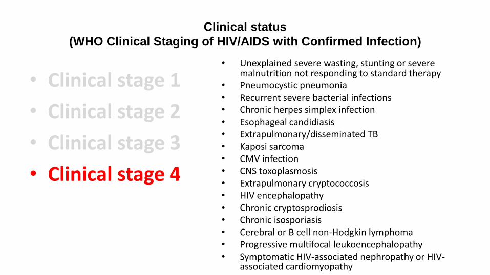
Clinical status
(WHO Clinical Staging of HIV/AIDS with Confirmed Infection)
• Clinical stage 1
• Clinical stage 2
• Clinical stage 3
• Clinical stage 4
• Unexplained severe wasting, stunting or severe malnutrition not responding to standard therapy
• Pneumocystic pneumonia• Recurrent severe bacterial infections• Chronic herpes simplex infection• Esophageal candidiasis• Extrapulmonary/disseminated TB• Kaposi sarcoma• CMV infection• CNS toxoplasmosis• Extrapulmonary cryptococcosis• HIV encephalopathy• Chronic cryptosprodiosis• Chronic isosporiasis• Cerebral or B cell non-Hodgkin lymphoma• Progressive multifocal leukoencephalopathy• Symptomatic HIV-associated nephropathy or HIV-
associated cardiomyopathy

Degree of immunologic impairment
(Severity of immmune suppression based on CD4 levels in children)
Immune Status Age
<11mo 12-35mo 36-59mo >5yr
Not significant >35% >30% >25% >500 cells/mm3
Mild 30-35% 25-30% 20-25% 350-499 cells/mm3
Advanced 25-30% 20-25% 15-20% 200-349 cells/mm3
Severe <25% or <1500cells/mm3
<20% or <750 cells/mm3
<15% or <350 cells/mm3
<15% or <200 cells/mm3

Respiratory diseases
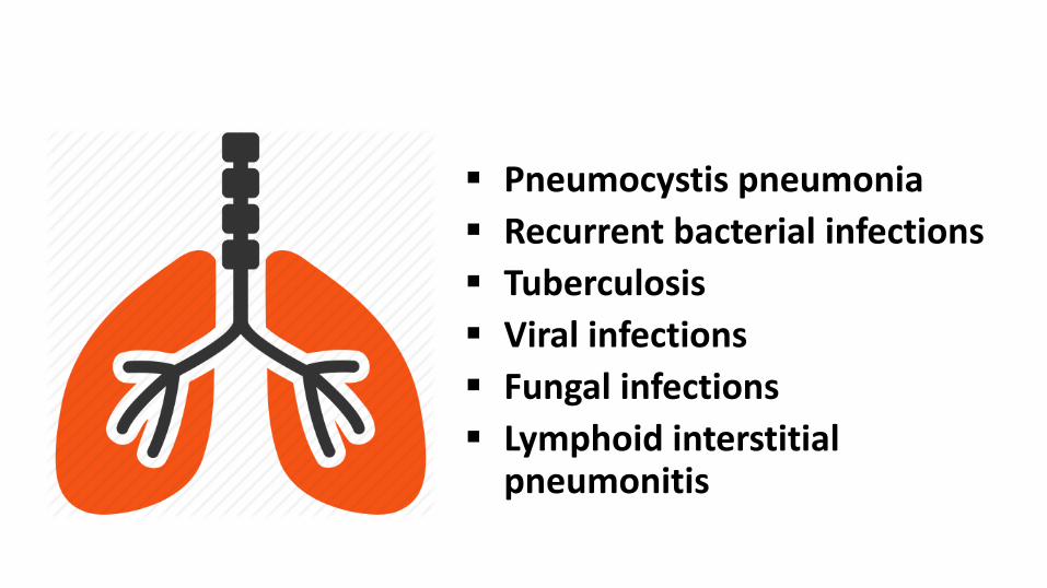
Pneumocystis pneumonia
Recurrent bacterial infections
Tuberculosis
Viral infections
Fungal infections
Lymphoid interstitial pneumonitis

Pneumocystis pneumonia Pneumocystis jiroveci
(previously P.carinii) pneumonia (PCP) is the opportunistic infection that led to the initial description of AIDS
Treatment: cotrimoxazole Untreated FATAL

Recurrent bacterial infection 90% had history of recurrent
pneumonias Initial episodes often occur before
the development of significant immunosupression
As the immunosuppression frequency increases, the frequency increases

Recurrent bacterial infection Community-acquired pneumonia:
Strep. Pneumoniae H. influenza Staph. Aureus
Hospital-acquired infection: Pseudomonas aeruginosa

Recurrent bacterial infection Treatment:
Combination of a broad spectrum cephalosporin + aminoglycoside
Nonsevere pneumonia: 2nd/3rd
generation cephalosporin or a combination like amoxicillin-clavulanic acid
Supportive care

Tuberculosis More likely to have
extrapulmonary and disseminated TB; course is likely to be more rapid
Coexistent TB + HIV accelerate the progression of both diseases

Tuberculosis The overall risk of active TB
in HIV-infected children is at least 5-10 fold higher
Should received longer duration of antituberculartherapy (9-12 month)

Viral infections Respiratory syncytial virus,
influenza and para influenzaviruses more often result in symptomatic disease
Adenovirus and measles virus are more likely to lead to serious sequelae

Fungal infections Primary infection is uncommon
Pulmonary candidiasis should be suspected in any sick HIV-infected child with LRTI that does not respond to the common therapeutic modalities

Lymphoid Interstitial pneumonitis (LIP) In absence of antiretroviral therapy,
nearly 20% of HIV-infected children developed LIP
Usually diagnosed in children with perinatally acquired HIV infection when they are older than 1yr of age
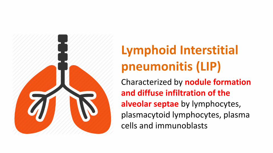
Lymphoid Interstitial pneumonitis (LIP)Characterized by nodule formation and diffuse infiltration of the alveolar septae by lymphocytes, plasmacytoid lymphocytes, plasma cells and immunoblasts

Lymphoid Interstitial pneumonitis (LIP) Etiology and pathogenesis is not well understood
Suggested etiologies are:
- Exagerrated immunologic response to inhaled/circulating antigens
- Primary infection of the lung with HIV, EBV, or both

Lymphoid Interstitial pneumonitis (LIP) Mostly are aymptomatic
Severe Tachypnea, cough, wheezing and hypoxemia
Advanced Clubbing
Can progress to chronic respiratory failure/bronchiectasis

Lymphoid Interstitial pneumonitis (LIP)Diagnosis:
Chest Xray: reticulonodular pattern, with/without hilar lymphadenopathy that persists =/>2 months
Unresponsive to antimicrobial therapy
Histopathology (definitive diagnosis)

Lymphoid Interstitial pneumonitis (LIP)Management:
Steroids – if children with LIP have symptoms and signs of chronic pulmonary disease, clubbing, hypoxemia
initial 4-12 week course of prednisolone (2mg/kg/day) tapering dose

Gastrointestinal diseases

Infections Bacteria – Salmonella,
Campylobacter, M. avium Protozoa* – Giardia,
Cryptosporidium, Isospora Fungi – Candida Virus – CMV, HSV, Rotavirus* most severe

AIDS enteropathy a syndrome of malabsorption
with partial villous atrophy not associated with a specific pathogen
• Result of direct HIV infection of the gut

Chronic liver inflammation Common in HIV infected children
In some children, hepatitis caused by CMV, hepatitis B or C viruses, or mycobacteria may lead to liver failure and portal hypertension.
*several of the antivirus drugs (eg: didanosine and protease inhibitors) may also cause reversible elevation of transaminases

Pancreatitis Uncommon May be the result of drug therapy
(eg: didanosine, lamivudine, nevirapine, or pentamidine)
Rarely, opportunistic infections such as mycobacteria or CMV may be responsible for acute pancreatitis

Neurologic diseases
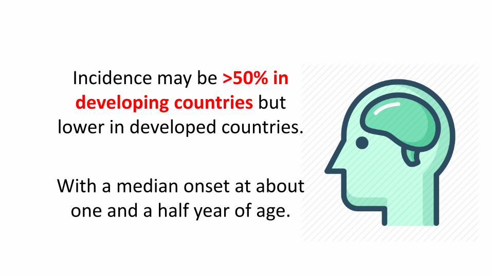
Incidence may be >50% in developing countries but
lower in developed countries.
With a median onset at about one and a half year of age.

Progressive encephalopathy Loss/plateau of developmental
milestones
Cognitive deterioration
Impaired brain growthacquiredmicrocephaly
Symmetric motor dysfunction

Meningitis Due to bacterial pathogens,
fungi (eg: Cryptococcus) and a number of viruses may be responsible

Toxoplasmosis Extremely rare in young
infants
But may occur in HIV-infected adolescents

Cardiovascular diseases

Cardiac abnormalities is common, persistent and often progressive in HIV-infected children

Left ventricular structure and function progressively may deteriorate in the first 3 years of life increased ventricular mass

Children with encephalopathyor other AIDS-defining conditions have the highest rates of adverse cardiac outcomes

Resting sinus tachycardia and marked sinus arrhythmia has been reported in upto nearly 2/3 and in 1/5 of the HIV-infected children

Congestive heart failure clinically indicated by gallop rhythm with tachypnea and hepatosplenomegaly

Renal diseases

Nephrotic syndrome
with azotemia & normal blood pressure
Renopathyunusual
(more common in older children)

• Principles of management of gastrointestinal and neurological diseasesare similar to those in non-HIV-infected children
• Electrocardiography and echocardiography are helpful in assessing cardiac function before the onset of clinical symptoms

DIAGNOSIS

Diagnosis can be made by:
HIV antibody testing
(beyond 18 months of age)
Virological testing
(before 18 months of age)

HIV antibody testing• All infants born to HIV-infected mothers are test antibody-
positive at birth (due to passive transfer of maternal HIV antibody across placenta)
• Most uninfected children lose maternal antibody between 6-12 months of age; only small proportion continue to have up to 18 months of age
• Hence, positive IgG antibody tests in infant younger than this age cannot be used to make definitive diagnosis

Methods:
Demonstration of IgG antibody to HIV: -- Reactive enzyme immunoassay (EIA)
Confirmatory test
- Western blot
- Immunofluorescence assay

Virological testing
• Essential for young infants born to a HIV-infected mother
• Methods:
- HIV DNA/RNA PCR
- HIV culture
- HIV p24 Ag immune dissociated p24

MANAGEMENT

• Management of HIV-infected child includes:
– Prophylaxis
– Antiretroviral therapy
– Treatment of opportunistic infection
– Adequate nutrition
– Immunization

Cotrimoxazole prophylaxis
• Recommended for:1. All HIV-exposed infants, starting at 4-6 weeks of
age; continued until HIV infection can be excluded
2. HIV-exposed breastfeeding children of any age; continued until can be excluded by HIV antibody testing or virological testing at least 6 weeks after complete cessation of breastfeeding
3. All children <1yr of age documented to be living with HIV
4. Symptomatic children >1yr of age
*all children who begin cotrimoxazole prophylaxis should continue until age of 5 yr, when they can be reassessed

The World Health Organization
now recommends initiation of ART
for all HIV infected children <2yr age
irrespective of clinical symptoms and
the immunologic stage
Antiretroviral therapy

The currently available therapy does not eradicate the virus and cure the child;
Rather it suppresses the virus replication for extended periods of time

Antiretroviral therapy
The main 3 groups of drugs:
1. Nucleoside reverse transcriptase inhibitors (NRTI)
2. Non-nucleoside reverse transcriptase inhibitors (NNRTI)
3. Protease inhibitors (PI)

NRTI
• Zidovudine (AZT)
• Lamivudine (3TC)
• Stavudine (d4T)
• Abacavir (ABC)
• Didanosine
• Zalcitabine
NNRTI
• Nevirapine (NVP)
• Efavirenz (EFV)
PI
• Lopinavir (LPV)
• Amprenavir
• Indinavir
• Nelfinavir
• Ritonavir
• Saquinavir
• Highly active antiretroviral therapy (HAART) is a combination of 2 NRTIs with a PI/NNRTI
• The national program recommends a combination of Zidovudine + Lamivudine + Nevirapine (1st line therapy)
• Alternative regimen: Stavudine + Lamivudine + Nevirapine
Nutrition
• It is important to provide adequate nutritionto HIV-infected children.
• Many of these children have failure to thrive.
• These children will need nutritional rehabilitation.
• Micronutrients like zinc may be useful

Immunization
• The vaccines that are recommended in the national schecule can be administered to HIV infected children except that symptomatic HIV children should not be given oral polio and BCG vaccines
Prevention of Mother to Child Transmission (PMTCT)

the risk of MTCT can be reduced to under 2%
Antiretroviral prophylaxis during
pregnancy
Intrapartum interventions
Regimens for infants
Avoidance of breastfeeding
1. Antiretroviral drug regimens for pregnant women
• FOR THEIR OWN HEALTH, ART should be administered irrespective of gestational age and is continued throughout pregnancy, delivery and thereafter
(recommended for all HIV-infected pregnant women with CD4 cell count <350cells/mm3 or WHO clinical stage 3 or 4)
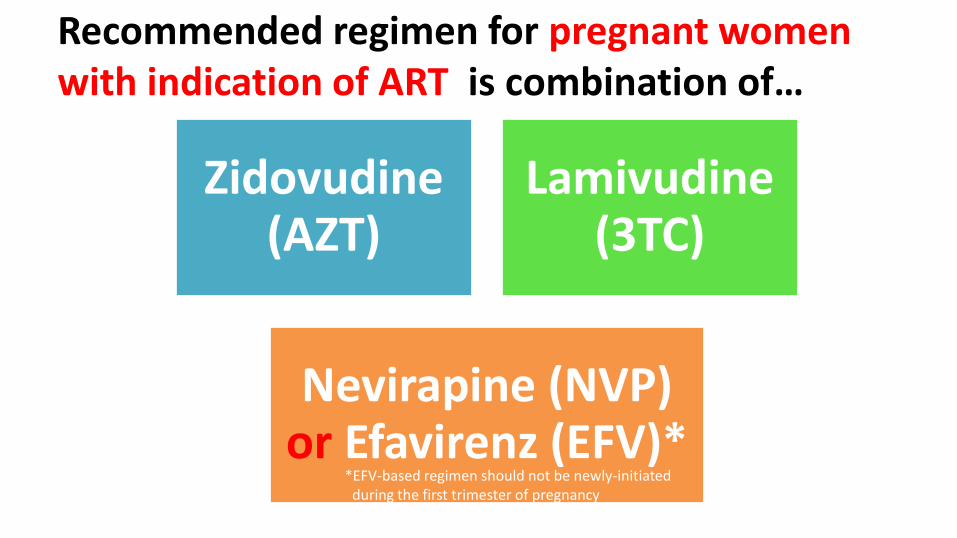
Recommended regimen for pregnant women with indication of ART is combination of…
Zidovudine (AZT)
Lamivudine (3TC)
Nevirapine (NVP) or Efavirenz (EFV)*
*EFV-based regimen should not be newly-initiated during the first trimester of pregnancy

Recommended regimen for pregnant women
who are NOT ELIGIBLE for ART , BUT FOR
PREVENTING MTCT is…
To start ART as early as 14 weeks gestation OR as soon as possible (when women present late
in pregnancy, in labor or at delivery)*EFV-based regimen should not be newly-initiated
during the first trimester of pregnancy

For them, 2 options are available:
Option 1:
• Daily AZT in antepartum period
• AZT + single dose NVP at onset of labor
• AZT + 3TC during labor
• AZT + 3TC for 7 days in
postpartum period
Option 2:
• Triple ART starting as early as 14
week of gestation until 1 week after exposure to breastmilk has ended
• AZT + 3TC + LPV (lopinavir)
• AZT + 3TC + ABC (abacavir)
• AZT + 3TC + EFV

2. Regimens for infants• If mother only received AZT during antenatal period:
– For BF infants: daily NVP from birth until 1 week after all exposure to breast milk has ended
– For non-BF infants: daily AZT/NVP from birth until 6 week
• If mother received triple drug ART during pregnancy and entire breastfeeding:– daily AZT/NVP from birth until 6 weeks irrespective of
feeding Dosage:NVP – 10mg/day (infants<2.5kg) or
15mg/day (infants>2.5kg) POAZT – 4mg/kg BD, PO
3. Intrapartum interventions• Delivery by elective cesarean section at 38 weeks before
onset of labor and rupture of membrane should be considered
• Avoid artificial rupture of membrane (ARMs) unless medically indicated
• Avoid procedures increasing risk of exposure of child to maternal blood and secretions like use of scalp electrodes

4. Breastfeeding • Important modality of transmission of HIV infection in
developing countries
• Risk of infection via BF highest in the early months of BF
• Increase Risks: – Detectable levels of HIV in breast milk – Presence of mastitis– Low maternal CD4+ T cell count
4. Breastfeeding • Mothers known to be HIV-infected should only
give commercial infant formula milk as a replacement feed when specific conditions are met:– referred earlier as affordable, feasible, acceptable,
sustainable and safe (AFASS) in 2006 WHO recommendations on HIV and infant feeding
– Otherwise, exclusive BF during first 6 months of life– Cessation of BF should be gradual

REFERENCEGhai Essential Paediatrics, 8th Edition, Vinod K Paul and Arvind Bagga

THANK YOUNUR HANISAH ZAINOREN