pacemaker neurons and neuronal networks: an integrative view
TRANSCRIPT

Pacemaker neurons and neuronal networks: an integrative viewJan-Marino Ramirez�1, Andrew K Tryba2 and Fernando Pena3
Rhythmically active neuronal networks give rise to rhythmic
motor activities but also to seemingly non-rhythmic behaviors
such as sleep, arousal, addiction, memory and cognition. Many
of these networks contain pacemaker neurons. The ability of
these neurons to generate bursts of activity intrinsically lies in
voltage- and time-dependent ion fluxes resulting from a
dynamic interplay among ion channels, second messenger
pathways and intracellular Ca2+ concentrations, and is
influenced by neuromodulators and synaptic inputs. This
complex intrinsic and extrinsic modulation of pacemaker
activity exerts a dynamic effect on network activity. The
nonlinearity of bursting activity might enable pacemaker
neurons to facilitate the onset of excitatory states or to
synchronize neuronal ensembles — an interactive
process that is intimately regulated by synaptic and
modulatory processes.
Addresses1 Department of Organismal Biology and Anatomy, Committees on
Neurobiology, Computational Neuroscience and Molecular Medicine,
The University of Chicago, Chicago, Illinois 60637, USA�e-mail: [email protected] Texas Tech University Health Sciences Center, Department of
Physiology, Lubbock TX 79416, USA
e-mail: [email protected] Departamento de Farmacobiologıa, CINVESTAV, Col. Granjas Coapa,
14330, Mexico D.F., Mexico
e-mail: [email protected]
Current Opinion in Neurobiology 2004, 14:665–674
This review comes from a themed issue on
Motor systems
Edited by Marjorie E Anderson and Ole Kiehn
Available online 5th November 2004
0959-4388/$ – see front matter
# 2004 Elsevier Ltd. All rights reserved.
DOI 10.1016/j.conb.2004.10.011
Abbreviations
CNS c
www.s
entral nervous system
Ih h
yperpolarization-activated currentPBC p
re-Botzinger complexIntroductionPacemaker neurons are defined by their intrinsic ability to
generate rhythmic bursting activity. The rhythmicity that
emerges through voltage- and time-dependent ion fluxes
in single neurons could be central to understanding how
neuronal networks generate many rhythmic activities
[1��,2]. This is an intriguing possibility, because many
mammalian neuronal networks generate rhythmic activ-
ity [1��,2,3��], including networks in the neocortex, basal
ciencedirect.com
ganglia, thalamus, locus coeruleus, hypothalamus, ventral
tegmentum area, hippocampus and amygdala. These
structures are associated with sleep, wakefulness, arousal,
motivation, addiction, memory consolidation, cognition
and fear. All of these neuronal networks contain rhythm-
ically active neurons that possess ion channels crucial for
generating pacemaker activity. For example, hippocam-
pal neurons [4] and dopaminergic ventral tegmentum area
neurons [5] possess a hyperpolarization-activated current
(Ih), the ‘pacemaker current’ that also produces the heart
beat.
But whereas nobody would question the importance of
heart pacemaker cells, the role of neurons with pace-
maker properties in many parts of the mammalian central
nervous system (CNS) remains a matter of debate [6]. In
fact, in many cases we do not even know what to make of
the rhythmicity generated in a given neuronal structure.
It is easy to imagine how strengthening synaptic inter-
actions could engrave memory, but how rhythmicity
contributes to memory, movement initiation, arousal,
motivation or addiction remains a mystery — an inter-
esting problem still waiting to be solved.
In discussing how pacemaker activity contributes to
rhythm generation in general, here we focus on the most
recent findings in various neuronal networks. In addition,
we refer to concrete examples found in the mammalian
respiratory network, specifically in the pre-Botzinger
complex (PBC) — an area that is essential for generating
inspiratory activity [7,8]. Our review shows that interac-
tions between pacemaker neurons and networks are at
least as plastic as the behavior that they underlie. The
significance of these interactions goes far beyond the
control of rhythmic motor activity, and the principles
found in simple motor circuits might be equally important
in the generation of higher brain functions.
Emergence of pacemaker properties from acomplex cellular environmentTypically, a neuron begins to burst because of the activa-
tion of inward currents or the cessation of outward cur-
rents [9]. The burst is subsequently terminated because
either the channels responsible for the inward current
inactivate or the ongoing burst activates calcium- or
sodium-dependent potassium currents that hyperpolarize
the membrane. The next burst can be initiated by the Ih
current, by voltage-independent intracellular signals, by
slow activation or inactivation properties of inward or
outward currents or by various other mechanisms [9].
However, whether or not any of the above mentioned
mechanisms will turn a neuron into a pacemaker neuron
Current Opinion in Neurobiology 2004, 14:665–674
666 Motor systems
will depend on a very fine and highly flexible balance
between various voltage-dependent ion channels and
voltage-independent factors. By varying the conduc-
tances of only eight voltage-dependent ion channels,
computational approaches yield 1.8 million single-
compartment model neurons with different discharge
properties, including pacemaker activity ranging from
irregular bursting, non-periodic bursting and one-spike
bursting to regular bursting [10��]. The modeling of eight
ion channels in a single neuronal compartment is a
simplification for practical purposes. Indeed, molecular
cloning techniques have uncovered a large diversity of ion
channel subtypes that are differentially distributed both
throughout the CNS and within the dendritic and somatic
compartments of the same neuron [11��,12��,13�].
The theoretical findings are consistent with the wide
spectrum of pacemaker properties recorded in real mam-
malian networks. Neurons in the respiratory network
constitute a continuum that can show irregular tonic
firing, irregular bursting and regular bursting. These
patterns sometimes make the discrimination between
non-pacemaker and bursting neurons ambiguous (JM
Ramirez et al., unpublished). To be able to handle this
heterogeneity, pacemaker properties are traditionally
typified according to only a few aspects of the response
or discharge pattern of a neuron [14–16]. Thus, neurons
within a single category are not identical with regard to
every property and do not possess the same combinations
of ion channels [17��,18]. Such heterogeneity is a common
characteristic of most types of neuron in the mammalian
nervous system [12��]; it is hoped that the introduction of
modern genetic and molecular tools will facilitate an
improved characterization of neuron types and their func-
tional roles in generating network activities [19��,20��].
This heterogeneity, however, is not only determined
genetically [21��,22�]. Activity-dependent and activity-
independent mechanisms can regulate the strength of
ionic conductances [23��,24��]. Ion channels that are
essential for generating pacemaker activity can be phos-
phorylated or dynamically modulated by intracellular
Ca2+ signaling networks using intracellular and extracel-
lular Ca2+ sources [13�,25��,26��]. Examples are the per-
sistent sodium current [22�,27], or leak currents [4] that
can be modulated by various intra- and extracellular
factors. Voltage-dependent and -independent processes
are directly interwoven in case of Ih channels that have
voltage-dependent and voltage-independent compo-
nents [28�]. Voltage-independent ion channels are
similarly modulated by voltage-dependent and voltage-
independent processes. An example is the calcium-
activated non-specific cation current termed the CAN
current [17��], which responds to increases in intracellular
calcium concentration. This current can depend on vol-
tage-dependent calcium currents that in turn are highly
modulated. These modulatory effects blur the distinction
Current Opinion in Neurobiology 2004, 14:665–674
between purely voltage-dependent and voltage-indepen-
dent influences on pacemaker activity. Thus, in conclu-
sion, pacemaker activity is very heterogeneous and
emerges through the complex interaction of voltage-
dependent and non-voltage dependent components of
ion channels within their intracellular and extracellular
environment.
Pacemaker properties depend on the neuromodulatory
milieu
Because ion channel properties are influenced by their
intracellular and extracellular environment, it should
not be surprising that a neuron can act as a non-pace-
maker under some conditions and express pacemaker
properties under different conditions. Neuromodulators
play an important part in the induction, enhancement
and suppression of pacemaker properties (Figure 1;
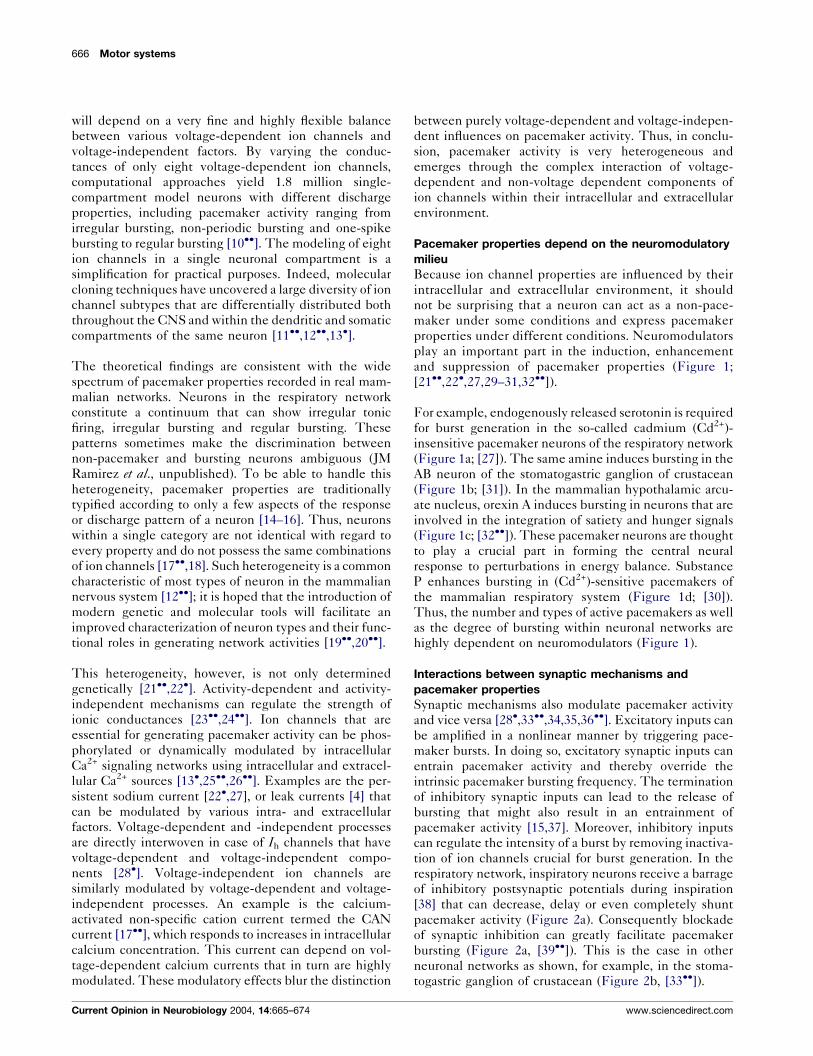
[21��,22�,27,29–31,32��]).
For example, endogenously released serotonin is required
for burst generation in the so-called cadmium (Cd2+)-
insensitive pacemaker neurons of the respiratory network
(Figure 1a; [27]). The same amine induces bursting in the
AB neuron of the stomatogastric ganglion of crustacean
(Figure 1b; [31]). In the mammalian hypothalamic arcu-
ate nucleus, orexin A induces bursting in neurons that are
involved in the integration of satiety and hunger signals
(Figure 1c; [32��]). These pacemaker neurons are thought
to play a crucial part in forming the central neural
response to perturbations in energy balance. Substance
P enhances bursting in (Cd2+)-sensitive pacemakers of
the mammalian respiratory system (Figure 1d; [30]).
Thus, the number and types of active pacemakers as well
as the degree of bursting within neuronal networks are
highly dependent on neuromodulators (Figure 1).
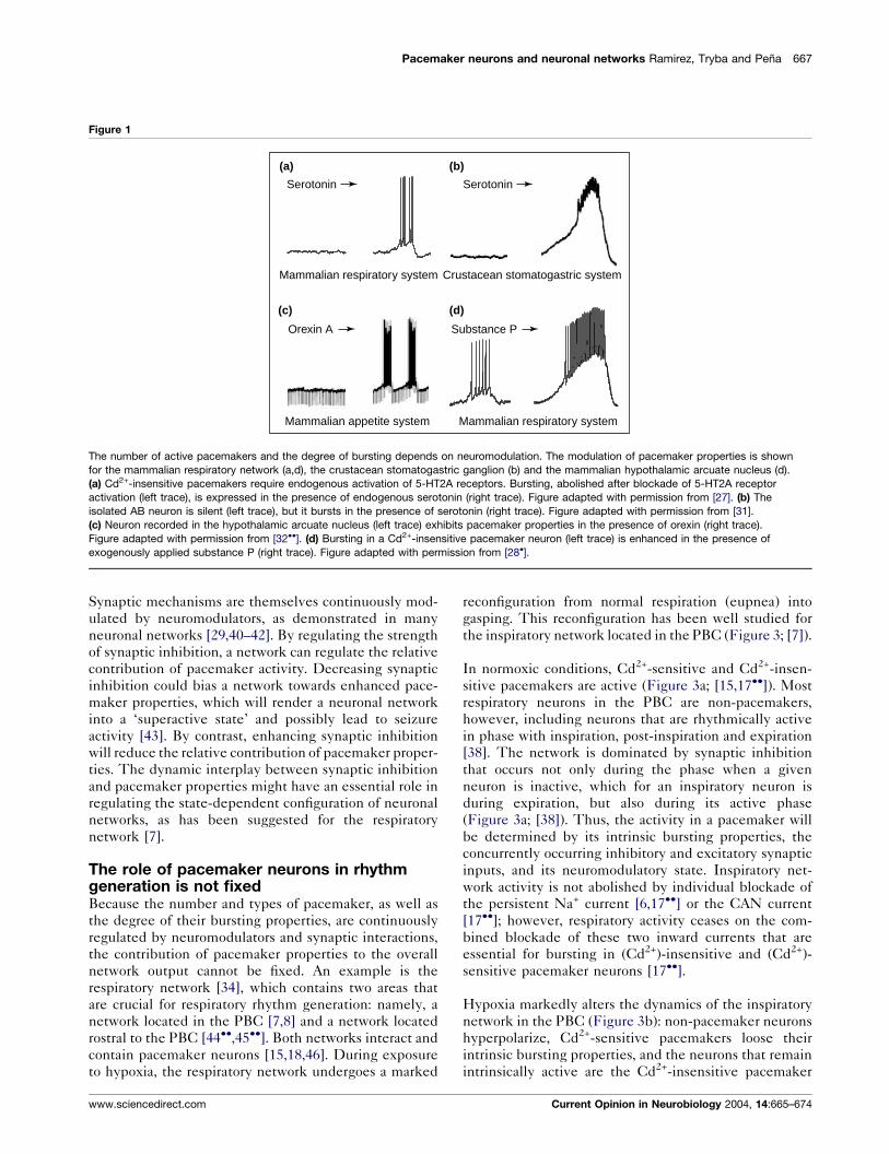
Interactions between synaptic mechanisms and
pacemaker properties
Synaptic mechanisms also modulate pacemaker activity
and vice versa [28�,33��,34,35,36��]. Excitatory inputs can
be amplified in a nonlinear manner by triggering pace-
maker bursts. In doing so, excitatory synaptic inputs can
entrain pacemaker activity and thereby override the
intrinsic pacemaker bursting frequency. The termination
of inhibitory synaptic inputs can lead to the release of
bursting that might also result in an entrainment of
pacemaker activity [15,37]. Moreover, inhibitory inputs
can regulate the intensity of a burst by removing inactiva-
tion of ion channels crucial for burst generation. In the
respiratory network, inspiratory neurons receive a barrage
of inhibitory postsynaptic potentials during inspiration
[38] that can decrease, delay or even completely shunt
pacemaker activity (Figure 2a). Consequently blockade
of synaptic inhibition can greatly facilitate pacemaker
bursting (Figure 2a, [39��]). This is the case in other
neuronal networks as shown, for example, in the stoma-
togastric ganglion of crustacean (Figure 2b, [33��]).
www.sciencedirect.com
Pacemaker neurons and neuronal networks Ramirez, Tryba and Pena 667
Figure 1
(a) (b)
Mammalian respiratory system
Crustacean stomatogastric system
Mammalian appetite system
Mammalian respiratory system
Substance P
SerotoninSerotonin
Orexin A
(c) (d)
The number of active pacemakers and the degree of bursting depends on neuromodulation. The modulation of pacemaker properties is shown
for the mammalian respiratory network (a,d), the crustacean stomatogastric ganglion (b) and the mammalian hypothalamic arcuate nucleus (d).
(a) Cd2+-insensitive pacemakers require endogenous activation of 5-HT2A receptors. Bursting, abolished after blockade of 5-HT2A receptor
activation (left trace), is expressed in the presence of endogenous serotonin (right trace). Figure adapted with permission from [27]. (b) The
isolated AB neuron is silent (left trace), but it bursts in the presence of serotonin (right trace). Figure adapted with permission from [31].
(c) Neuron recorded in the hypothalamic arcuate nucleus (left trace) exhibits pacemaker properties in the presence of orexin (right trace).
Figure adapted with permission from [32��]. (d) Bursting in a Cd2+-insensitive pacemaker neuron (left trace) is enhanced in the presence of
exogenously applied substance P (right trace). Figure adapted with permission from [28�].
Synaptic mechanisms are themselves continuously mod-
ulated by neuromodulators, as demonstrated in many
neuronal networks [29,40–42]. By regulating the strength
of synaptic inhibition, a network can regulate the relative
contribution of pacemaker activity. Decreasing synaptic
inhibition could bias a network towards enhanced pace-
maker properties, which will render a neuronal network
into a ‘superactive state’ and possibly lead to seizure
activity [43]. By contrast, enhancing synaptic inhibition
will reduce the relative contribution of pacemaker proper-
ties. The dynamic interplay between synaptic inhibition
and pacemaker properties might have an essential role in
regulating the state-dependent configuration of neuronal
networks, as has been suggested for the respiratory
network [7].
The role of pacemaker neurons in rhythmgeneration is not fixedBecause the number and types of pacemaker, as well as
the degree of their bursting properties, are continuously
regulated by neuromodulators and synaptic interactions,
the contribution of pacemaker properties to the overall
network output cannot be fixed. An example is the
respiratory network [34], which contains two areas that
are crucial for respiratory rhythm generation: namely, a
network located in the PBC [7,8] and a network located
rostral to the PBC [44��,45��]. Both networks interact and
contain pacemaker neurons [15,18,46]. During exposure
to hypoxia, the respiratory network undergoes a marked
www.sciencedirect.com
reconfiguration from normal respiration (eupnea) into
gasping. This reconfiguration has been well studied for
the inspiratory network located in the PBC (Figure 3; [7]).
In normoxic conditions, Cd2+-sensitive and Cd2+-insen-
sitive pacemakers are active (Figure 3a; [15,17��]). Most
respiratory neurons in the PBC are non-pacemakers,
however, including neurons that are rhythmically active
in phase with inspiration, post-inspiration and expiration
[38]. The network is dominated by synaptic inhibition
that occurs not only during the phase when a given
neuron is inactive, which for an inspiratory neuron is
during expiration, but also during its active phase
(Figure 3a; [38]). Thus, the activity in a pacemaker will
be determined by its intrinsic bursting properties, the
concurrently occurring inhibitory and excitatory synaptic
inputs, and its neuromodulatory state. Inspiratory net-
work activity is not abolished by individual blockade of
the persistent Na+ current [6,17��] or the CAN current
[17��]; however, respiratory activity ceases on the com-
bined blockade of these two inward currents that are
essential for bursting in (Cd2+)-insensitive and (Cd2+)-
sensitive pacemaker neurons [17��].
Hypoxia markedly alters the dynamics of the inspiratory
network in the PBC (Figure 3b): non-pacemaker neurons
hyperpolarize, Cd2+-sensitive pacemakers loose their
intrinsic bursting properties, and the neurons that remain
intrinsically active are the Cd2+-insensitive pacemaker
Current Opinion in Neurobiology 2004, 14:665–674
668 Motor systems
Figure 2
Mammalian respiratory system
Crustacean stomatogastric system
Removal ofglycinergic &GABAergicinhibition
Removal ofglutamatergic
inhibition
(a)
(b)
Pacemaker activity is controlled by synaptic inhibition. Examples of synaptic modulation are shown for (a) the mammalian respiratory system
and (b) the crustacean stomatogastric system. (a) Many respiratory neurons express no pacemaker properties when embedded in the
network that contains many inhibitory neurons (left trace), but their intrinsic pacemaker activity is revealed on blockade of synaptic inhibition
(right trace). Figure adapted with permission from [39��]. (b) The intraburst firing pattern of neurons, such as this LP neuron, is shaped by
synaptic inhibition (left trace). Bursting is enhanced on blockade of synaptic inhibition (right trace). Figure adapted with permission from [33��].
Figure 3
Inspiration Gasping
Hypoxia(a) (b)
CS
E
E
EE
I
I
I
I
II
I
I CICS
E
E
EE
I
I
I
I
II
I
I CI
Normoxia
CI
CS
(i)
(ii)
(iii)
5-HT
The expression of pacemaker properties is dependent on state. Examples are shown for the mammalian respiratory network (a,b) and the
crustacean stomatogastric system (c). (a) Under normal conditions, the inspiratory network located in the pre-Botzinger complex (PBC) is
dominated by synaptic inhibition. During inspiration, the kernel of inspiratory neurons is active (top panel). This kernel consists of excitatory
(E, blue) and inhibitory (I, grey) non-pacemaker neurons, and a heterogeneous pacemaker population of Cd2+-sensitive (CS, yellow; top trace) and
Cd2+-insensitive (CI, red; bottom trace) pacemaker neurons. Blockade of bursting in a single population of pacemaker neurons is not sufficient to
abolish inspiratory activity in this network configuration. (b) In hypoxia, the inhibitory (I, white) and excitatory non-pacemaker neurons (E, white),
as well as the Cd2+-sensitive pacemaker neurons (CS, white; top trace) cease to discharge intrinsically. The only neurons that remain intrinsically
active are the Cd2+-insensitive pacemakers (CI, red; bottom trace). These Cd2+-insensitive pacemaker neurons become the sole driver of
respiratory activity during hypoxia, generating gasping activity. Blockade of Cd2+-insensitive pacemaker neurons is sufficient to block gasping
in this network configuration. Traces adapted with permission from [17��]. (c) In the pyloric network the contribution of pacemaker neurons to the
frequency control depends on the neuromodulatory state. (i) In serotonin (5-HT) the frequency is determined by the AB neuron (red); (ii) in
dopamine (DA) by AB (red) and PD (yellow) coupled via a strengthened electrical synapse, (iii) and in octopamine (OCT) the frequency is
determined by AB (red), PD (yellow) and LP (blue). Figure adapted with permission from [31].
Current Opinion in Neurobiology 2004, 14:665–674 www.sciencedirect.com

Pacemaker neurons and neuronal networks Ramirez, Tryba and Pena 669
neurons (Figure 3b; [17��]). Under these reduced condi-
tions, Cd2+-insensitive pacemaker neurons become
essential for rhythm generation: blockade of the persis-
tent Na+ current is sufficient to abolish inspiratory net-
work activity during hypoxia [17��]. This leads to some
important conclusions.
First, the Cd2+-insensitive pacemaker neurons cannot be
considered as the principal drivers of the much more
complex respiratory network operating during normoxia
(Figure 3a), but they become the sole drivers of gasping
(Figure 3b). Second, hypoxia renders the respiratory net-
work more vulnerable to the blockade of a single ionic
mechanism: namely, the persistent Na+ current. Factors
that inhibit this current directly or indirectly — for
example, through its dependence on serotonin [27] —
could eliminate pacemaker activity in those neurons that
are responsible for driving gasping. Gasping is an impor-
tant autoresuscitation mechanism, and victims of sudden
infant death syndrome become hypoxic and show dis-
turbed gasping [47,48] and serotonergic brainstem
abnormalities [49]. Consistent with a change in the con-
figuration of the respiratory network (Figure 3), victims of
sudden infant death syndrome breathe normally during
normoxia, but do not gasp effectively when exposed to
hypoxic conditions [47,48].
The respiratory network is just one example in which the
relative contribution of different pacemaker neurons can
be altered in a state-dependent manner. In the stomato-
gastric ganglion of crustacean the relative importance of
different pacemaker neurons to determining the final
cycle frequency is not fixed but can vary under different
modulatory conditions. This is shown for the modulation
by 5-hydroxytryptophan, dopamine and octopamine
(Figure 3c [32��]).
Controlling the onset of network excitation:formulation of a hypothesisIf pacemakers are not simply ‘driving’ neuronal networks
under normal physiological conditions, what are their
specific functions in rhythm generation? Whereas it is
possible to attribute very specific functions to the activity
of individual neurons in invertebrates [50], such associa-
tions remain extremely difficult to demonstrate in the
mammalian nervous system, which explains, for example,
the uncertainty over whether pacemaker neurons are
important for respiratory rhythm generation [6]. Although
there are a few exceptions in which single mammalian
neurons can command a rhythmic motor output [51��],the manipulation of single pacemaker neurons generally
does not alter rhythmic network activity [6,17��,34].
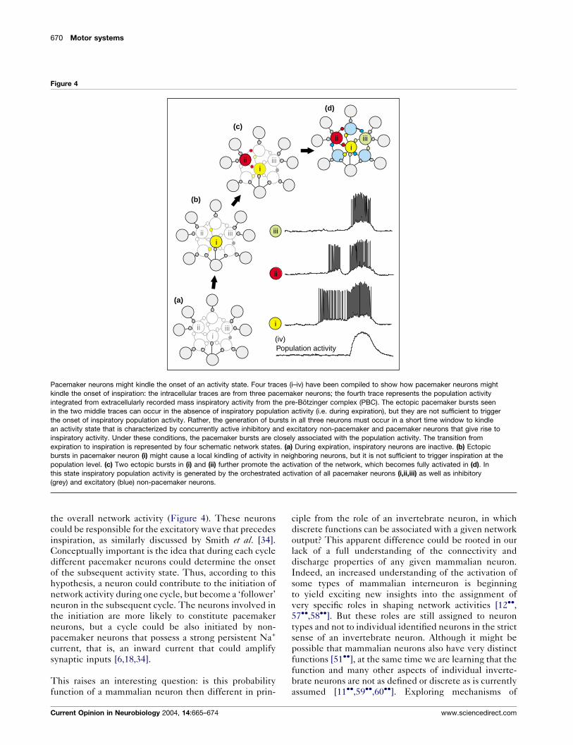
In the respiratory network, ‘ectopic’ pacemaker bursts
can be easily evoked in single neurons, but these bursts
are not sufficient to trigger an inspiration (Figure 4;
[6,14,17��]). This is not so surprising, because it is gen-
www.sciencedirect.com
erally assumed that mammalian behaviors emerge
through the orchestration of large populations of neurons
[52]. Orchestration can only work, however, if networks
prevent single neurons from initiating activity. Several
mechanisms could prevent a single neuron from trigger-
ing an activity state: first, a very weak coupling between
excitatory neurons [37], second, synaptic depression, and
third, concurrent inhibition and excitation [38]. These
mechanisms are characteristic of mammalian neuronal
networks [53,54]. For example, concurrent inhibition
and excitation characterize not only inspiratory activity
(Figures 2–4) but also upstates in the neocortex [55��] and
basal ganglia [56].
The mechanisms that prevent the initiation of activity
states can be overcome by pacemaker neurons that acti-
vate networks through their nonlinear and rhythmic
bursting properties. Closely timed bursts in several pace-
maker neurons might kindle a new activity state that is
associated with the synaptic recruitment of other pace-
maker and non-pacemaker neurons, including not only
excitatory but also inhibitory neurons (Figure 4). In the
respiratory network, the intrinsic inactivation properties
of pacemaker currents will reduce the probability of
bursting directly after an inspiration, thereby preventing
a premature kindling of the next inspiration. In the
absence of pacemaker properties — for example, after
the combined blockade of the CAN and persistent
sodium currents — excitatory synaptic interactions are
too weak to initiate an activity state in the inspiratory
network and respiration is abolished [17��]. By regulating
the number of active pacemaker neurons and their burst-
ing intensity, both neuromodulators (Figure 1) and synap-
tic interactions (Figure 2) could affect the probability of
initiating the onset of inspiration (Figure 4). By reducing
the number of active pacemaker neurons, the likelihood
of kindling an activity state might become unreliable and
the resulting rhythm irregular [27]; by contrast, increasing
the number of pacemaker neurons or their activity will
have the opposite effect (Figure 1d; [30]).
Pacemaker activity might similarly kindle network
ensembles in other neuronal networks, thereby facilitat-
ing the generation of regular rhythmic activity. For exam-
ple, pacemaker activity might be involved in the
neocortical gamma oscillations that crucially depend on
the intrinsic bursting properties of fast rhythmic bursting
neurons [36��]. Blockade of the persistent Na+ current is
associated with the suppression of bursting in these fast
rhythmic bursting neurons and the elimination of neo-
cortical gamma activity [36��].
Returning to the issue of the role that pacemaker neurons
play in mammalian rhythm generation, we speculate that
a single neuron could have only a probability function and
that a minimum number of pacemaker neurons have to
burst during a specific time window to exert an effect on
Current Opinion in Neurobiology 2004, 14:665–674
670 Motor systems
Figure 4
(d)
(c)
(a)
(b)
iii iii
iii iii
iii iii
iii iii
i
ii
iii
Population activity(iv)
Pacemaker neurons might kindle the onset of an activity state. Four traces (i–iv) have been compiled to show how pacemaker neurons might
kindle the onset of inspiration: the intracellular traces are from three pacemaker neurons; the fourth trace represents the population activity
integrated from extracellularly recorded mass inspiratory activity from the pre-Botzinger complex (PBC). The ectopic pacemaker bursts seen
in the two middle traces can occur in the absence of inspiratory population activity (i.e. during expiration), but they are not sufficient to trigger
the onset of inspiratory population activity. Rather, the generation of bursts in all three neurons must occur in a short time window to kindle
an activity state that is characterized by concurrently active inhibitory and excitatory non-pacemaker and pacemaker neurons that give rise to
inspiratory activity. Under these conditions, the pacemaker bursts are closely associated with the population activity. The transition from
expiration to inspiration is represented by four schematic network states. (a) During expiration, inspiratory neurons are inactive. (b) Ectopic
bursts in pacemaker neuron (i) might cause a local kindling of activity in neighboring neurons, but it is not sufficient to trigger inspiration at the
population level. (c) Two ectopic bursts in (i) and (ii) further promote the activation of the network, which becomes fully activated in (d). In
this state inspiratory population activity is generated by the orchestrated activation of all pacemaker neurons (i,ii,iii) as well as inhibitory
(grey) and excitatory (blue) non-pacemaker neurons.
the overall network activity (Figure 4). These neurons
could be responsible for the excitatory wave that precedes
inspiration, as similarly discussed by Smith et al. [34].
Conceptually important is the idea that during each cycle
different pacemaker neurons could determine the onset
of the subsequent activity state. Thus, according to this
hypothesis, a neuron could contribute to the initiation of
network activity during one cycle, but become a ‘follower’
neuron in the subsequent cycle. The neurons involved in
the initiation are more likely to constitute pacemaker
neurons, but a cycle could be also initiated by non-
pacemaker neurons that possess a strong persistent Na+
current, that is, an inward current that could amplify
synaptic inputs [6,18,34].
This raises an interesting question: is this probability
function of a mammalian neuron then different in prin-
Current Opinion in Neurobiology 2004, 14:665–674
ciple from the role of an invertebrate neuron, in which
discrete functions can be associated with a given network
output? This apparent difference could be rooted in our
lack of a full understanding of the connectivity and
discharge properties of any given mammalian neuron.
Indeed, an increased understanding of the activation of
some types of mammalian interneuron is beginning
to yield exciting new insights into the assignment of
very specific roles in shaping network activities [12��,57��,58��]. But these roles are still assigned to neuron
types and not to individual identified neurons in the strict
sense of an invertebrate neuron. Although it might be
possible that mammalian neurons also have very distinct
functions [51��], at the same time we are learning that the
function and many other aspects of individual inverte-
brate neurons are not as defined or discrete as is currently
assumed [11��,59��,60��]. Exploring mechanisms of
www.sciencedirect.com

Pacemaker neurons and neuronal networks Ramirez, Tryba and Pena 671
rhythmic generation is clearly a plastic endeavor in itself
and we must adjust our views continuously to keep up
with the increasing understanding of the complexity that
underlies interactions between pacemaker neurons and
networks.
ConclusionsPacemaker neurons are ubiquitous cellular components
of rhythm-generating networks, but their ability to burst
rhythmically is not a genetically fixed property. The same
neuron can be a pacemaker or non-pacemaker depending
on dynamic interactions between intrinsic and extrinsic
factors that are modulated in a state-dependent and
behaviorally regulated manner. Consequently, the con-
tribution of pacemaker activity to neuronal networks
cannot be fixed.
This principle is well illustrated by the mammalian
respiratory network. A reduced population of pacemaker
neurons rhythmically drives network activity during
hypoxia, resulting in the generation of gasping. During
normoxia, however, heterogeneous pacemaker and syn-
aptic properties are interwoven and continuously modu-
lated by amines and peptides that dynamically alter the
characteristics of these properties. In this network con-
figuration, pacemaker neurons cannot be regarded as
simple drivers of rhythmic activity; instead, the number
of active pacemaker neurons and their bursting properties
might influence the likelihood of kindling the onset of the
next rhythmic phase. This process is not one way, how-
ever, because the number of active pacemaker neurons
and their bursting properties are highly modulated by
synaptic and modulatory influences. By modulating pace-
maker activity, synaptic and modulatory mechanisms can
similarly kindle the onset of activity states in other
neuronal mammalian networks.
AcknowledgementsWe thank SP Lieske, K Nagel, M Thoby-Brisson and J-C Viemari forthoughts and contributions that inspired many ideas discussed in thisreview; and N Ramirez for help with the manuscript and figures.This study was supported by grants from the National Institutes ofHealth (HL60120, HL68860) to JM Ramirez.
References and recommended readingPapers of particular interest, published within the annual period ofreview, have been highlighted as:
� of special interest�� of outstanding interest
1.��
Arshavsky YI: Cellular and network properties in thefunctioning of the nervous system: from central patterngenerators to cognition. Brain Res Brain Res Rev 2003,41:229-267.
The author discusses the role of pacemaker activity and network proper-ties in the generation of rhythmic activity. The article shows in animpressive manner that rhythm generation is a fundamental propertyof the CNS and underlies behavioral functions ranging from motor controlto cognition.
2. Llinas RR: The intrinsic electrophysiological properties ofmammalian neurons: insights into central nervous systemfunction. Science 1988, 242:1654-1664.
www.sciencedirect.com
3.��
Buzsaki G, Draguhn A: Neuronal oscillations in corticalnetworks. Science 2004, 304:1926-1929.
‘‘Clocks tick, bridges and skyscrapers vibrate, neuronal network oscillate’’.This excerpt from the abstract sets the tone for this exciting review onneocortical rhythms that addresses the important question: are neuronalrhythms an inevitable by-product or an essential part of the brain’sdesign? The authors make a convincing case that network rhythmsare crucial for many higher brain functions including memory consolida-tion, temporal representation and input selection. This outstanding reviewis highly recommended not only for readers who thought that rhythms areonly important for generating rhythmic motor activities, but also for thosewho still suspect that fast oscillations are just an epiphenomenon.
4. Schweitzer P, Madamba SG, Siggins GR: The sleep-modulatingpeptide cortistatin augments the h-current in hippocampalneurons. J Neurosci 2003, 23:10884-10891.
5. Liu Z, Bunney EB, Appel SB, Brodie MS: Serotonin reduces thehyperpolarization-activated current (Ih) in ventral tegmentalarea dopamine neurons: involvement of 5-HT2 receptors andprotein kinase C. J Neurophysiol 2003, 90:3201-3212.
6. Del Negro CA, Morgado-Valle C, Feldman JL: Respiratoryrhythm: an emergent network property? Neuron 2002,34:821-830.
7. Lieske SP, Thoby-Brisson M, Telgkamp P, Ramirez JM:Reconfiguration of the neural network controlling multiplebreathing patterns: eupnea, sighs and gasps.Nat Neurosci 2000, 3:600-607.
8. Smith JC, Ellenberger HH, Ballanyi K, Richter DW, Feldman JL:Pre-Botzinger complex: a brainstem region that may generaterespiratory rhythm in mammals. Science 1991, 254:726-729.
9. Harris-Warrick RM: Voltage-sensitive ion channels in rhythmicmotor systems. Curr Opin Neurobiol 2002, 12:646-651.
10.��
Prinz AA, Billimoria CP, Marder E: Alternative to hand-tuningconductance-based models: construction and analysis ofdatabases of model neurons. J Neurophysiol 2003,90:3998-4015.
Whether a neuron is a pacemaker or not is determined by the densitiesand dynamics of ion channels. Currents flowing through these channelsdepend in a nonlinear manner on the membrane potential, which itself ischanged by the ion fluxes. This very innovative paper introduces acomputational approach that will aid our understanding of this complexdynamical interplay and how such interactions lead to a given dischargepattern. The authors create a huge database of model neurons in whichthe conductances of eight ion channels are varied independently. Thedatabase provides extensive information about steady-state voltagetraces, spontaneous behaviors and response properties of millions ofmodel neurons. The data can be used to learn how different combinationsof ion channels lead to discharge patterns typically found in realisticneurons. It is predicted that this approach will become an invaluable toolin cellular and systems neuroscience.
11.��
French LB, Lanning CC, Matly M, Harris-Warrick RM: Cellularlocalization of Shab and Shaw potassium channels in thelobster stomatogastric ganglion. Neuroscience 2004,123:919-930.
The discharge pattern of a neuron depends not only on the ion channels,but also on their location. This study is one of several exciting paperspublished this year by the Harris-Warrick laboratory exploring howdifferent ion channel subtypes are distributed on functionally identifiedneurons, how these channels interact, and how they are homeostaticallyregulated in order to generate a functionally identified activity pattern.
12.��
Whittington MA, Traub RD: Interneuron diversity series:inhibitory interneurons and network oscillations in vitro.Trends Neurosci 2003, 26:676-682.
In this outstanding review, the authors unravel the interaction betweennonlinear membrane properties and synaptic characteristics of differenttypes of hippocampal neuron to understand how these components leadto the differential generation of gamma and theta rhythms. Of importanceare the spatial distribution of ion channels and the contribution ofelectrical synapses. The authors conclude that different types of inter-neuron have different tasks in generating these rhythms.
13.�
Levi R, Samoilova M, Selverston AI: Calcium signalingcomponents of oscillating invertebrate neurons in vitro.Neuroscience 2003, 118:283-296.
This study explores the role of Ca2+ dynamics in regulating neuronalactivity in isolated neurons from the stomatogastric ganglion. The authorsshow how Ca2+ signals arriving from different spatial compartments and
Current Opinion in Neurobiology 2004, 14:665–674

672 Motor systems
sources contribute to the discharge pattern of neurons involved ingenerating rhythmic motor activity.
14. Thoby-Brisson M, Telgkamp P, Ramirez JM: The role of thehyperpolarization-activated current in modulating rhythmicactivity in the isolated respiratory network of mice.J Neurosci 2000, 20:2994-3005.
15. Thoby-Brisson M, Ramirez JM: Two types of inspiratorypacemaker neurons in the isolated respiratory network ofmice. J Neurophysiol 2001, 86:104-112.
16. Rekling JC, Champagnat J, Denavit-Saubie M:Electroresponsive properties and membrane potentialtrajectories of three types of inspiratory neurons in thenewborn mouse brain stem in vitro. J Neurophysiol 1996,75:795-810.
17.��
Pena F, Parkis MA, Tryba AK, Ramirez JM: Differentialcontribution of pacemaker properties to the generation ofrespiratory rhythms during normoxia and hypoxia.Neuron 2004, 43:105-117.
This study replaces the prevailing concept that respiratory rhythm isgenerated by just one neuronal mechanism with the more dynamicconcept that heterogeneous and state-dependent mechanisms areimportant for generating respiratory rhythm. The data suggest that thedependence on pacemaker neurons is not fixed but altered by state-dependent changes, in this case by hypoxia.
18. Del Negro CA, Koshiya N, Butera RJ Jr, Smith JC: Persistentsodium current, membrane properties and bursting behaviorof pre-Botzinger complex inspiratory neurons in vitro.J Neurophysiol 2002, 88:2242-2250.
19.��
Monyer H, Markram H: Interneuron diversity series: molecularand genetic tools to study GABAergic interneuron diversityand function. Trends Neurosci 2004, 27:90-97.
This up-to-date overview describes how modern molecular and genetictools can be combined with electrophysiological and neuroanatomicalapproaches to arrive at a better characterization of interneurons and theirdischarge pattern. The authors discuss novel ways to manipulate inter-neuronal activity to explore how genes, neurons and networks give rise tocomplex behaviors.
20.��
Kiehn O, Kullander K: Central pattern generators deciphered bymolecular genetics. Neuron 2004, 41:317-321.
The authors review modern molecular and genetic approaches for dis-secting the organization and development of rhythm-generating neuronalnetworks in invertebrate and vertebrate motor systems. These powerfulapproaches enable a very specific manipulation of neurons that are unitedby a common genetic or molecular marker, such as the presence of apromoter for a given transcription factor. It is easy to imagine that in thenear future many genetic markers will prove useful for differentiallymanipulating different subpopulations of interneurons, thereby increasingour understanding of how these neurons contribute to the generation ofcomplex network activity.
21.��
Steriade M: Neocortical cell classes are flexible entities.Nat Rev Neurosci 2004, 5:121-134.
Neocortical neuron types are not fixed but dependent on state. Thisconclusion is obtained through impressive intracellular recordings fromawake and sleeping cats, which indicate that state transitions are asso-ciated with a change in the incidence of neuron types and a transforma-tion in discharge patterns: for example, intrinsically bursting neurons canbe transformed into regular spiking neurons. This study emphasizes theimportance of recording from neurons in a functional and/or behavioralcontext and shows that a given discharge pattern is not a fixed entity.
22.�
Traub RD, Buhl EH, Gloveli T, Whittington MA: Fast rhythmicbursting can be induced in layer 2/3 cortical neurons byenhancing persistent Na+ conductance or by blocking BKchannels. J Neurophysiol 2003, 89:909-921.
In the quest to understand how cortical neurons could contribute to thegeneration of sensory-evoked gamma oscillations, the authors combinecomputational and experimental approaches to demonstrate howchanges in the persistent Na+ current and the Ca2+-dependent K+ currentcan transform the discharge pattern of a neuron from regular spiking tofast rhythmic bursting. It is likely that these transformations are mediatedin vivo by neuromodulators that alter Na+ and K+ conductances.
23.��
Zhang W, Linden DJ: The other side of the engram: experience-driven changes in neuronal intrinsic excitability.Nat Rev Neurosci 2003, 4:885-900.
This important review raises the awareness that neuronal plasticity isrestricted not only to changes in synaptic mechanisms, but also to
Current Opinion in Neurobiology 2004, 14:665–674
changes in the function of voltage-gated ion channels. Changes inintrinsic excitability might be a powerful mechanism for the consolidationand adaptive generalization of memories.
24.��
MacLean JN, Zhang Y, Johnson BR, Harris-Warrick RM:Activity-independent homeostasis in rhythmically activeneurons. Neuron 2003, 37:109-120.
With all the known plasticity that can alter the discharge pattern of aneuron, how can neurons maintain a preferred activity range that isfunctionally meaningful? An elegant series of experiments reveals a novelform of activity-independent regulation of ion channels that could have anessential role in promoting stability in the activity of individual neurons andnetworks.
25.��
Pape HC, Munsch T, Budde T: Novel vistas of calcium-mediatedsignaling in the thalamus. Pflugers Arch 2004, 448:131-138.
This outstanding review provides exciting insights into the dynamic Ca2+
signaling network that regulates neuronal excitability. The authors focuson the role of intracellularly and extracellularly derived Ca2+ in regulatingthalamic pacemaker activity. These Ca2+ interactions are state-depen-dently regulated and differentially distributed in somatic and dendriticcompartments.
26.��
Ladewig T, Kloppenburg P, Lalley PM, Zipfel WR, Webb WW,Keller BU: Spatial profiles of store-dependent calcium releasein motoneurones of the nucleus hypoglossus from newbornmouse. J Physiol 2003, 547:775-787.
Hypoglossal motor neurons have a low capacity for buffering endogen-ous Ca2+. This property accelerates the relaxation rates of Ca2+ transientsthat are necessary to follow the rapid depolarizations associated with thegeneration of rhythmic activity. The low Ca2+ buffering is potentiallydangerous, however, and the authors elegantly show that these neuronshave developed exquisite Ca2+-regulating mechanisms in form of orga-nelles such as mitochondria.
27. Pena F, Ramirez JM: Endogenous activation of serotonin 2Areceptors is required for normal respiratory rhythm generationin vitro. J Neurosci 2002, 22:11055-11064.
28.�
Frere SG, Luthi A: Pacemaker channels in mousethalamocortical neurones are regulated by distinct pathwaysof cAMP synthesis. J Physiol 2004, 554:111-125.
Ion channels that are essential for pacemaker activity are highly modu-lated by neuromodulators and synaptic mechanisms. In this study, theauthors describe how GABAergic inhibition and adrenergic mechanismsmodulate the hyperpolarization-activated Ih current in thalamocorticalpacemaker neurons. An important finding is that the Ih current is mod-ulated by distinct pathways of cAMP synthesis that are recruited bysynaptic and modulatory mechanisms.
29. Johnson BR, Kloppenburg P, Harris-Warrick RM: Dopaminemodulation of calcium currents in pyloric neurons of thelobster stomatogastric ganglion. J Neurophysiol 2003,90:631-643.
30. Pena FP, Ramirez JM: Substance P mediated modulation ofpacemaker properties in the mammalian respiratory network.J Neurosci 2004, 24:7549-7556.
31. Ayali A, Harris-Warrick RM: Monoamine control of thepacemaker kernel and cycle frequency in the lobster pyloricnetwork. J Neurosci 1999, 19:6712-6722.
32.��
van den Top M, Lee K, Whyment AD, Blanks AM, Spanswick D:Orexigen-sensitive NPY/AgRP pacemaker neurons in thehypothalamic arcuate nucleus. Nat Neurosci 2004, 7:493-494.
The authors describe a novel class of conditional pacemaker neurons thatcould play a crucial role in generating the central neuronal response toperturbations in energy balance. The pacemaker properties of theseneurons are modulated by substances that are released in response tosatiety and hunger.
33.��
Szucs A, Pinto RD, Rabinovich MI, Abarbanel HD, Selverston AI:Synaptic modulation of the interspike interval signatures ofbursting pyloric neurons. J Neurophysiol 2003, 89:1363-1377.
Whether pacemaker bursts are units of neuronal information or whethertheir internal structure plays an important part in the function of neuronalnetworks is largely unresolved. In this study, the authors show howsynaptic inhibition shapes burst discharges in pacemaker activity. As aresult, these neurons express remarkable flexibility, rich dynamical beha-vior and a plastic input sensitivity in the burst that could be important inregulating network activity.
34. Smith JC, Butera RJ, Koshiya N, Del Negro C, Wilson CG,Johnson SM: Respiratory rhythm generation in neonatal and
www.sciencedirect.com
Pacemaker neurons and neuronal networks Ramirez, Tryba and Pena 673
adult mammals: the hybrid pacemaker-network model.Respir Physiol 2000, 122:131-147.
35. Kiehn O, Kjaerulff O, Tresch MC, Harris-Warrick RM:Contributions of intrinsic motor neuron properties to theproduction of rhythmic motor output in the mammalian spinalcord. Brain Res Bull 2000, 53:649-659.
36.��
Cunningham MO, Whittington MA, Bibbig A, Roopun A,LeBeau FE, Vogt A, Monyer H, Buhl EH, Traub RD: A role for fastrhythmic bursting neurons in cortical gamma oscillationsin vitro. Proc Natl Acad Sci USA 2004, 101:7152-7157.
Gamma frequency oscillations are a hallmark of electroencephalographicactivity. These oscillations might establish a temporal framework for long-range synchronization that is essential for processing sensory and motorinformation. The authors use electrophysiological, pharmacological andcomputational approaches to investigate how fast rhythmic bursting(FRB) neurons contribute to generating these gamma oscillations. Block-ade of persistent Na+ channels reduces bursting in FRB neurons andgamma oscillations in the neocortex but not in the hippocampus, sug-gesting that FRB neurons have a crucial role in the generation of neo-cortical gamma oscillations.
37. Butera RJ Jr, Rinzel J, Smith JC: Models of respiratory rhythmgeneration in the pre-Botzinger complex. II. Populations ofcoupled pacemaker neurons. J Neurophysiol 1999, 82:398-415.
38. Ramirez JM, Telgkamp P, Elsen FP, Quellmalz UJ, Richter DW:Respiratory rhythm generation in mammals: synaptic andmembrane properties. Respir Physiol 1997, 110:71-85.
39.��
Tryba AK, Pena F, Ramirez JM: Stabilization of bursting inrespiratory pacemaker neurons. J Neurosci 2003,23:3538-3546.
This study indicates that respiratory pacemaker neurons possess abackground inward current that stabilizes their bursting against changesin extracellular K+ and imposed changes in the membrane potential. Sucha background inward current might be essential for stabilizing rhythmgeneration not only in the respiratory network but also in other rhythm-generating neuronal networks.
40. Bamford NS, Zhang H, Schmitz Y, Wu NP, Cepeda C, Levine MS,Schmauss C, Zakharenko SS, Zablow L, Sulzer D:Heterosynaptic dopamine neurotransmission selects sets ofcorticostriatal terminals. Neuron 2004, 42:653-663.
41. Lee KH, Broberger C, Kim U, McCormick DA: Histaminemodulates thalamocortical activity by activating a chlorideconductance in ferret perigeniculate neurons. Proc Natl AcadSci USA 2004, 101:6716-6721.
42. Sakurai A, Katz PS: Spike timing-dependent serotonergicneuromodulation of synaptic strength intrinsic to a centralpattern generator circuit. J Neurosci 2003, 23:10745-10755.
43. Van Drongelen W, Koch H, Marcuccilli C, Pena F, Ramirez JM:Synchrony levels during evoked seizure-like bursts in mouseneocortical slices. J Neurophysiol 2003, 90:1571-1580.
44.��
Onimaru H, Homma I: A novel functional neuron group forrespiratory rhythm generation in the ventral medulla.J Neurosci 2003, 23:1478-1486.
Optical recordings show that the respiratory rhythm-generating networkextends to a location more rostral than the pre-Botzinger complex. Theauthors suggest that pacemaker neurons (the so-called Pre-I neurons)located in the parafacial respiratory group interact with pre-Botzingercomplex neurons in the form of a coupled oscillatory system to regulaterespiratory rhythmic activity.
45.��
Mellen NM, Janczewski WA, Bocchiaro CM, Feldman JL:Opioid-induced quantal slowing reveals dual networks forrespiratory rhythm generation. Neuron 2003, 37:821-826.
The prevailing concept that a single medullary network generates respira-tory rhythmic activity is proved to be wrong in this important study. In avery convincing series of experiments, the authors show that two rhyth-mically active networks interact to generate respiratory rhythmic activity,and that under different conditions each is sufficient to generate arespiratory rhythm. Both networks have different modulatory sensitivities.The authors discuss the possibility that the differential sensitivities mightenhance the robustness of the respiratory rhythm.
46. Onimaru H, Arata A, Homma I: Intrinsic burst generation ofpreinspiratory neurons in the medulla of brainstem-spinalcord preparations isolated from newborn rats. Exp Brain Res1995, 106:57-68.
www.sciencedirect.com
47. Poets CF: Apparent life-threatening events and sudden infantdeath on a monitor. Paediatr Respir Rev 2004,5(Suppl A):S383-S386.
48. Sridhar R, Thach BT, Kelly DH, Henslee JA: Characterization ofsuccessful and failed autoresuscitation in human infants,including those dying of SIDS. Pediatr Pulmonol 2003,36:113-122.
49. Kinney HC, Randall LL, Sleeper LA, Willinger M, Belliveau RA,Zec N, Rava LA, Dominici L, Iyasu S, Randall B et al.: Serotonergicbrainstem abnormalities in Northern Plains Indians with thesudden infant death syndrome. J Neuropathol Exp Neurol 2003,62:1178-1191.
50. Dembrow NC, Jing J, Brezina V, Weiss KR: A specific synapticpathway activates a conditional plateau potential underlyingprotraction phase in the Aplysia feeding central patterngenerator. J Neurosci 2004, 24:5230-5238.
51.��
Brecht M, Schneider M, Sakmann B, Margrie TW: Whiskermovements evoked by stimulation of single pyramidal cellsin rat motor cortex. Nature 2004, 427:704-710.
Those readers who think that single neurons do not matter in the mam-malian nervous system are proved wrong in this exciting study. Stimulationof a single pyramidal neuron is able to reset and to initiate rhythmic whiskermovements. The authors propose that sparse cortical activity in thevibrissae motor cortex is sufficient to generate motor output by disynapt-ically activating a central pattern generator in the facial nucleus.
52. Buzsaki G: Large-scale recording of neuronal ensembles.Nat Neurosci 2004, 7:446-451.
53. Bartho P, Hirase H, Monconduit L, Zugaro M, Harris KD,Buzsaki G: Characterization of neocortical principal cells andinterneurons by network interactions and extracellularfeatures. J Neurophysiol 2004, 92:600-608.
54. Thomson AM: Presynaptic frequency- and pattern-dependentfiltering. J Comput Neurosci 2003, 15:159-202.
55.��
Shu Y, Hasenstaub A, McCormick DA: Turning on and offrecurrent balanced cortical activity. Nature 2003, 423:288-293.
By using isolated neocortical slices that spontaneously generate up-and-down states, this study demonstrates that local cortical circuits operatethrough a balance of synaptic excitation and inhibition generated throughlocal recurrent connections. The authors’ finding is consistent with thehypothesis that recurrent activity is a basic operational principle of thecerebral cortex.
56. Blackwell KT, Czubayko U, Plenz D: Quantitative estimate ofsynaptic inputs to striatal neurons during up and down statesin vitro. J Neurosci 2003, 23:9123-9132.
57.��
Klausberger T, Magill PJ, Marton LF, Roberts JD, Cobden PM,Buzsaki G, Somogyi P: Brain-state- and cell-type-specific firingof hippocampal interneurons in vivo. Nature 2003, 421:844-848.
Hippocampal network oscillations occurring at distinct frequencies havebeen implicated in the encoding, consolidation and retrieval of informa-tion. This study shows that three types of interneuron contribute differ-entially to different aspects of theta and ripple oscillations. The authorspropose that the large diversity of GABAergic interneurons evolved tocontrol pyramidal cells in a temporally distinct and brain-state dependentmanner.
58.��
Butt SJ, Kiehn O: Functional identification of interneuronsresponsible for left-right coordination of hindlimbs inmammals. Neuron 2003, 38:953-963.
The authors identify a class of interneurons that are crucial for thegeneration of locomotor activity. Interestingly, some of the connectionsdescribed in this study are reconfigured on switching from rest tolocomotion via a mechanism that might be associated with synapticplasticity in the spinal cord.
59.��
Weaver AL, Hooper SL: Relating network synaptic connectivityand network activity in the lobster (Panulirus interruptus)pyloric network. J Neurophysiol 2003, 90:2378-2386.
How do the activity of single pacemaker neurons and their synapticinteractions contribute to the generation of a network output? Thisimportant issue is addressed in the stomatogastric ganglion of crusta-cean, perhaps the best-understood motor network. Surprisingly, theauthors find that despite producing very similar rhythmic output, net-works from different individuals have varied cellular and synaptic proper-ties. Removal of single neurons often has little effect on the activity of itspostsynaptic partners and is inconsistent across preparations.
Current Opinion in Neurobiology 2004, 14:665–674

674 Motor systems
60.��
Horn CC, Zhurov Y, Orekhova IV, Proekt A, Kupfermann I,Weiss KR, Brezina V: Cycle-to-cycle variability ofneuromuscular activity in Aplysia feeding behavior.J Neurophysiol 2004, 92:157-180.
This intriguing paper investigates cycle-to-cycle variability in the feed-ing behavior of the sea slug Aplysia recorded in reduced preparationsand intact animals. The study demonstrates that variability has real
Current Opinion in Neurobiology 2004, 14:665–674
existence at all levels of this rhythmic behavior. The authors proposethat variability is a part of an optimal strategy of trial, error andstabilization that the CNS adopts in an uncertain environment. Thisbuilt-in variability is often overlooked because most studies focus on theaverage value; however, cycle-to-cycle variability might have importantfunctional significance for the operation of central pattern generators ingeneral.
www.sciencedirect.com