oxygen saturation can predict pediatric pneumonia in a resource-limited setting
TRANSCRIPT

The Journal of Emergency Medicine, Vol. -, No. -, pp. 1–9, 2013Copyright � 2013 Elsevier Inc.
Printed in the USA. All rights reserved0736-4679/$ - see front matter
http://dx.doi.org/10.1016/j.jemermed.2013.04.041
RECEIVED: 13 OACCEPTED: 30 A
InternationalEmergency Medicine
OXYGEN SATURATION CAN PREDICT PEDIATRIC PNEUMONIA INA RESOURCE-LIMITED SETTING
Payal Modi, MD, MPH,* Richard B. Mark Munyaneza, MD,† Elizabeth Goldberg, MD,* Garry Choy, MD,‡Randheer Shailam, MD,‡ Pallavi Sagar, MD,‡ Sjirk J. Westra, MD,‡ Solange Nyakubyara, BSN,§
Mathias Gakwerere, MD,§ Vanessa Wolfman, MD, MPH,jj{ Alexandra Vinograd, MD,jj Molly Moore, MD,jj{ andAdam C. Levine, MD, MPH*
*Department of Emergency Medicine, Brown University Medical School, Providence, Rhode Island, †Department of Community Health,Rwanda Ministry of Health, Kigali, Rwanda, ‡Department of Radiology, Massachusetts General Hospital, Harvard Medical School, Boston,Massachusetts, §Butaro Hospital, Rwanda Ministry of Health, Burera District, Rwanda, jjPartners in Health/Inshuti Mu Buzima, Rwinkwavu,
Rwanda, and {Division of General Pediatrics, Boston Children’s Hospital, Harvard Medical School, Boston, Massachusetts
Reprint Address: Payal Modi, MD, MPH, Department of Emergency Medicine, Brown University Medical School, 593 Eddy St, Claverick 274,Providence, RI 02903
, Abstract—Background: The World Health Organiza-tion (WHO) recommends using age-specific respiratoryrates for diagnosing pneumonia in children. Past studieshave evaluated theWHO criteria with mixed results. Objec-tive: We examined the accuracy of clinical and laboratoryfactors for diagnosing pediatric pneumonia in resource-limited settings. Methods: We conducted a retrospectivechart review of children under 5 years of age presentingwith respiratory complaints to three rural hospitals inRwanda who had received a chest radiograph. Data werecollected on the presence or absence of 31 historical, clinical,and laboratory signs. Chest radiographs were interpretedby pediatric radiologists as the gold standard for diagnosingpneumonia. Overall correlation and test characteristicswere calculated for each categorical variable as comparedto the gold standard. For continuous variables, we createdreceiver operating characteristic (ROC) curves to determinetheir accuracy for predicting pneumonia. Results: BetweenMay 2011 and April 2012, data were collected from 147charts of children with respiratory complaints. Approxi-mately 58% of our sample had radiologist-diagnosed pneu-monia. Of the categorical variables, a negative blood smearfor malaria (c2 = 6.21, p = 0.013) and the absence of historyof asthma (c2 = 4.48, p = 0.034) were statistically associatedwith pneumonia. Of the continuous variables, only oxygensaturation had a statistically significant area under the
ctober 2012; FINAL SUBMISSION RECEIVED: 15 Mapril 2013
1
ROC curve (AUC) of 0.675 (95% confidence interval [CI]0.581–0.769 and p = 0.001). Respiratory rate had an AUCof 0.528 (95% CI 0.428–0.627 and p = 0.588). Conclusion:Oxygen saturation was the best clinical predictor for pediat-ric pneumonia and should be further studied in a prospectivesample of children with respiratory symptoms in a resource-limited setting. � 2013 Elsevier Inc.
, Keywords—pediatric; pneumonia; developing coun-tries; international; child; diagnosis
INTRODUCTION
Pneumonia kills more children under the age of 5 yearsthan any other illness; it is responsible for over two mil-lion pediatric deaths each year (1). In the developingworld, about 29% of children under 5 years of age, orabout 151 million children each year, will develop clini-cal pneumonia. Nearly 10% of those, or 14 million chil-dren, will go on to have severe disease requiringhospitalization (1). Over 97% of new pneumonia caseseach year occur among children living in low- andmiddle-income countries, and nearly two-thirds of thosecases are in children living in Southeast Asia and
rch 2013;

2 P. Modi et al.
sub-Saharan Africa (1). Because it is the primary causefor one in every five child deaths, prompt identificationand treatment of pneumonia remains integral to achievingthe fourth Millennium Development Goal of reducing theunder-5 mortality by two-thirds before 2015.
To address the disease burden caused by pneumoniaand other common childhood diseases such as malnutri-tion, diarrhea, and malaria, the World Health Organiza-tion (WHO) released the Integrated Management ofChildhood Illnesses (IMCI) guidelines to establish a stan-dardized protocol for the diagnosis and management ofcommon and often fatal pediatric diseases (2). Giventhat in most resource-limited settings where child mortal-ity remains high and access to advanced diagnostic mo-dalities such as chest radiography are often limited, theIMCI guidelines appropriately focus on simple clinicalmeasures for diagnosing and treating these common pedi-atric diseases. Although a recent evaluation of the impactof IMCI in five countries found relatively consistentimprovements in health worker skills, community knowl-edge, and care-seeking behavior with the implementationof IMCI, the studies were mixed with regards to improve-ments in child mortality (3–7). Although the IMCIguidelines have proved to be both an effective andcost-effective tool for improving pediatric care inresource-limited settings, there is still room for furtherimprovement and refinement of the guidelines to bringabout more significant reductions in child mortality.
Specifically with regards to pneumonia, the IMCIguidelines recommend separating children with coughor difficulty breathing into one of three categories: chil-dren with chest indrawing (subcostal retractions) shouldbe considered to have severe pneumonia; children withrapid respiratory rate (tachypnea) alone should be treatedas having non-severe pneumonia; and children withouttachypnea should be treated as having no pneumonia (es-sentially as having a viral upper respiratory infection).According to the IMCI guidelines, children with severepneumonia should be given intravenous antibiotics andadmitted to a hospital; children with non-severe pneumo-nia should be treated as outpatients with oral antibiotics;and children with no pneumonia should be treated as out-patients without antibiotics (2).
As part of a preliminary analysis for a larger study todevelop a clinical prediction rule for pediatric pneumo-nia, we conducted a retrospective chart review of childrenunder 5 presenting with respiratory complaints to threerural hospitals in Rwanda. We developed a list of candi-date clinical variables that were associated with our out-come of interest: the presence of pneumonia on chestradiograph. Based on our review of the literature and ex-perience treating children with respiratory disease in ruralRwanda, we hypothesized that other clinical signs andsymptoms, including oxygen saturation, may be better
predictors of pneumonia in children than respiratoryrate alone.
METHODS
Study Design
We conducted a retrospective chart review of children ad-mitted with respiratory complaints (cough or difficultybreathing) to three rural hospitals in Rwanda. The re-search was approved by both the Rwanda National EthicsCommittee and the Lifespan (Rhode Island Hospital) In-stitutional Review Board, both of which waived informedconsent for this study given its retrospective nature andthe difficulty in contacting rural villagers up to a year af-ter their child’s admission to the hospital.
Setting
This study was conducted at three rural, government hos-pitals in Rwanda. Rwinkwavu Hospital is located inKayonza District and Kirehe Hospital is located in KireheDistrict, both in the Eastern Province of the country. Bu-taro Hospital is located in Burera District in the NorthernProvince. All three hospitals serve relatively impover-ished populations and are supported by Partners inHealth/Inshuti Mu Buzima (PIH/IMB), an internationalnon-government organization. Each hospital sees about15–25 pediatric patients per day in its outpatient clinicand Emergency Department, with a census of 25–45 pa-tients on the inpatient pediatric ward at any given time.Pneumonia is one of the most common reasons for pedi-atric admissions to each of the three hospitals. Together,these three hospitals serve a catchment area that includesapproximately 750,000 people, about 16% of which arechildren under 5 years of age (8).
Patient Selection
We reviewed all medical charts for children admitted withrespiratory symptoms to the pediatric wards of our threestudy hospitals in Rwanda between May 2011 and April2012. Charts were included for children who presentedwith respiratory complaints, including cough or difficultybreathing, regardless of their final discharge diagnosis.We excluded charts for children over 5 years of age be-cause the IMCI guidelines focus on children under 5. Ad-ditionally, this is the population at highest risk for deathfrom pneumonia, in whom an accurate diagnosis ismost important. We also excluded charts for those chil-dren who did not receive a chest X-ray study during theiradmission, because chest radiography was used as thegold standard for determining pneumonia in our study.In all cases, the decision to obtain a chest radiograph
Oxygenation Predicts Pneumonia 3
was at the discretion of the treating physician, althoughthe vast majority of patients admitted with respiratorycomplaints to our study hospitals received a chest X-raystudy.
Data Collection
We created a data abstraction tool based on our review ofthe published literature and in consultation with localphysicians to capture all available data from the medicalchart relevant to the diagnosis of pneumonia in our set-ting. Research assistants at each of the study hospitalswere trained to use the tool and to collect data from pa-tient charts. Clinical data collected included basic demo-graphic measures (age and sex), clinical symptoms(cough, wheezing, dyspnea, fever, feeding intolerance,vomiting, diarrhea, and duration of symptoms), past his-tory (immunization status, mother’s human immunodefi-ciency virus [HIV] status, tuberculosis [TB] contact, priorasthma diagnosis), vital signs (temperature, pulse, respi-ratory rate, and oxygen saturation), physical examinationsigns (weakness, cyanosis, pallor, intercostal or subcostalretractions, grunting, nasal flaring, wheezing, crepita-tions, or decreased breath sounds), and basic laboratorytests (white blood cell count, hemoglobin, C-reactive pro-tein, erythrocyte sedimentation rate, and malaria bloodsmear). We also collected data on the treating physician’sinitial clinical suspicion for pneumonia before chestX-ray study. All clinical data were collected from the ad-mitting physician’s note on the patient’s first day of hos-pitalization, whereas laboratory and radiologic tests werecollected from their first appearance in the chart.
In addition to collecting clinical data, research assis-tants also recorded the interpretation of chest radiographyas documented by the Rwandan general practice physi-cian caring for the patient. Chest radiographs at our studyhospitals were generally limited to a single, anterior-posterior view of the chest usually conducted with thechild upright (small children who cannot stand are usu-ally held up for the chest radiograph). Digital images ofall chest radiographs were also obtained by the researchassistant and transmitted to the United States to be inter-preted by two fellowship-trained pediatric radiologists.Any discrepancy in interpretation between the two radiol-ogists was reconciled by a third pediatric radiologist, andthe final consensus among the three pediatric radiologistson the presence or absence of pneumonia was consideredthe gold standard for pneumonia in our study, as recom-mended by the WHO Radiology Working Group (9).
Data Analysis
We summarized basic demographic measures for ourpopulation including age, gender, date of admission,
and the proportion of children from each of the threedistricts. We then summarized the data for each of the31 historical symptoms, clinical signs, and basic labora-tory tests included in our data abstraction tool, as wellas the Rwandan provider’s interpretation of the chest ra-diograph when available. Finally, we summarized clinicaloutcome data for each patient, including mortality andfinal clinical discharge diagnosis.
Based on the final consensus among our three pediatricradiologists, we calculated the proportion of childrenwith radiologist-confirmed pneumonia. As a secondaryoutcome, we also calculated the proportion of childrenwith radiographic evidence of other diseases that com-monly cause respiratory symptoms in our setting, includ-ing tuberculosis, pulmonary edema (due to congenital orrheumatic heart disease), or hyperexpansion (due toasthma or bronchiolitis).
Finally, for each of the categorical clinical and labora-tory variables collected by our data abstraction tool, weassessed its correlation with our outcome of interest,radiologist-confirmed pneumonia using chi-squaredanalysis. We also calculated its test characteristics, in-cluding sensitivity and specificity, as well as positiveand negative likelihood ratios for predicting pneumonia.For continuous variables, we constructed receiveroperating characteristic (ROC) curves as compared toradiologist-confirmed pneumonia, and calculated thearea under the ROC curve for each continuous variableto determine its accuracy for predicting pneumonia.Finally, we calculated the test characteristics for respira-tory rate using the pre-determined, age-specific cutoffsrecommended by the WHO.
RESULTS
Demographics
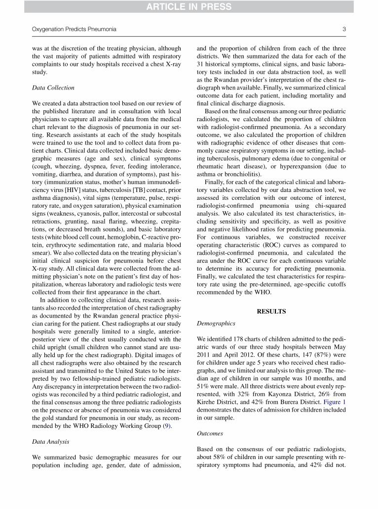
We identified 178 charts of children admitted to the pedi-atric wards of our three study hospitals between May2011 and April 2012. Of these charts, 147 (87%) werefor children under age 5 years who received chest radio-graphs, and we limited our analysis to this group. Theme-dian age of children in our sample was 10 months, and51% were male. All three districts were about evenly rep-resented, with 32% from Kayonza District, 26% fromKirehe District, and 42% from Burera District. Figure 1demonstrates the dates of admission for children includedin our sample.
Outcomes
Based on the consensus of our pediatric radiologists,about 58% of children in our sample presenting with re-spiratory symptoms had pneumonia, and 42% did not.

Figure 1. Date of admission.
4 P. Modi et al.
Those without pneumonia on their chest X-ray study hada range of other radiologic diagnoses, including hyperex-pansion (4%), tuberculosis (3%), pulmonary edema/car-diomegaly (1%), or normal chest X-ray studies (34%).There was a statistically significant correlation betweena final clinical discharge diagnosis of pneumonia in thechart and radiologist-confirmed pneumonia (c2 = 5.68,p = 0.017). However, there was no significant correlation
Table 1. Test Characteristics of Categorical Variables for Predictin
Symptoms Sensitivity Specificity Likelih
Respiratory distress 0.93 0.06Dyspnea 1 0.03Cough 0.98 0.01Wheezing 0.81 0Fever 0.93 0.06Feeding tolerance 0.31 0.84Vomiting 0.75 0.38Diarrhea 0.66 0.29Past medical history
Vaccinated 0.98 0.04Mother HIV positive 0.11 0.90Prior TB contact 0 0.8History of asthma 0.27 0.2
Clinical signsWeakness 0.80 0.26Cyanosis 0 0.5Pallor 0.21 0.57Intercostal retractions 0.85 0.08Subcostal retractions 0.81 0.11Grunting 0.4 1Nasal flaring 0.91 0.18Wheezing 0.44 0.4Crepitations/decreased breath sounds 0.70 0.38Suspicion of PNA 0.88 0.18
StudiesHIV 0 0.94Positive malaria smear 0 0.90CRP 0.65 0.38GP CXR read 0.55 0.72
HIV = human immunodeficiency virus; TB = tuberculosis; PNA = pneumchest X-ray study; CI = Confidence Interval.
between clinical suspicion for pneumonia (as charted bythe physician on admission before chest X-ray study orlaboratory tests) and radiologist-confirmed pneumonia(c2 = 1.54, p = 0.215). Overall, there was a strong corre-lation between the Rwandan general practitioner and ra-diologist chest X-ray study interpretation for the presenceof pneumonia (c2 = 7.68, p = 0.006). The overall case fa-tality ratio for children admitted with respiratory symp-toms to our study hospitals during this time period was2.3% (1.5% in children with radiologist-confirmed pneu-monia and 3.9% in children without radiologist-confirmed pneumonia).
Predictors of Pneumonia
Of all the historical categorical variables studied, onlyhistory of asthma was significantly associated with theabsence of pneumonia (c2 = 4.48, p = 0.034) on chest ra-diograph. Of all the categorical clinical signs and labora-tory tests studied, only a positive blood smear for malariawas statistically associated with the absence of pneumo-nia (c2 = 6.21, p = 0.013). Table 1 shows the sensitivity,specificity, and positive and negative likelihood ratios ascompared to our gold standard diagnosis of radiologist-confirmed pneumonia for each of the categorical vari-ables collected by our data abstraction tool.
g Pneumonia
ood Ratio Positive (95% CI) Likelihood Ratio Negative (95% CI)
1.00 (0.85–1.18) 0.90 (0.08–9.26)1.03 (0.97–1.09) 0 (0–0)1.00 (0.95–1.05) 0.68 (0.04–10.7)0.81 (0.61–1.08) 0 (0–0)0.99 (0.90–1.10) 1.00 (0.23–4.27)2.03 (0.46–8.81) 0.81 (0.54–1.21)1.22 (0.82–1.81) 0.63 (0.27–1.47)0.94 (0.60–1.46) 1.14 (0.44–2.95)
1.02 (0.96–1.09) 0.36 (0.03–3.94)1.29 (0.20–8.27) 0.97 (0.78–1.20)
0 (0–0) 1.25 (0.80–1.93)0.34 (0.14–0.82) 3.61 (0.61–21.3)
1.10 (0.85–1.41) 0.71 (0.31–1.66)0 (0–0) 2 (0.50–7.99)
0.49 (0.18–1.32) 1.36 (0.91–2.04)0.93 (0.73–1.19) 1.71 (0.19–14.7)0.91 (0.65–1.27) 1.68 (0.20–13.9)
0 (0–0) 0.6 (0.29–1.22)1.12 (0.82–1.51) 0.45 (0.07–2.84)0.74 (0.26–2.05) 1.38 (0.40–4.71)1.13 (0.86–1.50) 0.77 (0.45–1.30)1.09 (0.94–1.26) 0.60 (0.26–1.35)
0 (0–0) 1.05 (0.94–1.17)0 (0–0) 1.10 (1.00–1.21)
1.07 (0.68–1.68) 0.88 (0.41–1.90)2.06 (1.16–3.68) 0.60 (0.42–0.85)
onia; CRP = C-reactive protein; GP = general practitioner; CXR =

Table 2. Area under ROC Curve for Continuous Variablesfor Predicting Pneumonia
ContinuousVariables
Area underROC Curve
95% Confidence Interval
Lower Bound Upper Bound
Symptom duration 0.556 0.457 0.654Oxygen saturation 0.675 0.581 0.769Respiratory rate 0.528 0.428 0.627Temperature 0.480 0.379 0.581WBC 0.558 0.444 0.671Hemoglobin 0.530 0.424 0.636ESR 0.522 0.259 0.786
ROC = receiver operating characteristic; WBC = white blood cellcount; ESR = erythrocyte sedimentation rate.
Oxygenation Predicts Pneumonia 5
Table 2 demonstrates the area under the ROC curvesand 95% confidence intervals (CI) for each of the contin-uous variables studied. Of all the continuous variablesstudied, only oxygen saturation had an area under theROC curve (AUC) statistically different from the refer-ence line (i.e., better than chance), with an AUC of0.675 (95% CI 0.581–0.769 and p = 0.001). Respiratoryrate, on the other hand, had an AUC of 0.528 (95% CI0.428–0.627 and p = 0.588), not significantly differentfrom the reference line. Figure 2 demonstrates the ROCcurves for respiratory rate and oxygen saturation,respectively.
Using the age-specific cutoffs for respiratory raterecommended by the WHO IMCI guidelines for thediagnosis of pneumonia (respiratory rate above 60breaths/min in children under 2 months, above 50
Figure 2. Oxygen saturation and respiratory rate receiver operatin
breaths/min for children 2 months to 1 year, and above40 breaths/min for children over 1 year), respiratoryrate in our sample failed to demonstrate a statistical asso-ciation with the presence of pneumonia on the chest ra-diograph (c2 = 1.26, p = 0.261). Using the WHOcutoffs, respiratory rate had a sensitivity of 37%, a speci-ficity of 72%, a positive likelihood ratio of 1.33 (95% CI0.80–2.21), and a negative likelihood ratio of 0.87 (95%CI 0.69–1.10) for predicting pneumonia.
Based on our ROC curve for oxygen saturation, we de-termined the best cut-point for the prediction of pneumo-nia to be an oxygen saturation below 93%. Using thiscut-point, oxygen saturation was significantly associatedwith the presence of pneumonia on the chest radiograph(c2 = 9.87, p = 0.002). Additionally, oxygen saturationbelow 93%was found to have a sensitivity of 76%, a spec-ificity of 51%, a positive likelihood ratio of 1.54 (95% CI1.14–2.06), and a negative likelihood ratio of 0.47 (95%CI 0.29–0.76) for predicting pneumonia. A cut-point ofoxygen saturation below 90% had a sensitivity of 61%and specificity of 68%, and oxygen saturation below96% had a sensitivity of 86% and specificity of 28%.
DISCUSSION
The accuracy and reliability of respiratory rate and otherclinical indicators for the diagnosis of pneumonia is of farmore than just academic importance; the use of inaccu-rate or unreliable means for diagnosing pneumonia inchildren can have significant negative impacts on both
g characteristic curve.

6 P. Modi et al.
the effectiveness and cost-effectiveness of pediatric carein resource-limited settings. For instance, the low speci-ficity of respiratory rate alone for diagnosing pneumoniain certain settings can mean that many children aretreated unnecessarily with antibiotics, increasing antibi-otic resistance and wasting health care resources. Farmore important, however, is that children mistakenly di-agnosed with pneumonia due to an increased respiratoryrate alone may not be evaluated and treated appropriatelyfor other life-threatening childhood diseases that alsopresent with tachypnea such as malaria or asthma. Thisis evidenced by the trend toward higher mortality in chil-dren in our sample without pneumonia as compared tothose with pneumonia. Finally, the low sensitivity of re-spiratory rate alone for the diagnosis of pneumonia incertain pediatric populations, such as those with severemalnutrition, can mean that antibiotics may be inappro-priately withheld from those children who truly needthem.
Based on the IMCI guidelines, the diagnosis of pneu-monia in children hinges on the assessment of respiratoryrate by community health workers, nurses, or doctorsworking at first-level health facilities. Whereas a reviewof several early pneumonia diagnostic studies found respi-ratory rate itself to be a moderately good predictor ofpneumonia in children under the age of 5 years, with pos-itive likelihood ratios ranging from 1.5 to 3.2 and negativelikelihood ratios ranging from .32 to .75, several more re-cent studies have not found respiratory rate alone to be anaccurate predictor of pneumonia in children (10–13). Inparticular, studies have found respiratory rate to bea less accurate predictor of pneumonia in children over12 months, those suffering from severe malnutrition orHIV, and those presenting with wheezing (14–18).
The significant variability in the diagnostic accuracyof respiratory rate for predicting pneumonia in childrencan be explained in three ways: 1) variability in themethod of measurement of respiratory rate; 2) variabilityin the gold standard used for diagnosing pneumonia; and3) variability in the prevalence of other diseases in chil-dren that cause tachypnea. Prior studies for instance,have found high variability in measured respiratory ratein children depending on the method of counting (i.e., ob-servation vs. auscultation), the duration of the countingperiod (i.e., 30 vs. 60 s), and the state of the child (i.e.,sleeping, agitated, or feeding) (19,20). Perhaps evenmore important than the inter-study variability in themethod of determining respiratory rate in children isthe inter-study variability in the gold standard used forthe detection of pneumonia. A recent review found noless than 11 different gold standards used in 25 differentdiagnostic studies assessing various clinical and labora-tory predictors for pneumonia in children (21).
Finally, the sensitivity and specificity of respiratoryrate alone for the diagnosis of pneumonia can also be af-fected by the prevalence of other diseases in the pediatricpopulation (18). For instance, if all children enrolled ina particular study with cough or shortness of breathhave either pneumonia or a viral upper respiratory infec-tion, then respiratory rate may be an accurate means ofpredicting which children will have pneumonia andwhich will not. However, if significant numbers of chil-dren in the population studied have other diseases thatcause rapid breathing, such as bronchiolitis, asthma, ma-laria, heart failure, or tuberculosis, then respiratory ratewill have far lower specificity for the diagnosis of pneu-monia. This was clearly true in our population of childrenwhere lack of prior asthma diagnosis and negative bloodsmear for malaria were significant predictors of pneumo-nia, whereas respiratory rate was not. Similarly, in popu-lations where severe malnutrition is common (as it is inours, according to a recent Rwanda Demographic andHealth Survey), many children with pneumonia maynot be able to mount an increased respiratory drive, mak-ing rapid breathing a less sensitive predictor of pneumo-nia (8).
In this study, we retrospectively assessed more thantwo dozen clinical signs and symptoms for the presenceof pneumonia in a population of children presentingwith cough or difficulty breathing to three rural, Rwandanhospitals, and found that oxygen saturation was the mostpredictive clinical indicator of radiologist-confirmedpneumonia. Although many prior studies have analyzedthe accuracy of other clinical signs for predicting hypoxiain children diagnosed with pneumonia, no prior studieshave analyzed the accuracy of oxygen saturation itselffor detecting pneumonia in children, either alone or incombination with other clinical signs (22–25). Finally,whereas most prior diagnostic studies of pneumonia inchildren have been conducted at single urban referralhospitals, our research was conducted in several front-line rural hospitals in a low-income African country, in-creasing the generalizability of our results to childrenliving across sub-Saharan Africa.
Limitations
The primary limitation of this study is its retrospectivenature. This means that in studying the association of var-ious clinical signs and symptoms for the presence ofpneumonia on chest radiography, we were limited bythe data primary providers chose to record, or failed to re-cord, in the chart. The problem of missing data was great-est for certain historical symptoms and past medicalhistory, such as history of feeding intolerance, historyof asthma, and history of TB contact, which were missing
Oxygenation Predicts Pneumonia 7
from a majority of charts. These missing data likely com-promised the power of our study to detect significant as-sociations between these historical signs and the presenceof pneumonia (though history of asthma did still demon-strate a significant association). Similarly, a number ofclinical signs were also frequently missing from thecharts, including the presence or absence of cyanosis,grunting, or wheezing. However, vital signs, includingtemperature, respiratory rate, and oxygen saturation,were recorded in all charts, though we cannot verify theprecise methods used by providers to collect each of thesevariables. A future, prospective study will be necessary toensure that all data are collected for all children enrolledin a consistent manner, increasing the power to detect as-sociations between the various clinical predictors and thepresence of pneumonia. In addition, this study was con-ducted at hospitals supported by Partners in Health,which may have somewhat better resources and providertraining than government hospitals not supported by anoutside organization.
Another potential limitation is our decision to utilizechest radiography as our gold standard for the diagnosisof pneumonia. Although the ‘‘perfect’’ gold standard forbacterial pneumonia would likely be culture of an aspi-rate from the lower respiratory track obtained by bron-choalveolar lavage or lung puncture, this is incrediblyinvasive and rarely used in studies of pediatric pneumo-nia, whether in the developed or developing world. In-stead, a number of different gold standards have beenused for diagnostic studies of pneumonia in children,with the most common in the published literature beingchest radiography and blood cultures (21). Of thesetwo, only chest radiography is available in our settingin rural Rwanda. Furthermore, the largest meta-analysisof clinical indicators of pneumonia in children includedonly studies utilizing chest radiography as the referencestandard, as this was felt to be the most practical and validgold standard for pneumonia in children (10). As such,we have chosen to use chest radiography, interpreted byfellowship-trained pediatric radiologists, as the referencestandard for pneumonia in our study. A particular strengthof our study was that we had digital images of each chestradiograph reviewed by two separate pediatric radiolo-gists, with any discrepancy in their reads resolved bya third pediatric radiologist. This is recommended bythe WHO Radiology Working Group, given the inherentinter-rater variability they found in chest radiograph in-terpretation for the presence of pneumonia in childrenin resource-limited settings, due to the poor quality ofmany chest X-ray studies in these settings (9).
A final limitation of this study is that it only includedhospitalized children. Unfortunately, as in most ruralareas of sub-Saharan Africa, medical charts are onlykept for hospitalized children in our setting. In addition,
the use of chest radiography as our gold standard mayhave biased our sample towards sicker children, whowere more likely to receive a chest radiograph. However,of all children admitted with respiratory symptoms to thepediatric wards at our study hospitals, 87% were childrenunder 5 years of age who received a chest radiograph. Ofthe small percentage of children who did not receivea chest radiograph in our sample, most of these casesseemed to be due to malfunctioning of the X-ray ma-chine, a common occurrence at rural hospitals in our set-ting. However, as this is essentially a random event, itshould not bias our results.
CONCLUSION
Pneumonia is a major cause of morbidity and mortalityamong children in resource-limited settings. Currently,the IMCI guidelines rely on respiratory rate as the primaryindicator of pneumonia in children and advise initiatingor withholding treatment based on this variable alone.However, the low specificity of respiratory rate maylead to overtreatment with antibiotics while missing otherimportant diagnoses in children, and the low sensitivitydictates that antibiotics may be withheld from childrenrequiring them. Better-performing clinical variables thatcan be easily collected in resource-limited settings, suchas oxygen saturation, could improve diagnostic accuracyand thereby resource utilization. As pulse oximeters be-come increasingly available in resource-limited settingsover the coming years, they have the potential to be a rel-atively inexpensive method for diagnosing pneumonia inthese settings. We recommend a prospective study to de-rive a clinical prediction rule for the diagnosis of pneumo-nia in children under age 5 years in resource-limitedsettings that incorporates oxygen saturation and otherclinical variables found to be predictive of pneumonia.
Acknowledgments—We would like to thank the Rwanda Minis-try of Health and Partners in Health/Inshuti Mu Buzima for theirsupport of this project.
REFERENCES
1. Rudan I, Boschi-Pinto C, Biloglav Z, Mulholland K, Campbell H.Epidemiology and etiology of childhood pneumonia. Bull WorldHealth Organ 2008;86:408–16.
2. World Health Organization. Integrated management of childhoodillness. Geneva: World Health Organization; 2005.
3. Amaral J, Gouws E, Bryce J, Leite AJ, Cunha AL, Victora CG. Ef-fect of Integrated Management of Childhood Illness (IMCI) onhealth worker performance in Northeast-Brazil. Cad Saude Publica2004;20(Suppl 2):S209–19.
4. Bryce J, Victora CG, Habicht JP, Black RE, Scherpbier RW. Pro-grammatic pathways to child survival: results of a multi-countryevaluation of Integrated Management of Childhood Illness. HealthPolicy Plan 2005;20(Suppl 1):i5–17.

8 P. Modi et al.
5. Arifeen SE, Hoque DM, Akter T, et al. Effect of the IntegratedMan-agement of Childhood Illness strategy on childhood mortality andnutrition in a rural area in Bangladesh: a cluster randomised trial.Lancet 2009;374:393–403.
6. Huicho L, Davila M, Gonzales F, Drasbek C, Bryce J, Victora CG.Implementation of the Integrated Management of Childhood Illnessstrategy in Peru and its association with health indicators: an eco-logical analysis. Health Policy Plan 2005;20(Suppl 1):i32–41.
7. Armstrong Schellenberg J, Bryce J, de Savigny D, et al. The effectof Integrated Management of Childhood Illness on observed qualityof care of under-fives in rural Tanzania. Health Policy Plan 2004;19:1–10.
8. National Institute of Statistics of Rwanda (NISR). Rwanda demo-graphic and health survey 2010. Calverton, MD: NISR and ORCMacro; 2011.
9. Cherian T, Mulholland EK, Carlin JB, et al. Standardized interpre-tation of paediatric chest radiographs for the diagnosis of pneumo-nia in epidemiological studies. Bull World Health Organ 2005;83:353–9.
10. Margolis P, Gadomski A. The rational clinical examination. Doesthis infant have pneumonia? JAMA 1998;279:308–13.
11. Puumalainen T, Quiambao B, Abucejo-Ladesma E, et al. Clinicalcase review: a method to improve identification of true clinicaland radiographic pneumonia in children meeting the World HealthOrganization definition for pneumonia. BMC Infect Dis 2008;8:95.
12. Shah S, Bachur R, Kim D, Neuman MI. Lack of predictive value oftachypnea in the diagnosis of pneumonia in children. Pediatr InfectDis J 2010;29:406–9.
13. Hazir T, Nisar YB, Qazi SA, et al. Chest radiography in childrenaged 2–59months diagnosed with non-severe pneumonia as definedby World Health Organization: descriptive multicentre study inPakistan. BMJ 2006;333:629.
14. Redd SC, Vreuls R, Metsing M, Mohobane PH, Patrick E,Moteetee M. Clinical signs of pneumonia in children attendinga hospital outpatient department in Lesotho. Bull World Health Or-gan 1994;72:113–8.
15. Chisti MJ, Tebruegge M, La Vincente S, Graham SM, Duke T.Pneumonia in severely malnourished children in developingcountries – mortality risk, aetiology and validity of WHO clinicalsigns: a systematic review. Trop Med Int Health 2009;14:1173–89.
16. Urganci N, Polat T, Ozer N, Kayaalp N. The presence of clinicalsigns in malnourished infants with acute lower respiratory tract in-fections. Paediatr Child Health 2003;8:83–6.
17. Hazir T, Qazi S, Nisar YB, et al. Assessment and management ofchildren aged 1-59 months presenting with wheeze, fast breathing,and/or lower chest indrawing; results of a multicentre descriptivestudy in Pakistan. Arch Dis Child 2004;89:1049–54.
18. Graham SM, English M, Hazir T, Enarson P, Duke T. Challenges toimproving case management of childhood pneumonia at health fa-cilities in resource-limited settings. Bull World Health Organ 2008;86:349–55.
19. Berman S, Simoes EA, Lanata C. Respiratory rate and pneumonia ininfancy. Arch Dis Child 1991;66:81–4.
20. Simoes EA, Roark R, Berman S, Esler LL, Murphy J. Respiratoryrate: measurement of variability over time and accuracy at differentcounting periods. Arch Dis Child 1991;66:1199–203.
21. Lynch T, Bialy L, Kellner JD, et al. A systematic review on the di-agnosis of pediatric bacterial pneumonia: when gold is bronze.PLoS One 2010;5:e11989.
22. Weber MW, Usen S, Palmer A, Jaffar S, Mulholland EK. Predictorsof hypoxaemia in hospital admissions with acute lower respiratorytract infection in a developing country. Arch Dis Child 1997;76:310–4.
23. Usen S, Weber M, Mulholland K, et al. Clinical predictors of hypo-xaemia in Gambian children with acute lower respiratory tract in-fection: prospective cohort study. BMJ 1999;318:86–91.
24. Lodha R, Bhadauria PS, Kuttikat AV, et al. Can clinical symptomsor signs accurately predict hypoxemia in children with acute lowerrespiratory tract infections? Indian Pediatr 2004;41:129–35.
25. Basnet S, Adhikari RK, Gurung CK. Hypoxemia in children withpneumonia and its clinical predictors. Indian J Pediatr 2006;73:777–81.

Oxygenation Predicts Pneumonia 9
ARTICLE SUMMARY
1. Why is this topic important?Pediatric pneumonia is the leading cause of death in
children under 5 years of age worldwide. Improvedmethods of diagnosis are necessary as the current WorldHealth Organization guidelines have had mixed diagnos-tic results.2. What does this study attempt to show?
This study attempts to find new clinical indicators thatcan predict pneumonia.3. What are the key findings?
Of all the variables studied, a positive blood smear formalaria and a history of asthma were statistically associ-ated with the absence of pneumonia. Of the continuousvariables, only oxygen saturation had a statistically signif-icant area under the receiver operating characteristiccurve, whereas respiratory rate did not.4. How is patient care impacted?
By finding better methods of diagnosing pneumonia inchildren, possibly with the use of oxygen saturation, wecan initiate early treatment and hopefully reduce the mor-bidity and mortality worldwide.