oxidative stress and dna damage in nile tilapia
TRANSCRIPT

92 Journal of Bioscience and Applied Research , 2019, Vol.5, No. 1, P.92 -109 pISSN: 2356 - 9174, eISSN: 2356 –9182
BioBacta
Journal of Bioscience and Applied Research
www.jbaar.org
Oxidative stress and DNA damage in Nile Tilapia (Oreochromis niloticus) as
biomarkers of aquatic pollution
EL-Hassan Mokhamer, Eman H. Radwan and Moataz Elsaka
Zoologlogy Department, Faculty of science, Damanhour university,Egypt (Email: [email protected])
Abstract
The main purpose of this study is to evaluate the impact of heavy metals (Pb, Zn, Cu, Fe, and Cd) aquatic
pollution of EL-Mahmoudeyia canal on the antioxidant enzymatic activities, GSH content and lipid
peroxidation levels (MDA) in Oreochromis. niloticus muscles tissues collected from two areas EL-
Mahmoudeyia canal as Polluted area and Rosetta branch of river Nile as reference area in summer 2018
and winter 2019 as well as DNA damage was assessed in fish gills(erythrocytes) samples by applying
comet assay. EL-Mahmoudeyia canal exposed to excessive of industrial effluents which impact the living
organisms especially fish. The herein results showed that higher concentrations of heavy metals (Pb, Zn,
Cu, Fe, and Cd) were detected in water and fish samples collected from the polluted area in comparison
with the reference area, especially in winter. The accumulation patterns of heavy metals in muscles of O.
niloticus, were in the following order: Fe > Zn >Pb> Cu and Cd.The antioxidant enzymatic activities of
(SOD, CAT, GPx and GST) and the lipid peroxidation biomarker MDA levels in muscles of O. niloticus
collected from the polluted area were found to be significantly increased compared to that of the
reference area. Meanwhile, there was a significant decrease in the GSH content level in muscles of O.
niloticus collected from the polluted area compared to that of the reference area. A significant elevation
in DNA damage frequencies was observed in fish collected from the polluted areas compared with those
from the reference area. These noticeable alterations in the selected antioxidant enzymatic activities in
muscles of the O. niloticus go in parallel with the remarkable elevation in the levels of the detected heavy
metals in water from EL Mahmoudeyia canal, as a result of pollution in these areas. This study explored
the utility of the DNA damage, the altered antioxidant enzymatic activities, GSH content and MDA level
as biomarkers of aquatic pollution.
Keywords : Heavy metals, pollution, oxidative stress, DNA damage, antioxidants enzymes.

93 Journal of Bioscience and Applied Research , 2019, Vol.5, No. 1, P.92 -109 pISSN: 2356 - 9174, eISSN: 2356 –9182
Introduction
Pollution levels in aquatic environments have
greatly increased recently as a result of intense
human activities, which, in some areas have
resulted in a substantial impact.(van der Oost et
al., 1996).Contamination of the aquatic ecosystem
by industrial and agricultural pollutants affects the
health of fish, either directly by uptake from the
water, or indirectly through their diet of
vegetation, invertebrates or smaller fish. Since fish
are part of the natural diet of both aquatic
mammals and birds, as well as providing an
increasingly important protein source for humans,
their population and health is of major concern
(Kime, 1995).
The pollution problems in the surface water either
drainage or fresh water affect the quality of fish in
polluted areas. The specific contaminants leading
to pollution in water include a wide spectrum of
chemicals, pathogens and physical or sensory
changes such as elevated temperature and
discoloration (Pitt and Burton Jr, 2001).
Heavy metals pollution in aquatic environment has
become a worldwide problem during past few
decades. This fact is mainly attributed to their
persistent stability and toxic effect to aquatic as
well as terrestrial creatures(MacFarlane and
Burchet, 2000). Among environmental pollutants,
metals are of particular concern, due to their
potential toxic effect and ability to bioaccumulate
in aquatic ecosystems(Censi et al., 2006)Heavy
metal concentrations in aquatic ecosystems are
monitored by measuring their concentrations in
water, sediments and biota. (Namminga, 1976).
Electric power generation also play major role in
water pollution. Generally, more than 75% of
waste is disposed of in unlined, unmonitored
onsite landfills and surface impoundments. Toxic
substances in the waste - including arsenic,
mercury, chromium, and cadmium can
contaminate drinking water supplies and damage
vital human organs and the nervous
system(Tripathi et al., 2015).The essential use of
water in industry is the cooling system (Nriagu and
Pacyna, 1988)When water used as a coolant is
returned to the natural environment at a higher
temperature, the change in temperature impacts
organisms by decreasing oxygen supply, and
affecting ecosystem composition(El Safty and
Siha, 2013).The atmosphere is the major route of
Pb entry in natural waters, a fact that has been well
documented in the literature(Flegal and Patterson,
1983),(Veron et al., 1987)
Fish are largely being used for the assessment of
the aquatic environment quality and can serve as
bioindicators of environmental pollution.
(Dautremepuits et al., 2004, Lopes et al., 2001).
Fish muscle is commonly analyzed to determine
contaminant concentrations and to assess the
health risks because it is the main part consumed
by humans. Fish can be considered as one of the
most significant indicators in freshwater systems
for the impact of metal pollution (Begum et al.,
2005).Tilapia (Oreochromis niloticus) is a fresh
water fish that is hardy, prolific, fast growing
tropical fish that is farmed mainly in Africa and
Asia. Tilapia fish are beneficial to human's beings
as they make up a major part of the human diet
and provide humans with as much of needed
proteins as in meat (Ghorbani and Mirakabad,
2010).

94 Journal of Bioscience and Applied Research , 2019, Vol.5, No. 1, P.92 -109 pISSN: 2356 - 9174, eISSN: 2356 –9182
Heavy metals can be taken up into fish either from
digestion or contaminated food via alimentary
track or through gills or skin after the absorption it
transported through blood stream to the organs and
tissues, where they are accumulated. Fish can
regulate metal concentrations to a certain extent,
after the occurrence of bioaccumulation (Magdy et
al.)
Aquatic ecosystems are not usually able to
eliminate heavy metals from waste discharges by
their own natural processes. Mercury, cadmium,
arsenic, and copper tend to accumulate in bottom
sediments, from which they may be released by
various processes of remobilisation. They can
then, in different forms, move up through
biological food chains, eventually to humans in
whom they can produce both chronic and acute
ailments(Forstner). Metal accumulation causes an
increase in highly reactive oxygen species (ROS)
such as hydrogen peroxide, superoxide radical,
hydroxyl radical which leading to oxidative stress
in fish (Dautremepuits et al., 2002).
Oxidative stress is a situation characterized by an
imbalance between increased production of
oxidant species and/or decreased efficacy of the
antioxidant defense system(Gosmaro et al., 2013)
leading macromolecule to damage including
lipid peroxidation, protein cross linking, DNA
damage, changes in growth and function of cells
(Ehsaei et al., 2015). Many factors including heavy
metals in soil and waste water can affect the DNA
genetic material of organisms directly or indirectly
and not only to damage the integrity of the DNA
structure but also influence its expression and
eventually cause genotoxicity to organisms(Yu,
2000).
Comet assay is used as one of the best approaches
to study the genotoxic effects of pollutants on fish
(Nagarani et al., 2012), as it is used for the
estimation of DNA damage to evaluate the genetic
risk associated with xenobiotic exposures. The
comet assay possesses a number of advantages as
compared to other genotoxicity tests. In addition to
the capability of this assay to identify DNA
damage at the single cell level, other significant
advantages include its sensitivity for detecting low
levels of DNA damage, the requirement for only
small numbers of cells per sample, its ease of
application and low cost, and the short time
needed to perform the assay. (Tice et al., 2000)
2. MATERIALS AND METHODS
2.1 Study area
EL-Mahmoudeyia Canal, is a 45-mile-long sub-
canal from the Nile River which starts at the Nile-
port of EL-Mahmoudeyia and goes through
Alexandria to the Mediterranean Sea. It was built
to supply Alexandria with food and fresh water
from the Nile. This study was carried out in EL-
Mahmoudeyia province, El-Beheira governorate.
Samples were collected from two site areas
throughout summer 2018and winter 2019 as
shown in Figure 1:
Site 1:
The Rosetta branch of Nile river close to Alatf
village which considered as reference area free
from the industrial activities and wastes.
Site 2:
EL-Mahmoudeyia canalas polluted area where the
EL-Mahmoudeyia electric power station and a
textile mill dump directly their effluents to the

95 Journal of Bioscience and Applied Research , 2019, Vol.5, No. 1, P.92 -109 pISSN: 2356 - 9174, eISSN: 2356 –9182
stream of the canal. The distance between the two studied areas is about 2 Km.
Fig. 1: The two sites of the study area at El Mahmodia(El Behaira, Egypt)
2.2 Sample collection
Water and fish (Nile tilapia, Oreochromis
niloticus) samples were collected from the
previously mentioned areas during the first season
(summer, July 2018) and the second season
(winter, February2019). Fish samples were caught
by fisherman's net, the collected fish were with an
average body weight (120 ± 10 gm), and an
average body length (16±4 cm). After dissection of
fish, muscle tissues were separated for estimation
of heavy metals, antioxidant enzymes activity,
thiobarbituric acid-reactive substances
(MDA)concentration and DNA damage by comet
assay. Water samples were collected in clean
bottles from a 2-meter depth of the studied areas.
2.3 Chemicals
The assay kits used for biochemical estimation of
lipid peroxide (Malondialdehyde, MDA), Reduced
glutathione concentration (GSH) , catalase (CAT),
glutathione-s transferase (GST), superoxide
dismutase (SOD) and glutathione peroxidase
(GPx) activities were purchased from
Biodiagnostic Co., Egypt. All other chemicals and
reagents were of analytical grade and were
commercially available from local scientific
distributors in Egypt.
2.4. Chemical analysis
The concentration of heavy metals (Pb, Zn, Cu, Fe,
and Cd) in the samples were determined after
digestion by using Atomic Absorption
Spectrophotometer (Perkin Eelmer E. Analyst,
2000, USA).according to the method described
by(Vitošević et al., 2007). Results in water were
expressed in (mg/L) and in fish muscles in mg/kg
dry wt.
2.5. Antioxidants and lipid peroxidation
biomarkers
Tissue homogenates were prepared from muscle
samples in 10 volumes of 0.1 M Tris-EDTA buffer

96 Journal of Bioscience and Applied Research , 2019, Vol.5, No. 1, P.92 -109 pISSN: 2356 - 9174, eISSN: 2356 –9182
(pH7.4), centrifuged at 1,000 × g at 4°C for 30
min. Aliquots of the supernatant were utilized for
the following spectrophotometric assessments.
2.5.1. Superoxide Oxide Dismutase activity
(SOD) was determined spectrophotometrically at
560 nm according to the method of Nishikimi et al.
(1972). The method based on the ability of SOD
enzyme to inhibit the phenazine methosulphate-
mediated reduction of nitroblue tetrazolium dye.
Briefly, 0.05 mL sample was mixed with 1.0 mL
buffer (pH 8.5), 0.1 mL nitroblue tetrazolium
(NBT) and 0.1 mL NADH. The reaction was
initiated by adding 0.01 mL phenazine
methosulphate (PMs) and then increased in
absorbance was read at 560 nm for 5 min. SOD
activity was expressed as U/gm tissue.
2.5.2.The enzymatic activity of Catalase (CAT)
was measured according to the method described
by (Aebi, 1984). The CAT reacts with a known
quantity of H2O2, and the reaction is stopped after
1 min with a CAT inhibitor. In the presence of
peroxidase, the remaining H2O2 reacts with 3,5-
dichloro-2-hydroxybenzene sulfonic acid and 4-
aminophenazone to form a chromophore, with a
color intensity inversely proportional to the
amount of CAT in the sample. The absorbance
was measured at 510 nm.
2.5.3. The enzymatic activity of Glutathione
peroxidase(GPx) in tissue was estimated
colorimetrically using kits from Bio-diagnostic
Company. The assay is an indirect measure of the
activity of GPx. The GSSG that is produced upon
the reduction of organic peroxide by GPx is
recycled to its reduced state by GR. The oxidation
of NADPH to NADP+ is accompanied by a
decrease in absorbance at 340 nm (A340),
providing a spectrophotometric means for
monitoring GPx enzymatic activity. To assay GPx,
an aliquot of tissue homogenate was added to a
solution containing GSH, GR, and NADPH. The
enzyme reaction was initiated by adding the
substrate, tert-butyl hydroperoxide, and the A340
was recorded. The rate of the decrease in A340 is
directly proportional to the GPx activity in the
tested sample(Paglia and Valentine, 1967)
2.5.4.Glutathione-S-Transferase (GST)
enzymatic activity in tissue homogenates were
assayed spectrophotometrically according to the
method of (Habig et al., 1974). By measuring the
conjugation of 1- chloro- 2,4- dinitrobenzene
(CDNB) with reduced glutathione. The
conjugation is accompanied by an increase in
absorbance at 340 nm. The rate of increase is
directly proportional to the GST activity in the
sample.
2.5.5.The reduced glutathione (GSH) level was
assayed using a method based on the reductive
cleavage of 5, 5′-dithiobis (2-nitrobenzoic acid)
(DTNB) by a sulfhydryl (-SH) group to yield a
yellow color. The reduced chromogen (absorbance
measured at 412 nm) is directly proportional to the
GSH concentration.(Beutler et al., 1963)
2.5.6. lipid peroxidation (LPO): lipid
peroxidation (LPO) was determined by a
colorimetric method of (Kei, 1978) according to
the details given in the kit’s instructions. A
thiobarbituric acid reactive substance (TBARS)
was used for the estimation of LPO and expressed
in terms of malondialdehyde (MDA) content. For
this purpose, 0.5 mL of trichloroacetic acid (10%)
solution was added into 0.5 mL sample in test
tube. The mixture was centrifuged at 600×g for 10

97 Journal of Bioscience and Applied Research , 2019, Vol.5, No. 1, P.92 -109 pISSN: 2356 - 9174, eISSN: 2356 –9182
min and 0.2 mL of the supernatant was transferred
into a new test tube containing 1.0 mL of TBA (25
mmol /L) solutions and boiling for 30 min. The
solution was then cooled and a pink color
chromogen for samples and standard were read at
534 nm using spectrophotometer. The MDA
values were expressed as nmol of MDA/gm tissue.
2.6. DNA damage using the comet assay
Isolated gills cells of fish from both areas were
subjected to the modified single-cell gel
electrophoresis or comet assay(Fairbairn et al.,
1995). Before running the comet assay, cell
viability for erythrocytes and gill cells was
determined using the trypan blue exclusion
method. To obtain the cells, a small piece of the
gills was washed with an excess of ice-cold
Hank's balanced salt solution (HBSS) and
minced quickly into approximately 1 mm3
pieces while immersed in HBSS, with a pair of
stainless steel scissors. After several washings
with cold phosphate-buffered saline, the minced
tissues were dispersed into single cells using a
pipette(Lai and Singh, 1995). In brief, the
protocol for electrophoresis involved
embedding of the isolated cells in agarose gel
on microscopic slides and lysing them with
detergent at high salt concentrations overnight
(in the cold). The cells were treated with alkali
for 20 min to denature the DNA and
electrophoresis under alkaline conditions (30
min) at 300 mA, 25 V. The slides were stained
with ethidium bromide and examined using a
fluorescence microscopy (Zeiss, axiostar plus
USA)with a green filter at × 40 magnifications.
For each experimental condition, about 100
cells per a fish were examined to determine the
percentage of cells with DNA damage that
appear like comets. The non-overlapping cells
were randomly selected and were visually
assigned a score on an arbitrary scale of 0–3
(i.e., class 0 = no detectable DNA damage and
no tail; class 1 = tail with a length less than the
diameter of the nucleus; class 2 = tail with
length between 1× and 2× the nuclear diameter;
and class 3 = tail longer than 2× the diameter of
the nucleus) based on perceived comet tail
length migration and relative proportion of
DNA in the nucleus(Collins et al., 1997,
Kobayashi, 1995). A total damage score for
each slide was derived by multiplying the
number of cells assigned to each class of
damage by the numeric value of the class and
summing up the values. Slides were analyzed
by one observer to minimize the scoring
variability.
2.7. Statistical analysis:
The results were expressed as Mean±SE and
statistical significance was evaluated by one way
ANOVA using SPSS (version 20.0) program.
Values were considered statistically significant
when p < 0.05.
3. Results
The mean heavy metals concentrations in water
from both reference and polluted area are
presented in table (1). The concentration of Pb, Zn,
Cu, Fe and Cd in water from the selected areas
ranged between (0.009-0.035), (0.178- 0.281),
(0.00096-0.0034), (0.233-1.308) and (0.0032-
0.0128) mg/L in winter, and ranged between
(0.0086-0.28), (0.177-0.272), (0.00086-0.003),
(0.235-1.263) and (0.0029-0.0110) mg/L in
summer respectively. EL-Mahmoudeyia canal
(polluted area) had the highest levels of heavy

98 Journal of Bioscience and Applied Research , 2019, Vol.5, No. 1, P.92 -109 pISSN: 2356 - 9174, eISSN: 2356 –9182
metals while, the reference area exhibited the
lowest values during period of sampling. The
results demonstrated that, Fe had the highest
concentration (0.235-1.263 mg/l) among the tested
metals, while Cd exhibited the lowest one (0.0029-
0.0110 mg/l) during the study period. Also, the
concentrations of Pb, Zn, Cu, Fe and Cd in water
were elevated in winter compared to summer at
two studied areas.
Table (1): The concentration of heavy metals (mg/l) in water samples collected from both reference
and polluted area
Values are expressed as mean ± S.E
*: Significant difference in comparison with the reference area (P < 0.05)
All data of heavy metals ( Pb, Zn, Cu, Fe and Cd)
accumulations in muscles of Oreochromis
niloticus collected from the Rosetta River Nile
(Reference area ) and EL-Mahmoudeyia canal
(polluted area) (mg/kg dry wt.) during summer and
winterrecorded in table 2.The values of iron
concentrations in the muscles of Oreochromis
niloticus showed a highly significant increase (p <
0.05) at polluted area during winter and summer
seasons in comparison with non-polluted area.
Lead concentrations in the muscles of
Oreochromis niloticus samples showed a
significant increase (P < 0.05) at polluted area
during summer and winter in comparison with
non-polluted area. Lead concentrations in muscles
reached the value of 0.163 ± 0.005 mg/kg dry wt.
in summer to 0.809 ± 0.011 mg/kg dry wt. in
winter season.
Zinc concentrations in the muscles of Oreochromis
niloticus samples had a highly significant increase
at (p < 0.05) in polluted area during summer and
winter seasons in comparison with non-polluted
area. Zinc concentrations in muscles reached the
value of 2.974± 0.012 mg/kg dry wt. in summer to
3.530±0.047 mg/kg dry wt. in winter.
Copper concentrations in the muscles of
Oreochromis niloticus samples had a highly
significant increase (p < 0.05) at polluted area
during summer and winter seasons in comparison
with non-polluted area. Copper concentrations in
muscles reached the quantity of 0.086.207 ±
0.0085 mg/kg dry wt. in winter to 0.080± 0.0008
mg/kg dry wt. in summer.
The iron concentrations in muscles reached the
value of 3.939 ± 0.038 mg/kg dry wt. in winter to
3.632 ± 0.085 mg/kg dry wt. in summer, the iron
Heavy Metal
Metal concentration (mg/l)
Summer Winter
Reference area Polluted area Reference area Polluted area
Pb 0.0086±0.0005 0.028±0.0009* 0.009±0.0002 0.035±0.001*
Zn 0.177±0.006 0.272±0.005* 0.178±0.006 0.281±0.006*
Cu 0.00086±0.00003 0.003±0.00005* 0.00096±0.00005 0.0034±0.00007*
Fe 0.235±0.003 1.263±0.015* 0.233±0.004 1.308±0.020*
Cd 0.0029±0.0002 0.0110±0.0004* 0.0032±0.0.0001 0.0128±0.0003*
99 Journal of Bioscience and Applied Research , 2019, Vol.5, No. 1, P.92 -109 pISSN: 2356 - 9174, eISSN: 2356 –9182
concentrations in muscles of Oreochromis
niloticus showed highly significant increase (p <
0.05) at the polluted area during the two seasons
summer and winter in comparison with non-
polluted area.
The cadmium concentrations in muscles reached
the value of 0.0255 ± 0.0005 mg/kg dry wt. in
winter to 0.0224 ± 0.0008 mg/kg dry wt. in
summer, the cadmium concentrations in muscles
of Oreochromis niloticus showed a significant
increase (p < 0.05) at the polluted area during the
two seasons summer and winter in comparison
with non-polluted area.
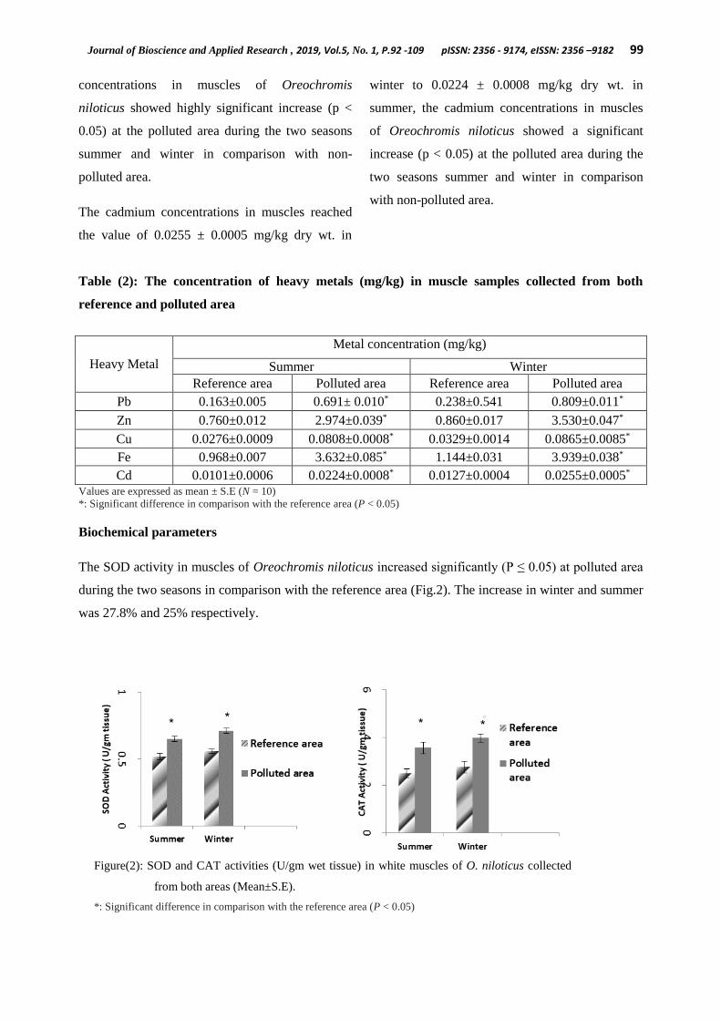
Table (2): The concentration of heavy metals (mg/kg) in muscle samples collected from both
reference and polluted area
Values are expressed as mean ± S.E (N = 10)
*: Significant difference in comparison with the reference area (P < 0.05)
Biochemical parameters
The SOD activity in muscles of Oreochromis niloticus increased significantly (P ≤ 0.05) at polluted area
during the two seasons in comparison with the reference area (Fig.2). The increase in winter and summer
was 27.8% and 25% respectively.
Heavy Metal
Metal concentration (mg/kg)
Summer Winter
Reference area Polluted area Reference area Polluted area
Pb 0.163±0.005 0.691± 0.010* 0.238±0.541 0.809±0.011*
Zn 0.760±0.012 2.974±0.039* 0.860±0.017 3.530±0.047*
Cu 0.0276±0.0009 0.0808±0.0008* 0.0329±0.0014 0.0865±0.0085*
Fe 0.968±0.007 3.632±0.085* 1.144±0.031 3.939±0.038*
Cd 0.0101±0.0006 0.0224±0.0008* 0.0127±0.0004 0.0255±0.0005*
Figure(2): SOD and CAT activities (U/gm wet tissue) in white muscles of O. niloticus collected
from both areas (Mean±S.E).
*: Significant difference in comparison with the reference area (P < 0.05)
*
* * * *
100 Journal of Bioscience and Applied Research , 2019, Vol.5, No. 1, P.92 -109 pISSN: 2356 - 9174, eISSN: 2356 –9182
The CAT activity in muscles of Oreochromis
niloticus increased significantly (P ≤ 0.05) at
polluted area during the two seasons in
comparison with the reference area (Fig.2). The
increase in winter and summer was 43.8% and
42.7% respectively.
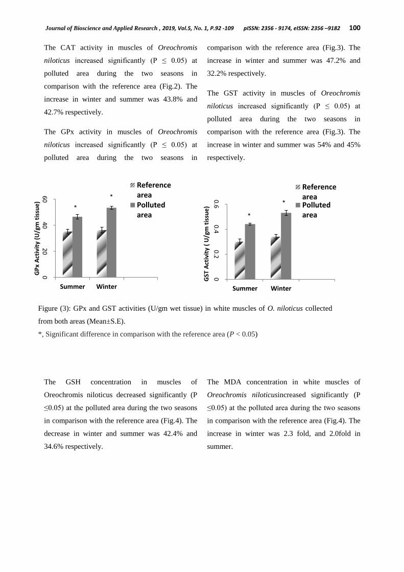
The GPx activity in muscles of Oreochromis
niloticus increased significantly (P ≤ 0.05) at
polluted area during the two seasons in
comparison with the reference area (Fig.3). The
increase in winter and summer was 47.2% and
32.2% respectively.
The GST activity in muscles of Oreochromis
niloticus increased significantly (P ≤ 0.05) at
polluted area during the two seasons in
comparison with the reference area (Fig.3). The
increase in winter and summer was 54% and 45%
respectively.
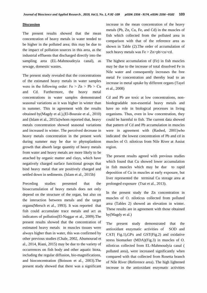
The GSH concentration in muscles of
Oreochromis niloticus decreased significantly (P
≤0.05) at the polluted area during the two seasons
in comparison with the reference area (Fig.4). The
decrease in winter and summer was 42.4% and
34.6% respectively.
The MDA concentration in white muscles of
Oreochromis niloticusincreased significantly (P
≤0.05) at the polluted area during the two seasons
in comparison with the reference area (Fig.4). The
increase in winter was 2.3 fold, and 2.0fold in
summer.
*
*
* *
Figure (3): GPx and GST activities (U/gm wet tissue) in white muscles of O. niloticus collected
from both areas (Mean±S.E).
*, Significant difference in comparison with the reference area (P < 0.05)
00
.20
.40
.6
Summer Winter
GST
Act
ivit
y (
U/g
m t
issu
e)
ReferenceareaPollutedarea
02
04
06
0
Summer Winter
GP
x A
ctiv
ity
(U/g
m t
issu
e)
ReferenceareaPollutedarea
*
*
*
*

101 Journal of Bioscience and Applied Research , 2019, Vol.5, No. 1, P.92 -109 pISSN: 2356 - 9174, eISSN: 2356 –9182
Comet assay
The results obtained in the current study using the
comet assay(Table 3) revealed that during both
seasons, the average scores for DNA damage in O.
niloticus from the polluted area (EL-Mahmoudyia
canal) were statistically significant (p<0.05) when
compared with that of reference area ( Rosetta
branch of Nile River ). There was a predominance
of comets in Classes 2 and 3, with medium and
high DNA damage in cells from fish collected
from the polluted area (Fig.5). The results revealed
that blood cells from fish collected from reference
area showed lower comet score than that collected
from the polluted area (Fig.6).
Table (3): Visual score of DNA damage in fish samples.
Values are expressed as mean ± S.E
*: Significant difference in comparison with the reference area (P < 0.05)
Season Area No. of cells
Class**
DNA
damaged
cells%
(Mean±SEM)
Analyzed* Comets 0 1 2 3
Summer Reference 300 41 259 24 11 6 13.67±1.53
Polluted 300 68 232 33 24 11 22.72±1.15*
Winter Reference 300 53 247 31 14 7 17.67±1.52
Polluted 300 70 230 27 29 14 23.33±1.50*
*
* *
*
Figure(4): GSH content (mmol/gm wet tissue) and MDA concentration (nmol/gm wet tissue) in muscles of
O. niloticus collected from both areas (Mean±S.E).
*,: Significant difference in comparison with the reference area (P < 0.05)
0
0.5
1
1.5
2
2.5
Summer Winter
GSH
co
nte
nt
( m
mo
l/gm
tiss
ue
) ReferenceareaPollutedarea
0
1
2
3
4
5
6
Summer Winter
MD
A C
on
cen
trat
ion
(n
mo
l/gm
ti
ssu
e)
Reference areaPollutedarea
*
* *
*

102 Journal of Bioscience and Applied Research , 2019, Vol.5, No. 1, P.92 -109 pISSN: 2356 - 9174, eISSN: 2356 –9182
Figure 5: Visual score of normal DNA (class 0) and comet (class 1, class 2) using comet assay
in fish cells collected from Rosetta branch on Nile River
Figure6: Visual score of comet (class1, class 2, class 3) using comet assay in fish cells
collected from EL-Mahmoudeyia canal
103 Journal of Bioscience and Applied Research , 2019, Vol.5, No. 1, P.92 -109 pISSN: 2356 - 9174, eISSN: 2356 –9182
Discussion
The present results showed that the mean
concentration of heavy metals in water tended to
be higher in the polluted area; this may be due to
the impact of pollution sources in this area, as the
industrial effluents that discharged directly into the
sampling area (EL-Mahmoudeyia canal), as
sewage, domestic wastes.
The present study revealed that the concentrations
of the estimated heavy metals in water samples
were in the following order: Fe ˃ Zn ˃ Pb ˃ Cu
and Cd. Furthermore, the heavy metal
concentrations in water samples demonstrated
seasonal variations as it was higher in winter than
in summer. This in agreement with the results
obtained by(Magdy et al.),(El-Bouraie et al., 2010)
and (Islam et al., 2015a)whom reported that, heavy
metals concentration showed seasonal variations
and increased in winter. The perceived decrease in
heavy metals concentration in the present work
during summer may be due to phytoplankton
growth that absorb large quantity of heavy metals
from water and heavy metals are more likely to be
attached by organic matter and clays, which have
negatively charged surface functional groups that
bind heavy metal that are positively charged and
settled down in sediments. (Islam et al., 2015b)
Preceding studies presented that the
bioaccumulation of heavy metals does not only
depend on the structure of the organ, but also on
the interaction between metals and the target
organs(Mersch et al., 1993). It was reported that
fish could accumulate trace metals and act as
indicators of pollution(El-Naggar et al., 2009).The
present results showed that the concentration of
estimated heavy metals in muscles tissues were
always higher than in water, this was confirmed by
other previous studies (Chale, 2002, Abumourad et
al., 2014, Riani, 2015) may be due to the variety of
occurrences on fish body and other aquatic biota,
including the regular diffusion, bio-magnifications,
and bioconcentration (Boisson et al., 2003).The
present study showed that there was a significant
increase in the mean concentration of the heavy
metals (Pb, Zn, Cu, Fe, and Cd) in the muscles of
fish which collected from the polluted area in
comparison with that of the reference area as
shown in Table (2).The order of accumulation of
such heavy metals was Fe > Zn>pb>cu>cd.
The highest accumulation of (Fe) in fish muscles
may be due to the increase of total dissolved Fe in
Nile water and consequently increases the free
metal Fe concentration and thereby lead to an
increase in metal uptake by different organs (Tayel
et al., 2008)
Cd and Pb are toxic at low concentrations, non-
biodegradable non-essential heavy metals and
have no role in biological processes in living
organisms. Thus, even in low concentration, they
could be harmful to fish. The current data showed
that pattern of Cd and Pb accumulation in muscles
were in agreement with (Rashed, 2001)who
indicated the lowest concentration of Pb and cd in
muscles of O. niloticus from Nile River at Assiut
region.
The present results agreed with previous studies
which found that Cu showed lower accumulation
in fish muscles which may be due to rapid
deposition of Cu in muscles at early exposure, but
liver represented the terminal Cu storage area at
prolonged exposure (Tsai et al., 2013).
In the present study the Zn concentration in
muscles of O. niloticus collected from polluted
area (Tables 2) showed an elevation in winter.
These results are in agreement with those obtained
by(Magdy et al.)
The present study demonstrated that the
antioxidant enzymatic activities of SOD and
CAT( Fig.1),GPx and GST(Fig.2) and oxidative
stress biomarker (MDA)(Fig.3) in muscles of O.
niloticus collected from EL-Mahmoudyia canal (
polluted area), were increased significantly when
compared with that collected from Rosetta branch
of Nile River (Reference area). The high lightened
increase in the antioxidant enzymatic activities

104 Journal of Bioscience and Applied Research , 2019, Vol.5, No. 1, P.92 -109 pISSN: 2356 - 9174, eISSN: 2356 –9182
demonstrated the adaptive responses of fish to the
oxidative damage caused by the generated reactive
oxygen species from exposure to water pollutants
(dos Santos Carvalho et al., 2012).
The current study revealed that the antioxidant
enzymatic activities of (SOD, CAT, GPx and
GST) as well as oxidative stress biomarker
(MDA), were increased in winter in comparison to
that in summer, in agreement with (Magdy et al.).
This explained that, more free radical are produced
at lower temperature, which results in oxidative
damage in tissues exposed to pollutants and
increased antioxidant enzymes expression to
protect the exposed tissues against the oxidative
damage of free radicals.
The variation in the precedent antioxidants
enzymatic activities and oxidative stress biomarker
MDA during winter and summer may be due to
decreased ROS elimination systems at lower
temperature (Lushchak, 2011) , also fish at lower
temperatures have higher polyunsaturated fatty
acids content in their membrane lipids to maintain
its function which increase the risk for lipid
peroxidation and oxidative damage affecting its
membrane integrity(Guderley and St-Pierre, 2002).
The pronounced elevation of the antioxidants
enzymatic activity of SOD, CAT, GPx and GST in
the current study was in agreement with the results
presented in gold fish, Carassius auratus(Aliko et
al., 2018), in O. niloticus (Magdy et al 2017).
The highlighted elevation of the enzymatic
activities of SOD, CAT, GST and GPx in the
muscles of O.niloticus collected from the polluted
area could be due to the high accumulation level of
the estimated heavy metals ( Pb, Zn Cu, Fe and
Cd) in fish muscles that generate free radicals
which disturb the pro-oxidants/antioxidants
balance creating a situation of oxidative stress in
muscles tissues which in response to that situation
increase the expression of precedent antioxidants
enzymes as a type of defense mechanism against
the free radicals produced by the increased level
of heavy metals accumulation in the fish muscles.
Our results are in agreement with the findings of
Farombi et al (Farombi et al., 2007b)
GSH is well known as a metal protective molecule
that has the ability to change the tendency to metal
(Killa and Rabenstein, 1989). GSH is taken into
consideration as a first line of protection against
metals via chelating and detoxifying them,
scavenging and detoxification of oxyradicals via
reactions catalysed by GPx (Sies, 1999). The
current results showed a significant decrease in the
GSH content in fish muscles collected from the
polluted area in comparison with that of fish
collected from the reference area, this could be
linked to the increased GST activity which uses
GSH for converting xenobiotics into more
hydrophilic compounds. Other factors such as
GST may induce the consumption of GSH (Eroglu
et al., 2015). Some studies reported the GSH
reduction with a significant increase in GST
activity, which is in accordance with the present
data(Zhang et al., 2004) , others reported that
GSH levels decrease in response to oxidative
stress after initial exposure to toxicants and can
also increase as a compensatory action (Guyonnet
et al., 1999, Tan et al., 1998)
Malondialdehyde (MDA) is widely used as an
indicator of lipid peroxidation and oxidative stress
in cells and tissues(Esterbauer et al., 1991).The
herein results revealed that there was a significant
increase in MDA level in muscles collected from
the polluted area when compared with that of the
reference area, the increased level of MDA in the
polluted area may be resulted from the high levels
of ROS produced by the accumulated heavy
metals in fish (Dautremepuits et al., 2002).Same
results were recorded in C. gariepinus collected
from heavily polluted river nearby major industries
in Nigeria(Farombi et al., 2007a).
It has been reported that there is an association
between DNA damage in aquatic animals and
aquatic environment pollution (Klobučar et al.,
2010, Fatima et al., 2014). Fish is the best
accessible vertebrate model to estimate potential

105 Journal of Bioscience and Applied Research , 2019, Vol.5, No. 1, P.92 -109 pISSN: 2356 - 9174, eISSN: 2356 –9182
risks, due to their capability to metabolize and
accumulate contaminants in their
bodies(Diekmann et al., 2004), furthermore, fish
blood erythrocytes are the most suitable for
DNA damage analyses as it displays the complete
health status of the organism so fish blood cells
have attained specific attention as their
erythrocytes are nucleated
and, therefore, suitable for acquiring nucleoids
for single cell gel electrophoresis(Costa et al.,
2011).The herein results showed that there is a
significant increase in the average scores for DNA
damage in O. niloticus from the polluted area
when compared with that of from the reference
area in agreement with(Badr et al., 2014) who had
found that the DNA strand breaks increased
statistically in tilapia collected from polluted area
compared to fish collected from reference area.
During the present study significantly higher
percentages of DNA damage in O. niloticus
indicate its higher susceptibility to metals. In
addition, metal accumulation causes an elevation
in production of ROS such as hydrogen peroxide,
superoxide radical, hydroxyl radical which results
in oxidative stress in fish(Dautremepuits et al.,
2002), and hence more MDA is produced that
capable of attacking biomolecules such as protein
and DNA producing a variety of adducts (Adeyeye
et al., 1996) such as that with DNA bases which
considered as mutagenic (Becker et al., 2008)
Conclusion
Continuous discharge of industrial effluents
without treatment into fresh canals can cause
physiological changes in fish as Nile tilapia and
hence the human health. Fish biomarkers as
genotoxic damage in peripheral blood erythrocytes
and oxidative stress in Nile tilapia are necessary to
assess the impact of xenobiotic compounds (i.e.
heavy metals) on fish. Moreover, the treatment of
all kinds of waste waters, sewage and agricultural
wastes is recommended before discharge into the
aquatic systems.
References
ABUMOURAD, I., ABBAS, W., AUTHMAN, M.
& GIRGIS, S. 2014. Environmenta l
impact of heavy metal pollution on
metallothionein expression in Nile Tilapia.
Res J Pharm Biol Chem Sci, 5, 998-1005.
ADEYEYE, E. I., AKINYUGHA, N. J., FESOBI,
M. E. & TENABE, V. O. 1996.
Determination of some metals in Clarias
gariepinus (Cuvier and Vallenciennes),
Cyprinus carpio (L.) and Oreochromis
niloticus (L.) fishes in a polyculture fresh
water pond and their environments.
Aquaculture, 147, 205-214.
AEBI, H. 1984. [13] Catalase in vitro. Methods in
enzymology. Elsevier.
ALIKO, V., QIRJO, M., SULA, E., MORINA, V.
& FAGGIO, C. 2018. Antioxidant defense
system, immune response and erythron
profile modulation in gold fish, Carassius
auratus, after acute manganese treatment.
Fish Shellfish Immunol, 76, 101-109.
BADR, A. M., MAHANA, N. A. & EISSA, A.
2014. Assessment of Heavy Metal Levels
in Water and Their Toxicity in Some
Tissues of Nile Tilapia
(Oreochromisniloticus) in River Nile
Basin at Greater Cairo, Egypt. Global
Veterinaria, 13, 432-443.
BECKER, J., FULLNER, K., SEELING, U.,
FORNALCZYK, G. & KUHN, A. 2008.
Measuring magnesium, calcium and
potassium isotope ratios using ICP-QMS
with an octopole collision cell in tracer
studies of nutrient uptake and
translocation in plants. Analytical and
bioanalytical chemistry, 390, 571-578.
BEGUM, A., AMIN, M. N., KANECO, S. &
OHTA, K. 2005. Selected elemental
composition of the muscle tissue of three
species of fish, Tilapia nilotica, Cirrhina
mrigala and Clarius batrachus, from the
fresh water Dhanmondi Lake in
Bangladesh. Food chemistry, 93, 439-443.
BEUTLER, E., DURON, O. & KELLY, M. 1963.
Colorimetric method for determination of
106 Journal of Bioscience and Applied Research , 2019, Vol.5, No. 1, P.92 -109 pISSN: 2356 - 9174, eISSN: 2356 –9182
glutathione reduced. J. Lab. Clin. Med, 61,
882.
BOISSON, F., COTRET, O., TEYSSIE, J.-L., EL-
BARADEı, M. & FOWLER, S. 2003.
Relative importance of dissolved and food
pathways for lead contamination in
shrimp. Marine Pollution Bulletin, 46,
1549-1557.
CENSI, P., SPOTO, S., SAIANO, F.,
SPROVIERI, M., MAZZOLA, S.,
NARDONE, G., DI GERONIMO, S.,
PUNTURO, R. & OTTONELLO, D.
2006. Heavy metals in coastal water
systems. A case study from the
northwestern Gulf of Thailand.
Chemosphere, 64, 1167-1176.
CHALE, F. 2002. Trace metal concentrations in
water, sediments and fish tissue from Lake
Tanganyika. Science of the total
environment, 299, 115-121.
COLLINS, A., DUŠINSKá, M., FRANKLIN, M.,
SOMOROVSKá, M., PETROVSKá, H.,
DUTHIE, S., FILLION, L.,
PANAYIOTIDIS, M., RAŠLOVá, K. &
VAUGHAN, N. 1997. Comet assay in
human biomonitoring studies: reliability,
validation, and applications.
Environmental and molecular
mutagenesis, 30, 139-146.
COSTA, P. M., NEUPARTH, T. S., CAEIRO, S.,
LOBO, J., MARTINS, M., FERREIRA,
A. M., CAETANO, M., VALE, C.,
DELVALLS, T. A. & COSTA, M. H.
2011. Assessment of the genotoxic
potential of contaminated estuarine
sediments in fish peripheral blood:
laboratory versus in situ studies.
Environmental Research, 111, 25-36.
DAUTREMEPUITS, C., BETOULLE, S. &
VERNET, G. 2002. Antioxidant response
modulated by copper in healthy or
parasitized carp (Cyprinus carpio L.) by
Ptychobothrium sp.(Cestoda). Biochimica
et Biophysica Acta (BBA)-General
Subjects, 1573, 4-8.
DAUTREMEPUITS, C., PARIS-PALACIOS, S.,
BETOULLE, S. & VERNET, G. 2004.
Modulation in hepatic and head kidney
parameters of carp (Cyprinus carpio L.)
induced by copper and chitosan.
Comparative Biochemistry and Physiology
Part C: Toxicology & Pharmacology, 137,
325-333.
DIEKMANN, M., WALDMANN, P.,
SCHNURSTEIN, A., GRUMMT, T.,
BRAUNBECK, T. & NAGEL, R. 2004.
On the relevance of genotoxicity for fish
populations II: genotoxic effects in
zebrafish (Danio rerio) exposed to 4-
nitroquinoline-1-oxide in a complete life-
cycle test. Aquatic toxicology, 68, 27-37.
DOS SANTOS CARVALHO, C., BERNUSSO,
V. A., DE ARAúJO, H. S. S.,
ESPíNDOLA, E. L. G. & FERNANDES,
M. N. 2012. Biomarker responses as
indication of contaminant effects in
Oreochromis niloticus. Chemosphere, 89,
60-69.
EHSAEI, M., KHAJAVI, M., ARJMAND, M. H.,
ABUEE, M. A., GHAYOUR-
MOBARHAN, M. & ALAMDARI, D. H.
2015. Prooxidant–antioxidant balance in
patients with traumatic brain injury. Acta
Neurologica Belgica, 115, 69-73.
EL-BOURAIE, M., EL-BARBARY, A., YEHIA,
M. & MOTAWEA, E. 2010. Heavy metal
concentrations in surface river water and
bed sediments at Nile Delta in Egypt. Suo,
61, 1-12.
EL-NAGGAR, A. M., MAHMOUD, S. A. &
TAYEL, S. I. 2009. Bioaccumulation of
some heavy metals and histopathological
alterations in liver of Oreochromis
niloticus in relation to water quality at
different localities along the River Nile,
Egypt. World Journal of Fish and Marine
Sciences, 1, 105-114.
EL SAFTY, A. & SIHA, M. 2013.
ENVIRONMENTAL AND HEALTH
IMPACT OF COAL USE FOR ENERGY

107 Journal of Bioscience and Applied Research , 2019, Vol.5, No. 1, P.92 -109 pISSN: 2356 - 9174, eISSN: 2356 –9182
PRODUCTION. Egyptian Journal of
Occupational Medicine, 37, 181-194.
EROGLU, A., DOGAN, Z., KANAK, E., ATLI,
G. & CANLI, M. 2015. Effects of heavy
metals (Cd, Cu, Cr, Pb, Zn) on fish
glutathione metabolism. Environmental
Science and Pollution Research, 22, 3229-
3237.
ESTERBAUER, H., SCHAUR, R. J. &
ZOLLNER, H. 1991. Chemistry and
biochemistry of 4-hydroxynonenal,
malonaldehyde and related aldehydes.
Free radical Biology and medicine, 11,
81-128.
FAIRBAIRN, D. W., OLIVE, P. L. & O'NEILL,
K. L. 1995. The comet assay: a
comprehensive review. Mutation
Research/Reviews in Genetic Toxicology,
339, 37-59.
FAROMBI, E., ADELOWO, O. & AJIMOKO, Y.
2007a. Biomarkers of oxidative stress and
heavy metal levels as indicators of
environmental pollution in African cat fish
(Clarias gariepinus) from Nigeria Ogun
River. International Journal of
Environmental Research and Public
Health, 4, 158-165.
FAROMBI, E. O., ADELOWO, O. A. &
AJIMOKO, Y. R. 2007b. Biomarkers of
oxidative stress and heavy metal levels as
indicators of environmental pollution in
African cat fish (Clarias gariepinus) from
Nigeria Ogun River. Int J Environ Res
Public Health, 4, 158-65.
FATIMA, M., USMANI, N., HOSSAIN, M. M.,
SIDDIQUI, M. F., ZAFEER, M. F.,
FIRDAUS, F. & AHMAD, S. 2014.
Assessment of genotoxic induction and
deterioration of fish quality in commercial
species due to heavy-metal exposure in an
urban reservoir. Archives of environmental
contamination and toxicology, 67, 203-
213.
FLEGAL, A. & PATTERSON, C. 1983. Vertical
concentration profiles of lead in the
Central Pacific at 15 N and 20 S. Earth
and Planetary Science Letters, 64, 19-32.
FORSTNER, U. GTW Wittman. 1983. Metal
Pollution In The Aquatic Environment.
Springer-Verlag, Berlin.
GHORBANI, M. & MIRAKABAD, H. Z. 2010.
Factors influencing on trout production in
Khorasan Razavi Province. Trends Agric.
Econ, 3, 19-27.
GOSMARO, F., BAGNATI, M., BERTO, S.,
BELLOMO, G. & PRENESTI, E. 2013.
Measurement of total antioxidant capacity
of human plasma: Setting and validation
of the CUPRAC–BCS method on routine
apparatus ADVIA 2400. Talanta, 115,
526-532.
GUDERLEY, H. & ST-PIERRE, J. 2002. Going
with the flow or life in the fast lane:
contrasting mitochondrial responses to
thermal change. Journal of Experimental
Biology, 205, 2237-2249.
GUYONNET, D., SIESS, M.-H., LE BON, A.-M.
& SUSCHETET, M. 1999. Modulation of
phase II enzymes by organosulfur
compounds from allium vegetables in rat
tissues. Toxicology and applied
pharmacology, 154, 50-58.
HABIG, W. H., PABST, M. J. & JAKOBY, W. B.
1974. Glutathione S-transferases the first
enzymatic step in mercapturic acid
formation. Journal of biological
Chemistry, 249, 7130-7139.
ISLAM, M. S., AHMED, M. K., HABIBULLAH-
AL-MAMUN, M. & MASUNAGA, S.
2015a. Assessment of trace metals in fish
species of urban rivers in Bangladesh and
health implications. Environmental
toxicology and pharmacology, 39, 347-
357.
ISLAM, M. S., AHMED, M. K.,
RAKNUZZAMAN, M., HABIBULLAH-
AL-MAMUN, M. & MASUNAGA, S.
2015b. Metal speciation in sediment and
their bioaccumulation in fish species of
three urban rivers in Bangladesh. Archives

108 Journal of Bioscience and Applied Research , 2019, Vol.5, No. 1, P.92 -109 pISSN: 2356 - 9174, eISSN: 2356 –9182
of environmental contamination and
toxicology, 68, 92-106.
KEI, S. 1978. Serum lipid peroxide in
cerebrovascular disorders determined by a
new colorimetric method. Clinica chimica
acta, 90, 37-43.
KILLA, H. & RABENSTEIN, D. L. 1989.
Separation and detection of bis (alkylthio)
selenides of penicillamine and glutathione
by liquid chromatography with
electrochemical detection. Journal of
chromatography, 465, 359-368.
KIME, D. E. 1995. The effects of pollution on
reproduction in fish. Reviews in Fish
Biology and Fisheries, 5, 52-95.
KLOBUČAR, G. I., ŠTAMBUK, A., PAVLICA,
M., PERIĆ, M. S., HACKENBERGER, B.
K. & HYLLAND, K. 2010. Genotoxicity
monitoring of freshwater environments
using caged carp (Cyprinus carpio).
Ecotoxicology, 19, 77.
KOBAYASHI, H. 1995. A comparison between
manual microscopic analysis and
computerized image analysis in the single
cell gel electrophoresis. MMS Commun.,
3, 103-115.
LAI, H. & SINGH, N. P. 1995. Acute
low‐intensity microwave exposure
increases DNA single‐strand breaks in rat
brain cells. Bioelectromagnetics, 16, 207-
210.
LOPES, P. A., PINHEIRO, T., SANTOS, M. C.,
DA LUZ MATHIAS, M., COLLARES-
PEREIRA, M. J. & VIEGAS-CRESPO, A.
M. 2001. Response of antioxidant
enzymes in freshwater fish populations
(Leuciscus alburnoides complex) to
inorganic pollutants exposure. Science of
the total environment, 280, 153-163.
LUSHCHAK, V. I. 2011. Environmentally
induced oxidative stress in aquatic
animals. Aquatic toxicology, 101, 13-30.
MACFARLANE, G. & BURCHET, M. 2000.
Cellular distribution of Cu, Pb, and Zn in
the Grey Mangrove, Avicemnia marina
(Forsk.), Vierh Aquatic Botanic 68: 45-59.
MAGDY, T., NAHED, S. G., NASR, A. &
SALLY, S. M. Antioxidant Defense
System Alternations in Fish as a Bio-
Indicator of Environmental Pollution.
MERSCH, J., DUBOST, N. & PIHAN, J. Year.
Comparison of several inert and biological
substrata to assess the trace metal
contamination in the reservoir of the
nuclear power plant in Cattenom, France.
In: Annales de limnologie. Toulouse,
1993. 325-337.
NAGARANI, N., DEVI, V. J. &
KUMARAGURU, A. 2012. Identification
of DNA damage in marine fish Therapon
jarbua by comet assay technique. Journal
of environmental biology, 33, 699.
NAMMINGA, H. E. Year. Effects of high
discharge and an oil refinery cleanup
operation on heavy metals in water and
sediments in Skeleton Creek. In:
Proceedings of the Oklahoma Academy of
Science, 1976. 133-138.
NRIAGU, J. O. & PACYNA, J. M. 1988.
Quantitative assessment of worldwide
contamination of air, water and soils by
trace metals. nature, 333, 134.
PAGLIA, D. E. & VALENTINE, W. N. 1967.
Studies on the quantitative and qualitative
characterization of erythrocyte glutathione
peroxidase. The Journal of laboratory and
clinical medicine, 70, 158-169.
PITT, R. & BURTON JR, G. A. 2001. Stormwater
effects handbook: A toolbox for watershed
managers, scientists, and engineers, CRC
Press.
RASHED, M. 2001. Cadmium and lead levels in
fish (Tilapia nilotica) tissues as biological
indicator for lake water pollution.
Environmental monitoring and
assessment, 68, 75-89.
RIANI, E. 2015. The Effect of Heavy Metals on
Tissue Damage in Different Organs of
Goldfish Cultivated in Floating Fish Net in

109 Journal of Bioscience and Applied Research , 2019, Vol.5, No. 1, P.92 -109 pISSN: 2356 - 9174, eISSN: 2356 –9182
Cirata Reservoir, Indonesia Marine
Science.
SIES, H. 1999. Glutathione and its role in cellular
functions. Free radical Biology and
medicine, 27, 916-921.
TAN, S., SAGARA, Y., LIU, Y., MAHER, P. &
SCHUBERT, D. 1998. The regulation of
reactive oxygen species production during
programmed cell death. The Journal of
cell biology, 141, 1423-1432.
TAYEL, S., YACOUB, A. & MAHMOUD, S.
2008. Histopathological and
haematological responses to freshwater
pollution in the Nile catfish Clarias
gariepinus. Journal of Egyptian Academic
Society for Environmental Development,
9, 43-60.
TICE, R. R., AGURELL, E., ANDERSON, D.,
BURLINSON, B., HARTMANN, A.,
KOBAYASHI, H., MIYAMAE, Y.,
ROJAS, E., RYU, J. C. & SASAKI, Y.
2000. Single cell gel/comet assay:
guidelines for in vitro and in vivo genetic
toxicology testing. Environmental and
molecular mutagenesis, 35, 206-221.
TRIPATHI, L., MISHRA, A., DUBEY, A. &
TRIPATHI, C. 2015. Water pollution
through energy sector. International
Journal of Technology Enhancements and
Emerging Engineering Research, 3, 2347-
4289.
TSAI, J.-W., JU, Y.-R., HUANG, Y.-H., DENG,
Y.-S., CHEN, W.-Y., WU, C.-C. & LIAO,
C.-M. 2013. Toxicokinetics of tilapia
following high exposure to waterborne
and dietary copper and implications for
coping mechanisms. Environmental
Science and Pollution Research, 20, 3771-
3780.
VAN DER OOST, R., GOKSøYR, A.,
CELANDER, M., HEIDA, H. &
VERMEULEN, N. P. 1996.
Biomonitoring of aquatic pollution with
feral eel (Anguilla anguilla) II.
Biomarkers: pollution-induced
biochemical responses. Aquatic
toxicology, 36, 189-222.
VERON, A., LAMBERT, C. E., ISLEY, A.,
LINET, P. & GROUSSET, F. 1987.
Evidence of recent lead pollution in deep
north-east Atlantic sediments. nature, 326,
278.
VITOŠEVIĆ, B., SAMARDŽIĆ, S.,
ANTONIJEVIĆ, V. & JAKOVLJEVIĆ,
V. 2007. Heavy metals in some imported
food products and their potential toxic
implications. Medicus, 8, 62-66.
YU, R. 2000. Cadmium and DNA damage,
oncogene expression as well as cell
apoptosis. Overseas Medicine Hygienics
Fascicule, 27, 359-363.
ZHANG, J., WANG, X., GUO, H., WU, J. &
XUE, Y. 2004. Effects of water-soluble
fractions of diesel oil on the antioxidant
defenses of the goldfish, Carassius
auratus. Ecotoxicology and Environmental
Safety, 58, 110-116.