overview of molecular diagnostics part 1 - laboratory · pdf fileoverview of molecular...
TRANSCRIPT

10/16/2014
1
1
Overview of Molecular DiagnosticsPart 1
Jennifer L. Hunt, MD, MEdAubrey J. Hough Jr, MD, Endowed Professor of PathologyChair of Pathology and Laboratory MedicineUniversity of Arkansas for Medical [email protected]
2
Agenda
•Basic Techniques in Molecular Diagnostics•Difficult starting materials
•Polymerase chain reaction
•Reverse transcriptase – PCR
•Detection methods
•Sequencing
•Fluorescent in situ hybridization
Kary Mullis (1983): Polymerase Chain Reaction (Nobel Prize, 1993)
Watson & Crick (1953): DNA (Nobel Prize, 1962)
Alfred Knudson (1971): Tumor suppressor genes
Frederick Sanger (1975): Sequencing
First human genome sequenced (2000‐2003)
1950
1970
1960 1980
1990
2000
5
Source of DNA
• Blood
• Body fluids
• Cytology samples
• Fresh or frozen cells and tissue
• Paraffin embedded tissue• Cell blocks
6
Potential Issues With Samples
• Sample purity • Normal cell contamination
• Sample size and template concentration
• Template quality and degradation

10/16/2014
2
7
Nucleic Acids in Tissues
What have you done to your specimen?
8
Fixatives Are Our Friend
• Stabilize cell morphology and tissue architecture
• Disable proteolytic enzymes
• Strengthen samples to withstand further processing and staining
• Protect samples against microbial contamination and decomposition
9
Fixatives in AP
•Cross-linking fixatives•Formaldehyde
•Glutaraldehyde
•Paraformaldehyde
•Precipitating fixatives•Alcohol
•Methanol
•Acetone
12
DNA Damage in Formalin
300 to 400Basepair Fragments

10/16/2014
3
13
Tissue Fixation
Fixative Components DNA Quality RNA Quality
Unbuffered formalin Formaldehyde Poor Poor
Buffered formalin FormaldehydePhosphate buffers
Fair Fair
Ethanol 70% - 100% Good Good
Decalcifying acids Various acids Poor Poor
Bouin’s Picric acidFormaldehydeGlacial acetic acid
Poor Poor
Mercury solutions Mercuric chloride Sodium acetate
Poor Poor
14
DNA Degradation
69%
17%
5%0%
10%
20%
30%
40%
50%
60%
70%
152 b 268 bp 676 bp
Gillio-Tos, Pathology, 39:345, 2007
25 year-old tissue blocks
15
Agenda
•Basic Techniques in Molecular Diagnostics•Difficult starting materials
•Polymerase chain reaction
•Reverse transcriptase – PCR
•Detection methods
•Sequencing
•Fluorescent in situ hybridization
16
Analysis of DNA
17
Analysis of DNA
•Fresh tissue, blood, or cytology fluids
•Frozen tissue
•Fixed tissue
•Paraffin embedded tissue•Archival
18
Polymerase Chain Reaction
•Discovered in 1983 (Kary Mullis, Cetus)
•Patented technique
•Patent sold to Roche in 1991 for $300 Million

10/16/2014
4
19
•Problems before PCR•Quantity of DNA was small
•The target was a tiny fraction of all DNA
Polymerase Chain Reaction
Quiz
22
•PCR enabled•Amplification for quantity
•Target specific enrichment
Polymerase Chain Reaction
23
•Template DNA
•Primers
•Taq enzyme
•Nucleotide building blocks
Polymerase Chain Reaction
24
Primer Design
•Primers•Short DNA sequence (18-24 nucleotides)
• Forward and Reverse•“Sense & Antisense”

10/16/2014
5
25
Primer Design
26
Primer Binding
27
Polymerase
•Taq Enzyme•Thermus Aquaticus
•DNA polymerase
•Heat activated
TaqTaqTaqTaqTaqTaqTaqTaq
C G AT
C
G
G
G
T
TT
A
A
A
A
CC CTTT AA GG C
G
G
G
T
T
A
A
A
C CGG AAT A G T T T C C CA A G T
29
PCR: Thermal Cyclers
30
Cycle 2
DenaturationCycle 1
DenaturationAnnealingExtension
90
72
55
DenaturationAnnealingExtension Denaturation
A
G
C
T A
G
T
AG
C
T
A
G
C
T
A
GC
T
AG
C
T
AG
C
TAA
G
T
G
C T
A
G
C
T
Annealing

10/16/2014
6
31
PCR Amplification
Time
PCR Product Exponential phase
Plateau phase
32
Agenda
•Basic Techniques in Molecular Diagnostics•Difficult starting materials
•Polymerase chain reaction
•Detection methods
•Reverse transcriptase – PCR
•Sequencing
•Fluorescent in situ hybridization
33
PCR Product Analysis
• What do we do after PCR?• Detect the sequence in the product
• The actual sequence (Sequencing)
• Differences that cause altered migration (MSI)
• Detect the amount of product• Quantitative (real time-PCR)
• Semi-quantitative (LOH)
34
PCR product detection
•Gel: Agarose or polyacrylamide
•Capillary electrophoresis
•Quantitative PCR
35
Agarose Gel Electrophoresis
36
Capillary Electrophoresis

10/16/2014
7
37
Capillary Electrophoresis
38
Capillary Gel Electrophoresis
2000
1800
1600
1400
Size of PCR product (basepairs)aaaaaaaaaa
aaaaaaaa
Relative amount of PCR product
41
Quantitative Real-Time PCR(not RT-PCR)
• Polymerase chain reaction with a reporter• Double stranded DNA binding dye
• Fluorescent reporter probe
Quantitative PCR
Fluorescence
Cycle Number

10/16/2014
8
Quantitative PCR
Fluorescence
Cycle Number
Fluorescent Reporter Probe
45
Agenda
•Basic Techniques in Molecular Diagnostics•Difficult starting materials
•Polymerase chain reaction
•Reverse transcriptase – PCR
•Detection methods
•Sequencing
•Fluorescent in situ hybridization
46
Nucleotide
Ribose
Nucleo
side
H
CH2
H
HH
H
O
3’
5’
O
NH2
Cytosine
OH
RNA: Bases (A, C, G, U)
47
Gene Structure and Translation
mRNA
DNA
Protein
Intron 1 Intron 2 Intron 3
EX
ON
2
EX
ON
3
EX
ON
4
EX
ON
1
5’ UTRPromoter
3’ UTR
EX
ON
4
EX
ON
3
EX
ON
2
EX
ON
1
48
Reverse Transcriptase – PCR
Extract RNAfrom sample
Reverse TranscribemRNA to cDNA
Perform PCR on
cDNA sample
Analyze PCR
products

10/16/2014
9
49
Primers for Reverse Transcriptase
• Random hexamers• Anneals randomly
• Oligo dT • Anneals to polyA tail of mRNA
• Product specific primers• Anneals to location of interest
50
mRNA
Primers for Reverse Transcriptase
AAAAAA
TTTTTT
51
RT
Reverse Transcriptase - PCR
CC C CGGG TT TTT AAAAA GGC CGG AAU A G U U U C C CA A G U
cDNA
mRNA
52
RT-PCR Product Analysis
• What do we do after RT-PCR?• Detect qualitative product
• Abnormal transcripts (Translocation analysis)
• Detect the amount of product, which represents the amount of expression of a gene• Quantitative (Real time or quantitative RT-PCR)
53
Detecting Translocations
• DNA based PCR testing
• RNA based RT-PCR testing
• Fluorescent in situ hybridization (FISH)
54
Gene 1DNA
Gene 2DNA
Fusion mRNA
X
Product
X

10/16/2014
10
55
Agenda
•Basic Techniques in Molecular Diagnostics•Difficult starting materials
•Polymerase chain reaction
•Reverse transcriptase – PCR
•Detection methods
•Sequencing
•Fluorescent in situ hybridization
56
Mutation Detection Techniques
• PCR detection and screening methods• Heteroduplex formation
• Allele specific PCR
• SSCP
• Full sequencing methods• Sanger sequencing
• Single base extension
• Pyrosequencing
• Next generation sequencing
57
Detecting Point Mutations
• Full Gene Sequencing Approaches• Dideoxy sequencing (“first generation”)
• Sanger sequencing
• Single base extension sequencing (“SNaPshot”)
58
Sanger Sequencing: The Gold Standard
Template DNA
PCR Product
Sequence
PCR with primers
Sequencing reaction
59
Frederick Sanger (1918 - )
• University of Cambridge, England
• Nobel Laureate twice• Insulin and unique protein structures (1958)
• Dideoxy method of sequencing “Sanger method” (1980)
60
Sequencing Reaction
• Primer
• Polymerase
• Labeled bases (A T C G)• Abundant normal nucleotides
• Low concentration modified bases • Dideoxy instead of deoxy: stops extension (chain
terminator)
• Added fluorescent tag: product visualization

10/16/2014
11
Nucleotide
Deoxyribose
Nucleo
side
H
CH2
H
H
HH
H
5’
O
Cytosine
Dideoxynucleotide Chain Terminator
NH2
H
P
P
P
P
P
Dideoxynucleotide Nucleotides
C G ATT
A
CC CTTT AA GG
G
G
G
T
T
A
A
A
C
Sanger Sequencing
C CGG AAT A G T T T C C CA A G T

10/16/2014
12
Sanger Sequencingttgatttgagattaatctacttttgatttgagattaatctactttgatttgagattaatctacttgatttgagattaatctattgatttgagattaatctttgatttgagattaatcttgatttgagattaatttgatttgagattaattgatttgagattattgatttgagattttgatttgagatttgatttgagattgatttgagttgatttgattgatttgttgatttttgattttgatttgattgttt
A G C T
T T G A T T T G A G A T T A A T C T A C T T
ttgatTttgaTttgA
CC CTTT AA GG
SNaPshot Sequencing
G
T
C
A
C CGG AAT A G T T T C C CA A G T
Patient’s tumorNormal Control
PIK3CA Gene
72
Pitfalls in Sequencing
• Performance characteristics• Sensitivity is variable
• Deletion detection not optimal
• Technical• Expensive and labor intensive
• Time consuming
• Tissue fixation may be an issue

10/16/2014
13
Normal and Mutant
74
3/8
2/8
Normal Contamination
100%
75%
50%
Mt Wt Mt Wt Mt Wt Mt Wt
Mt Wt Mt Wt Mt Wt Wt
Mt Wt Mt Wt
Wt
WtWtWtWt
4/8
75
Allele Specific PCR
CC CTTT AA GGC CGG AAT A G T T T C C CA A G T
CC CTCT AA GGC CGG GAT A G T T T C C CA A G T
Normal Sequence (A allele)
Mutant Sequence (G allele)
Allele Specific PCR
Allele Specific PCR for BRAF Mutation
Normal BRAF
Mutant BRAF
BRAF gene mutation
Pyrosequencing for BRAF Mutation
Normal BRAF
Mutant BRAF
BRAF gene mutation
10/16/2014
14
79
Agenda
•Basic Techniques in Molecular Diagnostics•Difficult starting materials
•Polymerase chain reaction
•Reverse transcriptase – PCR
•Detection methods
•Sequencing
•Fluorescent in situ hybridization
80
Basic Techniques
• Fluorescent in situ hybridization• Quantitative
• Amplification
• Deletion
• Qualitative • Translocation analysis
• Presence foreign nucleic acid (virus)
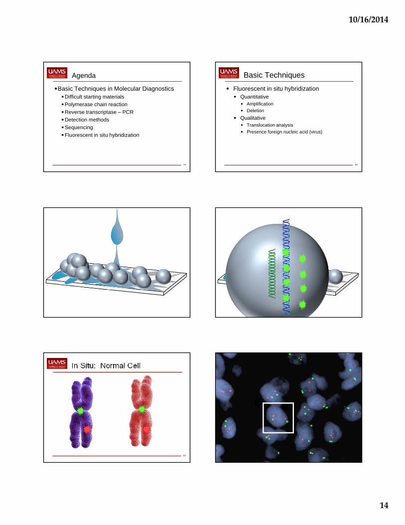
83
In Situ: Normal Cell

10/16/2014
15
85
In Situ for Amplification
86
In Situ for Deletion
87
In Situ for Virus
88
In Situ for Translocations
• Fusion probes• One probe on each
partner
• Both genes must be known
• Will only pick up consistent partner genes
• Break-apart probes• Probes flank the break
point on one partner
• Only one gene must be known
• Will pick up variable translocations
89
Fusion for Translocation
90
Break-Apart for Translocation
10/16/2014
16
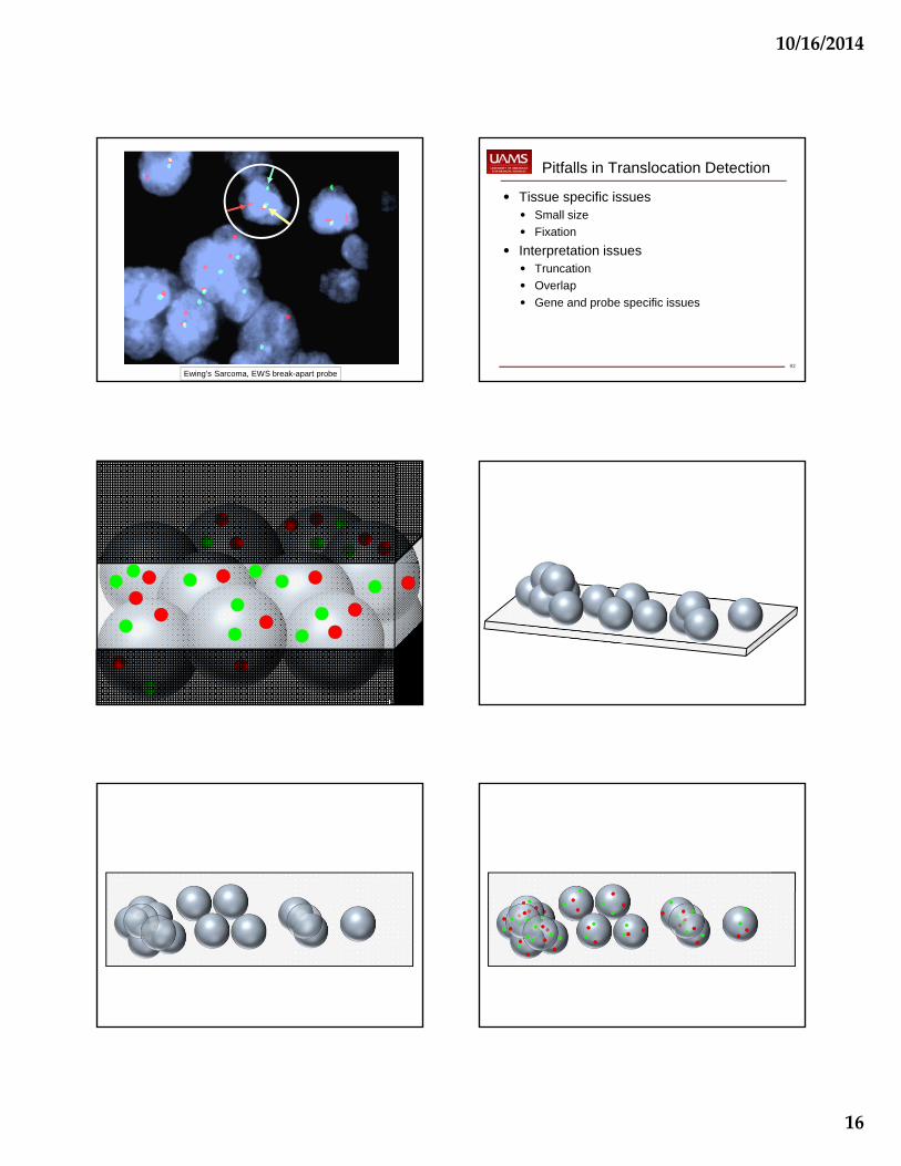
Ewing’s Sarcoma, EWS break-apart probe92
Pitfalls in Translocation Detection
• Tissue specific issues• Small size
• Fixation
• Interpretation issues• Truncation
• Overlap
• Gene and probe specific issues

10/16/2014
17
97
Gene Specific Issues
• RET-PTC and EML4-ALK• Intrachromosomal rearrangements
99
Summary
•Basic Techniques in Molecular Diagnostics•Difficult starting materials
•Polymerase chain reaction
•Reverse transcriptase – PCR
•Detection methods
•Sequencing
•Fluorescent in situ hybridization