overexpression of mutant prp induce amyloidogenesis in ... file · web viewdissociation of prion...
TRANSCRIPT

Dissociation of prion protein amyloid seeding from transmission of a spongiform
encephalopathy
Running Title: PrP misfolding dissociated from infectivity
Pedro Piccardo1,2, Declan King2, Glenn Telling3, Jean C Manson2, and Rona M Barron2#
1Laboratory of Bacterial and TSE Agents, Food and Drug Administration, Kensington,
Maryland, USA,
2Neurobiology Division, The Roslin Institute and R(D)SVS, University of Edinburgh,
Easter Bush, Midlothian, UK,
3Colorado State University, CO, US
corresponding author
Rona M Barron ([email protected])
Tel 44 131 527 4200
Fax 44 131 440 0434
Abstract Word Count : 248
Text Word Count : 4422
1
1
2
3
4
5
6
7
8
9
10
11
12
13
14
15
16
17
18
19
20
21
Abstract
Misfolding and aggregation of proteins is a common pathogenic mechanism of a group of
diseases called proteinopathies. The formation and spread of proteinaceous lesions within
and between individuals was first described in prion diseases and proposed as the basis of
its infectious nature. Recently a similar “prion-like” mechanism of transmission has been
proposed in other neurodegenerative diseases such as Alzheimer’s disease. We
investigated if misfolding and aggregation of corrupted prion protein (PrPTSE) is always
associated with horizontal transmission of disease. Knock-in transgenic mice (101LL)
expressing mutant PrP (PrP-101L) that are susceptible to disease but do not develop any
spontaneous neurological phenotype were inoculated with (i) brain extracts containing
PrPTSE from healthy 101LL mice with PrP plaques in the corpus callosum or (ii) mice
overexpressing PrP-101L with neurological disease, severe spongiform encephalopathy
and formation of proteinase-K-resistant PrPTSE. In all instances, 101LL mice developed
PrP plaques in the area of inoculation and vicinity in the absence of clinical disease or
spongiform degeneration of the brain. Importantly 101LL mice did not transmit disease
on serial passage ruling out the presence of subclinical infection. Thus, in both
experimental models formation of PrPTSE is not infectious. These results have
implications for the interpretation of tests based on the detection of protein aggregates
and suggest that de novo formation of PrPTSE in the host does not always result in a
transmissible prion disease. In addition, these results question the validity of assuming
that all diseases due to protein misfolding can be transmitted between individuals.
2
22
23
24
25
26
27
28
29
30
31
32
33
34
35
36
37
38
39
40
41
42
43
Introduction
Several studies suggest that neurodegenerative diseases due to protein misfolding
(proteinopathies) share a common pathogenesis with seeded aggregation of native
proteins, and the potential for horizontal transmission between individuals. Prion diseases
serve as a paradigm for proteinopathies. The pathologic hallmark of prion diseases is
widespread spongiform encephalopathy with deposition of disease-associated misfolded
prion protein (PrPTSE) which results from the post-translational conversion of a normal
host-encoded cellular isoform of the prion protein (PrPC) (1). Numerous investigators
have concluded that PrPTSE is the sole component of the infectious agent (“prion”) causing
disease in the complete absence of nucleic acid. Studies using 263K hamster scrapie have
shown a strong correlation between PrPTSE and infectivity (1-3). However, prion diseases
can develop with high levels of infectivity and very low levels of PrP deposition and,
conversely, PrPTSE accumulates in the brain in some situations unaccompanied by the
other typical neuropathologic changes or any clinical signs of disease (4-10). Thus, both
the molecular basis of prion propagation between individuals and the mechanism by
which PrPC converts into PrPTSE and spreads from cell to cell in the affected host remain
unclear.
Misfolding and aggregation of other host proteins similar to that seen in prion
diseases, is the molecular hallmark of several neurodegenerative diseases (e.g., β-amyloid
in Alzheimer’s disease (AD), alpha-synuclein in Parkinson’s disease (PD)). It has been
reported that in all proteinopathies, misfolded proteins can elicit seeded aggregation of
native proteins and spread through the brain in a “prion-like” manner (11). This
3
44
45
46
47
48
49
50
51
52
53
54
55
56
57
58
59
60
61
62
63
64
65
66

possibility raises the potential for diseases due to protein misfolding to be transmissible
between individuals. However, there may be significant differences between the
mechanism by which misfolded protein spreads from cell to cell to that in which PrPTSE
acquires infectious properties resulting in horizontal transmission of disease.
Based on the hypothesis that PrPTSE is infectious, conversion of PrPC into PrPTSE
has been analyzed as a model of replication of prion infectivity using several
methodologies including in vitro amplification of PrPTSE (12-14). Recent studies
concluded that the ratio of the infectivity titer to the amount of PrPTSE (specific
infectivity) is much lower when the PrPTSE is generated by amplification in vitro than in
infected brain-derived samples (15). Thus, while misfolded protein is readily generated in
these assays it is clear that not all the misfolded protein is infectious. Similarly, attempts
to produce de-novo infectivity from refolding or fibrillisation of recombinant PrP into
PrPTSE has resulted in poor transmission rates probably due to low levels of infectivity
and often require a passage through transgenic mice or hamsters before disease can be
induced in recipient animals (16, 17). However, others reported that recombinant PrP in
the presence of lipids and RNA can efficiently transmit prion disease to mice suggesting
that co-factors are important for prion infectivity (18).
PrPTSE in the form of PrP-amyloid is consistently deposited in multiple areas of
the cerebrum and cerebellum of patients with Gerstmann-Sträussler-Scheinker (GSS)
disease, a prion disease that is most commonly present in individuals who have a proline
- to - leucine (P-to-L) substitution at codon 102 (GSS P102L) of the prion-protein-
4
67
68
69
70
71
72
73
74
75
76
77
78
79
80
81
82
83
84
85
86
87
88
89

encoding gene (PRNP). In most patients with GSS P102L (“typical” phenotype) brain
lesions include spongiform degeneration and widespread amyloid deposits. However
other patients present with an “atypical” GSS P102L phenotype, with large amounts of
PrP amyloid accumulating in multiple areas of the cerebrum and cerebellum in the
absence of spongiform encephalopathy (19-21). By inoculation of brain extracts from
patients with typical and atypical GSS P102L into gene targeted transgenic mice
expressing the equivalent mutation in murine PrP (101LL) (22), we have demonstrated
differences in the ability of misfolded PrP from these two forms of GSS to generate
disease in these mice, where typical GSS produces high transmission rates but atypical
GSS transmits poorly (7). Unexpectedly, several asymptomatic mice (101LL-8a) which
received atypical GSS P102L inoculum were found on subsequent histological analysis to
have large PrP amyloid plaques only in the corpus callosum (7). However, low
transmission rates and long incubation periods are often observed when attempting to
transmit prion disease to a new species in what is known as “species barrier” (e.g., from
human to mouse), or in models of synthetic prion transmission, raising the possibility that
the apparent absence of transmission is due to low level agent replication and subclinical
infection. This can be addressed by subsequent subpassage in the same mouse line,
where replication of infectivity and disease is observed following adaptation of the agent
to the new species.
We hypothesize that there is a difference between cell to cell spread of misfolded proteins
(described in AD, PD and other diseases) within a host and transmission of an infectious
agent between individuals. Infection is defined as invasion and multiplication of a micro-
5
90
91
92
93
94
95
96
97
98
99
100
101
102
103
104
105
106
107
108
109
110
111
112

organism in body tissues. Disease can arise if the host immune response fails to eradicate
the pathogen and damage is inflicted on the host. However for such organisms to survive
and repeat the infectious cycle in other hosts, they must be able to leave the host and
transmit to a new susceptible individual. In TSE diseases such as scrapie in sheep and
goats, and chronic wasting disease (CWD) in deer, infection appears to be spread by
direct contact between infected animals. In the case of BSE (and vCJD) the infection is
spread via indirect routes in contaminated feed and blood. However in diseases such as
sCJD and GSS, the misfolding of protein is thought to arise “spontaneously” in the brain
in the absence of invasion by an exogenous infectious organism. Spread of these diseases
between individuals does not normally occur and happens only in rare occurrences via
iatrogenic intervention. To explore the relationship between PrPTSE and infectivity, we
performed transmission studies in 101LL mice using as inoculum brain extracts from (i)
healthy 101LL mice with PrP plaques (101LL-8a) and (ii) mice overexpressing PrP-101L
with neurological disease, spongiform degeneration and PrPTSE deposits.
Materials and Methods
Transgenic mouse lines
The 101LL mice generated by gene targeting (expressing wild-type levels of mutant PrP
(PrP-101L) and GSS-22 mice generated by microinjection of fertilized eggs
(overexpressing ~12-fold levels of mutant PrP (PrP-101L) were previously described (22,
23). Previous studies have shown that GSS-22 mice develop neurological signs due to
6
113
114
115
116
117
118
119
120
121
122
123
124
125
126
127
128
129
130
131
132
133
134
overexpression of PrP-101L (23). Conventional wild-type 129Ola mice served as
controls.
Preparation of the inocula and challenge
Inoculation of 101LL-8a brain extracts: A third serial passage of 101LL-8a was carried
out by intracerebral (ic) inoculation of 20 μl of 10% brain homogenate prepared from the
second passage of atypical GSS P102L in 101LL mice (7) into groups (n=24) of 101LL
and 129/Ola control mice (Fig. 1A). The 101LL mouse used to prepare the homogenate
was culled 689 days post inoculation, had no clinical signs or spongiform degeneration,
but had large PrP amyloid plaques in the corpus callosum and vicinity.
Inoculation of GSS-22 brain extracts: Twenty-μl aliquots of 10% homogenates prepared
from brains of two terminally ill GSS-22 mice were inoculated ic into groups (n=24) of
101LL and 129Ola control mice (Fig. 1B). Brain tissues were selected from two
recipient 101LL mice, culled at the end of their expected normal lifespan and inoculated
into groups of 101LL and 129/Ola mice (Fig. 1B). They showed no evidence of
neurological disease or spongiform degeneration but had immunopositive PrP deposits in
the brain. Brains were aseptically removed and inoculations performed as described
previously (22).
Scoring of clinical TSE
Clinical signs of TSE were assessed, and incubation times were calculated according to
previously described protocols (24). Mice were killed either during terminal disease, at
7
135
136
137
138
139
140
141
142
143
144
145
146
147
148
149
150
151
152
153
154
155
156
157

the end of the expected normal lifespan, or earlier if they developed an intercurrent non-
neurological illness. The left half of each brain was fixed in 10% formol saline. Fixed
brain tissue was processed and tissue sections were prepared as described (7). The
remaining half brains were frozen at -70oC for biochemical analysis (22). All mouse
experiments were reviewed and approved by the Local Ethical Review Committee and
performed under License from the UK Home Office in accordance with the United
Kingdom Animal (Scientific Procedures) Act 1986.
Lesion profiles and immunohistochemical analyses
Tissue sections were assessed for spongiform degeneration following previously
described procedures (24). Selected sections were immunostained with monoclonal
antibody (Mab) 6H4 (Prionics, Zurich, Switzerland) recognizing residues 143-151 of
murine PrP (2 μg/ml). Amyloid plaques were visualized with thioflavin-s (25). Selected
sections were probed with anti-PrP polyclonal antibodies (generously provided by B.
Caughey, Rocky Mountain Laboratory, NIAID, NIH, Hamilton, MT); all antibodies were
used at 1:4000 dilution. The selected antibodies are directed against several regions of
PrP: R24 (amino acids 23-47), R30 (amino acids 89-103), R18 (amino acids 142-155)
and R20 (amino acids 218-232). The presence of gliosis was assessed by incubating brain
sections with an antibody to glial fibrillary acidic protein (GFAP; DAKO UK Ltd.,
1.45µg/ml) and anti-Iba1 antibody (Wako Chemicals, 0.05µg/ml-1).
PrP immunoblotting
8
158
159
160
161
162
163
164
165
166
167
168
169
170
171
172
173
174
175
176
177
178
179
Brain tissues from terminally ill GSS-22 and asymptomatic 101LL mice (used for
subpassage) were digested with PK 20μg/ml incubated at 37oC for 1h and prepared for
immunoblotting as described (26). Brain tissue from 129Ola mice inoculated with the
ME7 strain of mouse-adapted TSE (scrapie), and uninoculated 101LL mice were
processed similarly as controls. In brief, samples were electrophoresed on 12%
Tris/glycine gels or 4-20% Tris/glycine gels, immunoblotted, and filters probed with a
series of anti-PrP-monoclonal and polyclonal antibodies directed against various PrP
epitopes. The following antibodies were used: 7A12 (1mg/ml, monoclonal antibody
recognizing amino acids 90-145, 1:20,000), 8H4 (1.5mg/ml, monoclonal antibody
recognizing amino acids 145-180, Sigma; 1:200), R30 (polyclonal antibody recognizing
amino acids 89-103; 1:20,000), Mab6664 (polyclonal antibody recognizing amino acids
79-97, Abcam; 1:2,000), 1E4 (0.5mg/ml monoclonal antibody recognizing amino acids
108-119, Abcam; 1:500), 3C10 (1mg/ml monoclonal antibody recognizing amino acids
97-102, Jena-Bioscience; 1:5,000) or 6G3 (25µg/ml monoclonal antibody recognizing
amino acids 130-150, Santa Cruz; 1:200). Bands were visualized using a secondary
antibody labelled with horseradish peroxidase (HRP) (Jackson Immuno Research
Laboratories, UK) and a chemiluminescent substrate (Roche).
Identification of PrPTSE using ligand-coated PrP antigen-capture plates
Brain and spleen extracts from terminally ill GSS-22 and asymptomatic 101LL mice
inoculated with brain extracts from terminally ill GSS-22 mice were screened by the
IDEXX HerdChek© assay following the manufacturer’s guidelines with minor
modifications to optimize the test for mouse brain and spleen extracts (IDEXX West
9
180
181
182
183
184
185
186
187
188
189
190
191
192
193
194
195
196
197
198
199
200
201
202
Yorkshire, UK). Buffer volumes were adjusted whenever possible to obtain 30% (w/v)
homogenates. Selected brain and spleen homogenates from all test groups were assayed
in duplicate whenever possible both before and after protease digestion using PK
20µg/ml. Tissues were also assayed from well-characterized mouse-adapted scrapie
agents: ME7, 79A and 139A. A 101LL uninfected brain extract was used as negative
control.
Results
Prion infectivity does not replicate in 101LL-8a mice
Inefficient agent replication in a new host can produce a subclinical infection, where no
clinical signs of disease are observed, but tissues are able to transmit disease to recipient
animals on sub-passage. Therefore, to rule out subclinical infection in the 101LL-8a mice
described previously (7) we obtained brains from 101LL-8a mice with no spongiform
degeneration, but large PrP amyloid plaques in the corpus callosum following second
passage of atypical GSS P102L in 101LL mice (7), and performed serial passage into
101LL mice and 129Ola controls (Fig. 1). Except for animals sacrificed due to
intercurrent illness (n:3) all mice remained asymptomatic, and the experiment was
terminated 602 days post-inoculation. Despite the absence of clinical signs, spongiform
degeneration, or widespread gliosis (reactive astrocytes were seen in the periphery of
amyloid plaques) all 101LL mice available for examination showed PrP amyloid plaques
in the corpus callosum and vicinity (Table 1). To the best of our knowledge 101LL-8a
mice represent the first experimental model in which PrPC is consistently converted (over
3 passages) into PrP-amyloid in restricted areas of the brain in the absence of any other
10
203
204
205
206
207
208
209
210
211
212
213
214
215
216
217
218
219
220
221
222
223
224
225
signs of prion disease. Collectively the results show that formation of PrP-amyloid can be
dissociated from replication of infectivity, due to the lack of clinical disease or
spongiform degeneration of the brain following 3 passages in mice. Importantly, 129Ola
mice used as controls did not develop any prion disease and did not form PrPTSE (Table
1).
Tg overexpressing PrP-101L (GSS-22) develop the hallmarks of prion disease
GSS-22 transgenic mice were previously engineered (23) to express PrP-101L at high
levels (~12 fold). A group of 12 mice were aged and monitored for signs of clinical prion
disease. All mice were culled due to intercurrent illness between 147 and 221 days of age
(Table 1). Nine of these mice presented with a neurological phenotype that was neither
consistent between animals, nor consistent with clinical prion disease. Histopathologic
studies of brain from uninoculated GSS-22 mice showed widespread spongiform
degeneration (Table 1). The degree of spongiform degeneration in the brain as
determined by lesion profile analysis is an important parameter to define prion disease.
Comparative analysis of the lesion profiles seen in uninoculated aged GSS-22 with that
described in terminally ill 101LL mice inoculated with brain homogenates from patients
with “typical” GSS P102L (having severe spongiform degeneration of the brain) were
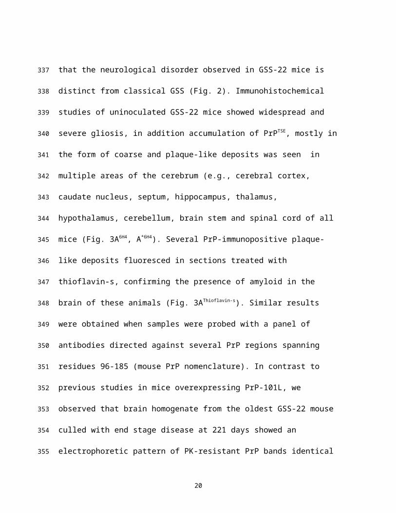
clearly distinct, indicating that the neurological disorder observed in GSS-22 mice is
distinct from classical GSS (Fig. 2). Immunohistochemical studies of uninoculated GSS-
22 mice showed widespread and severe gliosis, in addition accumulation of PrPTSE,
mostly in the form of coarse and plaque-like deposits was seen in multiple areas of the
cerebrum (e.g., cerebral cortex, caudate nucleus, septum, hippocampus, thalamus,
11
226
227
228
229
230
231
232
233
234
235
236
237
238
239
240
241
242
243
244
245
246
247
248
hypothalamus, cerebellum, brain stem and spinal cord of all mice (Fig. 3A6H4, A*6H4).
Several PrP-immunopositive plaque-like deposits fluoresced in sections treated with
thioflavin-s, confirming the presence of amyloid in the brain of these animals (Fig.
3AThioflavin-s). Similar results were obtained when samples were probed with a panel of
antibodies directed against several PrP regions spanning residues 96-185 (mouse PrP
nomenclature). In contrast to previous studies in mice overexpressing PrP-101L, we
observed that brain homogenate from the oldest GSS-22 mouse culled with end stage
disease at 221 days showed an electrophoretic pattern of PK-resistant PrP bands identical
to that observed in most prion diseases (Fig. 4). However we were unable to detect
similar PK-res PrP bands in any of the younger mice analyzed, despite the identification
of PrP amyloid plaques by immunohistochemistry. Although prion infectivity can be
detected in the presence of low levels of PK-res PrP (6, 27), the identification of protease
resistant PrPTSE in oldest GSS-22 mice indicates that this mouse model reproduces some
aspects of prion disease. However the inconsistency of clinical signs and general lack of
PK-resistant PrP confirms that this model may instead develop a disorder caused by
overexpression of the mutant PrP and amyloid accumulation, and not replication of an
infectious, transmissible prion disease.
GSS-22 mice do not transmit disease to 101LL
As the disease characterized in the GSS-22 mice did not resemble a classical TSE
phenotype, we assayed for the presence of a transmissible prion agent in brain tissue of
these mice by serial passage into 101LL mice, a knock-in model of the same PrP
mutation. Homogenates were prepared from brains of two GSS-22 mice culled at 197 and
12
249
250
251
252
253
254
255
256
257
258
259
260
261
262
263
264
265
266
267
268
269
270
271

221 days of age, and each inoculated ic into groups of 101LL (101LL-SP1) and 129Ola
mice (129Ola-SP1) (Table 1 and Fig. 1B). The GSS-22 mice used to generate each
inoculum showed severe spongiform degeneration and high levels of immunopositive PrP
in most brain areas. All 101LL-SP1 and 129Ola-SP1 mice were either culled for
intercurrent illness or at the end of their expected normal lifespan over 520 days post
inoculation. None of these animals developed neurological signs or spongiform
degeneration of the brain (Table 1). A total of 37 101LL mice (101LL-SP1) and 37
129/Ola mice (129/Ola-SP1) were available from both transmissions for analysis. All
animals were examined for PrP deposition by immunohistochemistry using antibody
6H4. We observed that, in contrast to the widespread and severe PrP deposition seen in
overexpressor GSS-22 mice, brains of 101LL-SP1 (29/37) mice showed a restricted
pattern of PrP accumulation in the form of PrP amyloid plaques mostly limited to the
corpus callosum and stratum lacunosum-moleculare of the hippocampus (Fig. 3B6H4,
B*6H4). In few animals, limited PrP immunopositivity was seen in the subependymal
region of the lateral ventricle. Some plaques showed unstained centers, probably due to
failure of the antibody to penetrate the amyloid cores. However, the poorly-stained
plaques fluoresced in sections treated with thioflavin-s, indicating that they were amyloid
deposits (Fig. 3Thioflavin-s). In 101LL-SP1 mice astro and microgliosis was seen in the
vicinity of amyloid plaques, but was less than the gliosis seen in uninoculated GSS-22
mice (Fig. 3BIBA1, BGFAP). No PrP deposits were observed in the brains of any 129Ola-SP1
mice (Table 1). These findings show that expression of mutant PrP in the host is
important for amyloidogenesis and that amyloid deposition occurs in the inoculated
13
272
273
274
275
276
277
278
279
280
281
282
283
284
285
286
287
288
289
290
291
292
293
101LL-SP1 mice in the absence of clinical disease or spongiform degeneration of the
brain.
Are 101LL SP-1 mice asymptomatic carriers?
To investigate the possibility of subclinical infection, we prepared brain homogenates
from two 101LL-SP1 mice (776 and 807 days old) for further passage (Fig. 1B). To
increase the chances of disease transmission, we prepared brain homogenates from
selected mice with large PrP deposits in the corpus callosum and vicinity (the only
regions of the brain having moderate to abundant PrP accumulations in this group of
animals). Each brain homogenate was inoculated ic into 101LL mice (101LL-SP2) and
129Ola mice (129Ola-SP2) (Fig. 1B and Table 1). None of those mice developed
neurological signs, and were finally culled due to intercurrent illness or at the end of their
expected normal lifespan at over 540 days post inoculation (Table 1). Neuropathologic
examination revealed no spongiform degeneration in any of those mice. No
accumulations of PrP were detected in 129Ola-SP2 mice; however, 101LL-SP2 mice
showed small number of PrP-positive plaques located mostly in the hippocampus and
corpus callosum (Fig. 3C6H4, C*6H4 and Table 1). Lower amounts of PrP accumulation
was seen in 101LL-SP2 mice than in 101LL-SP1 mice. In a few animals, limited PrP
immunopositivity was also seen in the subependymal region of the lateral ventricle. The
level of gliosis detected in 101LL-SP2 mice is comparable to that observed in age
matched uninoculated 101LL mice (control) (Fig 3CIBA1, CGFAP, DIBA1, DGFAP). In contrast
to the immunopositive PrP seen in tissue sections, no protease-resistant PrPTSE was
detected in the brains of 101LL mice from subpassages (101LL-SP1 and 101LL-SP2)
14
294
295
296
297
298
299
300
301
302
303
304
305
306
307
308
309
310
311
312
313
314
315
316
tested by immunoblotting. Thus, inoculation of PrPTSE from GSS-22 mice into 101LL
mice induces the formation of PrP amyloid but no disease on 2 passages.
Some prion strains have been shown to replicate preferentially in lymphoid tissue without
generating a fatal neurological disease. Under this scenario the apparent absence of
transmission could represent subclinical infection (28). A ligand-coated PrP antigen
capture immunoassay (IDEXX HerdCheck®) has been widely utilized for the screening of
natural prion diseases in animals. Therefore, we used this immunoassay (that has been
shown to be highly sensitive for the detection of aggregated PrP) to explore the presence
of PrPTSE in the brain and spleen of spontaneously sick GSS-22 and in asymptomatic
101LL-SP1 and 101LL-SP2 mice. Using IDEXX HerdCheck®, we detected PrPTSE in
brains of aged/sick, uninoculated GSS-22 mice, brains of mice in the first subpassage
(101LL-SP1) and in control animals following inoculation with mouse adapted scrapie
agents (ME7 or 79A with high amounts of PrPTSE and infectivity titers). Brains from mice
in the second sub-passage experiments (101LL-SP2 mice) yielded negative results. In
addition, spleens from all experimental groups (GSS22, 101LL-SP1 and 101LL-SP2)
were negative for PrPTSE. The results argue for absence of PrPTSE in the periphery of GSS-
22, 101LL-SP1 and 101LL-SP2 mice, despite the formation of plaques in the brain.
Discussion
The original concept that prion diseases constitute a special group among proteinopathies,
because the seeded polymerization of PrPTSE replicates its misfolded conformation
indefinitely and provides the molecular basis for spread within defined anatomical
15
317
318
319
320
321
322
323
324
325
326
327
328
329
330
331
332
333
334
335
336
337
338
339

pathways and transmission between individuals has more recently been expanded to
include other neurodegenerative diseases (29, 30). The mechanism of transport of
misfolded protein aggregates from one cell to another remains to be determined but could
include endocytosis, tunneling nanotubes, or transynaptic. The latter was proposed for the
cell to cell spread of hyperphosphorylated microtubule associated protein (tau) in patients
with AD (31). However, our results suggest that there is a difference between cell-to-cell
spread of a misfolded protein, and the transfer of infectivity from one organism to
another, a process that clearly requires a different kind of interaction between the
transmissible agent and its host.
101LL-8a mice do not have subclinical infection
In our original study (7) we found no evidence of replication of infectivity in 101LL-8a
mice with PrP amyloid plaques following inoculation with PrPTSE purified from a patient
with atypical GSS P102L. Absence of cross species transmission to host species that are
susceptible to many prion agents has been observed in few instances. Importantly,
analysis following serial subpassage of hamster prion agent into mice has demonstrated
that in some instances subclinical infections have two distinct phases, a persistent phase
followed by a replicative phase (32). This issue has practical consequences because in
wildlife and agricultural settings prion agents might both persist and adapt over long
periods of time (years). Here, we demonstrate, even following subpassages, that the
formation of mouse PrP amyloid plaques triggered by the inoculation of PrPTSE obtained
from a patient with atypical GSS P102L is dissociated from disease. In fact, the only
form of GSS shown to be transmissible to experimental animals is typical GSS P102L.
16
340
341
342
343
344
345
346
347
348
349
350
351
352
353
354
355
356
357
358
359
360
361
362

GSS-22 mice overexpressing PrP 101L develop a non-transmissible prion disease
The dramatic differences in the amounts and distribution of PrPTSE seen in 101LL-SP1
and 101LL-SP2 compared with GSS-22 mice can be interpreted in several ways. PrPTSE in
the GSS-22-derived inoculum might be unstable and readily degraded, preventing the
misfolding and accumulation of endogenous PrP that would cause disease. However the
presence of amyloid plaques in the corpus callosum and vicinity in many animals
strongly suggest that PrPTSE in the inoculum is stable and remains capable of seeding
further amyloid formation. Another possibility is that low levels of infectivity in the GSS-
22 inoculum caused subclinical infection in 101LL animals. Other investigators reported
long incubation time and slow progression of clinical disease in mice and hamsters
inoculated with synthetic prions (32), lack of clinical disease in animals expressing
anchorless PrP inoculated with mouse adapted scrapie agent (33) or in transmission
experiments across species (34, 35). Interestingly, in all these models disease was
consistently detected after serial passage in wild-type animals demonstrating efficient
adaptation and spread of the infectious agent. To rule out the occurrence of subclinical
infection in 101LL mice after the initial injection with GSS-22 brain suspension, we
performed subpassage experiments. We observed that, although 101LL recipient animals
remained asymptomatic and without any spongiform encephalopathy, they again
accumulated PrPTSE but only in restricted areas of the brain. These results differ
substantially from those obtained by others where generation of prion infectivity and
disease transmission was evident following subpassage (32-35).
17
363
364
365
366
367
368
369
370
371
372
373
374
375
376
377
378
379
380
381
382
383
384

The data presented in this work could be interpreted to show an infection, where
introduction of the amyloid seed from brains of patients with atypical GSS P102L or
GSS-22 mice leads to the propagation of further amyloid in the brains of recipient mice.
However this “transmission” is iatrogenic, and we have yet to determine whether such
seeding can be initiated following “natural” routes such as oral exposure. Such infection
would also appear to be non-productive, as plaques are restricted to a limited brain area,
and following up to 3 subpassages, no disease is observed in 101LL mice. Limited spread
is also seen within the brain, and no spread to the periphery is apparent. We also failed to
induce the plaque forming phenotype in wild-type mice. In addition, previous
ultrastructural analysis showed that cell membrane alterations consistently seen in murine
scrapie and other infectious prion diseases were not present in 101LL-8a mice with
amyloid plaques, suggesting differences in the pathogenesis of these conditions (9).
Therefore, our model would appear to represent a different mechanism from what is
generally understood as infection.
Relevance to natural protein misfolding diseases
Most proteinopathies (e.g., AD, PD) have previously not been considered to be infectious
diseases and indeed there is no epidemiological evidence to suggest that they are. It
would seem important therefore to try to understand when a misfolded protein has the
potential to transmit between individuals which should be considered a very different
scenario from a cell to cell spread within an individual; the former having a consequence
for a population the later for an individual. If even within prion diseases some affect the
individual only, then this would provide precedence for other protein misfolding diseases
18
385
386
387
388
389
390
391
392
393
394
395
396
397
398
399
400
401
402
403
404
405
406
407

to be specific to individuals rather than having consequences for populations. We believe
the experiments outlined in this paper start to address this important issue, namely protein
seeding within an individual and transmission of disease between individuals represents
different processes.
In our experiments we consider that the 101LL mouse model would, with extended
lifespan, overcome the long lag phase required to form endogenous PrP amyloid plaques
naturally via a nucleated polymerisation mechanism. This would reflect what is seen in
atypical GSS P102L where PrP plaques are seen in the absence of spongiform change in
the brains of older patients. By introducing the PrP-8a or GSS-22 amyloid seed into
101LL mice, the lag phase is reduced, accelerating plaque formation in restricted areas of
the brain. GSS-22 mice ubiquitously overexpress PrP-101L and therefore are likely to
shift the balance of seed nucleation due to unregulated overproduction of the mutated
protein. These data mirror what has been described in overexpressing transgenic models
of human APP, seeded with AD brain (30).
In vitro studies suggested that infectious and non-infectious aggregates of PrP might be
structurally similar but that co-factors such as lipids and nucleic acids might also be
critical in determining the infectious characteristics of PrPTSE (36). Another possibility for
the lower specific infectivity associated with PrP amyloid is that extracts with smaller
PrPTSE aggregates might be more infectious than the larger ones (37). While disease
associated PrP (i.e., PrPTSE) was originally described as insoluble in detergents and PK-
resistant, it is increasingly recognized that multiple distinct disease related PrP isoforms
19
408
409
410
411
412
413
414
415
416
417
418
419
420
421
422
423
424
425
426
427
428
429
430
may be important in the pathogenesis of prion diseases (5, 23, 38-40). Our data suggest
that the formation of PrPTSE does not necessarily correlate with the replication of prion
infectivity. The results from bioassays reported here have implications for the
interpretation of tests based on the detection of protein aggregates. Thus, determining the
difference between PrPTSE aggregates associated with infectivity and those aggregates that
are not infectious will be essential to determine which diseases associated with protein
misfolding are threats to public health (transmissible between individuals).
Acknowledgments
We thank Dr David M Asher (Laboratory of Bacterial and TSE Agents, US FDA) for
critical reading of the manuscript; V Thomson, S Cumming, K Hogan, S Carpenter and R
Greenan for care and scoring of the animals; A Coghill, A Boyle, S Mack and G
McGregor for tissue processing and lesion profile analysis. These studies were partially
funded by the Biotechnology and Biological Sciences Research Council (BBSRC)
Institute Strategic Grant BB/J004332/1; NIH-NIAID Agreement No.Y1-AI-4893-02;
FDA Agreement No. 224-05-1307; Public Health Service Cooperative Research and
Development Agreement between US FDA and The Roslin Institute, University of
Edinburgh, UK. The findings and conclusions in this article have not been formally
disseminated by the Food and Drug Administration and should not be construed to
represent any Agency determination of policy.
20
431
432
433
434
435
436
437
438
439
440
441
442
443
444
445
446
447
448
449
450
451
452
453
References
1. Prusiner S. 1982. Novel proteinaceous infectious particles cause scrapie. Science
216:136 - 144.
2. McKinley MP, Bolton DC, Prusiner SB. 1983. A protease-resistant protein is a
structural component of the scrapie prion. Cell 35:57-62.
3. Beekes M, Baldauf E, Diringer H. 1996. Sequential Appearance and
Accumulation Of Pathognomonic Markers In the Central-Nervous-System Of
Hamsters Orally Infected With Scrapie. J. Gen. Virol. 77:1925-1934.
4. Lasmezas CI, Deslys J, Robain O, Jaegly A, Beringue V, Peyrin J, Fournier
J, Hauw J, Rossier J, Dormont D. 1997. Transmission of the BSE Agent to
Mice in the Absence of Detectable Abnormal Prion Protein. Science 275:402-405.
5. Chiesa R, Piccardo P, Quaglio E, Drisaldi B, Si-Hoe SL, Takao M, Ghetti B,
Harris DA. 2003. Molecular distinction between pathogenic and infectious
properties of the prion protein. J. Virol. 77:7611-7622.
6. Barron RM, Campbell SL, King D, Bellon A, Chapman KE, Williamson RA,
Manson JC. 2007. High titres of TSE infectivity associated with extremely low
levels of PrPSc in vivo. J. Biol. Chem. 282:35878-35886.
7. Piccardo P, Manson JC, King D, Ghetti B, Barron RM. 2007. Accumulation
of prion protein in the brain that is not associated with transmissible disease. Proc.
Natl. Acad. Sci. U. S. A. 104:4712-4717.
8. Balkema-Buschmann A, Eiden M, Hoffmann C, Kaatz M, Ziegler U, Keller
M, Groschup MH. 2011. BSE infectivity in the absence of detectable PrPSc
21
454
455
456
457
458
459
460
461
462
463
464
465
466
467
468
469
470
471
472
473
474
475
accumulation in the tongue and nasal mucosa of terminally diseased cattle. J. Gen.
Virol. 92:467-476.
9. Jeffrey M, McGovern G, Chambers EV, King D, González L, Manson JC,
Ghetti B, Piccardo P, Barron RM. 2011. Mechanism of PrP-Amyloid
Formation in Mice Without Transmissible Spongiform Encephalopathy. Brain
Pathol. 22:58-66.
10. Miyazawa K, Emmerling K, Manuelidis L. 2011. High CJD Infectivity
Remains After Prion Protein Is Destroyed. J. Cell. Biochem. 112:3630-3637.
11. Frost B, Diamond MI. 2010. Prion-like mechanisms in neurodegenerative
diseases. Nat. Rev. Neurosci. 11:155-159.
12. Saborio GP, Permanne B, Soto C. 2001. Sensitive detection of pathological
prion protein by cyclic amplification of protein misfolding. Nature 411:810-813.
13. Colby DW, Zhang Q, Wang S, Groth D, Legname G, Riesner D, Prusiner SB.
2007. Prion detection by an amyloid seeding assay. Proc. Natl. Acad. Sci. U. S. A.
104:20914-20919.
14. Atarashi R, Wilham JM, Christensen L, Hughson AG, Moore RA, Johnson
LM, Onwubiko HA, Priola SA, Caughey B. 2008. Simplified ultrasensitive
prion detection by recombinant PrP conversion with shaking. Nat Meth 5:211-
212.
15. Klingeborn M, Race B, Meade-White KD, Chesebro B. 2011. Lower specific
infectivity of protease-resistant prion protein generated in cell-free reactions.
Proceedings of the National Academy of Sciences 108:E1244–E1253.
22
476
477
478
479
480
481
482
483
484
485
486
487
488
489
490
491
492
493
494
495
496
497
16. Makarava N, Kovacs GG, Bocharova O, Savtchenko R, Alexeeva I, Budka H,
Rohwer RG, Baskakov IV. 2010. Recombinant prion protein induces a new
transmissible prion disease in wild-type animals. Acta Neuropathol. (Berl).
119:177-187.
17. Raymond GJ, Race B, Hollister JR, Offerdahl DK, Moore RA, Kodali R,
Raymond LD, Hughson AG, Rosenke R, Long D, Dorward DW, Baron GS.
2012. Isolation of novel synthetic prion strains by amplification in transgenic
mice coexpressing wild-type and anchorless prion proteins. J. Virol. 86:11763-
11778.
18. Wang F, Wang X, Yuan C-G, Ma J. 2010. Generating a Prion with Bacterially
Expressed Recombinant Prion Protein. Science 327:1132-1135.
19. Ghetti B, Tagliavini F, Kovacs GG, Piccardo P. 2011. Gerstmann-Straussler-
Sheinker disease. In Dickinson DW, Weller RO (ed.), Neurodegeneration: The
Molecular Pathology of Dementia and Movement Disorders. Blackwell
Publishing Ltd.
20. Piccardo P, Dlouhy SR, Lievens PMJ, Young K, Thomas DP, Nochlin D,
Dickson DW, Vinters HV, Zimmerman TR, Mackenzie IRA, Kish SJ, Ang
LC, De Carli C, Pocchiari M, Brown P, Gibbs CJ, Gajdusek DC, Bugiani O,
Ironside J, Tagliavini F, Ghetti B. 1998. Phenotypic variability of Gerstmann-
Straussler-Scheinker disease is associated with prion protein heterogeneity. J.
Neuropathol. Exp. Neurol. 57:979-988.
21. Parchi P, Chen SG, Brown P, Zou WQ, Capellari S, Budka H, Hainfellner J,
Reyes PF, Golden GT, Hauw JJ, Gajdusek DC, Gambetti P. 1998. Different
23
498
499
500
501
502
503
504
505
506
507
508
509
510
511
512
513
514
515
516
517
518
519
520
patterns of truncated prion protein fragments correlate with distinct phenotypes in
P102L Gerstmann- Straussler-Scheinker disease. Proc. Natl. Acad. Sci. U. S. A.
95:8322-8327.
22. Manson JC, Jamieson E, Baybutt H, Tuzi NL, Barron R, McConnell I,
Somerville R, Ironside J, Will R, Sy MS, Melton DW, Hope J, Bostock C.
1999. A single amino acid alteration (101L) introduced into murine PrP
dramatically alters incubation time of transmissible spongiform encephalopathy.
EMBO J. 18:6855-6864.
23. Nazor KE, Kuhn F, Seward T, Green M, Zwald D, Pürro M, Schmid J,
Biffiger K, Power AM, Oesch B, Raeber A, Telling G. 2005. Immunodetection
of disease-associated mutant PrP, which accelerates disease in GSS transgenic
mice EMBO J. 24:2472-2480.
24. Fraser H, Dickinson AG. 1967. Distribution of experimentally induced scrapie
lesions in the brain. Nature 216:1310-1311.
25. Schmidt ML, Robinson KA, Lee VMY, Trojanowski JQ. 1995. Chemical and
immunological heterogeneity of fibrillar amyloid in plaques of Alzheimer's
disease and Downs Syndrome brains revealed by confocal microscopy. Am. J.
Pathol. 147:503-515.
26. Plinston C, Hart P, Chong A, Hunter N, Foster J, Piccardo P, Manson JC,
Barron RM. 2011. Increased Susceptibility of Human-PrP Transgenic Mice to
Bovine Spongiform Encephalopathy Infection following Passage in Sheep. J.
Virol. 85:1174-1181.
24
521
522
523
524
525
526
527
528
529
530
531
532
533
534
535
536
537
538
539
540
541
542
27. Tzaban S, Friedlander G, Schonberger O, Horonchik L, Yedidia Y, Shaked
G, Gabizon R, Taraboulos A. 2002. Protease-sensitive scrapie prion protein in
aggregates of heterogeneous sizes. Biochemistry (Mosc). 41:12868-12875.
28. Beringue V, Herzog L, Jaumain E, Reine F, Sibille P, Le Dur A, Vilotte JL,
Laude H. 2012. Facilitated Cross-Species Transmission of Prions in Extraneural
Tissue. Science 335:472-475.
29. Westermark GT, Westermark P. 2010. Prion-like aggregates: infectious agents
in human disease. Trends in Molecular Medicine 16:501-507.
30. Brundin P, Melki R, Kopito R. 2010. Prion-like transmission of protein
aggregates in neurodegenerative diseases. Nat. Rev. Mol. Cell Biol. 11:301-307.
31. de Calignon A, Polydoro M, Suarez-Calvet M, William C, Adamowicz DH,
Kopeikina KJ, Pitstick R, Sahara N, Ashe KH, Carlson GA, Spires-Jones TL,
Hyman BT. 2012. Propagation of Tau Pathology in a Model of Early Alzheimer's
Disease. Neuron 76:461-461.
32. Makarava N, Kovacs GG, Savtchenko R, Alexeeva I, Budka H, Rohwer RG,
Baskakov IV. 2012. Stabilization of a Prion Strain of Synthetic Origin Requires
Multiple Serial Passages. J. Biol. Chem. 287:30205-30214.
33. Chesebro B, Trifilo M, Race R, Meade-White K, Teng C, LaCasse R,
Raymond L, Favara C, Baron G, Priola S, Caughey B, Masliah E, Oldstone
M. 2005. Anchorless Prion Protein Results in Infectious Amyloid Disease
Without Clinical Scrapie. Science 308:1435-1439.
25
543
544
545
546
547
548
549
550
551
552
553
554
555
556
557
558
559
560
561
562
563

34. Race R, Meade-White K, Raines A, Raymond GJ, Caughey B, Chesebro B.
2002. Subclinical scrapie infection in a resistant species: Persistence, replication,
and adaptation of infectivity during four passages. J. Infect. Dis. 186:S166-S170.
35. Hill AF, Antoniou M, Collinge J. 1999. Protease-resistant prion protein
produced in vitro lacks detectable infectivity. J. Gen. Virol. 80:11-14.
36. Deleault NR, Kascsak R, Geoghegan JC, Supattapone S. 2010. Species-
Dependent Differences in Cofactor Utilization for Formation of the Protease-
Resistant Prion Protein in Vitro. Biochemistry (Mosc). 49:3928-3934.
37. Silveira JR, Raymond GJ, Hughson AG, Race R, Sim VL, Hayes SF,
Caughey B. 2005. The most infectious prion protein particles. Nature 437:257-
261.
38. Biasini E, Seegulam ME, Patti BN, Solforosi L, Medrano AZ, Christensen
HM, Senatore A, Chiesa R, Williamson RA, Harris DA. 2008. Non-infectious
aggregates of the prion protein react with several PrP<sup>Sc</sup>-directed
antibodies. J. Neurochem. 105:2190-2204.
39. Miller MB, Geoghegan JC, Supattapone S. 2011. Dissociation of Infectivity
from Seeding Ability in Prions with Alternate Docking Mechanism. PLoS Pathog.
7.
40. Chiesa R, Piccardo P, Biasini E, Ghetti B, Harris DA. 2008. Aggregated,
Wild-Type Prion Protein Causes Neurological Dysfunction and Synaptic
Abnormalities. J. Neurosci. 28:13258-13267.
26
564
565
566
567
568
569
570
571
572
573
574
575
576
577
578
579
580
581
582
583
584
585586
Table 1. PrP-amyloid formation in 101LL mice inoculated with brain extracts from
101LL-8a and overexpressor GSS-22 mice
Inoculum Source
Recipient (n)*
Survival (days ± SEM)¶
Clinical prion
disease
Spongiform degeneratio
n
PrP Plaques
Output tissue ID#
101LL-8a 101LL (16) 602ǂ 0/16 0/16 16/16 101LL-8a101LL-8a 129/Ola (22) 602ǂ 0/22 0/22 0/22 129Ola-8an/a GSS-22 (9) 181±8 0/9 9/9 8/9† GSS-22GSS-22 101LL (37) 528±23 0/37 0/37 29/37 101LL-SP1GSS-22 129/Ola (37) 526±24 0/37 0/37 0/37 129Ola-SP1101LL-SP1 101LL (47) 529±14 0/47 0/47 40/47 101LL-SP2101LL-SP1 129/Ola (46) 558±13 0/46 0/46 0/46 129Ola-SP2
* Number of mice available for analysis
¶ Average survival ± SEM
# Identifying tag given to tissues produced in each transmission
ǂ Experiment terminated
† Widespread accumulation of PrP positive plaques and diffuse deposits in the brain
27
587
588
589590
591
592
593
594
595
596
597
Figure Legends.
Figure 1. Flow chart outlining the serial passage of brain extract from a patient with
“atypical” GSS P102L and uninoculated GSS-22 mice overexpressing PrP-101L into
101LL and 129Ola mice
Panel A: Brain extract from a patient with “atypical” GSS P102L was inoculated
intracerebrally into 101LL and 129Ola mice. The absence of prion disease in 101LL(8a)
and 129Ola mice was confirmed neuropathologically. Brain extract from selected
101LL(8a) mice without disease but with PrP-amyloid plaques was serially passaged into
101LL and 129Ola mice. 1st and 2nd pass were described (reference 7). 3rd pass performed
in the work described here. Panel B: Brain extract from uninoculated GSS-22 mice
overexpressing PrP-101L showing spongiform degeneration and diffuse and amyloid PrP
deposits in the brain was serially passaged 101LL and 129Ola mice (101LL SP-1). The
absence of prion disease was confirmed neuropathologically. Selected 101LL SP-1 tissue
without prion disease showing PrP amyloid plaques in the corpus callosum and vicinity
was used for serial passage into 101LL and 129Ola mice (101LL SP-2).
Figure 2. Pattern of vacuolation in 101LL mice with prion disease after inoculation
of brain extract from a patient with “typical” GSS P102L and in uninoculated GSS-
22 mice overexpressing PrP-101L
Lesion profile comparison of uninoculated mice overexpressing mutant PrP (GSS-22 red)
and 101LL mice with prion disease (blue). Data shows mean lesion profile ± standard
error of the mean. Areas 1-9, grey matter scoring regions: (1) dorsal medulla, (2)
28
598
599
600
601
602
603
604
605
606
607
608
609
610
611
612
613
614
615
616
617
618
619
620
cerebellar cortex, (3) superior colliculus, (4) hypothalamus, (5) thalamus, (6)
hippocampus, (7) septum, (8) retrosplenial and adjacent motor cortex, (9) cingulate and
adjacent motor cortex; 1*-3*, white matter scoring regions: (1*) cerebellar white matter,
(2*) mesencephalic tegmentum, (3*) pyramidal tract.
Figure 3. Contrast between abundant PrPTSE accumulation and gliosis in the brain
of uninoculated GSS-22 mice and sparse PrP deposition in the brain of 101LL mice
after serial subpassage
Abundant PrPTSE accumulation in multiple areas of the cerebral cortex, hippocampus and
thalamus of uninoculated GSS-22 mice with terminal disease (A6H4, A*6H4). PrPTSE
accumulation restricted to the corpus callosum and hippocampus in 101LL-SP1 (B6H4,
B*6H4). Small amounts of PrPTSE in the corpus callosum of 101LL-SP2 (C6H4, C*6H4). PrP
fluorescent amyloid deposits in the corpus callosum and hippocampus of GSS-22
(AThioflavin-s) and 101LL-SP1 mice (BThioflavin-s). Severe microgliosis (AIBA1) and astrogliosis
(AGFAP) in the hippocampus of GSS-22 mice. Milder microgliosis (BIBA1) and astrogliosis
(BGFAP) in the hippocampus and vicinity of 101LL-SP1. Minimal microgliosis (CIBA1) and
astrogliosis (CGFAP) in 101LL-SP2 mice and in 101LL aged controls (DIBA1) and (DGFAP).
29
621
622
623
624
625
626
627
628
629
630
631
632
633
634
635
636
637
638
Figure 4 Presence of PK-resistant PrP in uninoculated GSS-22 mice
Western blotting using the 7A12 anti-PrP antibody was carried out in brain homogenates
of uninoculated GSS-22 mice (lanes 1 and 2); 101LL-SP1 mice (lanes 3 and 4) and
101LL-SP2 mice (lanes 5 and 6). Positive control 101LL inoculated with ME7 scrapie-
agent (lanes 7 and 8), negative control 101LL non-inoculated mouse (lanes 9 and 10).
PK-resistant PrP is observed in GSS-22 (lane 2) and in 101LL mouse inoculated with
ME7 scrapie-agent (lane 8). Non PK-treated samples (lanes 1, 3, 5, 7 and 9); PK-treated
samples (lanes 2, 4, 6, 8 and 10). Image cropped from single blot to remove irrelevant
lanes.
30
639
640
641
642
643
644
645
646
647
648
649