overcoming treatment challenges in myelofibrosis and
TRANSCRIPT

1 3
Cancer Chemother Pharmacol (2016) 77:1125–1142DOI 10.1007/s00280-016-3012-z
REVIEW ARTICLE
Overcoming treatment challenges in myelofibrosis and polycythemia vera: the role of ruxolitinib
Jeffrey C. Bryan1 · Srdan Verstovsek2
Received: 11 September 2015 / Accepted: 15 March 2016 / Published online: 26 March 2016 © The Author(s) 2016. This article is published with open access at Springerlink.com
than standard therapy in controlling hematocrit levels and improving splenomegaly and PV-related symptoms. Only 1 of 110 patients in the ruxolitinib arm compared with 6 of 112 patients in the control arm experienced a thrombo-embolic event through week 32. Grade ≥3 cytopenias were uncommon.
Keywords JAK inhibition · Myelofibrosis · Myeloproliferative neoplasm · Polycythemia vera · Ruxolitinib
Introduction
Philadelphia chromosome–negative myeloproliferative neoplasms (MPNs), including myelofibrosis (MF), poly-cythemia vera (PV), and essential thrombocythemia (ET), are genetically related, but heterogeneous chronic diseases characterized by overactive signaling through the Janus kinase (JAK)-signal transducer and activator of transcrip-tion (STAT) pathway as the central pathogenic mechanism [1–5]. The JAK–STAT pathway plays an essential role in normal hematopoiesis by mediating incoming signals from hematopoietic growth factors such as erythropoietin and thrombopoietin in hematopoietic stem cells [6]. The term “myelofibrosis” refers collectively to patients with de novo, primary myelofibrosis (PMF) and those with myelofibrotic transformation from PV to post-PV MF or from ET to post-ET MF [7, 8].
Although our understanding of the relationship between MPN genotype and phenotype remains incomplete, tremen-dous progress has been made in recent years in elucidating the complexity of somatic mutations that drive MPN patho-genesis and influence patients’ prognoses. Prompted by the discovery of the JAK2V617F gain-of-function mutation in
Abstract Myelofibrosis (MF) and polycythemia vera (PV) are BCR-ABL1-negative myeloproliferative neo-plasms associated with somatic hematopoietic stem cell mutations leading to over activation of JAK–STAT signal-ing. MF and PV are pathogenically related and share spe-cific clinical features such as splenomegaly and constitu-tional symptoms. The MF phenotype is dominated by the effects of progressive bone marrow fibrosis resulting in shortened survival. In contrast, elevated thrombosis risk due to erythrocytosis is the primary clinical concern in PV. Ruxolitinib, an oral JAK1/JAK2 inhibitor, is approved in the USA for the treatment of patients with intermedi-ate- or high-risk MF and patients with PV who have had an inadequate response to or are intolerant of hydroxyurea. For MF, results of two phase III studies demonstrated that ruxolitinib therapy reduced spleen volume and MF-related symptom burden, improved quality-of-life measures, and was associated with prolonged overall survival. Treatment benefits were generally sustained with continued therapy. Dose-dependent cytopenias were common but generally manageable with transfusions (for anemia), dose reduction, or treatment interruption. Optimal dosing management is critical to maintain long-term treatment benefit, because cessation of therapy resulted in rapid return of symp-toms to baseline levels. Results of the phase III PV trial showed that ruxolitinib was significantly more effective
* Jeffrey C. Bryan [email protected]
1 Pharmacy Clinical Programs, Division of Pharmacy, The University of Texas MD Anderson Cancer Center, 1515 Holcombe Boulevard, Houston, TX 77030, USA
2 Division of Cancer Medicine, Department of Leukemia, The University of Texas MD Anderson Cancer Center, Houston, TX, USA

1126 Cancer Chemother Pharmacol (2016) 77:1125–1142
1 3
more than 50 % of patients with PMF or ET and at least 95 % of patients with PV [4], the importance of JAK–STAT pathway dysregulation in the pathogenesis of MPNs is now well established [5]. In addition to the JAK2V617F muta-tion, which promotes constitutive activation of JAK2, other somatic stem cell mutations have been identified in patients with MPNs that lead to over activation of JAK–STAT sign-aling, including mutations in the thrombopoietin receptor gene (MPL) [4, 5, 9] and the calreticulin gene (CALR) [5, 10, 11] (Fig. 1 [10, 12]). In the vast majority of patients, JAK2, MPL, and CALR mutations appear to be mutually exclusive and are considered principal drivers of neoplastic myeloproliferation [10, 11]. However, the diagnostic value of these mutations is limited, as none is specific for any one type of MPN and a small minority of patients with MPNs lack JAK2, MPL, and CALR mutations [13, 14]. Further-more, patients with MPNs often carry multiple additional mutations that may affect clinical phenotype and/or prog-nosis [5, 14, 15].
In addition to its essential role in hematopoiesis, the JAK–STAT pathway is central to cytokine activation and signaling in the immune system [6]. It is well documented that patients with MF have abnormally high levels of cir-culating inflammatory cytokines, including tumor necrosis factor alpha (TNF-α) and interleukin (IL)-6 [16], which appear to be fueled by aberrant cytokine secretion of both malignant and nonmalignant cells in the bone marrow [17]. Moreover, JAK1 hyperactivity has been noted in patients
with MF [18] and may be due to cytokine hyperstimula-tion. It is believed that abnormally high levels of circulating inflammatory cytokines are a major cause for the burden of constitutional symptoms in patients with MPNs [16, 19].
Ruxolitinib, an orally bioavailable inhibitor of JAK1 and JAK2, is currently the only pharmacotherapy with approved indications in MF, and it has been recently approved by the US Food and Drug Administration for the treatment of patients with PV with an inadequate response to or intolerant of hydroxyurea. Ruxolitinib oral tablets are available in strengths of 5, 10, 15, 20, and 25 mg, allowing for individualized dosing regimens (per dosing recommen-dations in the prescribing information [20]). In this review, we summarize the efficacy and safety data for ruxolitinib in both indications and discuss specific pharmacologic prop-erties relevant for its safe and effective administration.
Ruxolitinib: general pharmacology
Ruxolitinib is an equipotent inhibitor of JAK1 [mean half maximal inhibitory concentration (IC50) = 3.3 nM] and JAK2 (mean IC50 = 2.8 nM) in vitro, with at least 100-fold less inhibitory activity against JAK3 [18]. Early results obtained with a mouse model of JAK2V617F-induced MPN showed that ruxolitinib was highly effective in reduc-ing splenomegaly and lowering IL-6 and TNF-α levels and prolonged survival. The effects of ruxolitinib on circulating
Primary Myelofibrosis(N = 203)
NonmutatedJAK2, MPL,and CALR
MPL mutation
CALRmutation
JAK2mutation
b
CALR mutations
Regulatedactivation ofJAK-STAT
TPO
TPO
MPL mutations
AdditiveSTAT
activation
MyeloproliferationCell SurvivalInflammatorycytokineexpression
Overactivationof JAK-STAT
JAK2V617F activityresults in chromatindemethylation anddecreased binding of HP1α
Epigenetic mutations (eg, TET2, ASXL1, EZH2)
?
TPO
JAK2
TPO
JAK2 JAK2
JAK2V617F
JAK1/JAK2
STATSTATSTAT
STAT
STATSTAT
JAK2 JAK2
JAK2 JAK2
Ovveractivatiove
a
Fig. 1 a Mutations underlying the pathobiology of myelofibrosis. Mutations commonly affect the JAK–STAT pathway, including those that occur directly in JAK2 such as the JAK2V617F mutation, or indirectly such as those in MPL, which encodes the thrombopoietin receptor, or in CALR, which may activate STATs through an unknown mechanism. Somatic mutations in epigenetic modifiers can also result in increased myeloproliferation, survival, and cytokine expression in
myelofibrosis. From [12]. Copyright © 2014. b The approximate pro-portion of patients with JAK2, MPL, and CALR mutations was exam-ined in 203 patients with primary myelofibrosis. From [10].Copyright © 2013 Massachusetts Medical Society. Reprinted with permission from Massachusetts Medical Society. CALR calreticulin, JAK Janus kinase, MPL myeloproliferative leukemia virus oncogene, STAT sig-nal transducer and activator of transcription
1127Cancer Chemother Pharmacol (2016) 77:1125–1142
1 3
cytokine levels were subsequently confirmed in phase II and III clinical studies and are believed to be integral to the efficacy of ruxolitinib [16, 21].
Ruxolitinib has a terminal half-life of approximately 3 h [22] and is predominantly metabolized by CYP3A4 and to a lesser extent by CYP2C9, with 74 and 22 % of a ruxolitinib dose recovered in urine and feces, respectively, within 24 h of oral administration in healthy human vol-unteers (<1 % was unchanged drug) [20, 23]. Ruxolitinib plasma exposure [area under the curve from time 0 extrapo-lated to infinite time (AUC0–inf)] increased by 91 and 27 % following concomitant administration of ketoconazole (a potent CYP3A4 inhibitor) and erythromycin (a moderate CYP3A4 inhibitor), respectively [24]. Pretreatment with the potent CYP3A4 inducer rifampin resulted in a 71 % decrease in ruxolitinib AUC0–inf, but had a limited impact on the overall pharmacodynamic activity, which decreased by 10 % [24]. This may be explained in part by an increase in the relative abundance of active ruxolitinib metabolites with rifampin coadministration [24].
Myelofibrosis
Natural history and prognosis
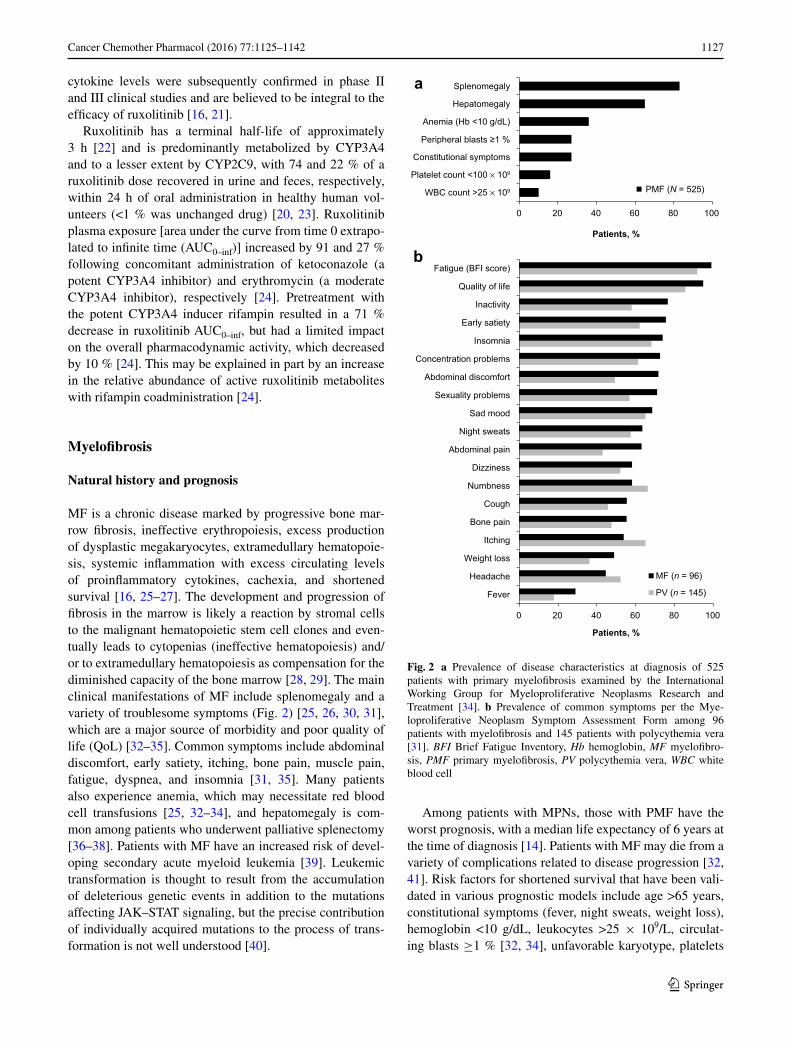
MF is a chronic disease marked by progressive bone mar-row fibrosis, ineffective erythropoiesis, excess production of dysplastic megakaryocytes, extramedullary hematopoie-sis, systemic inflammation with excess circulating levels of proinflammatory cytokines, cachexia, and shortened survival [16, 25–27]. The development and progression of fibrosis in the marrow is likely a reaction by stromal cells to the malignant hematopoietic stem cell clones and even-tually leads to cytopenias (ineffective hematopoiesis) and/or to extramedullary hematopoiesis as compensation for the diminished capacity of the bone marrow [28, 29]. The main clinical manifestations of MF include splenomegaly and a variety of troublesome symptoms (Fig. 2) [25, 26, 30, 31], which are a major source of morbidity and poor quality of life (QoL) [32–35]. Common symptoms include abdominal discomfort, early satiety, itching, bone pain, muscle pain, fatigue, dyspnea, and insomnia [31, 35]. Many patients also experience anemia, which may necessitate red blood cell transfusions [25, 32–34], and hepatomegaly is com-mon among patients who underwent palliative splenectomy [36–38]. Patients with MF have an increased risk of devel-oping secondary acute myeloid leukemia [39]. Leukemic transformation is thought to result from the accumulation of deleterious genetic events in addition to the mutations affecting JAK–STAT signaling, but the precise contribution of individually acquired mutations to the process of trans-formation is not well understood [40].
Among patients with MPNs, those with PMF have the worst prognosis, with a median life expectancy of 6 years at the time of diagnosis [14]. Patients with MF may die from a variety of complications related to disease progression [32, 41]. Risk factors for shortened survival that have been vali-dated in various prognostic models include age >65 years, constitutional symptoms (fever, night sweats, weight loss), hemoglobin <10 g/dL, leukocytes >25 × 109/L, circulat-ing blasts ≥1 % [32, 34], unfavorable karyotype, platelets
0 20 40 60 80 100
WBC count >25 × 109
Platelet count <100 × 109
Constitutional symptoms
Peripheral blasts ≥1 %
Anemia (Hb <10 g/dL)
Hepatomegaly
Splenomegaly
PMF (N = 525)
Patients, %
a
0 20 40 60 80 100
Fever
Headache
Weight loss
Itching
Bone pain
Cough
Numbness
Dizziness
Abdominal pain
Night sweats
Sad mood
Sexuality problems
Abdominal discomfort
Concentration problems
Insomnia
Early satiety
Inactivity
Quality of life
Fatigue (BFI score)
MF (n = 96)
PV (n = 145)
b
Patients, %
Fig. 2 a Prevalence of disease characteristics at diagnosis of 525 patients with primary myelofibrosis examined by the International Working Group for Myeloproliferative Neoplasms Research and Treatment [34]. b Prevalence of common symptoms per the Mye-loproliferative Neoplasm Symptom Assessment Form among 96 patients with myelofibrosis and 145 patients with polycythemia vera [31]. BFI Brief Fatigue Inventory, Hb hemoglobin, MF myelofibro-sis, PMF primary myelofibrosis, PV polycythemia vera, WBC white blood cell

1128 Cancer Chemother Pharmacol (2016) 77:1125–1142
1 3
<100 × 109/L, and the need for red blood cell transfusions [33]. Median survival varies from approximately 11 years for those with low-risk disease to 2 years for those with high-risk disease [32]. Additional variables that have dem-onstrated prognostic value outside of these models include mutations associated with worse (ASXL1) or better (CALR) prognosis [15, 42, 43], the number of mutations present [43], and the levels of circulating cytokines [44]. Current prognostic models also do not consider the potential impact of splenomegaly, fibrosis grade, low cholesterol, or comor-bidities on survival [45–49].
Limitations of traditional therapies
Allogeneic hematopoietic stem cell transplantation is the only potentially curative option for MF and provides reso-lution of bone marrow fibrosis in the majority of successful transplants [26, 50, 51]. However, because of the high risks of treatment-related morbidity and mortality, allogeneic hematopoietic stem cell transplantation is generally limited to younger patients with a survival expectancy of <5 years, with consideration of the patient’s overall benefit-risk pro-file [26, 52]. Before the development of targeted inhibitors, conventional therapies addressing specific signs or symp-toms of MF were used in a multimodal approach essen-tially intended to provide palliation. Treatment with immu-nomodulatory agents such as thalidomide, lenalidomide, or pomalidomide may benefit some patients with MF and anemia; however, recent studies suggest that these agents often have modest activity and/or provide limited long-term benefit with no or marginal effects on splenomegaly [53–57]. Chemotherapies such as hydroxyurea, melpha-lan, busulfan, and cladribine have been used mainly for the treatment of symptomatic splenomegaly; however, their efficacy was also modest and their use was associated with an increased burden of adverse events [58]. Importantly, conventional therapies have not demonstrated alleviation of constitutional symptoms such as fatigue [35] and have not been shown to result in improved overall survival or dis-ease modification.
For patients with symptomatic splenomegaly who are intolerant of or refractory to pharmacotherapy, splenic irra-diation and splenectomy are alternative palliative treatment options. However, both options have significant limitations. Palliative cytoreductive radiotherapy to the spleen, liver, or other sites of extramedullary hematopoiesis often pro-vides nondurable responses and may cause or exacerbate cytopenias [59]. Splenectomy is associated with increased risk of complications and poor postoperative prognosis [37, 60]. Therefore, splenectomy should be considered only for patients who have splenomegaly associated with portal hypertension, severe cytopenias, or other severe symptom, and who have no other treatment options.
Ruxolitinib for myelofibrosis
In November 2011, ruxolitinib was approved in the USA for the treatment of patients with intermediate- or high-risk MF [20]. Outside the USA, ruxolitinib is approved for the treatment of MF-related splenomegaly and symptoms in more than 80 countries. The initial approval of ruxolitinib was based on the results of two pivotal phase III clinical tri-als, COntrolled MyeloFibrosis study with ORal JAK inhib-itor Treatment (COMFORT)-I [21] and -II [61].
Efficacy
Patients with intermediate-2 or high-risk MF, platelet counts ≥100 × 109/L, and splenomegaly received ruxolitinib or pla-cebo in a randomized double-blind study (COMFORT-I), or ruxolitinib or best available therapy (BAT, most commonly hydroxyurea, 47 % of patients) in a randomized open-label study (COMFORT-II) [21, 61]. Based on assessments by magnetic resonance imaging or computed tomography, the primary endpoint, a ≥35 % reduction in total spleen volume (from baseline to week 24 for COMFORT-I and week 48 for COMFORT-II), was achieved by 41.9 % of patients in the ruxolitinib group versus 0.7 % in the placebo group in COM-FORT-I and in 28 % in the ruxolitinib group versus 0 % in the BAT group in COMFORT-II [21, 61]. Overall, 97 % of patients in both studies experienced some degree of reduction in spleen volume upon treatment with ruxolitinib [21, 61]. Long-term ruxolitinib therapy was associated with marked and durable reductions in splenomegaly. In COMFORT-I, the median reduction from baseline in spleen volume was 34.9 % at week 96 and 34.1 % at week 144 in patients randomized to ruxolitinib [41, 62]. Patients in COMFORT-II who achieved a ≥35 % reduction in spleen volume had a 50 % probability of maintaining this level of improvement at week 144 [63]. At the time of the 3-year analyses, 49.7 and 45 % of patients randomized to ruxolitinib in COMFORT-I and COMFORT-II, respectively, were still being treated [41, 62, 63].
An important secondary objective of the COMFORT trials was the assessment of changes in symptom burden, which is poorly addressed with traditional therapies. In COMFORT-I, 45.9 % of patients who received ruxolitinib versus 5.3 % of those who received placebo (P < 0.001) achieved a ≥50 % improvement in total symptom score at week 24, as assessed by the modified Myelofibrosis Symptom Assessment Form ver-sion 2 [21]. Marked improvements in role functioning and QoL per the European Organization for Research and Treatment of Cancer (EORTC) Quality-of-Life questionnaire core model (QLQ-C30) were also noted in ruxolitinib-treated patients (Fig. 3) [21, 64]. Similarly, in COMFORT-II, patients treated with ruxolitinib experienced mean improvements in fatigue, pain, dyspnea, insomnia, and appetite loss at week 48 per their responses on the EORTC-QLQ-C30, whereas patients treated

1129Cancer Chemother Pharmacol (2016) 77:1125–1142
1 3
with BAT experienced mean worsening of those symptoms (Fig. 3) [61]. Some of the reduction in symptom burden may be due to a decrease in the proinflammatory state, because rux-olitinib has been shown to decrease cytokine levels in patients with MF, which coincided with improvement in symptoms and splenomegaly [16, 65, 66]. Ruxolitinib-mediated symptom improvement and QoL benefits were durable among patients remaining on therapy based on recent 2- and 3-year follow-up data from the COMFORT studies [41, 62, 63].
In both COMFORT studies, efficacy was not depend-ent on the presence of the JAK2V617F mutation [21, 61]. Moreover, the rapid reductions in splenomegaly and symp-tom burden seen with ruxolitinib were not reflected by cor-responding changes in allele burden, which generally were modest at the time of the primary analyses of COMFORT-I and COMFORT-II [21, 61, 63]. The absence of JAK2V617F should not preclude treatment with ruxolitinib and change in JAK2V617F allele burden is not useful as an indicator or predictor of short-time treatment success or as a surrogate measure of spleen size or symptom burden reduction.
Effect on biomarkers
A hallmark of advanced MF is the preponderance of cachexia and systemic inflammation, manifested by unwanted weight loss, hypocholesterolemia, low-grade fever, fatigue, and other constitutional symptoms [31, 45, 67]. In addition, MF is characterized by increased plasma levels of proinflammatory cytokines such as IL-6, IL-8, TNF-α, and lipocalin-2 [16, 44, 68]. Some of these cytokines, such as lipocalin-2 and IL-8, have been impli-cated directly in MF pathogenesis. Increased IL-8 expres-sion in megakaryocytes derived from malignant stem cells may contribute to excess fibroblast proliferation in the bone marrow [69], whereas lipocalin-2 may promote oxidative damage of normal but not malignant CD34+ cells as well as osteoclastogenesis and fibrosis [68]. IL-6 is recognized as a mediator of cancer-related cachexia [70] and thus may be a driver of cachexia in MF.
Results from clinical studies of ruxolitinib in MF dem-onstrated that ruxolitinib not only mitigated cachexia but also had a positive effect on various markers of inflamma-tion. In the phase 1/2 study of ruxolitinib in MF, ruxolitinib promoted rapid reductions in plasma levels of a large num-ber of inflammatory markers, including but not limited to IL-6, TNF-α, and C-reactive protein, an acute-phase marker of inflammation [16]. In COMFORT-I, ruxolitinib ver-sus placebo was associated with significant improvements in weight (mean weight gain of 3.9 kg with ruxolitinib vs mean weight loss of 1.9 kg with placebo, P < 0.0001) and metabolic status (e.g., mean increase in total choles-terol of 26.4 % vs mean decrease in 3.3 %, P < 0.0001) at 24 weeks, and sustained improvement was observed with long-term therapy [71]. These metabolic improvements were accompanied by median reductions in plasma levels of close to 40 % for IL-6 and TNF-α and more than 70 % for C-reactive protein at week 24 [21]. In contrast, inflam-mation markers showed no (TNF-α) or marginal improve-ment (IL-6, C-reactive protein) with placebo. Furthermore, median plasma leptin levels increased approximately two-fold with ruxolitinib and decreased slightly with placebo [21].
Results from animal models of MF suggest that increased production of proinflammatory cytokines origi-nates not only from malignant cells but also from nonma-lignant cells and is rapidly and potently inhibited by rux-olitinib [17]. Of note, deletion of STAT3 in mutant clones has been shown to be insufficient to suppress inflamma-tory signaling [17]. This finding supports the importance of inhibiting JAK–STAT signaling in nonmalignant cells to mitigate inflammation-related symptoms and would explain the effectiveness of ruxolitinib in providing rapid symptom mitigation without clinically significant reduction in mutant allele burden.
b
−12.8
0.4
−1.9
3.0
−6.3
4.8
−12.3
6.0
−8.2
9.5
Mea
n ch
ange
from
bas
elin
e 15
5
10
0
−5
−10
−15Fatigue Pain Dyspnea Appetite
lossInsomnia
Improvem
entW
orsening
a
Ruxolitinib BAT
Global healthstatus/quality
of life
Physicalfunctioning
Rolefunctioning
Emotionalfunctioning
Cognitivefunctioning
Socialfunctioning
Mea
n (S
EM) c
hang
e fr
om b
asel
ine
15
5
10
0
−5
−10
−15
Improvem
entW
orsening
Ruxolitinib Placebo
N = 136N = 104
N = 137N = 107
N = 137N = 105
N = 137N = 105
N = 136N = 103
N = 135N = 106
Fig. 3 a Mean change from baseline to week 24 in EORTC-QLQ-C30 global health status and functional scales results in COMFORT-I . From [21]. Copyright © 2012 Massachusetts Medical Society. Reprinted with permission from Massachusetts Medical Society. b Mean changes from baseline to week 48 in EORTC-QLQ-C30 symp-tom scores in COMFORT-II. EORTC-QLQ-C30 European Organiza-tion for Research and Treatment of Cancer Quality-of-Life question-naire core model 30 . From [61]. Copyright © 2012 Massachusetts Medical Society. Reprinted with permission from Massachusetts Medical Society

1130 Cancer Chemother Pharmacol (2016) 77:1125–1142
1 3
Survival and disease modification
Overall, data from the COMFORT studies suggest that rux-olitinib was associated with improved overall survival com-pared with placebo or BAT in patients with MF [21, 41, 61, 72, 73]. Kaplan–Meier analyses of overall survival based on the intention-to-treat principle showed hazard ratios in favor of ruxolitinib versus placebo or BAT despite a potential bias in favor of the control arms caused by patient crossover to ruxolitinib [21, 61–63, 74]. In COMFORT-II, the survival advantage observed was independent of the mutation pro-file, including prognostically detrimental mutations [75]. Furthermore, an ad hoc comparison of COMFORT-II data with those from a historical control group determined that patients with PMF receiving ruxolitinib had longer overall survival from the time of diagnosis than patients receiving conventional therapy [median survival 5 vs 3.5 years; hazard ratio (95 % CI) 0.61 (0.41–0.91), P = 0.0148], suggesting that ruxolitinib may modify the natural history of PMF [76]. This hypothesis is supported by long-term follow-up data from the phase II and III trials of ruxolitinib in MF show-ing treatment-associated changes in bone marrow fibrosis and/or allele burden. Comparative analyses of bone mar-row biopsies from patients enrolled in the ruxolitinib phase II study and a matched cohort treated with BAT showed that the proportions of patients who had improved or stabi-lized bone marrow fibrosis after 1–5 years of treatment was greater with ruxolitinib than BAT [77]. Conversely, the odds of worsening fibrosis were lower with ruxolitinib versus BAT [odds ratio at 5 years (95 % CI) 0.07 (0.01–0.34)] [77]. A recent post hoc analysis from the COMFORT-I study fur-ther showed that long-term therapy with ruxolitinib resulted in partial or complete molecular remission of JAK2V617F allele burden in 20 and six patients, respectively [78]. Of note, complete resolution of bone marrow fibrosis docu-mented in two case reports of patients with post-PV MF appeared to be accompanied by complete molecular remis-sion [79, 80]. Together, these findings suggest that long-term therapy can result in profound disease modification, including sustained remission of bone marrow fibrosis and malignant clonal burden. Nevertheless, the general prolon-gation of survival observed with ruxolitinib versus placebo or BAT in the COMFORT trials may be the result of overall improvement in patients’ health status, including the reduc-tion of cachexia (unwanted weight loss) and other constitu-tional symptoms, which are known prognostic factors in MF [32].
Safety and tolerability
The most common nonhematologic adverse reactions occurring in a greater proportion of patients receiving rux-olitinib versus placebo were ecchymosis, dizziness, and
headache (mostly grade 1 or 2), whereas abdominal pain and fatigue, which are typical disease-related symptoms, occurred less frequently in the ruxolitinib arm versus the placebo arm (Table 1) [21]. In COMFORT-II, the most common nonhematologic adverse events occurring in more patients treated with ruxolitinib versus BAT were diarrhea (23 vs 12 %) and asthenia (18 vs 10 %); most nonhemato-logic adverse events were grade 1 or 2 [61].
Ruxolitinib inhibits normal erythropoietin and throm-bopoietin signaling through JAK2 (through the erythropoi-etin and thrombopoietin receptors, respectively) [6], often resulting in dose-dependent anemia and thrombocytopenia [21, 61]. Indeed, in the COMFORT studies, the most com-mon hematologic adverse reactions with ruxolitinib were thrombocytopenia and anemia, some of which were grade 3 or 4 (Table 2) [21, 61]. However, of 301 patients rand-omized to ruxolitinib treatment in the two trials, only 1 patient discontinued ruxolitinib for anemia and 2 patients discontinued ruxolitinib for thrombocytopenia. Instead, cytopenias were managed by dose modifications and treat-ment interruptions or with red blood cell transfusions for anemia [21, 61]. In COMFORT-I, 77 % of patients with a baseline platelet count of 100–200 × 109/L and 39 % of patients with a baseline platelet count >200 × 109/L were receiving a lower dose at week 24 than at baseline [81]. Overall the incidence of adverse events decreased over time, with most occurring during the first 12 weeks
Table 1 Nonhematologic adverse events in ≥10 % of patients who received ruxolitinib in COMFORT-I From [21]. Copyright © 2012 Massachusetts Medical Society. Reprinted with permission from Massachusetts Medical Society
Percent of patients
Ruxolitinib (n = 155) Placebo (n = 151)
All grades Grade 3 or 4 All grades Grade 3 or 4
Fatigue 25.2 5.2 33.8 6.6
Diarrhea 23.2 1.9 21.2 0
Peripheral edema
18.7 0 22.5 1.3
Ecchymosis 18.7 0 9.3 0
Dyspnea 17.4 1.3 17.2 4.0
Dizziness 14.8 0.6 6.6 0
Nausea 14.8 0 19.2 0.7
Headache 14.8 0 5.3 0
Constipation 12.9 0 11.9 0
Vomiting 12.3 0.6 9.9 0.7
Pain in extrem-ity
12.3 1.3 9.9 0
Insomnia 11.6 0 9.9 0
Arthralgia 11.0 1.9 8.6 0.7
Pyrexia 11.0 0.6 7.3 0.7
Abdominal pain 10.3 2.6 41.1 11.3

1131Cancer Chemother Pharmacol (2016) 77:1125–1142
1 3
of treatment [21, 41, 61, 63]. In both trials, mean hemo-globin levels decreased initially, but recovered after the first 8–12 weeks to new steady-state levels slightly lower than baseline [21, 61]. Similarly, ruxolitinib-treated patients initially required more blood transfusions than placebo-treated patients, but transfusion rates gradually returned close to baseline values when assessed over a 36-week period [21]. In COMFORT-I, patients treated with rux-olitinib who experienced grade 3 or 4 anemia had similar improvements in symptoms as those who did not have ane-mia [21]. Mandatory dose reductions for thrombocytopenia occurred mostly during the first 8–12 weeks of treatment when mean platelet count decreased; after this period, a stabilization of mean platelet counts was observed [82].
In COMFORT-I, at the 3-year follow-up, four patients originally randomized to ruxolitinib and four patients ran-domized to placebo experienced disease progression to secondary acute myeloid leukemia [21, 41, 62]. In COM-FORT-II, at the 3-year follow-up, five patients (3.4 %) in the ruxolitinib arm and four patients (5.5 %) in the BAT arm experienced leukemic transformation [73].
Although rare adverse events of fever, respiratory dis-tress, hypotension, and multi-organ failure have been reported after treatment discontinuation [20], experience from the placebo-controlled COMFORT-I study pro-vided no evidence that treatment discontinuation per se was associated with serious adverse events [21, 41]. If a patient experiences one of these adverse events after the drug has been withdrawn or while tapering the dose, the intercurrent illness should be evaluate and treated, and restarting or increasing the dose of ruxolitinib should be considered [20]. If a patient needs to discontinue the use of ruxolitinib for a reason other than cytopenia, a gradual tapering of the dose by 5 mg twice daily each week may be considered to reduce the severity of returning symp-toms [20]. Furthermore, the use of corticosteroids fol-lowing discontinuation of ruxolitinib may be considered in specific cases where tapering of ruxolitinib is not pos-sible (e.g., in cases of severe thrombocytopenia requiring immediate treatment discontinuation) and abrupt rux-olitinib withdrawal results in an acute return of systemic inflammatory symptoms.
Dose management to maximize efficacy and minimize treatment‑related cytopenias
The recommended starting and maintenance dose of ruxoli-tinib for the treatment of patients with MF is dependent on the baseline platelet count (Table 3) [20]. If response is insuf-ficient, the dose may be increased beginning 4 weeks after initiation of therapy to a maximum of 25 mg twice daily for patients with starting platelet counts ≥100 × 109/L and to a maximum of 10 mg twice daily for patients with starting plate-let counts of 50 to <100 × 109/L [82]. These increases may be undertaken in patients who fail to achieve a ≥50 % reduc-tion in palpable spleen length or a ≥35 % reduction in spleen volume, as long as adequate platelet and absolute neutrophil counts are maintained [20]. Once-daily doses are not as effec-tive as twice-daily dosing and should not be used unless spe-cifically indicated [20]. If a dose is missed, the patient should take the next scheduled dose at the regular time [20]. Dose modifications should not be implemented on the basis of allele burden, because, as discussed above, allele burden reduction is not a clinically validated outcome marker in MF.
Experience from the COMFORT studies showed that careful monitoring of complete blood counts and adjustment of the dosing regimen accordingly are key to long-term ther-apeutic success in patients with MF, as adherence to therapy and avoidance of extended interruptions are important fac-tors in maintaining response to therapy [21, 61]. If ruxolitinib needs to be stopped, symptoms can be expected to return within 1 week, usually to pretreatment levels [20]. There-fore, timely management of cytopenias with dose reductions, particularly during the first 3 months of therapy, is generally preferable to the risk of extended treatment interruptions that may reverse treatment gains. Prior to initiating ruxolitinib, a complete blood count must be performed, with subsequent monitoring during therapy every 2–4 weeks until doses sta-bilize, and then as clinically indicated [20, 82]. For patients with a starting platelet count of ≥100 × 109/L, if at any point the platelet count falls below 50 × 109/L, treatment should be interrupted until the count recovers; for patients with a baseline platelet count of 50 to <100 × 109/L, treatment should be interrupted if platelet count falls below 25 × 109/L [20]. Frequent monitoring and prompt dose adjustments
Table 2 Hematologic adverse eventsa in the phase III COMFORT trials, regardless of relation to study drug [21, 61]
BAT best available therapy, COMFORT COntrolled MyeloFibrosis study with ORal JAK inhibitor Treatment, NR not reporteda New or worsening hematologic events based on laboratory values
Percent of patients with any grade event (percent with grade 3 or 4 event)
COMFORT-I COMFORT-II
Ruxolitinib (n = 155) Placebo (n = 151) Ruxolitinib (n = 146) BAT (n = 73)
Anemia 96.1 (45.2) 86.8 (19.2) 96 (42) 94 (31)
Thrombocytopenia 69.7 (12.9) 30.5 (1.3) 68 (8) 29 (7)
Neutropenia 18.7 (7.1) 4.0 (2.0) NR NR

1132 Cancer Chemother Pharmacol (2016) 77:1125–1142
1 3
should be employed to avoid these occurrences. Detailed dosing recommendations for cytopenias can be found in the prescribing information [20, 82].
In patients requiring modifications from the starting dose, clinical evidence suggests that ruxolitinib can be effective long-term at titrated doses as low as 10 mg twice daily [82–84]. In trials of patients with MF with low plate-let counts (between 50 and 100 × 109/L), dosing of ruxoli-tinib was initiated at 5 mg twice daily followed by esca-lation by 5 mg daily every 4 weeks to 10 mg twice daily or higher in patients with adequate platelet counts [83, 85]. Preliminary findings suggest that this dosing strategy is effective in reducing spleen volume and improving symp-toms [83]. Although there are no contraindications for the use of ruxolitinib, treatment is not recommended in patients with baseline platelet counts <50 × 109/L [20].
Patients developing anemia may require blood transfu-sions and/or dose modifications of ruxolitinib. Patients treated with ruxolitinib in COMFORT-II who also received erythropoietin-stimulating agents (ESA) had a decrease in the rate of grade 3 or 4 anemia within 6 weeks of the first administration of ESA, and administration of ESA did not appear to affect the efficacy of ruxolitinib in reducing spleen volume [86]. Severe neutropenia (absolute neutro-phil count <0.5 × 10⁹/L) is generally reversible with inter-ruption of ruxolitinib therapy [81], which may be restarted once absolute neutrophil count recovers [20].
Polycythemia vera
Clinical manifestation and prognosis
By definition [87], patients with PV have no or only minor degrees of bone marrow fibrosis, and with a median life expectancy of 13.5 years from the time of diagnosis, their prognosis is much more favorable than that of patients
with PMF [14]. However, patients with PV who have some degree of bone marrow fibrosis at diagnosis have a 22 % chance of developing post-PV MF within 10 years, com-pared with a 7 % chance for patients with no bone marrow fibrosis at diagnosis [88]. Patients with PV typically have high levels of red blood cell mass, and keeping hemato-crit levels below 45 % has been shown to be instrumental in minimizing the risk of cardiovascular death and major thrombosis [89]. However, although controlling hematocrit levels is the main concern in patients with PV, the disease is also associated with splenomegaly in approximately 40 % of patients [90], and with a considerable symptom burden, including high prevalence of fatigue (92 %) and pruritus (65 %) reported in a large survey (Fig. 2) [31].
First‑line therapy
Low-dose aspirin is the therapy of choice to reduce the overall risk of vascular events [91], and phlebotomy and/or cytoreductive therapy are commonly used to maintain hema-tocrit levels of <45 % [89]. Hydroxyurea is the most common first-line cytoreductive therapy for patients at high risk of thrombosis who cannot undergo phlebotomy, require frequent phlebotomy, and/or have splenomegaly or PV-related symp-toms [19]. However, some evidence suggests that hydrox-yurea is possibly leukemogenic [92] and some patients may not tolerate hydroxyurea or may develop clinical resistance to hydroxyurea [93]. In addition to hydroxyurea, pegylated interferon is frequently used as first-line therapy for patients with PV, based on phase II clinical results that showed high rates of hematologic and molecular responses [94, 95].
Ruxolitinib for polycythemia vera
In December 2014, ruxolitinib was approved in the USA for the treatment of patients with PV who have had an
Table 3 Ruxolitinib regular starting doses and starting doses for patients with concomitant use of strong CYP3A4 inhibitors or fluconazole [20]
Population Platelet count Ruxolitinib starting dose
Myelofibrosis
Regular starting doses based on platelet count >200 × 109/L 20 mg twice daily
100 × 109 to 200 × 109/L 15 mg twice daily
50 × 109 to <100 × 109/L 5 mg twice daily
<50 × 109/L Not recommended
Concomitant use of strong CYP3A4 inhibitors or fluconazole ≤200 mg
≥100 × 109 to 200 × 109/L 10 mg twice daily
50 × 109 to <100 × 109/L 5 mg twice daily
Fluconazole >200 mg Any Concomitant use not recommended
Polycythemia vera
Regular starting dose Any 10 mg twice daily
Concomitant use of strong CYP3A4 inhibitors or fluconazole ≤200 mg Any 5 mg twice daily
Fluconazole >200 mg Any Concomitant use not recommended

1133Cancer Chemother Pharmacol (2016) 77:1125–1142
1 3
inadequate response to or are intolerant of hydroxyurea [20]. Approval was based on the demonstration of clinical benefits in phase II and III clinical studies [96, 97].
Efficacy
In the open-label, multicenter phase III Randomized Study of Efficacy and Safety in Polycythemia Vera with JAK Inhibitor INCB018424 versus Best Supportive Care (RESPONSE), 222 patients with PV and resistance to or intolerance of hydroxyurea were randomized to receive ruxolitinib (starting dose: 10 mg twice daily) or stand-ard therapy. Standard therapy consisted of hydroxyu-rea (at tolerated doses) in 58.9 %, interferon in 11.6 %,
pipobroman in 1.8 %, anagrelide in 7.1 %, immunomodu-lators (e.g., lenalidomide, thalidomide) in 4.5 %, and no medication in 15.2 % of patients in this treatment arm [98]. Per inclusion criteria, patients had splenomegaly, exhibited phlebotomy dependence (defined as two or more phlebotomies during the 24 weeks before screen-ing and one or more phlebotomies during the 16 weeks before screening), and did not receive prior treatment with a JAK inhibitor. The composite primary endpoint was the proportion of patients with both hematocrit control and a ≥35 % reduction in spleen volume from baseline to week 32. To achieve the endpoint of hematocrit control, a patient had to have ≤1 instance of phlebotomy eligibil-ity during the first 8 weeks after randomization and no
−49.6
–4.2 –2.1
−94.9 −100
−4.4
−51.5
11.1
−44.0
16.7
−80.2
7.9
−64.1
5.0
−41.8
10.9
−37.1
15.7
−93.9
0
−65.9
1.4017.2
−99.5
3.9
−61.1
0.4
62
N=
11
80
64
74581
49
74
17
7163
13
80
37
71
b
a
4020
0
–40–60
–100
–20
–80
Tirednes
s
Itching
Muscle
ache
Night swea
ts
Sweatin
g while
awak
e
Headac
he
Dizzines
s
Skin re
dness
Vision pro
blems
Ringing in ea
rs
Early s
atiety
Abdominal disc
omfort
Concentra
tion pro
blems
Ruxolitinib Standard therapy
70
60
40
30
10
50
20
0
Ruxolitinib Standard therapy
Patie
nts
with
≥50
% re
duct
ion
in s
ympt
om s
core
, %M
edia
n ch
ange
in s
ympt
om s
core
from
bas
elin
e to
wee
k 32
, %
Splenomegalysymptom cluster
Hyperviscositysymptom cluster
Cytokine symptomcluster
All 14 symptomson MPN-SAF
Numbness o
r tinglin
g
in hands o
r fee
t
Fig. 4 Symptom assessment in the RESPONSE study [98]. a The percentage of patients with polycythemia vera who had ≥50 % reduction in the MPN-SAF total symptom score (with regard to 14 symptoms; higher scores indicate greater severity of symptoms) and in total scores for the cytokine symptom cluster (tiredness, itching, muscle ache, night sweats, sweating while awake), the hypervis-cosity symptom cluster (vision problems, dizziness, concentration problems, headache, numbness or tingling in the hands or feet, ring-ing in the ears, skin redness), and the splenomegaly symptom clus-
ter (abdominal discomfort, early satiety) at week 32. b The median percentage change from baseline to week 32 in the score for each of the 14 symptoms on the MPN-SAF. Patients with data at both base-line (value >0) and week 32 were included in the analyses for both panels. Negative values indicate a reduction in the severity of symp-toms. MPN-SAF Myeloproliferative Neoplasm Symptom Assessment Form From [98]. Copyright © 2015 Massachusetts Medical Society. Reprinted with permission from Massachusetts Medical Society

1134 Cancer Chemother Pharmacol (2016) 77:1125–1142
1 3
instance of phlebotomy eligibility during the remainder of the 32-week study period.
The composite primary endpoint was achieved by 20.9 % of patients in the ruxolitinib arm compared with 0.9 % in the standard therapy arm (P < 0.001). Further analysis of the individual components of the primary end-point showed that 60.0 % of patients in the ruxolitinib arm versus 19.6 % in the standard therapy arm had hematocrit control and 38.2 % in the ruxolitinib versus 0.9 % in the standard therapy arm (i.e., a single patient who received hydroxyurea) had a ≥35 % reduction in spleen volume from baseline to week 32. In addition, 49 % of patients receiving ruxolitinib compared with 5 % receiving standard therapy had a ≥50 % reduction in total symptom score, as assessed with a 14-item MPN Symptom Assessment Form patient diary. Median reductions in symptom score from baseline to week 32 for each of the 14 symptoms ranged from 37.1 % (numbness or tingling in hands or feet) to >90 % (itching, night sweats, sweating while awake, and early satiety). In contrast, standard therapy was associated with no or minimal symptom improvement or with symp-tom worsening (Fig. 4). The difference in efficacy between the two arms was reflected in the mean changes from base-line in EORTC-QLQ-C30 QoL and functioning scores, whereas ruxolitinib therapy resulted in overall improve-ment, standard therapy was associated with worsening (Fig. 5) [98]. Recent longer-term follow-up data suggest that the benefits of ruxolitinib therapy are durable. Among patients who achieved the composite primary endpoint,
only one patient had lost response at the 80-week follow-up, and 82.7 % of patients were still receiving ruxolitinib after a median exposure of 111 weeks [99].
The superior benefit of ruxolitinib versus standard ther-apy was also reflected in the discontinuation rates for the two treatment arms. After a median treatment exposure of 81 weeks in the ruxolitinib arm, 15.5 % of patients had dis-continued and none discontinued for protocol-defined lack of efficacy. In contrast, 96.4 % of patients randomized to standard therapy discontinued treatment or crossed over to ruxolitinib after a median treatment exposure of 34 weeks, and 87.5 % did so for lack of efficacy.
Safety and tolerability
The hematologic safety profile in the RESPONSE study was similar for both treatment arms (Table 4) [98]. The most common grade 3 and 4 hematologic events were lymphopenia (ruxolitinib: 15.5 % and 0.9 %, respec-tively; standard therapy: 16.2 and 1.8 %, respectively). Two patients in the ruxolitinib arm (1.8 %) and no patients receiving standard therapy experienced grade 3 or 4 ane-mia. Grade 3 or 4 thrombocytopenia occurred in six (5.4 %) patients receiving ruxolitinib and four (3.6 %) patients receiving standard therapy.
Nonhematologic adverse events were generally grade 1 or 2 in both treatment arms [98]. Adverse events that were more common in the ruxolitinib versus the standard therapy arm (all-grade rates) included diarrhea (14.5 vs 7.2 %), muscle spasms (11.8 vs 4.5 %), and dyspnea (10.0 vs 1.8 %). In contrast, adverse events of pruritus, one of the most common symptoms of PV, occurred less frequently in the ruxolitinib versus the standard therapy arm (13.6 vs 22.5 %), likely reflecting the superior efficacy of ruxoli-tinib in symptom mitigation.
Within the 32-week study period, one patient treated with ruxolitinib versus six patients receiving standard ther-apy experienced thromboembolic events. Two patients died after crossover from standard therapy to ruxolitinib. The causes of death were central nervous system hemorrhage and multi-organ failure-associated hypovolemic shock and a sudden unexplained decrease in hemoglobin [98].
Dosing
The recommended starting dose of ruxolitinib for the treat-ment of patients with PV is 10 mg twice daily. Because of the different hematologic characteristics of PV and advanced MF, ruxolitinib-induced anemia and throm-bocytopenia are much less common in patients with PV than they are in patients with intermediate- or high-risk MF. In the ruxolitinib arm of the RESPONSE study, only 10 of 98 patients assessed at week 32 received doses that
Global health status/QoL
Physical functioning
Role functioning
Emotional functioning
Cognitive functioning
Social functioning
−10 −5 0 5 10 15
Mean change from baseline at week 32
10.86−4.82
6.44−1.51
5.3−0.41
7.921.04
4.17−3.33
7.66−0.42
RuxolitinibStandard therapy
Improvement
Fig. 5 Mean change from baseline in EORTC-QLQ-C30 QoL and functioning scores at week 32 in patients with polycythemia vera in the RESPONSE study [98]. EORTC-QLQ-C30 European Organiza-tion for Research and Treatment of Cancer Quality-of-Life ques-tionnaire core model 30, QoL quality of life From [98]. Copyright © 2015 Massachusetts Medical Society. Reprinted with permission from Massachusetts Medical Society

1135Cancer Chemother Pharmacol (2016) 77:1125–1142
1 3
were lower than the starting dose, whereas 55 patients received higher doses (15, 20, or 25 mg twice daily) [98]. Dose reduction should be considered when a patient has a hemoglobin value in the range of 100 to <120 g/L and a platelet count of 75 to <100 × 109/L. Dose reductions and interruptions are recommended if hemoglobin is >100 and >80 g/L, respectively, or platelet count is <75 × 109 and <50 × 109/L, respectively (for additional detail, see full prescribing information [20]).
General safety considerations
Based on the pharmacokinetic profile of ruxolitinib, it is recommended that for concurrent administration with strong CYP3A4 inhibitors, the starting dose of ruxolitinib
be reduced (Table 3) [20]. Patients already on a stable dose of ruxolitinib should also have their dose reduced if they begin taking a strong CYP3A4 inhibitor. Strong CYP3A4 inhibitors include but are not limited to boceprevir, clarithromycin, conivaptan, indinavir, itraconazole, keto-conazole, lopinavir, mibefradil, nefazodone, nelfinavir, posaconazole, ritonavir, saquinavir, telaprevir, telithro-mycin, and voriconazole. Because fluconazole is both a CYP3A4 and CYP2C9 inhibitor, it should not be taken at doses greater than 200 mg daily concomitantly with rux-olitinib. Furthermore, ingestion of grapefruit or grapefruit juice should be avoided in patients receiving ruxolitinib. Apart from grapefruit, ruxolitinib can be taken with or without food, as administration with food including high-fat, high-calorie meals versus administration without food met the bioequivalence criteria for bioavailability [20].
Renal and hepatic impairment
Patients with renal or hepatic impairment exhibit altered pharmacokinetic properties of ruxolitinib and/or its metab-olites [100]. Renal impairment had no effect on ruxolitinib pharmacokinetics but increased the exposure of ruxolitinib metabolites, with the greatest effect seen in patients with end-stage renal disease. Because these changes were asso-ciated with increased pharmacodynamic activity (inhibi-tion of STAT3 phosphorylation) [100], dose reductions are recommended for patients with moderate or severe renal disease. Ruxolitinib is not recommended for patients with end-stage renal disease, unless they are on dialysis (Table 5) [20]. Hepatic impairment did not affect maximal
Table 4 Hematologic adverse eventsa from start of study drug to week 32 of the RESPONSE trial, regardless of relation to study drug [98]
a New or worsening hematologic events based on laboratory values
Percent of patients with any grade event (percent with grade 3/grade 4 event)
Ruxolitinib (n = 110)
Standard therapy (n = 111)
Anemia 43.6 (0.9/0.9) 30.6 (0/0)
Thrombocytopenia 24.5 (4.5/0.9) 18.9 (2.7/0.9)
Lymphopenia 43.6 (15.5/0.9) 50.5 (16.2/1.8)
Leukopenia 9.1 (0.9/0) 12.6 (1.8/0)
Neutropenia 1.8 (0/0.9) 8.1 (0.9/0)
Table 5 Dosing of ruxolitinib in patients with renal or hepatic impairment [20]
CrCl creatinine clearance, MF myelofibrosis, PV polycythemia vera
Population Platelet count Ruxolitinib starting dose
Renal impairment
MF: moderate (CrCl 30–59 mL/min) or severe (CrCl 15–29 mL/min) impairment
>150 × 109/L No dose modification
100 × 109 to 150 × 109/L 10 mg twice daily
50 × 109 to <100 × 109/L 5 mg daily
<50 × 109/L Not recommended
PV: moderate or severe impairment Any 5 mg twice daily
MF or PV: end-stage renal disease (CrCl < 15 mL/min) on dialysis
>200 × 109/L 20 mg once after dialysis session
100 × 109 to 200 × 109/L 15 mg once after dialysis session
MF or PV: end-stage renal disease (CrCl < 15 mL/min) not requiring dialysis
Any Not recommended
Hepatic impairment (mild to severe)
MF >150 × 109/L No dose modification
100 × 109 to 150 × 109/L 10 mg twice daily
50 × 109 to <100 × 109/L 5 mg daily
<50 × 109/L Not recommended
PV Any 5 mg twice daily
1136 Cancer Chemother Pharmacol (2016) 77:1125–1142
1 3
serum concentration but increased exposure (by a factor of approximately 1.5–2) and decreased clearance, with no obvious quantitative relationship to the severity of impair-ment [100]. Accordingly, lower starting doses are recom-mended for all patients with MF or PV who have mild to severe hepatic impairment (Table 5) [20].
Risk of infection
In vitro and ex vivo evidence of inhibition of dendritic cell function and downregulation of T regulatory cells with ruxolitinib [101–103] suggests that ruxolitinib may have immunosuppressive activity. However, ruxolitinib does not appear to inhibit the formation of T cells, as evidenced by the successful immune reconstitution experienced by patients who received ruxolitinib and stem cell transplan-tation [104]. Nonetheless, isolated cases of serious infec-tions have been noted in patients with MF who were treated with ruxolitinib in clinical practice, including progressive multifocal leukoencephalopathy [105], increase in hepa-titis B viral titers [106], reactivation of herpes simplex virus [107], and disseminated tuberculosis [108]. Further-more, herpes zoster and urinary tract infections occurred in patients treated with ruxolitinib in phase 3 trials in MF and PV [20]. Because of the potential for bacterial, fungal, or viral infections to occur or be reactivated in patients receiv-ing ruxolitinib, special care should be taken when treat-ing patients with MF who have an already compromised immune system due to other underlying medical problems or comorbidities, or those who have a history of atypical infections. Patients should be observed for signs of infec-tion so that proper treatment can begin promptly, and rux-olitinib should not be started until serious infections have resolved. Patients at risk of tuberculosis (i.e., patients who have a history of tuberculosis, have lived in or traveled to countries with a high prevalence of tuberculosis, or have had close contact with a person with active tuberculosis) should be tested for latent infection prior to starting rux-olitinib. Those who test positive should consult with their physician regarding the use of ruxolitinib based on their overall benefit-risk ratio [20].
Cardiac safety
The effect of single-dose ruxolitinib (25 and 200 mg) on the QTc interval was evaluated in a randomized, placebo- and active-controlled (moxifloxacin 400 mg) four-period crossover thorough QT study in 47 healthy human par-ticipants [109]. In this study, which had a demonstrated ability to detect small effects, the upper bound of the one-sided 95 % CI for the largest placebo-adjusted, baseline-corrected QTc (Fridericia correction method) was <10 ms, which is below the level of regulatory concern. The dose of
200 mg is adequate to represent the high-exposure clinical scenario [20].
Future directions
Although ruxolitinib has proven to provide unprecedented clinical benefits for patients with MF in terms of spleen size reduction, symptom alleviation, and improvements in measures of quality of life, it has also become clear in recent years that single target-directed therapies are unlikely to provide high levels of responses against the underlying clonal neoplasm in a majority of patients with advanced disease. Despite the fact that ruxolitinib targets the most critical pathway driving neoplastic proliferation in MPNs (i.e., the JAK2-STAT pathway), its capacity to elicit significant molecular responses (i.e., allele burden reduc-tions) and/or bone marrow morphologic responses such as reversal of fibrosis in MF appears to be limited to a minor-ity of patients and are generally only achieved with sus-tained long-term therapy [77, 78]. It has been hypothesized that inflammation-imprinting of the bone marrow stroma in PMF by mutant clonal hematopoietic cells eventually results in changes in the stroma that protect the mutant clones against JAK2 inhibitor therapy [110]. This may explain, at least in part, why ruxolitinib is highly effec-tive in providing rapid relief from MF-related symptoms, which to a large extent are attributed to excess inflamma-tory cytokine expression, without having a major effect on clonal proliferation in patients with advanced MF [21, 61]. Similarly, the survival benefit observed with ruxolitinib in these patients is likely the result of effective disruption of the vicious cycle of self-sustaining inflammation and the consequent improvement of overall clinical status rather than of the moderate effects on myeloproliferation. Further improvement of survival in patients with MF may come from combination therapies of ruxolitinib with agents that target complementary pathogenic pathways, inhibit aber-rant epigenetic modification (e.g., histone deacetylation), and help to reverse fibrosis in the bone marrow; these novel therapeutic approaches are currently being evaluated in numerous early-phase clinical trials [111].
Conclusions
Results of phase III clinical trials have demonstrated that the JAK1 and JAK2 inhibitor ruxolitinib provides signifi-cant clinical benefits for patients with advanced MF and patients with PV who have had inadequate response to or are intolerant of hydroxyurea, including but not limited to reduction of splenomegaly and symptom burden in MF and PV and hematocrit control in PV. These findings are

1137Cancer Chemother Pharmacol (2016) 77:1125–1142
1 3
consistent with the critical involvement of dysregulated JAK–STAT in the pathobiology of both diseases, irrespec-tive of the type of pathogenic mutations. Although rux-olitinib is not curative, current evidence suggests that it improves overall QoL, has prolonged survival in controlled clinical trials of patients with MF, and in some cases may lead to complete resolution of bone marrow fibrosis. Rux-olitinib is generally well tolerated but its myelosuppres-sive activity may lead to de novo or worsening cytope-nias, particularly in patients with advanced MF, who often have disease-related anemia and/or low platelet counts. Careful monitoring of complete blood counts and prompt dose adjustments, particularly during the first 3 months of therapy, are essential tools of patient management to avoid dose-dependent cytopenias and ensure sustained, uninter-rupted therapy, which in turn is required to maintain treat-ment responses.
In summary, ruxolitinib is appropriate for the treatment of splenomegaly and MF-related symptoms in patients with intermediate- or high-risk MF who have a platelet count of ≥50 × 109/L and do not qualify or opt for allogeneic hema-tologic stem cell transplantation. Ruxolitinib-mediated reduc-tions in spleen size and symptom burden, and even improve-ment of survival appear not to be dependent on the patient’s mutation profile [75]. However, the ultimate treatment deci-sion should be based for each patient on the totality of patient- and disease-specific criteria, including age, performance-sta-tus, risk category, mutation profile, degree of symptoms and splenomegaly, and presence of disease-related cytopenias. In PV, ruxolitinib should be considered as a treatment option for patients with uncontrolled hematocrit who are refractory to or do not tolerate hydroxyurea, particularly if the patient also has splenomegaly and/or PV-related symptoms.
Acknowledgments Medical writing support, funded by Incyte Corporation, was provided by Beth Burke, PhD, and Roland Tacke, PhD, of Evidence Scientific Solutions, Philadelphia, PA, USA. Drs Bryan and Verstovsek had full editorial control of the content of this manuscript.
Compliance with ethical standards
Conflict of interest J.C. Bryan has no conflicts of interest. S. Ver-stovsek has received research support for conduct of clinical studies from AstraZeneca, Bristol-Myers Squibb, Celgene, CTI BioPharma Corp, Galena BioPharma, Geron, Gilead, Incyte Corporation, Infin-ity Pharmaceuticals, Lilly Oncology, NS Pharma, Pfizer, Promedior, Roche, and Seattle Genetics.
Open Access This article is distributed under the terms of the Creative Commons Attribution 4.0 International License (http://crea-tivecommons.org/licenses/by/4.0/), which permits unrestricted use, distribution, and reproduction in any medium, provided you give appropriate credit to the original author(s) and the source, provide a link to the Creative Commons license, and indicate if changes were made.
References
1. Levine RL, Wadleigh M, Cools J, Ebert BL, Wernig G, Huntly BJ, Boggon TJ, Wlodarska I, Clark JJ, Moore S, Adelsperger J, Koo S, Lee JC, Gabriel S, Mercher T, D’Andrea A, Fröhling S, Döhner K, Marynen P, Vandenberghe P, Mesa RA, Tefferi A, Griffin JD, Eck MJ, Sellers WR, Meyerson M, Golub TR, Lee SJ, Gilliland DG (2005) Activating mutation in the tyrosine kinase JAK2 in polycythemia vera, essential thrombocythemia, and myeloid metaplasia with myelofibrosis. Cancer Cell 7:387–397. doi:10.1016/j.ccr.2005.03.023
2. Kralovics R, Passamonti F, Buser AS, Teo SS, Tiedt R, Passweg JR, Tichelli A, Cazzola M, Skoda RC (2005) A gain-of-func-tion mutation of JAK2 in myeloproliferative disorders. N Engl J Med 352:1779–1790. doi:10.1056/NEJMoa051113
3. Mesa RA, Kiladjian JJ, Verstovsek S, Al-Ali HK, Gotlib J, Gisslinger H, Levy R, Siulnik A, Gupta V, Khan M, DiPersio JF, McQuitty M, Catalano JV, Hunter DS, Knoops L, Deininger M, Cervantes F, Miller C, Vannucchi AM, Silver RT, Barbui T, Talpaz M, Barosi G, Winton EF, Mendeson E, Harvey JH Jr, Arcasoy MO, Hexner E, Lyons RM, Paquette R, Raza A, Sun W, Sandor V, Kantarjian HM, Harrison C (2014) Comparison of placebo and best available therapy for the treatment of mye-lofibrosis in the phase 3 COMFORT studies. Haematologica 99:292–298. doi:10.3324/haematol.2013.087650
4. Cross NC (2011) Genetic and epigenetic complexity in myelo-proliferative neoplasms. Hematology Am Soc Hematol Educ Program 2011:208–214. doi:10.1182/asheducation-2011.1.208
5. Rampal R, Al-Shahrour F, Abdel-Wahab O, Patel JP, Brunel JP, Mermel CH, Bass AJ, Pretz J, Ahn J, Hricik T, Kilpivaara O, Wadleigh M, Busque L, Gilliland DG, Golub TR, Ebert BL, Levine RL (2014) Integrated genomic analysis illustrates the central role of JAK–STAT pathway activation in myelo-proliferative neoplasm pathogenesis. Blood 123:e123–e133. doi:10.1182/blood-2014-02-554634
6. Quintás-Cardama A, Kantarjian H, Cortes J, Verstovsek S (2011) Janus kinase inhibitors for the treatment of myelopro-liferative neoplasias and beyond. Nat Rev Drug Discov 10:127–140. doi:10.1038/nrd3264
7. Barosi G, Mesa RA, Thiele J, Cervantes F, Campbell PJ, Ver-stovsek S, Dupriez B, Levine RL, Passamonti F, Gotlib J, Reilly JT, Vannucchi AM, Hanson CA, Solberg LA, Orazi A, Tefferi A, on behalf of the International Working Group for Myelofi-brosis Research and Treatment (IWG-MRT) (2008) Proposed criteria for the diagnosis of post-polycythemia vera and post-essential thrombocythemia myelofibrosis: a consensus state-ment from the International Working Group for Myelofibrosis Research and Treatment. Leukemia 22:437–438. doi:10.1038/sj.leu.2404914
8. Vardiman JW, Thiele J, Arber DA, Brunning RD, Borowitz MJ, Porwit A, Harris NL, Le Beau MM, Hellström-Lindberg E, Tef-feri A, Bloomfield CD (2009) The 2008 revision of the World Health Organization (WHO) classification of myeloid neo-plasms and acute leukemia: rationale and important changes. Blood 114:937–951. doi:10.1182/blood-2009-03-209262
9. Vainchenker W, Delhommeau F, Constantinescu SN, Ber-nard OA (2011) New mutations and pathogenesis of myelo-proliferative neoplasms. Blood 118:1723–1735. doi:10.1182/blood-2011-02-292102
10. Klampfl T, Gisslinger H, Harutyunyan AS, Nivarthi H, Rumi E, Milosevic JD, Them NC, Berg T, Gisslinger B, Pietra D, Chen D, Vladimer GI, Bagienski K, Milanesi C, Casetti IC, Sant’Antonio E, Ferretti V, Elena C, Schischlik F, Cleary C, Six M, Schalling M, Schönegger A, Bock C, Malcovati L, Pascutto
1138 Cancer Chemother Pharmacol (2016) 77:1125–1142
1 3
C, Superti-Furga G, Cazzola M, Kralovics R (2013) Somatic mutations of calreticulin in myeloproliferative neoplasms. N Engl J Med 369:2379–2390. doi:10.1056/NEJMoa1311347
11. Nangalia J, Massie CE, Baxter EJ, Nice FL, Gundem G, Wedge DC, Avezov E, Li J, Kollmann K, Kent DG, Aziz A, Godfrey AL, Hinton J, Martincorena I, Van Loo P, Jones AV, Gug-lielmelli P, Tarpey P, Harding HP, Fitzpatrick JD, Goudie CT, Ortmann CA, Loughran SJ, Raine K, Jones DR, Butler AP, Teague JW, O’Meara S, McLaren S, Bianchi M, Silber Y, Dimi-tropoulou D, Bloxham D, Mudie L, Maddison M, Robinson B, Keohane C, Maclean C, Hill K, Orchard K, Tauro S, Du MQ, Greaves M, Bowen D, Huntly BJ, Harrison CN, Cross NC, Ron D, Vannucchi AM, Papaemmanuil E, Campbell PJ, Green AR (2013) Somatic CALR mutations in myeloproliferative neo-plasms with nonmutated JAK2. N Engl J Med 369:2391–2405. doi:10.1056/NEJMoa1312542
12. Savona MR (2014) Are we altering the natural history of pri-mary myelofibrosis? Leuk Res 38:1004–1012. doi:10.1016/j.leukres.2014.04.012
13. Lundberg P, Karow A, Nienhold R, Looser R, Hao-Shen H, Nissen I, Girsberger S, Lehmann T, Passweg J, Stern M, Beisel C, Kralovics R, Skoda RC (2014) Clonal evolution and clinical correlates of somatic mutations in myelopro-liferative neoplasms. Blood 123:2220–2228. doi:10.1182/blood-2013-11-537167
14. Tefferi A, Guglielmelli P, Larson DR, Finke C, Wassie EA, Pieri L, Gangat N, Fjerza R, Belachew AA, Lasho TL, Ketter-ling RP, Hanson CA, Rambaldi A, Finazzi G, Thiele J, Barbui T, Pardanani A, Vannucchi AM (2014) Long-term survival and blast transformation in molecularly annotated essential throm-bocythemia, polycythemia vera, and myelofibrosis. Blood 124:2507–2513; quiz 2615. doi:10.1182/blood-2014-05-579136
15. Vannucchi AM, Lasho TL, Guglielmelli P, Biamonte F, Parda-nani A, Pereira A, Finke C, Score J, Gangat N, Mannarelli C, Ketterling RP, Rotunno G, Knudson RA, Susini MC, Laborde RR, Spolverini A, Pancrazzi A, Pieri L, Manfredini R, Taglia-fico E, Zini R, Jones A, Zoi K, Reiter A, Duncombe A, Pietra D, Rumi E, Cervantes F, Barosi G, Cazzola M, Cross NC, Tefferi A (2013) Mutations and prognosis in primary myelofibrosis. Leu-kemia 27:1861–1869. doi:10.1038/leu.2013.119
16. Verstovsek S, Kantarjian H, Mesa RA, Pardanani AD, Cortes-Franco J, Thomas DA, Estrov Z, Fridman JS, Bradley EC, Erickson-Viitanen S, Vaddi K, Levy R, Tefferi A (2010) Safety and efficacy of INCB018424, a JAK1 and JAK2 inhibitor, in myelofibrosis. N Engl J Med 363:1117–1127. doi:10.1056/NEJMoa1002028
17. Kleppe M, Kwak M, Koppikar P, Riester M, Keller M, Bastian L, Hricik T, Bhagwat N, McKenney AS, Papalexi E, Abdel-Wahab O, Rampal R, Marubayashi S, Chen JJ, Romanet V, Fridman JS, Bromberg J, Teruya-Feldstein J, Murakami M, Radimerski T, Michor F, Fan R, Levine RL (2015) JAK–STAT pathway activation in malignant and nonmalignant cells con-tributes to MPN pathogenesis and therapeutic response. Cancer Discov 5:316–331. doi:10.1158/2159-8290.CD-14-0736
18. Quintás-Cardama A, Vaddi K, Liu P, Manshouri T, Li J, Scherle PA, Caulder E, Wen X, Li Y, Waeltz P, Rupar M, Burn T, Lo Y, Kelley J, Covington M, Shepard S, Rodgers JD, Haley P, Kantarjian H, Fridman JS, Verstovsek S (2010) Preclinical char-acterization of the selective JAK1/2 inhibitor INCB018424: therapeutic implications for the treatment of myeloprolif-erative neoplasms. Blood 115:3109–3117. doi:10.1182/blood-2009-04-214957
19. Geyer HL, Mesa RA (2014) Therapy for myeloproliferative neoplasms: when, which agent, and how? Blood 124:3529–3537. doi:10.1182/blood-2014-05-577635
20. Jakafi (ruxolitinib) tablets [prescribing information] (2014). Incyte Corporation, Wilmington, DE
21. Verstovsek S, Mesa RA, Gotlib J, Levy RS, Gupta V, DiPersio JF, Catalano JV, Deininger M, Miller C, Silver RT, Talpaz M, Winton EF, Harvey JH Jr, Arcasoy MO, Hexner E, Lyons RM, Paquette R, Raza A, Vaddi K, Erickson-Viitanen S, Koumenis IL, Sun W, Sandor V, Kantarjian HM (2012) A double-blind, placebo-controlled trial of ruxolitinib for myelofibrosis. N Engl J Med 366:799–807. doi:10.1056/NEJMoa1110557
22. Shi JG, Chen X, McGee RF, Landman RR, Emm T, Lo Y, Scherle PA, Punwani NG, Williams WV, Yeleswaram S (2011) The pharmacokinetics, pharmacodynamics, and safety of orally dosed INCB018424 phosphate in healthy volunteers. J Clin Pharmacol 51:1644–1654. doi:10.1177/0091270010389469
23. Shilling AD, Nedza FM, Emm T, Diamond S, McKeever E, Punwani N, Williams W, Arvanitis A, Galya LG, Li M, Shepard S, Rodgers J, Yue TY, Yeleswaram S (2010) Metabolism, excre-tion, and pharmacokinetics of [14C]INCB018424, a selective Janus tyrosine kinase 1/2 inhibitor, in humans. Drug Metab Dis-pos 38:2023–2031. doi:10.1124/dmd.110.033787
24. Shi JG, Chen X, Emm T, Scherle PA, McGee RF, Lo Y, Land-man RR, McKeever EG Jr, Punwani NG, Williams WV, Yeleswaram S (2012) The effect of CYP3A4 inhibition or induction on the pharmacokinetics and pharmacodynam-ics of orally administered ruxolitinib (INCB018424 phos-phate) in healthy volunteers. J Clin Pharmacol 52:809–818. doi:10.1177/0091270011405663
25. Tefferi A, Lasho TL, Jimma T, Finke CM, Gangat N, Vaidya R, Begna KH, Al-Kali A, Ketterling RP, Hanson CA, Pardan-ani A (2012) One thousand patients with primary myelofibro-sis: the Mayo Clinic experience. Mayo Clin Proc 87:25–33. doi:10.1016/j.mayocp.2011.11.001
26. Barbui T, Barosi G, Birgegard G, Cervantes F, Finazzi G, Griesshammer M, Harrison C, Hasselbalch HC, Hehlmann R, Hoffman R, Kiladjian JJ, Kröger N, Mesa R, McMullin MF, Pardanani A, Passamonti F, Vannucchi AM, Reiter A, Silver RT, Verstovsek S, Tefferi A (2011) Philadelphia–negative classical myeloproliferative neoplasms: critical concepts and manage-ment recommendations from European LeukemiaNet. J Clin Oncol 29:761–770. doi:10.1200/JCO.2010.31.8436
27. Tefferi A (2000) Myelofibrosis with myeloid metaplasia. N Engl J Med 342:1255–1265. doi:10.1056/NEJM200004273421706
28. Le Bousse-Kerdilès MC, Martyré MC, Samson M (2008) Cellular and molecular mechanisms underlying bone mar-row and liver fibrosis: a review. Eur Cytokine Netw 19:69–80. doi:10.1684/ecn.2008.0127
29. Hasselbalch HC (2013) The role of cytokines in the initiation and progression of myelofibrosis. Cytokine Growth Factor Rev 24:133–145. doi:10.1016/j.cytogfr.2013.01.004
30. Abdel-Wahab OI, Levine RL (2009) Primary myelofibrosis: update on definition, pathogenesis, and treatment. Annu Rev Med 60:233–245. doi:10.1146/annurev.med.60.041707.160528
31. Scherber R, Dueck AC, Johansson P, Barbui T, Barosi G, Van-nucchi AM, Passamonti F, Andreasson B, Ferarri ML, Rambaldi A, Samuelsson J, Birgegard G, Tefferi A, Harrison CN, Radia D, Mesa RA (2011) The Myeloproliferative Neoplasm Symp-tom Assessment Form (MPN-SAF): international prospective validation and reliability trial in 402 patients. Blood 118:401–408. doi:10.1182/blood-2011-01-328955
32. Cervantes F, Dupriez B, Pereira A, Passamonti F, Reilly JT, Morra E, Vannucchi AM, Mesa RA, Demory JL, Barosi G, Rumi E, Tefferi A (2009) New prognostic scoring system for primary myelofibrosis based on a study of the International Working Group for Myelofibrosis Research and Treatment. Blood 113:2895–2901. doi:10.1182/blood-2008-07-170449
1139Cancer Chemother Pharmacol (2016) 77:1125–1142
1 3
33. Gangat N, Caramazza D, Vaidya R, George G, Begna K, Schwager S, Van Dyke D, Hanson C, Wu W, Pardanani A, Cer-vantes F, Passamonti F, Tefferi A (2011) DIPSS plus: a refined Dynamic International Prognostic Scoring System for primary myelofibrosis that incorporates prognostic information from karyotype, platelet count, and transfusion status. J Clin Oncol 29:392–397. doi:10.1200/JCO.2010.32.2446
34. Passamonti F, Cervantes F, Vannucchi AM, Morra E, Rumi E, Pereira A, Guglielmelli P, Pungolino E, Caramella M, Maffioli M, Pascutto C, Lazzarino M, Cazzola M, Tefferi A (2010) A dynamic prognostic model to predict survival in primary myelofibrosis: a study by the IWG-MRT (Inter-national Working Group for Myeloproliferative Neoplasms Research and Treatment). Blood 115:1703–1708. doi:10.1182/blood-2009-09-245837
35. Mesa RA, Niblack J, Wadleigh M, Verstovsek S, Camoriano J, Barnes S, Tan AD, Atherton PJ, Sloan JA, Tefferi A (2007) The burden of fatigue and quality of life in myeloproliferative dis-orders (MPDs): an international Internet-based survey of 1179 MPD patients. Cancer 109:68–76. doi:10.1002/cncr.22365
36. Benjamini O, Jain P, Estrov Z, Kantarjian HM, Verstovsek S (2012) Therapeutic effects of ruxolitinib in patients with mye-lofibrosis without clinically significant splenomegaly. Blood 120:2768–2769. doi:10.1182/blood-2012-07-446849
37. Mesa RA, Tefferi A (2001) Palliative splenectomy in myelofi-brosis with myeloid metaplasia. Leuk Lymphoma 42:901–911. doi:10.3109/10428190109097709
38. Mughal TI, Vaddi K, Sarlis NJ, Verstovsek S (2014) Myelofi-brosis-associated complications: pathogenesis, clinical mani-festations, and effects on outcomes. Int J Gen Med 7:89–101. doi:10.2147/IJGM.S51800
39. Mesa RA, Li CY, Ketterling RP, Schroeder GS, Knudson RA, Tefferi A (2005) Leukemic transformation in myelofibrosis with myeloid metaplasia: a single-institution experience with 91 cases. Blood 105:973–977. doi:10.1182/blood-2004-07-2864
40. Rampal R, Mascarenhas J (2014) Pathogenesis and man-agement of acute myeloid leukemia that has evolved from a myeloproliferative neoplasm. Curr Opin Hematol 21:65–71. doi:10.1097/MOH.0000000000000017
41. Verstovsek S, Mesa RA, Gotlib J, Levy RS, Gupta V, DiPersio JF, Catalano JV, Deininger MW, Miller CB, Silver RT, Talpaz M, Winton EF, Harvey JH Jr, Arcasoy MO, Hexner EO, Lyons RM, Paquette R, Raza A, Vaddi K, Erickson-Viitanen S, Sun W, Sandor V, Kantarjian HM (2013) Efficacy, safety and sur-vival with ruxolitinib in patients with myelofibrosis: results of a median 2-year follow-up of COMFORT-I. Haematologica 98:1865–1871. doi:10.3324/haematol.2013.092155
42. Tefferi A, Guglielmelli P, Lasho TL, Rotunno G, Finke C, Man-narelli C, Belachew AA, Pancrazzi A, Wassie EA, Ketterling RP, Hanson CA, Pardanani A, Vannucchi AM (2014) CALR and ASXL1 mutations-based molecular prognostication in primary myelofibrosis: an international study of 570 patients. Leukemia 28:1494–1500. doi:10.1038/leu.2014.57
43. Guglielmelli P, Lasho TL, Rotunno G, Score J, Mannarelli C, Pancrazzi A, Biamonte F, Pardanani A, Zoi K, Reiter A, Dun-combe A, Fanelli T, Pietra D, Rumi E, Finke C, Gangat N, Ketterling RP, Knudson RA, Hanson CA, Bosi A, Pereira A, Manfredini R, Cervantes F, Barosi G, Cazzola M, Cross NC, Vannucchi AM, Tefferi A (2014) The number of prognostically detrimental mutations and prognosis in primary myelofibrosis: an international study of 797 patients. Leukemia 28:1804–1810. doi:10.1038/leu.2014.76
44. Tefferi A, Vaidya R, Caramazza D, Finke C, Lasho T, Parda-nani A (2011) Circulating interleukin (IL)-8, IL-2R, IL-12, and IL-15 levels are independently prognostic in primary
myelofibrosis: a comprehensive cytokine profiling study. J Clin Oncol 29:1356–1363. doi:10.1200/JCO.2010.32.9490
45. Mesa RA, Schwager S, Huang J, Pardanani AD, Hussein K, Camoriano J, Tefferi A (2009) Weight loss, splenomegaly, and hypocholesterolemia in myeloproliferative neoplasms: patterns and relevance from the pre JAK2 inhibitor era [abstract]. Blood (ASH Annu Meet Abstr) 114:3918
46. Sulai N, Mengistu B, Gangat N, Hanson CA, Ketterling RP, Pardanani A, Tefferi A (2012) Decreased levels of total or HDL cholesterol in primary myelofibrosis are associated with shortened survival: DIPSS-Plus independent prognostic value [abstract]. Blood (ASH Annu Meet Abstr) 120:2851
47. Lekovic D, Gotic M, Perunicic-Jovanovic M, Vidovic A, Bog-danovic A, Jankovic G, Cokic V, Milic N (2014) Contribution of comorbidities and grade of bone marrow fibrosis to the prog-nosis of survival in patients with primary myelofibrosis. Med Oncol 31:869. doi:10.1007/s12032-014-0869-8
48. Newberry KJ, Naqvi K, Nguyen KT, Cardenas-Turanzas M, Florencia Tanaka M, Pierce S, Verstovsek S (2014) Comorbidi-ties predict worse prognosis in patients with primary myelofi-brosis. Cancer 120:2996–3002. doi:10.1002/cncr.28857
49. Gianelli U, Vener C, Bossi A, Cortinovis I, Iurlo A, Frac-chiolla NS, Savi F, Moro A, Grifoni F, De Philippis C, Radice T, Bosari S, Lambertenghi Deliliers G, Cortelezzi A (2012) The European Consensus on grading of bone marrow fibro-sis allows a better prognostication of patients with primary myelofibrosis. Mod Pathol 25:1193–1202. doi:10.1038/modpathol.2012.87
50. Alchalby H, Yunus DR, Zabelina T, Kobbe G, Holler E, Born-häuser M, Schwerdtfeger R, Bethge W, Kvasnicka HM, Büsche G, Ayuk F, Bacher U, Zander AR, Kröger N (2012) Risk models predicting survival after reduced-intensity trans-plantation for myelofibrosis. Br J Haematol 157:75–85. doi:10.1111/j.1365-2141.2011.09009.x
51. Lussana F, Rambaldi A, Finazzi MC, van Biezen A, Scholten M, Oldani E, Carobbio A, Iacobelli S, Finke J, Nagler A, Volin L, Lamy T, Arnold R, Mohty M, Michallet M, de Witte T, Ola-varria E, Kröger N (2014) Allogeneic hematopoietic stem cell transplantation in patients with polycythemia vera or essential thrombocythemia transformed to myelofibrosis or acute mye-loid leukemia: a report from the MPN Subcommittee of the Chronic Malignancies Working Party of the European Group for Blood and Marrow Transplantation. Haematologica 99:916–921. doi:10.3324/haematol.2013.094284
52. Gupta V, Hari P, Hoffman R (2012) Allogeneic hemat-opoietic cell transplantation for myelofibrosis in the era of JAK inhibitors. Blood 120:1367–1379. doi:10.1182/blood-2012-05-399048
53. Daver N, Shastri A, Kadia T, Newberry K, Pemmaraju N, Jabbour E, Zhou L, Pierce S, Cortes J, Kantarjian H, Ver-stovsek S (2014) Phase II study of pomalidomide in com-bination with prednisone in patients with myelofibrosis and significant anemia. Leuk Res 38:1126–1129. doi:10.1016/j.leukres.2014.06.015
54. Daver N, Shastri A, Kadia T, Quintás-Cardama A, Jabbour E, Konopleva M, O’Brien S, Pierce S, Zhou L, Cortes J, Kantar-jian H, Verstovsek S (2013) Modest activity of pomalidomide in patients with myelofibrosis and significant anemia. Leuk Res 37:1440–1444. doi:10.1016/j.leukres.2013.07.007
55. Burgstaller S, Fridrik M, Hojas S, Kuhr T, Ludwig H, Mayr-baurl B, Pohnl R, Potscher M, Schlogl E, Zauner D, Thaler J, Gisslinger H (2013) Experience with lenalidomide in an Austrian non-study population with advanced myelofi-brosis. Wien Klin Wochenschr 125:196–199. doi:10.1007/s00508-013-0345-z

1140 Cancer Chemother Pharmacol (2016) 77:1125–1142
1 3
56. Begna KH, Pardanani A, Mesa R, Litzow MR, Hogan WJ, Han-son CA, Tefferi A (2012) Long-term outcome of pomalidomide therapy in myelofibrosis. Am J Hematol 87:66–68. doi:10.1002/ajh.22233
57. Jabbour E, Thomas D, Kantarjian H, Zhou L, Pierce S, Cortes J, Verstovsek S (2011) Comparison of thalidomide and lena-lidomide as therapy for myelofibrosis. Blood 118:899–902. doi:10.1182/blood-2010-12-325589
58. Vannucchi AM (2011) Management of myelofibrosis. Hema-tology Am Soc Hematol Educ Program 2011:222–230. doi:10.1182/asheducation-2011.1.222
59. Mesa RA, Tefferi A (2005) Surgical and radiotherapeutic approaches for myelofibrosis with myeloid metaplasia. Semin Oncol 32:403–413. doi:10.1053/j.seminoncol.2005.04.012
60. Rialon KL, Speicher PJ, Ceppa EP, Rendell VR, Vaslef SN, Beaven A, Tyler DS, Blazer DG 3rd (2015) Outcomes following splenectomy in patients with myeloid neoplasms. J Surg Oncol 111:389–395. doi:10.1002/jso.23846
61. Harrison C, Kiladjian JJ, Al-Ali HK, Gisslinger H, Waltzman R, Stalbovskaya V, McQuitty M, Hunter DS, Levy R, Knoops L, Cervantes F, Vannucchi AM, Barbui T, Barosi G (2012) JAK inhibition with ruxolitinib versus best available therapy for myelofibrosis. N Engl J Med 366:787–798. doi:10.1056/NEJMoa1110556
62. Verstovsek S, Mesa RA, Gotlib J, Levy RS, Gupta V, DiPersio JF, Catalano JV, Deininger MW, Miller CB, Silver RT, Talpaz M, Winton EF, Harvey JH Jr, Arcasoy MO, Hexner EO, Lyons RM, Raza A, Vaddi K, Sun W, Peng W, Sandor V, Kantar-jian H (2015) Efficacy, safety, and survival with ruxolitinib in patients with myelofibrosis: results of a median 3-year follow-up of COMFORT-I. Haematologica 100:479–488. doi:10.3324/haematol.2014.115840
63. Cervantes F, Vannucchi AM, Kiladjian JJ, Al-Ali HK, Sirul-nik A, Stalbovskaya V, McQuitty M, Hunter DS, Levy RS, Passamonti F, Barbui T, Barosi G, Harrison CN, Knoops L, Gisslinger H, on behalf of the COMFORT-II investigators (2013) Three-year efficacy, safety, and survival findings from COMFORT-II, a phase 3 study comparing ruxolitinib with best available therapy for myelofibrosis. Blood 122:4047–4053. doi:10.1182/blood-2013-02-485888
64. Mesa RA, Gotlib J, Gupta V, Catalano JV, Deininger MW, Shields AL, Miller CB, Silver RT, Talpaz M, Winton EF, Har-vey JH, Hare T, Erickson-Viitanen S, Sun W, Sandor V, Levy RS, Kantarjian HM, Verstovsek S (2013) Effect of ruxolitinib therapy on myelofibrosis-related symptoms and other patient-reported outcomes in COMFORT-I: a randomized, double-blind, placebo-controlled trial. J Clin Oncol 31:1285–1292. doi:10.1200/JCO.2012.44.4489
65. Dueck AC, Cleeland CS, Dantzer R, Sloan J, Verstovsek S, M. ER, Geyer HL, Mesa RA (2013) Cytokine profile changes in 309 myelofibrosis patients: comparison of JAK1/JAK2 inhibitor therapy vs. placebo—correlative analysis from the COMFORT-I trial [abstract]. Blood 122:4074
66. Squires M, Harrison CN, Barosi G, Vannucchi AM, Barbui T, Gisslinger B, Passamonti F, Al-Ali HK, Kiladijan JJ, Marker MT, Mendelson ET, Stalbovskaya V, Cervantes F, Knoops L (2013) The relationship between cytokine levels and symptoms in patients (Pts) with myelofibrosis (MF) from COMFORT-II, a phase 3 study of ruxolitinib (RUX) vs best available therapy (BAT) [abstract]. Blood 122:4070
67. Geyer HL, Scherber RM, Dueck AC, Kiladjian JJ, Xiao Z, Slot S, Zweegman S, Sackmann F, Fuentes AK, Hernandez-Mar-aver D, Dohner K, Harrison CN, Radia D, Muxi P, Besses C, Cervantes F, Johansson PL, Andreasson B, Rambaldi A, Bar-bui T, Vannucchi AM, Passamonti F, Samuelsson J, Birgegard G, Mesa RA (2014) Distinct clustering of symptomatic
burden among myeloproliferative neoplasm patients: retro-spective assessment in 1470 patients. Blood 123:3803–3810. doi:10.1182/blood-2013-09-527903
68. Hosoi M, Kumano K, Taoka K, Arai S, Kataoka K, Ueda K, Kamikubo Y, Takayama N, Otsu M, Eto K, Nakauchi H, Kurokawa M (2014) Generation of induced pluripotent stem cells derived from primary and secondary myelofibro-sis patient samples. Exp Hematol 42:816–825. doi:10.1016/j.exphem.2014.03.010
69. Lu M, Xia L, Liu YC, Hochman T, Bizzari L, Aruch D, Lew J, Weinberg R, Goldberg JD, Hoffman R (2015) Lipocalin pro-duced by myelofibrosis cells affects the fate of both hematopoi-etic and marrow microenvironmental cells. Blood. doi:10.1182/blood-2014-12-618595
70. Fearon KC, Glass DJ, Guttridge DC (2012) Cancer cachexia: mediators, signaling, and metabolic pathways. Cell Metab 16:153–166. doi:10.1016/j.cmet.2012.06.011
71. Mesa RA, Verstovsek S, Gupta V, Mascarenhas JO, Atallah E, Burn T, Sun W, Sandor V, Gotlib J (2015) Effects of ruxolitinib treatment on metabolic and nutritional parameters in patients with myelofibrosis from COMFORT-I. Clin Lymphoma Mye-loma Leuk 15(214–221):e211. doi:10.1016/j.clml.2014.12.008
72. Mascarenhas J, Hoffman R (2013) A comprehensive review and analysis of the effect of ruxolitinib therapy on the sur-vival of patients with myelofibrosis. Blood 121:4832–4837. doi:10.1182/blood-2013-02-482232
73. Cervantes F, Kiladjian J-J, Niederwieser D, Sirulnik A, Stal-bovskaya V, McQuity M, Hunter DS, Levy RS, Passamonti F, Barbui T, Barosi G, Gisslinger B, Vannucchi AM, Knoops L, Harrison CN (2012) Long-term safety, efficacy, and survival findings from Comfort-II, a phase 3 study comparing ruxolitinib with best available therapy (BAT) for the treatment of myelofi-brosis (MF) [abstract]. Blood 120:801
74. Cervantes F, Mesa R, Harrison C (2013) JAK inhibitors: beyond spleen and symptoms? Haematologica 98:160–162. doi:10.3324/haematol.2012.083543
75. Guglielmelli P, Biamonte F, Rotunno G, Artusi V, Artuso L, Bernardis I, Tenedini E, Pieri L, Paoli C, Mannarelli C, Fjerza R, Rumi E, Stalbovskaya V, Squires M, Cazzola M, Manfredini R, Harrison C, Tagliafico E, Vannucchi AM, on behalf of the COMFORT-II Investigators and the Associazione Italiana per la Ricerca sul Cancro Gruppo Italiano Malattie Mieloproliferative (AGIMM) Investigators (2014) Impact of mutational status on outcomes in myelofibrosis patients treated with ruxolitinib in the COMFORT-II study. Blood 123:2157–2160. doi:10.1182/blood-2013-11-536557
76. Passamonti F, Maffioli M, Cervantes F, Vannucchi AM, Morra E, Barbui T, Caramazza D, Pieri L, Rumi E, Gisslinger H, Knoops L, Kiladjian JJ, Mora B, Hollaender N, Pascutto C, Harrison C, Cazzola M (2014) Impact of ruxolitinib on the natural history of primary myelofibrosis: a comparison of the DIPSS and the COMFORT-2 cohorts. Blood 123:1833–1835. doi:10.1182/blood-2013-12-544411
77. Kvasnicka HM, Thiele J, Bueso-Ramos CE, Sun W, Cortes JE, Kantarjian HM, Verstovsek S (2013) Effects of five-years of ruxolitinib therapy on bone marrow morphology in patients with myelofibrosis and comparison with best available therapy [abstract]. Blood 122:4055
78. Deininger M, Radich J, Burn T, Huber R, Paranagama D, Ver-stovsek S (2015) Ruxolitinib reduces JAK2p.V617F allele burden in patients with myelofibrosis [abstract P674]. Paper presented at the 20th Congress of the European Hematology Association, Vienna, Austria, June 11–14, 2015
79. Wilkins BS, Radia D, Woodley C, Farhi SE, Keohane C, Harrison CN (2013) Resolution of bone marrow fibrosis in a patient receiving JAK1/JAK2 inhibitor treatment with

1141Cancer Chemother Pharmacol (2016) 77:1125–1142
1 3
ruxolitinib. Haematologica 98:1872–1876. doi:10.3324/haematol.2013.095109
80. Molica M, Serrao A, Saracino R, Zacheo I, Stingone C, Ali-mena G, Breccia M (2014) Disappearance of fibrosis in sec-ondary myelofibrosis after ruxolitinib treatment: new end-point to achieve? Ann Hematol 93:1951–1952. doi:10.1007/s00277-014-2096-y
81. Verstovsek S, Gotlib J, Gupta V, Atallah E, Mascarenhas J, Quintás-Cardama A, Sun W, Sarlis NJ, Sandor V, Levy RS, Kantarjian HM, Mesa RA (2013) Management of cytopenias in patients with myelofibrosis treated with ruxolitinib and effect of dose modifications on efficacy outcomes. Onco Targets Ther 7:13–21. doi:10.2147/OTT.S53348
82. Mesa RA, Cortes J (2013) Optimizing management of ruxoli-tinib in patients with myelofibrosis: the need for individualized dosing. J Hematol Oncol 6:79. doi:10.1186/1756-8722-6-79
83. Talpaz M, Paquette R, Afrin L, Hamburg SI, Prchal JT, Jamie-son K, Terebelo HR, Ortega GL, Lyons RM, Tiu RV, Winton EF, Natrajan K, Odenike O, Claxton D, Peng W, O’Neill P, Erickson-Viitanen S, Leopold L, Sandor V, Levy RS, Kantarjian HM, Verstovsek S (2013) Interim analysis of safety and efficacy of ruxolitinib in patients with myelofibrosis and low platelet counts. J Hematol Oncol 6:81. doi:10.1186/1756-8722-6-81
84. Tabarroki A, Lindner D, Visconte V, Rogers HJ, Desamito J, Duong HK, Lichtin AE, Advani AS, Sobecks RM, Cinalli T, Dodd K, Ai J, Saunthararajah Y, Kalaycio M, Stein BL, Sekeres MA, Tiu RV (2013) Modified dose escalation of ruxolitinib: a feasible therapeutic approach in the management of myelofibro-sis [abstract]. Blood 122:1586
85. Harrison CN, Gisslinger B, Miller CB, Kiladijan JJ, Atienza E, Stalbovskaya V, Sirulnik A, Al-Ali HK, Barosi G, Mc Mullin MF, Verstovsek S, Vannucchi AM (2012) Expand: a phase 1b, open-label, dose-finding study of ruxolitinib in patients with myelofibrosis and baseline platelet counts between 50 × 109/L and 99 × 109/L [abstract]. ASH Annual Meeting Abstracts 120:177
86. McMullin MF, Harrison CN, Niederwieser D, Demuynck H, Jakel N, Sirulnik A, McQuity M, Stalbovskaya V, Recher C, Theunissen K, Gisslinger H, Kiladjian JJ, Al- Ali HK (2012) The use of erythropoietic-stimulating agents (ESAs) with rux-olitinib in patients with primary myelofibrosis (PMF), post-polycythemia vera myelofibrosis (PPV-MF), and post-essential thrombocythemia myelofibrosis (PET-MF) [abstract]. Blood 120:2838
87. Tefferi A, Thiele J, Vardiman JW (2009) The 2008 World Health Organization classification system for myeloprolif-erative neoplasms: order out of chaos. Cancer 115:3842–3847. doi:10.1002/cncr.24440
88. Barbui T, Thiele J, Passamonti F, Rumi E, Boveri E, Randi ML, Bertozzi I, Marino F, Vannucchi AM, Pieri L, Rotunno G, Gisslinger H, Gisslinger B, Müllauer L, Finazzi G, Carob-bio A, Gianatti A, Ruggeri M, Nichele I, D’Amore E, Rambaldi A, Tefferi A (2012) Initial bone marrow reticulin fibrosis in polycythemia vera exerts an impact on clinical outcome. Blood 119:2239–2241. doi:10.1182/blood-2011-11-393819
89. Marchioli R, Finazzi G, Specchia G, Cacciola R, Cavazzina R, Cilloni D, De Stefano V, Elli E, Iurlo A, Latagliata R, Lunghi F, Lunghi M, Marfisi RM, Musto P, Masciulli A, Musolino C, Cas-cavilla N, Quarta G, Randi ML, Rapezzi D, Ruggeri M, Rumi E, Scortechini AR, Santini S, Scarano M, Siragusa S, Spadea A, Tieghi A, Angelucci E, Visani G, Vannucchi AM, Barbui T, for the CYTO-PV Collaborative Group (2013) Cardiovascular events and intensity of treatment in polycythemia vera. N Engl J Med 368:22–33. doi:10.1056/NEJMoa1208500
90. Tefferi A, Rumi E, Finazzi G, Gisslinger H, Vannucchi AM, Rodeghiero F, Randi ML, Vaidya R, Cazzola M, Rambaldi A,
Gisslinger B, Pieri L, Ruggeri M, Bertozzi I, Sulai NH, Casetti I, Carobbio A, Jeryczynski G, Larson DR, Müllauer L, Parda-nani A, Thiele J, Passamonti F, Barbui T (2013) Survival and prognosis among 1545 patients with contemporary poly-cythemia vera: an international study. Leukemia 27:1874–1881. doi:10.1038/leu.2013.163
91. Landolfi R, Marchioli R, Kutti J, Gisslinger H, Tognoni G, Patrono C, Barbui T, for the European Collaboration on Low-Dose Aspirin in Polycythemia Vera Investigators (2004) Effi-cacy and safety of low-dose aspirin in polycythemia vera. N Engl J Med 350:114–124. doi:10.1056/NEJMoa035572
92. Kiladjian JJ, Chevret S, Dosquet C, Chomienne C, Rain JD (2011) Treatment of polycythemia vera with hydroxyurea and pipobroman: final results of a randomized trial initiated in 1980. J Clin Oncol 29:3907–3913. doi:10.1200/JCO.2011.36.0792
93. Barosi G, Birgegard G, Finazzi G, Griesshammer M, Har-rison C, Hasselbalch H, Kiladijan JJ, Lengfelder E, Mesa R, Mc Mullin MF, Passamonti F, Reilly JT, Vannucchi AM, Barbui T (2010) A unified definition of clinical resistance and intolerance to hydroxycarbamide in polycythaemia vera and primary myelofibrosis: results of a European Leukemi-aNet (ELN) consensus process. Br J Haematol 148:961–963. doi:10.1111/j.1365-2141.2009.08019.x
94. Kiladjian JJ, Cassinat B, Chevret S, Turlure P, Cambier N, Roussel M, Bellucci S, Grandchamp B, Chomienne C, Fenaux P (2008) Pegylated interferon-alfa-2a induces com-plete hematologic and molecular responses with low toxic-ity in polycythemia vera. Blood 112:3065–3072. doi:10.1182/blood-2008-03-143537
95. Quintás-Cardama A, Kantarjian H, Manshouri T, Luthra R, Estrov Z, Pierce S, Richie MA, Borthakur G, Konopleva M, Cortes J, Verstovsek S (2009) Pegylated interferon alfa-2a yields high rates of hematologic and molecular response in patients with advanced essential thrombocythemia and poly-cythemia vera. J Clin Oncol 27:5418–5424. doi:10.1200/jco.2009.23.6075
96. Verstovsek S, Passamonti F, Rambaldi A, Barosi G, Rosen PJ, Rumi E, Gattoni E, Pieri L, Guglielmelli P, Elena C, He S, Con-tel N, Mookerjee B, Sandor V, Cazzola M, Kantarjian HM, Bar-bui T, Vannucchi AM (2014) A phase 2 study of ruxolitinib, an oral JAK1 and JAK2 inhibitor, in patients with advanced poly-cythemia vera who are refractory or intolerant to hydroxyurea. Cancer 120:513–520. doi:10.1002/cncr.28441
97. Verstovsek S, Kiladjian J-J, Griesshammer M, Masszi T, Dur-rant STS, Passamonti F, Harrison CN, Pane F, Zachee P, Mesa RA, He S, Jones M, Garrett WM, Li J, Pirron U, Lawniczek T, Vannucchi AM Results of a prospective, randomized, open-label phase 3 study of ruxolitinib (RUX) in polycythemia vera (PV) patients resistant to or intolerant of hydroxyurea (HU): the RESPONSE trial [abstract 7026]. Presented at: 50th Annual Meeting of the American Society of Clinical Oncology, Chi-cago, IL, May 29–June 2, 2015
98. Vannucchi AM, Kiladjian JJ, Griesshammer M, Masszi T, Dur-rant S, Passamonti F, Harrison CN, Pane F, Zachee P, Mesa R, He S, Jones MM, Garrett W, Li J, Pirron U, Habr D, Verstovsek S (2015) Ruxolitinib versus standard therapy for the treatment of polycythemia vera. N Engl J Med 372:426–435. doi:10.1056/NEJMoa1409002
99. Kiladjian J-J, Vannucchi AM, Griesshammer M, Masszi T, Dur-rant S, Passamonti F, Harrison CN, Pane F, Zachee P, Kirito K, Cazzola M, Besses C, Blau I, Mesa R, Jones MM, He S, Zhen H, Li J, Francillard N, Habr D, Verstovsek S (2015) Ruxolitinib versus best available therapy in patients with polycythemia vera: 80-week follow-up from the RESPONSE trial [S447]. Paper presented at the 20th Congress of the European Hematol-ogy Association, Vienna, Austria, June 11–14, 2015
1142 Cancer Chemother Pharmacol (2016) 77:1125–1142
1 3
100. Chen X, Shi JG, Emm T, Scherle PA, McGee RF, Lo Y, Land-man RR, Punwani NG, Williams WV, Yeleswaram S (2014) Pharmacokinetics and pharmacodynamics of orally-adminis-tered ruxolitinib (INCB018424 phosphate) in renal and hepatic impairment patients. Clin Pharmacol Drug Dev 3:34–42
101. Heine A, Held SA, Daecke SN, Wallner S, Yajnanarayana SP, Kurts C, Wolf D, Brossart P (2013) The JAK-inhibitor ruxoli-tinib impairs dendritic cell function in vitro and in vivo. Blood 122:1192–1202. doi:10.1182/blood-2013-03-484642
102. Barosi G, Campanelli R, Fois G, Poletto V, Villani L, Bonetti E, Catarsi P, Rosti V, Massa M (2013) Decrease of T regula-tory cells in patients with myelofibrosis receiving ruxolitinib [abstract]. Blood 122:4057
103. Massa M, Rosti V, Campanelli R, Fois G, Barosi G (2014) Rapid and long-lasting decrease of T-regulatory cells in patients with myelofibrosis treated with ruxolitinib. Leukemia 28:449–451. doi:10.1038/leu.2013.296
104. Stübig T, Alchalby H, Ditschkowski M, Wolf D, Wulf G, Zabe-lina T, Wolschke C, Ayuk F, Kröger N (2014) JAK inhibition with ruxolitinib as pretreatment for allogeneic stem cell trans-plantation in primary or post-ET/PV myelofibrosis. Leukemia 28:1736–1738. doi:10.1038/leu.2014.86
105. Wathes R, Moule S, Milojkovic D (2013) Progressive multifo-cal leukoencephalopathy associated with ruxolitinib. N Engl J Med 369:197–198. doi:10.1056/NEJMc1302135
106. Caocci G, Murgia F, Podda L, Solinas A, Atzeni S, La Nasa G (2014) Reactivation of hepatitis B virus infection following ruxolitinib treatment in a patient with myelofibrosis. Leukemia 28:225–227. doi:10.1038/leu.2013.235
107. Tong LX, Jackson J, Kerstetter J, Worswick SD (2014) Reac-tivation of herpes simplex virus infection in a patient undergo-ing ruxolitinib treatment. J Am Acad Dermatol 70:e59–e60. doi:10.1016/j.jaad.2013.09.035
108. Hopman RK, Lawrence SJ, Oh ST (2014) Disseminated tuber-culosis associated with ruxolitinib. Leukemia 28:1750–1751. doi:10.1038/leu.2014.104
109. Punwani N, Yeleswaram S, Chen X, Bowman J, Soloviev M, Williams W (2014) Evaluation of the effect of ruxolitinib on cardiac repolarization: a thorough QT study. Clin Pharmacol Drug Dev 3:207–214. doi:10.1002/cpdd.90
110. Desterke C, Martinaud C, Ruzehaji N, Le Bousse-Kerdiles MC (2015) Inflammation as a keystone of bone marrow stroma alterations in primary myelofibrosis. Mediators Inflamm 2015:415024. doi:10.1155/2015/415024
111. Mascarenhas J (2015) Looking forward: novel therapeutic approaches in chronic and advanced phases of myelofibrosis. Hematology Am Soc Hematol Educ Program 2015:329–339. doi:10.1182/asheducation-2015.1.329