outside-in signaling of cellulose synthesis by a spore ...ec.asm.org/content/1/2/281.full.pdf ·...
TRANSCRIPT

EUKARYOTIC CELL, Apr. 2002, p. 281–292 Vol. 1, No. 21535-9778/02/$04.00�0 DOI: 10.1128/EC.1.2.281–292.2002Copyright © 2002, American Society for Microbiology. All Rights Reserved.
Outside-In Signaling of Cellulose Synthesis by a Spore CoatProtein in Dictyostelium
Christopher M. West,* Ping Zhang, Aiko C. McGlynn, and Lee KaplanDepartment of Anatomy and Cell Biology, College of Medicine, University of Florida, Gainesville, Florida 32610-0235
Received 10 October 2001/Accepted 24 January 2002
The spore coat of Dictyostelium is formed de novo from proteins secreted from vesicles and cellulosesynthesized across the plasma membrane as differentiating spores rise up the stalk. The mechanism by whichthese events are coordinated is not understood. In the course of experiments designed to test the function ofthe inner layer coat protein SP85 (PsB), expression of a specific partial length fragment was found to interruptcoat assembly after protein secretion and prior to cellulose synthesis in 85% of the cells. This fragmentconsisted of SP85’s N-terminal domain, containing prespore vesicle targeting information, and its Cys-rich C1domain. The effect of the NC1 fusion was not cell autonomous in interstrain chimeras, suggesting that it actedat the cell surface. SP85-null spores presented an opposite phenotype in which spores differentiated prema-turely before reaching the top of the stalk, and cellulose was slightly overproduced in a disorganized fashion.A similar though less severe phenotype occurred when a fusion of the N and C2 domains was expressed. In adouble mutant, absence of SP85 was epistatic to NC1 expression, suggesting that NC1 inhibited SP85 function.Together, these results suggest the existence of an outside-in signaling pathway that constitutes a checkpointto ensure that cellulose synthesis does not occur until coat proteins are properly organized at the cell surfaceand stalk formation is complete. Checkpoint execution is proposed to be regulated by SP85, which is in turnunder the influence of other coat proteins that interact with SP85 via its C1 and C2 domains.
Cell walls of plants, animals, and fungi and other microbialeukaryotes are composed of polysaccharides and proteins.Long, linear polysaccharides such as cellulose, chitin, �-1,3-glucans, and hyaluronan are typically synthesized at the cyto-plasmic face of the plasma membrane and simultaneouslytranslocated to the cell surface. In contrast, the proteins andshorter or branched polysaccharides are primarily secreted viaexocytosis from post-Golgi vesicles of the secretory pathway.To ensure appropriate interactions between these two groupsof molecules, their delivery systems are regulated relative toone another. For example, the quantity and organization ofcellulose, hemicellulose, pectins, and proteins are distinct inthe primary, secondary, and tertiary cell walls of plants (5, 10).In Saccharomyces cerevisiae, the synthesis of polysaccharidessuch as chitin and �-1,3-linked glucans is coordinated at thebud tip with secretion of mannoproteins, �-1,6-linked glucans,and cross-linking enzymes (6, 18). Evidence is accumulatingthat remodeling of the cell walls of both plants and fungi isinfluenced by outside-in signaling pathways that involve cellsurface transmembrane proteins (17, 25).
In Dictyostelium, the spore coat is formed de novo at thesurface of prespore cells at the end of the fruiting body devel-opmental cycle, about 24 h after starvation (34). The sporecoat is a relatively simple cell wall consisting of about 50%cellulose and 50% protein, and 2% is a galuran polysaccharidecontaining Gal, GalNAc, and possibly GalUA (39). Cellulose,which is distributed throughout most of the thickness of thecoat, is sandwiched between an outer, protein-rich layer, which
comprises a permeability barrier toward exogenous macromol-ecules, and a more diminutive, proteinaceous inner layer nearthe plasma membrane (34) (see summary diagram in Fig. 9A).Ultrastructural analysis of differentiating wild-type and cellu-lose-null spores (34, 38) suggests that coat assembly involves anordered series of events including (i) secretion of the proteinsand the galuran, which are previously stored in prespore ves-icles (PSVs) (stage I), (ii) cell shrinkage and deposition ofcellulose (stage II), and (iii) formation of the electron-denseouter layer (stage III) (see Fig. 9A). This sequence makes itpossible for secreted proteins to influence later steps, includingcellulose synthesis.
SP85 is an abundant protein that localizes to the inner layerof the coat near the plasma membrane and was previouslysuggested to perform a cross-bridging role in spore coat struc-ture (22, 39, 40). Biochemical studies show that SP85 specifi-cally binds cellulose and another coat protein, SP65 (39), andthat its recombinant C-terminal domain can bind both simul-taneously (40). The N-terminal region contains information fortargeting SP85 to the PSV (40). Biochemical analysis of SP85from crude extracts of prespore cells suggests that SP85 alsointeracts with other coat proteins, including SP60, SP70, andSP96, and possibly cellulose, by direct and indirect contacts(22). Genetic ablation of SP85 results in a functionally defi-cient coat based on increased coat permeability, decreasedbuoyant density, failure to incorporate SP65, and reduced ger-mination time (40). Thus, SP85 contributes substantially to theassembly and structure of the coat, possibly via interactionswith cellulose, SP65, and other coat proteins.
To further examine the role of SP85, we carried out a do-main expression study in vivo to augment the biochemical andgene deletion findings. The C-terminal domain of SP85, 197amino acids, is separated from the remainder of the protein by
* Corresponding author. Mailing address: P.O. Box 100235, 1600SW Archer Rd., University of Florida College of Medicine, Gaines-ville, FL 32610-0235. Phone: (352) 392-3329. Fax: (352) 392-3305.E-mail: [email protected].
281
on May 7, 2018 by guest
http://ec.asm.org/
Dow
nloaded from

a series of 10 TXPP tetrapeptide repeats (Fig. 1A). It consistsof a Cys-rich C1 region of 118 amino acids and a C2 region of79 amino acids that lacks Cys residues. The C1 region containsfour tandem copies of the so-called C4C repeat, which containspredicted �-turns and four conserved Cys residues similar tofeatures of the N-terminal subdomain of the epidermal growthfactor (EGF) module (39). The predicted C1 and C2 domainswere fused to the N-terminal domain because the N domain isby itself targeted to the PSV but is not incorporated into thecoat (40) and might facilitate folding of the short C1 and C2domains. This study examines the expression of these NC1 andNC2 domain fusions and focuses on an early specific pheno-type of NC1-expressing cells: suppression of terminal sporula-tion. The findings suggest the existence of a novel signalingpathway, originating at the cell surface, that regulates terminalsteps of sporulation including cellulose synthesis. A role for
SP85 and interacting coat proteins in this signaling pathwayexplains many of the defects of SP85-null spores and the dom-inant negative effects of the partial length fragments.
MATERIALS AND METHODS
Cell manipulations. Cell strains are listed in Table 1. Cells were suspensiongrown in axenic medium (HL-5) and induced to develop by washing in 10 mMpotassium phosphate (KP), pH 6.5, and depositing in KP on nonnutrient agarplates as described previously (28). To induce synchronous terminal differenti-ation in dcsA-null strains, cell aggregates (22 to 26 h) were dissociated into singlecells and resuspended in 20 mM 8-bromocyclic AMP (8-Br-cAMP) (SigmaChemical Co., St. Louis, Mo.) in KPS (80 mM sucrose in KP) as describedpreviously (21, 38) and incubated on coverslips placed in six-well plates. Thesewere monitored on an inverted Nikon Diaphot phase contrast microscope.
To quantitate spore differentiation and cellulose production, 10-cm-diameternonnutrient agar plates were inoculated with 1.2 � 108 cells. To produce inter-strain chimeras, cells were mixed at the indicated ratios prior to plating. After 36to 48 h, culminants were scraped off with a spatula and resuspended in 0.5%
FIG. 1. Domain motifs in SP85 and expression constructs. (A) Domain model for SP85 based on amino acid sequence motifs and functionalexpression studies (39, 40). S.P., cleavable signal sequence. TXPP refers to the sequence of the tandem tetrapeptide repeats, which are likely tobe O-glycosylated. Boxes numbered 1 to 5 are EGF-like C4C repeats. (B) Sequence organization of a general expression construct for myc-taggedsecretory proteins in prespore cells (40) derived originally from pVEII. (C) Expression constructs examined in this study. N and C1C2 werepreviously described (40). N was expressed by ligating a cDNA for the N-domain into the BglII site of pVSB, yielding pVSBN. The NC1 and NC2domain fusion constructs were created by ligating cDNAs encoding C1 or C2 into the BamHI site of pVSBN.
TABLE 1. Strains employed
Strain Informal namea Parental strain Description Reference
Ax3 NC-4 Normal 19HW60 AH Ax3 Expresses NC1C2 40HW61 AC Ax3 Expresses C1C2 40HW62 AN Ax3 Expresses N 40HW65 ANC1 Ax3 Expresses NC1 This studyHW66 ANC2 Ax3 Expresses NC2 This studyDG1099 D1 Ax3 dcsA (cellulose synthase) null 4HW67 DNC1 DG1099 dcsA null, expresses NC1 This studyHW68 DNC2 DG1099 dcsA null, expresses NC2 This studyHW70 B1 Ax3 pspB (SP85) null 40HW71 BNC1 HW70 SP85�, expresses NC1 This studyHW72 BNC2 HW70 SP85�, expresses NC2 This studyTL56 Ax3 cotABC null 12
a Strains are named informally by using an acronym in which the first letter denotes the parental strain (A, Ax3; B, B1; D, DG1099) and the subsequent letters denotethe SP85 domains expressed.
282 WEST ET AL. EUKARYOT. CELL
on May 7, 2018 by guest
http://ec.asm.org/
Dow
nloaded from

(vol/vol) NP-40 (to lyse nonencapsulated cells)–20 mM EDTA in KP buffer (pH6.5) by vortexing. For mutant strains that produced sticky spores, cell suspensionswere subjected to mild probe sonication to generate a single-cell suspensionwhile avoiding spore lysis as determined by phase contrast microscopy. Stalkswere removed by filtration through a screen. Spores were centrifuged, resus-pended with vortexing in 0.1% Calcofluor White ST in KP buffer, and counted ina hemacytometer under epifluorescence illumination through the DAPI (4�,6�-diamidino-2-phenylinole) filter channel, to visualize Calcofluor-induced fluores-cence of cellulose-encased spores or spore coats (to account for apparent au-togermination). Cell suspensions were plated in association with Klebsiellaaerogenes on SM agar to examine germination efficiency (36).
Cells were collected in a similar manner for Western blot analysis, except thatnonnutrient agar plates were scraped in 20 mM EDTA in KP supplemented with90 mM sucrose (to protect dcsA-null cells) and cells were dissociated by repeatedpipetting. Cell pellets were separated from the soluble fraction (interspore ma-trix) by centrifugation at 13,000 � g for 15 s. In some cases, cells were resus-pended in 1 M ammonium acetate (NH4Ac) and centrifuged again in an attemptto remove loosely associated protein. These supernatants were dried in a vacuumcentrifuge, resuspended in water, and dried again to remove NH4Ac.
For viewing spore differentiation, individual sorocarps (fruiting bodies) werelifted from their base with a scalpel blade and transferred into an 18-�l drop of0.1% Calcofluor White ST in KPS on a glass slide and covered with a 0.17-mm-thick coverslip. For viewing many sori, the coverslip was touched to the tops ofa lawn of sorocarps and then deposited with the adsorbed sori onto a drop ofCalcofluor on a slide.
Expression of NC1 and NC2. PCR primers were designed to amplify the C1region, amino acids 309 to 437, and the C2 region, amino acids 437 to 512, frompspB cDNA in p14E6Pst73#1 (39). Primer sequences were as follows, whereunderlining denotes sequences homologous to SP85-encoding DNA: C1 upperprimer, 5�-TCTCGCGGATCCGAATTCACACAACCACCAAGAGCATCA;C1 lower primer, 5�-TCTCGCGGATCCTGGTCTAACGTATACACAATGGAG; C2 upper primer, 5�-TCTGGAAGATCTCCATGGCCAACAGGTAGATGGGGTGA; and C2 lower primer, 5�-AGACCTAGATCTAAAACCATTGAGATCGTTTACGTC. BamHI or BglII sites located at the 5� ends of the primerswere used to ligate the resulting fragments into the BamHI site of pVSBN (40),a previously described prespore cell expression plasmid for the N-terminal regionof SP85 whose BamHI site resided after the C-terminal c-myc epitope tag (Fig.1). The encoded NC1 fusion protein consisted of, starting at the N terminus, thecleavable celA signal peptide (for rough endoplasmic reticulum [rER] targeting),the SP85 N-terminal region (for PSV targeting), the c-myc epitope (for detec-tion), amino acids GSGFTQPP, the C1 region, and amino acids GS. NC2 wassimilar except that, following the c-myc epitope, it contained amino acids GSPW,the C2 region, and amino acids RS. Expected sequences were confirmed in bothdirections.
pVSBNC1 and pVSBNC2 were introduced into strains Ax3, B1, and DG1099by electroporation, and transformants were selected in 5 to 10 �g of G418/ml inHL-5 as previously described (24). Clonal isolates expressing maximal levels ofNC1 and NC2 in slugs based on Western blot analysis were examined further.
Cellulose assays. Spore samples to be analyzed for cellulose were centrifugedat 13,000 � g for 10 min, resuspended in 8 M urea–50 mM dithiothreitol in KPbuffer, boiled for 3 min, and washed twice in water by centrifugation. Anhydroustrifluoroacetolysis has been used previously to convert wood and spore celluloseinto Glc (11, 39). For trifluoroacetolysis, cell pellets were taken to dryness in avacuum centrifuge, resuspended in undiluted trifluoroacetic acid (TFA) (Pierce),and heated for 1 h at 100°C followed by dilution with 2 volumes of H2O andcontinued heating at 100°C for 3 h. The hydrolyzed samples were then dried ina vacuum centrifuge. For cellulase digestion, cell pellets were resuspended in 100�l of 100-U/ml cellulase enzyme complex from Trichoderma reesei (Sigma Chem-ical Co.) in 26 mM KP, pH 5.0, and incubated at 37°C for 1 h. Total Glc wasassayed amperometrically on a Dionex PA-10 column as described previously(39). Controls showed that cellulase contributed negligible Glc and that washedspore coats contained negligible free Glc (�2% of the level seen in cellulose)(data not shown).
To test the susceptibility of total glucan to mild acid hydrolysis, aliquots wereincubated in 4 M TFA for 4 h at 100°C and Glc was assayed as described above.
Immunofluorescence. Cells were processed for immunofluorescence as previ-ously described (38). Antibodies are listed in Table 2. Strain comparisons werecarried out on samples processed in parallel. Examples shown are representativeof two or more independent trials.
Western blot analysis of coat protein expression. Cell pellets and supernatantsfrom 0.5 � 106 to 2 � 106 input cell or spore equivalents were separated on asodium dodecyl sulfate (SDS)-polyacrylamide (7 to 15% linear gradient) gel,electroblotted to 0.45-�m-pore-size nitrocellulose, probed sequentially with pri-
mary antibodies (Abs) and alkaline phosphatase-conjugated secondary antibod-ies, and developed colorimetrically in the presence of 5-bromo-4-chloro-3-in-dolylphosphate (BCIP) and nitroblue tetrazolium as described previously (40).Anti-SpiA was adsorbed against an excess of a particulate fraction from strainAx3 cells grown in HL-5 for 18 h at 4°C and recovered as the supernatant aftercentrifugation at 100,000 � g for 1 h.
RESULTS
Expression of NC1 and NC2 domain fusions of SP85. Thepredicted domains of SP85 are depicted in Fig. 1A. In a pre-vious study (40), expression of DNA encoding the N domainwith the celA signal peptide and a c-myc epitope tag, undercontrol of the prespore-specific cotB promoter (pVSBN) (Fig.1B and C), had resulted in a protein that was targeted properlyto the PSV and subsequently secreted but was not incorpo-rated into the coat. To examine the functional properties of theC1 and C2 domains, they were separately fused downstream ofthe N domain and its C-terminal myc tag (Fig. 1C), to directtargeting to the PSV as for native SP85. An alternative ap-proach to express C1 and C2 alone has thus far been unsuc-cessful in vegetative cells, with most protein remaining intra-cellular and insoluble in extracts (P. Zhang and C. M. West,unpublished data). The resulting plasmids, pVSBNC1 andpVSBNC2, were electroporated as described in Materials andMethods into the normal strain Ax3, the SP85-null strain B1,and the cellulose synthase (dcsA)-null strain DG1099. Theresults from one representative high-expressing clone of eachstrain are presented here.
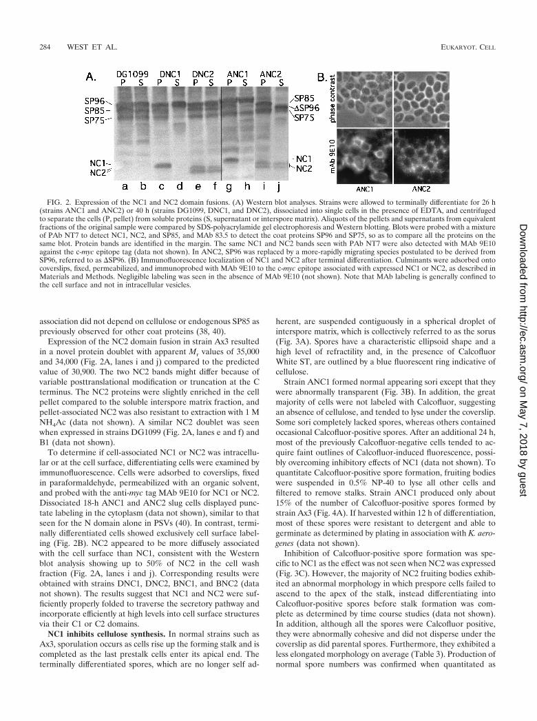
When induced to terminally differentiate, clonal strains de-rived from transfections of strain Ax3 with pVSBNC1 ex-pressed a novel, developmentally regulated protein with anapparent Mr of 38,000 that was similar to the predicted value of37,400 after removal of the signal peptide (data not shown).This new protein could be detected by polyclonal Ab (PAb)CT7 and PAb NT7 against SP85, and monoclonal Ab (MAb)9E10 against the myc epitope tag (Table 2). When cells fromterminally differentiated culminants were dissociated and cen-trifuged to separate cell-associated proteins from soluble pro-teins, the NC1 protein was almost exclusively associated withthe cells (Fig. 2A, lanes g and h). Strain names are defined inTable 1. Cell-associated NC1 was resistant to extraction withhigh salt (1 M NH4Ac) (data not shown). The level of NC1 wassimilar to that of endogenous SP85 based on labeling intensitywith PAb NT7. Similar results were obtained when NC1 wasexpressed in the dcsA-null strain DG1099 (Fig. 2A, lanes c andd) and the SP85-null strain B1 (data not shown). Thus, cell
TABLE 2. Abs employed
Ab Epitope Target protein Reference
MAbs83.5 Fuc�-1-PO4 SP96, SP80, SP75 1316.2 O-�GlcNAc SP85 (not NC1, NC2) 349E10 c-myc tag Expressed NC1, NC2 domains
of SP8540
PAbsCT7 Protein C-terminal domain of SP85 40NT7 Protein N-terminal domain of SP85 40Anti-SpiA Protein SpiA (DD31) 26
VOL. 1, 2002 DICTYOSTELIUM SPORULATION CHECKPOINT 283
on May 7, 2018 by guest
http://ec.asm.org/
Dow
nloaded from
association did not depend on cellulose or endogenous SP85 aspreviously observed for other coat proteins (38, 40).
Expression of the NC2 domain fusion in strain Ax3 resultedin a novel protein doublet with apparent Mr values of 35,000and 34,000 (Fig. 2A, lanes i and j) compared to the predictedvalue of 30,900. The two NC2 bands might differ because ofvariable posttranslational modification or truncation at the Cterminus. The NC2 proteins were slightly enriched in the cellpellet compared to the soluble interspore matrix fraction, andpellet-associated NC2 was also resistant to extraction with 1 MNH4Ac (data not shown). A similar NC2 doublet was seenwhen expressed in strains DG1099 (Fig. 2A, lanes e and f) andB1 (data not shown).
To determine if cell-associated NC1 or NC2 was intracellu-lar or at the cell surface, differentiating cells were examined byimmunofluorescence. Cells were adsorbed to coverslips, fixedin paraformaldehyde, permeabilized with an organic solvent,and probed with the anti-myc tag MAb 9E10 for NC1 or NC2.Dissociated 18-h ANC1 and ANC2 slug cells displayed punc-tate labeling in the cytoplasm (data not shown), similar to thatseen for the N domain alone in PSVs (40). In contrast, termi-nally differentiated cells showed exclusively cell surface label-ing (Fig. 2B). NC2 appeared to be more diffusely associatedwith the cell surface than NC1, consistent with the Westernblot analysis showing up to 50% of NC2 in the cell washfraction (Fig. 2A, lanes i and j). Corresponding results wereobtained with strains DNC1, DNC2, BNC1, and BNC2 (datanot shown). The results suggest that NC1 and NC2 were suf-ficiently properly folded to traverse the secretory pathway andincorporate efficiently at high levels into cell surface structuresvia their C1 or C2 domains.
NC1 inhibits cellulose synthesis. In normal strains such asAx3, sporulation occurs as cells rise up the forming stalk and iscompleted as the last prestalk cells enter its apical end. Theterminally differentiated spores, which are no longer self ad-
herent, are suspended contiguously in a spherical droplet ofinterspore matrix, which is collectively referred to as the sorus(Fig. 3A). Spores have a characteristic ellipsoid shape and ahigh level of refractility and, in the presence of CalcofluorWhite ST, are outlined by a blue fluorescent ring indicative ofcellulose.
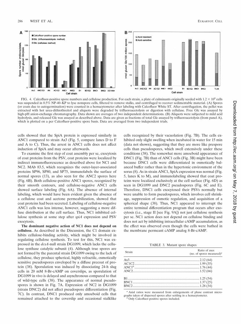
Strain ANC1 formed normal appearing sori except that theywere abnormally transparent (Fig. 3B). In addition, the greatmajority of cells were not labeled with Calcofluor, suggestingan absence of cellulose, and tended to lyse under the coverslip.Some sori completely lacked spores, whereas others containedoccasional Calcofluor-positive spores. After an additional 24 h,most of the previously Calcofluor-negative cells tended to ac-quire faint outlines of Calcofluor-induced fluorescence, possi-bly overcoming inhibitory effects of NC1 (data not shown). Toquantitate Calcofluor-positive spore formation, fruiting bodieswere suspended in 0.5% NP-40 to lyse all other cells andfiltered to remove stalks. Strain ANC1 produced only about15% of the number of Calcofluor-positive spores formed bystrain Ax3 (Fig. 4A). If harvested within 12 h of differentiation,most of these spores were resistant to detergent and able togerminate as determined by plating in association with K. aero-genes (data not shown).
Inhibition of Calcofluor-positive spore formation was spe-cific to NC1 as the effect was not seen when NC2 was expressed(Fig. 3C). However, the majority of NC2 fruiting bodies exhib-ited an abnormal morphology in which prespore cells failed toascend to the apex of the stalk, instead differentiating intoCalcofluor-positive spores before stalk formation was com-plete as determined by time course studies (data not shown).In addition, although all the spores were Calcofluor positive,they were abnormally cohesive and did not disperse under thecoverslip as did parental spores. Furthermore, they exhibited aless elongated morphology on average (Table 3). Production ofnormal spore numbers was confirmed when quantitated as
FIG. 2. Expression of the NC1 and NC2 domain fusions. (A) Western blot analyses. Strains were allowed to terminally differentiate for 26 h(strains ANC1 and ANC2) or 40 h (strains DG1099, DNC1, and DNC2), dissociated into single cells in the presence of EDTA, and centrifugedto separate the cells (P, pellet) from soluble proteins (S, supernatant or interspore matrix). Aliquots of the pellets and supernatants from equivalentfractions of the original sample were compared by SDS-polyacrylamide gel electrophoresis and Western blotting. Blots were probed with a mixtureof PAb NT7 to detect NC1, NC2, and SP85, and MAb 83.5 to detect the coat proteins SP96 and SP75, so as to compare all the proteins on thesame blot. Protein bands are identified in the margin. The same NC1 and NC2 bands seen with PAb NT7 were also detected with MAb 9E10against the c-myc epitope tag (data not shown). In ANC2, SP96 was replaced by a more-rapidly migrating species postulated to be derived fromSP96, referred to as SP96. (B) Immunofluorescence localization of NC1 and NC2 after terminal differentiation. Culminants were adsorbed ontocoverslips, fixed, permeabilized, and immunoprobed with MAb 9E10 to the c-myc epitope associated with expressed NC1 or NC2, as described inMaterials and Methods. Negligible labeling was seen in the absence of MAb 9E10 (not shown). Note that MAb labeling is generally confined tothe cell surface and not in intracellular vesicles.
284 WEST ET AL. EUKARYOT. CELL
on May 7, 2018 by guest
http://ec.asm.org/
Dow
nloaded from

described above (Fig. 4A). Instead of inhibiting cellulose for-mation as for NC1, NC2 might cause premature sporulation,resulting in stranding the spores partway up the stalk withnegative consequences for cell dissociation and spore shape.
Expression of the N domain alone had no effect on fruitingbody morphology, Calcofluor-positive spore number, or cellu-lose deposition (Fig. 4A). Although N is not incorporated intothe coat, expression of this domain does cause permanentchanges in the coat, including decreased buoyant density andbarrier functions (40; C. M. West, K. Kelley, and G. W. Erdos,unpublished data). Furthermore, the C1C2 construct had noeffect on these properties (Fig. 4A), including spore shape(Table 3), although previous work showed that C1C2 was in-corporated into the coat (40). Thus, the effect of NC1 could beattributed specifically to C1, and C1 needed to be isolated fromC2. The role of N is likely to target C1 to the PSV andsubsequently to the cell surface and possibly to promote fold-ing and or stability, but an additional, more direct role for N inNC1 action cannot be excluded.
To confirm that strain ANC1 underproduced cellulose dur-ing sporulation, cells from the sorus were assayed for cellulosecontent both chemically and enzymatically (Fig. 4A). The sam-ples were first extracted with urea to remove interfering ma-terial (39), and parallel aliquots were subjected to either an-
hydrous trifluoroacetolysis or cellulase digestion. The releasedGlc was measured by high-pH anion-exchange chromatogra-phy on a Dionex PA-10 column and quantitated by pulsedamperometry. Using either method, similar levels of Glc wereassayed from aliquots of strain Ax3 spores (Fig. 4A). Thisdemonstrated that nearly all urea-insoluble Glc-containingpolymer in these cells released by the acid treatment was cel-lulose-like. Consistent with this interpretation, negligible Glcwas recovered from the dcsA-null strain DG1099, which lacksthe catalytic subunit of cellulose synthase. Strains expressingthe N domain, the C1C2 fusion, or NC2 produced levels ofcellulose per spore similar to that of Ax3. In contrast, ANC1produced a low level of cellulose corresponding roughly to thesmall number of Calcofluor-positive spores produced. Thus,presumptive spores of strain ANC1 were inhibited in cellulosesynthesis, and inhibition appeared to be an all-or-nothing effectat the level of the individual cell.
NC1 does not inhibit spiA expression or PSV exocytosis. Toinvestigate the step at which sporulation was blocked by NC1,expression of the late sporulation marker gene spiA, which isinduced before cellulose synthesis, was examined. SpiA isthought to encode a prespore cell surface transmembrane pro-tein that is not necessary for coat formation but is required forspore stability (26). Western blot analysis of differentiating
FIG. 3. Sporulation in NC1 and NC2 domain expression strains. Representative culminants from strains Ax3 (parental), ANC1, and ANC2 areshown in the panels on the left. Ax3 forms a normal fruiting body with a sorus, consisting of spores, perched on top of the cellular stalk. The unusualmorphology of strain ANC2 varied in frequency from 50 to 90%, with the remaining culminants having a more normal appearance as shown inpanel A. Spores were found only in the lower sorus when present. Panels on the right show images from the same field of view of a representativesquash preparation of a single sorus. The field was imaged using either phase contrast (middle panels) or epifluorescence to reveal labeling withCalcofluor White ST (right panels) to show cellulose. Note that most ANC1 cells were Calcofluor negative and tended to lyse, that both ANC1and ANC2 Calcofluor-positive cells tended to be less elongate than parental Ax3 cells (Table 3), and that ANC2 spores did not disperse as wellunder the coverslip owing to abnormal self-cohesiveness.
VOL. 1, 2002 DICTYOSTELIUM SPORULATION CHECKPOINT 285
on May 7, 2018 by guest
http://ec.asm.org/
Dow
nloaded from
cells showed that the SpiA protein is expressed similarly inANC1 compared to strain Ax3 (Fig. 5, compare lanes D to Fand A to C). Thus, the arrest in ANC1 cells does not affectinduction of SpiA and may occur afterwards.
To examine the first step of coat assembly per se, exocytosisof coat proteins from the PSV, coat proteins were localized byindirect immunofluorescence as described above for NC1 andNC2. MAb 83.5, which recognizes the outer-layer-associatedproteins SP96, SP80, and SP75, immunolabels the surface ofnormal spores (13), as also seen for the ANC2 spores here(Fig. 6B). Both cellulose-positive ANC1 spores, recognized bytheir smooth contours, and cellulose-negative ANC1 cellsshowed surface labeling (Fig. 6A). The absence of internallabeling, which would have been evident given the absence ofa cellulose coat and acetone permeabilization, showed thatcoat proteins had been secreted. Labeling of cellulose-negativeANC1 cells was less intense, however, suggesting a more dif-fuse distribution at the cell surface. Thus, NC1 inhibited cel-lulose synthesis at some step after spiA expression and PSVexocytosis.
The dominant negative action of NC1 does not depend oncellulose. As described in the Discussion, the C1 domain ex-hibits cellulose-binding activity, which might be involved inregulating cellulose synthesis. To test for this, NC1 was ex-pressed in the dcsA-null strain DG1099, which lacks the cellu-lose synthase catalytic subunit (4). Although true spores arenot formed by the parental strain DG1099 owing to the lack ofcellulose, they produce spherical, highly refractile, osmoticallysensitive pseudospores enveloped by a diffuse precoat of pro-tein (38). Sporulation was induced by dissociating 24-h slugcells in 20 mM 8-Br-cAMP on coverslips, as sporulation ofDG1099 in vivo is delayed and asynchronous compared to thatof wild-type cells (38). The appearance of normal pseudo-spores is shown in Fig. 7A. Expression of NC2 in DG1099(strain DNC2) did not affect pseudospore differentiation (Fig.7C). In contrast, DNC1 produced only amoeboid cells thatremained attached to the coverslip and occasional stalklike
cells recognized by their vacuolation (Fig. 7B). The cells ex-hibited only slight swelling when incubated in water for 15 min(data not shown), suggesting that they are more like presporecells than pseudospores, which swell extensively under theseconditions (38). The somewhat more amoeboid appearance ofDNC1 (Fig. 7B) than of ANC1 cells (Fig. 3B) might have beenbecause DNC1 cells were differentiated in osmotically bal-anced buffer rather than in the hypertonic environment of thesorus (8). As in strain ANC1, SpiA expression was normal (Fig.5, lanes K to M), and immunolabeling showed that coat pro-teins were localized exclusively at the cell surface (Fig. 6D) asseen in DG1099 and DNC2 pseudospores (Fig. 6C and E).Therefore, DNC1 cells exocytosed their PSVs normally butwere unable to form pseudospores, which involves cell shrink-age, suppression of osmotic regulation, and acquisition of aspherical shape (38). Thus, NC1 appeared to interrupt theentire terminal differentiation program that occurs after exo-cytosis (i.e., stage II [see Fig. 9A]) not just cellulose synthesisper se. NC1 action does not depend on cellulose binding anddoes not act by inhibiting intracellular cAMP accumulation, asthe effect was observed even though the cells were bathed inthe membrane permeant cAMP analog 8-Br-cAMP.
FIG. 4. Calcofluor-positive spore numbers and cellulose production. For each strain, a plate of culminants originally seeded with 1.2 � 108 cellswas suspended in 0.5% NP-40–KP to lyse nonspore cells, filtered to remove stalks, and centrifuged to recover sedimentable material. (A) Spores(or coats due to autogermination) were counted in a hemacytometer after labeling with Calcofluor White ST. After centrifugation, the pellet wasextracted with hot urea-dithiothreitol and aliquots were degraded by trifluoroacetolysis or digestion with cellulase. Free Glc was assayed byhigh-pH anion-exchange chromatography. Data shown are averages of two independent determinations. (B) Aliquots were subjected to mild acidhydrolysis, and released Glc was assayed as described above. Data are given as fractions of total Glc assayed by trifluoroacetolysis (from panel A),which is plotted on a per Calcofluor-positive spore basis. Data are averaged from two independent trials.
TABLE 3. Mutant spore shapes
Strain Ratio of axes(no. of spores measured)a
Ax3.....................................................................................2.12 (64)AC1C2...............................................................................1.99 (35)ANC1b ...............................................................................1.76 (44)ANC2.................................................................................1.52 (66)
B1.......................................................................................1.25 (54)BNC1.................................................................................1.37 (25)BNC2.................................................................................1.28 (34)
a Axial ratios were measured from enlargements of phase contrast micro-graphs taken of dispersed spores after settling in a hemacytometer.
b Only Calcofluor-positive spores included.
286 WEST ET AL. EUKARYOT. CELL
on May 7, 2018 by guest
http://ec.asm.org/
Dow
nloaded from

NC1 inhibits cellulose deposition transcellularly. To inves-tigate whether NC1 inhibits cellulose deposition and otherstage II sporulation events before or after it is secreted, the cellautonomy of the NC1 phenotype was examined. If the pheno-type were not cell autonomous, this would support action atthe cell surface that had transferred to neighboring cells, as aprevious study showed that many coat proteins, including prob-ably SP85, intermingle between neighboring spores during coatassembly (35). This was tested by mixing DNC1 cells (whichexpress NC1) with normal Ax3 cells during development. SinceDNC1 cannot produce cellulose-positive spores because theyare dcsA null and cellulose deposition is cell autonomous (38),all cellulose-positive spores would have derived from strainAx3. Chimeras between dcsA-null DG1099 and Ax3 cells wereused as a control. A 9:1 mixture of DG1099:Ax3 produced sixtimes as many Calcofluor-positive spores as the DNC1:Ax3mixture (Ax3, 5.2 � 107 spores/108 input cells; DG1099, 0.00 �106 spores/108 input cells; DNC1, 0.00 � 106 spores/108 inputcells; DG1099:Ax3 (9:1), 4.0 � 106 spores/108 input cells;DNC1:Ax3 (9:1), 0.70 � 106 spores/108 input cells [data arefrom a single representative experiment]). The simplest inter-pretation is that the suppressive effect of DNC1 cells on cel-lulose deposition by neighboring normal spores was mediatedby NC1 diffusing between cells. This suggests that NC1 also
inhibits cellulose deposition by the cells in which it is expressedfrom the outside in.
SP85 is required for orderly cellulose deposition. The effectsof NC1 implicated its parent protein, SP85, in the normaltiming of terminal (stage II) sporulation events, including cel-lulose synthesis. To evaluate the role of full-length SP85,sporulation was reexamined in the SP85-null mutant B1 (40).Unlike strain ANC1, strain B1 produced normal numbers ofcellulose-positive spores (Fig. 4A and 8A). However, thespores differentiated before stalk formation was complete andwere self cohesive, effectively stranding them at half mast (Fig.8A) as seen in strain ANC2 above (Fig. 3C). Furthermore, B1spores tended to be spherical in contrast to the ellipsoid shapeof wild-type parental spores (Table 3), suggesting a defect incellulose as cellulose is required to maintain the initial elon-gate shape of spores during sporulation (38). In B1 sori thatwere less than 1 day old, empty spore coats were frequentlyseen, suggesting premature germination or spore lysis consis-tent with a defective coat (data not shown). B1 spores werealso observed to fragment more easily than Ax3 spores whenexposed to probe sonication (data not shown). Since the levelof cellulose per cell was not reduced in these coats (Fig. 4), theorganization of coat cellulose might be aberrant.
Spore coat cellulose is normally resistant to hydrolysis in
FIG. 5. Expression of the early sporulation marker SpiA. Cells were developed for 15, 20, 24, or 32 h as indicated and harvested for analysisby SDS-polyacrylamide gel electrophoresis and Western blotting with a preabsorbed antiserum against the SpiA protein. Culminants derived fromplating 106 cells were loaded per lane. The position of the SpiA protein at an Mr of 31,000 is shown in the margin. The lower Mr value bands arelikely to be degradation products seen previously (26). As expected, cells did not accumulate SpiA until after 20 h of development.
FIG. 6. Immunofluorescence localization of coat proteins in terminally differentiated cells. Cells from the indicated strains were processed asdescribed in the legend to Fig. 2B except that they were immunoprobed with MAb 83.5 to localize the coat proteins SP96, SP80, and SP75. Shownare phase contrast (upper panels) and immunofluorescence images (lower panels) of the same fields. Note that MAb labeling is generally confinedto the cell surface and not in intracellular vesicles. For ANC1 cells (A), labeling is seen around both Calcofluor-positive (Calc�) cells, recognizedby their more oval, phase-bright profiles, and Calcofluor-negative (Calc�) cells, which are more flattened against the coverslip.
VOL. 1, 2002 DICTYOSTELIUM SPORULATION CHECKPOINT 287
on May 7, 2018 by guest
http://ec.asm.org/
Dow
nloaded from

aqueous 4 M TFA (39), consistent with crystalline packing ofits linear �-1,4-glucan chains in microfibrils. This suggestedthat susceptibility to hydrolysis in 4 M TFA might be used toassess the organization of the glucan chains in the mutants.Aliquots of spores used to measure total cellulose above werehydrolyzed in 4 M TFA for 4 h at 100°C. Less than 25% of thecellulose from strain Ax3 was converted to Glc under theserelatively mild conditions (Fig. 4B). The greater fraction ofacid-labile Glc than previously reported (39) may be due to thepretreatment with hot urea to ensure removal of interferingsubstances. In contrast to Ax3 cellulose, greater than half ofthe B1 cellulose was hydrolyzed in 4 M TFA, suggesting thatthe glucan chains of B1 cellulose were more solvent exposed.In addition, cellulose was slightly overexpressed on a per sporebasis (Fig. 4). Increased synthesis of improperly organized cel-lulose might have resulted from premature cellulose synthesis,as suggested by the morphology of B1 fruiting bodies. Asdetailed in the Discussion, we speculate that coat cellulose ismore acid labile because cellulose synthesis was initiated priorto the proper spatial organization of cellulose synthase in themembrane and of potential cofactors at the cell surface. Thus,
absence of SP85 appears to have an effect opposite to that ofNC1, to promote rather than delay terminal sporulation.
NC1 acts upstream of SP85. The inhibitory effect of NC1 onterminal sporulation might result from an interfering effect onSP85 function, or represent a novel function. To distinguishbetween these possibilities, the phenotype of the double mu-tant in which NC1 was expressed in the SP85-null (B1) back-ground was examined. In contrast to when NC1 was expressedin the Ax3 background, all cells in the (lower) sorus wereCalcofluor positive (Fig. 8B) and normal numbers of Cal-cofluor-positive spores were produced (Fig. 4A), indicatingthat the absence of SP85 was epistatic to NC1. As seen also forB1 spores, BNC1 spores were defective based on the appear-ance of empty coats in the sorus, suggesting spontaneous ger-mination (Fig. 8B), a roundish shape (Table 3), and increasedlevels of cellulose which was also acid labile (Fig. 4B). Thefurther increase in the fraction of cellulose that was acid labile(Fig. 4B), seen also when NC2 and C1C2 were expressed,indicated that these constructs had additional effects in theabsence of SP85. With respect to checkpoint execution, NC1thus appears to act primarily by interfering with a naturalfunction of SP85 itself, possibly by competing with other pro-teins with which SP85 normally interacts (see Discussion).
DISCUSSION
The two main conclusions of this study are that the timing ofcoat protein secretion and cellulose deposition are coordinatedby a checkpoint, and that checkpoint execution is influenced bythe coat protein SP85. Based on previous evidence and sum-marized in Fig. 9A, coat assembly is a multistep process, be-ginning with protein secretion (stage I), followed by cell shrink-age and cellulose synthesis (stage II), and subsequently,formation of the outer proteinaceous layer (stage III). Proteinand cellulose are synthesized by separate mechanisms that arelikely to be under separate regulation, with evidence that cel-lulose synthesis is selectively controlled by the MEK-like ki-nase SplA (23; C. M. West, unpublished data). As depicted inFig. 9A, we propose that the synthesis of cellulose is alsoinfluenced by a cell surface checkpoint in which the coat struc-tural protein SP85 exerts an inhibitory effect until proteinsrearrange after secretion. Two postulated factors, C1 and C2,are proposed to inhibit and enhance SP85 function, respec-tively, and NC1 and NC2, two partial SP85 sequences, affectcheckpoint execution by interfering with their regulation ofSP85 (Fig. 9A). The evidence is as follows.
A cell surface checkpoint in coat assembly: evidence fromNC1. Previous time course studies indicated that exocytosis ofcoat proteins occurs before cellulose synthesis (34), and thiswas reinforced by the recent observation that exocytosis doesnot depend on cellulose in a cellulose synthase (dcsA)-nullstrain (38). In cells expressing the NC1 domain fusion of SP85,a sporulation arrest or delay occurs after exocytosis (Fig. 6) butbefore cellulose synthesis (Fig. 3 and 4A). Arrested cells ex-press the last known gene to be up-regulated during sporula-tion, spiA (Fig. 5), which is normally transcribed as presporecells rise up the extending stalk (27), and culmination is com-pleted normally. However, the arrested cells are fragile andtend to lyse when removed from the sorus for examination(Fig. 3B). A phenotype is also detected when NC1 is expressed
FIG. 7. Effects of NC1 and NC2 on terminal differentiation in thedcsA-null strain DG1099. Strains DG1099, DNC1, and DNC2 wereinduced to sporulate on coverslips in the presence of 20 mM 8-Br-cAMP and examined by phase contrast microscopy. Note that strainsDG1099 and DNC2 formed normal appearing pseudospores, charac-terized by their small, spherical, phase-bright shape, while DNC1 cellsremained amoeboid. Occasional vacuolated stalk cells are labeled St.
288 WEST ET AL. EUKARYOT. CELL
on May 7, 2018 by guest
http://ec.asm.org/
Dow
nloaded from

in dcsA-null cells: when such cells are induced to sporulate,they remain amoeboid (Fig. 7B), fail to shrink, and are resis-tant to hypotonic lysis. This suggests that in the sorus, arrestedcells might be unstable because of the hypertonic environmentof the interspore matrix (8) or because they attempt to germi-nate in situ (30).
Inhibition by NC1 occurs at a discrete step in the sporulationprogram, between stages I and II (Fig. 9A), which we refer toas a cell surface checkpoint. The existence of a checkpointduring normal sporulation would allow time for (i) coat pro-teins and galuran to hydrate into conformations suitable forinteraction with each other and nascent cellulose, (ii) finalassembly of cellulose synthase subunits into a multisubunitcomplex and organization of the terminal complex (5, 10, 14)to support deposition of crystalline microfibrils, and (iii) thedifferentiating spores to reach the stalk apex. Checkpoint ex-ecution may be actively suppressed in the PSV even beforeexocytosis of the coat proteins.
The NC1-dependent arrest seems to be mediated by anextracellular pathway, as an excess of DNC1 cells can arrestcellulose synthesis by normal spores in interstrain chimeras(see Results). Since the arrest is not rescued by incubation ofDNC1 cells in 8-Br-cAMP, NC1 expression does not appear to
interfere with activation of protein kinase A (PK-A) by thespore differentiation factor 2 (SDF-2)3DhkA pathway (32).The arrest may affect a target of PK-A that is regulated bySplA, the MEK-like kinase cited above.
The effect of NC1 appears to be attributable to native func-tions of the C1 and N domains and not to misfolded or oth-erwise aberrant protein configurations. NC1 is expressed at alevel comparable to that of other coat proteins and accumu-lates at the cell surface coordinately with other coat proteins(Fig. 2). The C1 domain confers retention of the fusion proteinat the cell surface since the N domain, expressed alone, isreleased in soluble form (40). The fraction of NC1 that issecreted in soluble form by induced dcsA-null cells is compe-tent to bind Avicel cellulose (L. Kaplan and C. M. West,unpublished data), indicating that its folding is not grosslyaberrant. Moreover, C4C motifs are expressed in a variety ofsequence contexts in other coat proteins (36), suggesting thattheir local environments might not be critical for folding. In-deed, C4C-folding may be intrinsically driven as the absence ofN-glycosylation motifs indicates that it is not subject to glyco-sylation-dependent quality control in the rER (16), and inde-pendent folding seems to be a general feature of EGF-likemodules (15). NC1-mediated arrest occurs at a discrete step
FIG. 8. Effects of NC1 and NC2 on terminal differentiation in SP85-null strain B1. Strains B1 (SP85-null), BNC1, and BNC2 were adsorbedto a coverslip, bathed in 0.1% Calcofluor White ST in KP buffer, and examined by phase contrast (left panels) or epifluorescence illumination (rightpanels) to identify cellulose-positive spores. Inset for strain B1 shows the appearance of a typical B1 fruiting body. Spores are present only in thelower sorus. Note that B1-derived spores tended to be round and did not disperse well under the coverslip owing to self cohesion and that therewas a tendency for autogermination as evidenced by the presence of Calcofluor-positive, phase-empty coats (two examples are labeled sc in panel B).
VOL. 1, 2002 DICTYOSTELIUM SPORULATION CHECKPOINT 289
on May 7, 2018 by guest
http://ec.asm.org/
Dow
nloaded from

many hours after accumulation of NC1, arguing that NC1 doesnot nonspecifically interfere with rER or Golgi processes. Fi-nally, the effect of NC1 depends on the presence of endoge-nous SP85 (Fig. 8), which also argues that it is not simply dueto an accumulation of denatured NC1.
Checkpoint model. A biochemical mechanism to explain theSP85 mutant phenotypes is proposed in Fig. 9B. According tothis model, an unidentified plasma membrane sensor protein,the checkpoint sensor, transfers inhibitory information intra-cellularly. When SP85 binds to the sensor, inhibition is exertedand the cell cannot proceed to stage II. SP85 itself is regulated,positionally or conformationally, in a positive sense by a hypo-thetical C2 factor and in a negative sense by a hypothetical C1factor, which bind to the C2 and C1 domains, respectively. TheC2 factor aids in positioning SP85 with respect to the check-point sensor, while the C1 factor prevents SP85 from bindingto the sensor as a result of hydration-dependent positional orconformational changes following its exocytosis. NC2 and NC1are interpreted to interfere with checkpoint regulation by com-peting with the activities of the C2 and C1 factors, respectively.
SP85 is thought to inhibit passage through the checkpointbecause in its absence, cellulose synthesis appears to occurprecociously. The earliest SP85-null phenotype is that sporesdeposit cellulose before stalk formation is complete, leavingthem stranded below the stalk apex (Fig. 8A). This phenotypeis consistent with evidence from myosin light chain mutantsthat the migration of prespore cells up the stalk appears torequire active cell motility (7), which would be blocked bypremature completion of the coat. SP85-null spores are alsoabnormally self-cohesive (Fig. 8A), less elongated (Table 3),and more permeable (40), and they contain excess, improperlyorganized cellulose (Fig. 4) and tend to break down in the
fruiting body (data not shown). These defects are not seen inother coat protein deletion mutants such as strain TL56 (Fig.4B). The simplest interpretation is that stages II and III of coatassembly are executed prematurely, resulting in strandedspores with defective coats. Thus, SP85 appears to normallysustain the checkpoint as a negative effector.
The NC1-induced arrest acts upstream of SP85 as it does notoccur in SP85-null cells (Fig. 8B). This suggests that NC1interferes with the normal down-regulation of SP85 to sustainblockage of the checkpoint. The checkpoint-inhibiting effect ofNC1 is not mimicked by NC2 or C1C2 (Fig. 4A), indicatingthat the phenotype is not the result of simply trapping the Ndomain in the coat and that it depends on the isolation of C1from the C2 domain of the protein. The C1 domain of NC1may competitively interfere with the normal inhibitory bindingof the hypothetical C1 factor to the C1 domain of SP85 (Fig.9B).
In contrast to that of NC1, NC2 expression mostly mimicsSP85-null phenotypes. NC2 cells differentiate into sticky sporesthat fail to rise to the apex of the stalk (Fig. 3C). Although theyproduce normal levels of cellulose, the wall is partially defec-tive (Fig. 4B) and spores tend to be round (Table 3). It isessential for these effects that C2 is expressed separately fromC1, as strain AC (expressing C1C2) produces normal appear-ing fruiting bodies and spores (Fig. 4, Table 3, and data notshown). The dominant negative nature of NC2’s action sug-gests that NC2 interferes with activation of SP85, leading us topropose the existence of an activating C2 factor. As illustratedin the model (Fig. 9B), the C2 factor might sequester SP85near the plasma membrane for optimal presentation to thecheckpoint sensor, similar to the role postulated for syndecanor other heparan sulfate proteoglycans in the presentation of
FIG. 9. Model for the terminal checkpoint and its regulation by SP85. (A) Delivery of the two major components of the coat, protein andcellulose, are proposed to be under control of separate signaling pathways which depend upon PK-A (20) and SplA (23), respectively. SDF-2 (32)is suggested to initiate coat formation by stimulating exocytosis of future coat proteins from PSVs (upper arm; stage I). The pathway and timingof arrival of cellulose synthase (lower arm) at the plasma membrane are not known. Subsequent stage II events, including expulsion of water andcellulose synthesis await checkpoint execution. The checkpoint is suppressed from the time of PSV formation by SP85, a resident PSV protein thatis later secreted to contribute to the coat. SP85 itself is regulated by the opposing activities of a C2 factor, which supports SP85-mediatedsuppression, and a C1 factor, which overcomes SP85 suppression, resulting in checkpoint activation. The effects of expressing NC1 and NC2 areinterpreted as competitive inhibition of the actions of the C1 and C2 factors, respectively. Deposition of cellulose results in organization of theouter and inner layers (stage III), which depends on a second function of SP85. Locations of the inner, middle, and outer layers and the plasmamembrane (pl. mem.) are noted. (B) SP85 is postulated to inhibit the checkpoint via a plasma membrane sensor, which transduces the inhibitionsignal intracellularly. The M or N domain of SP85 may bind the receptor directly or indirectly. Regulation of SP85 by the C2 and C1 factors arerepresented as locational. The deinhibiting influence of the C1 factor, itself presumably connected to a coat protein network, is proposed torelocate SP85 away from the sensor as a result of overall hydration. Inhibition of the checkpoint is relieved, and sporulation continues to stage II.
290 WEST ET AL. EUKARYOT. CELL
on May 7, 2018 by guest
http://ec.asm.org/
Dow
nloaded from
growth factors to animal cells (3). The somewhat milder phe-notype of ANC2 spores than of SP85-null spores suggests thatthe dominant negative mode of NC2 action is not as deleteri-ous as the complete absence of SP85. Expression of NC2 inSP85-null cells produces a stronger cellulose defect (Fig. 4B)than the absence of SP85 alone, however, indicating that NC2has additional targets.
SP85 ligands. The distinct sequence motifs present in the C1and C2 regions (Fig. 1) suggest that they fold separately andexhibit discrete binding activities. Binding studies utilizing se-cretions from DNC1 and DNC2 cells induced in suspension inthe presence of 8-Br-cAMP suggest that the cellulose and SP65binding activities of the C1C2 fusion (40) map to NC1, whereasNC2 binds two unidentified coat proteins (Zhang et al., un-published data). SP65 is thus the prime candidate for the C1factor that is proposed to relieve SP85-mediated inhibition ofcheckpoint execution (Fig. 9B). Additional receptor candi-dates for SP85 are other coat proteins, including SP70 andSP60 (22), and several proteins specifically expressed on theprespore cell plasma membrane, including SpiA (26), gp150/LagC (31), SP29/PsA (37), and WGA80B (33). However, SP70and SP60 might not be involved in checkpoint regulation, ascellulose synthesis is nearly normal in strain TL56 (Fig. 4B),which is genetically deficient in these and another coat proteins(12).
The checkpoint model predicts the existence of a sensor inthe plasma membrane and an intracellular pathway to trans-duce the signal to regulate cell volume and cellulose synthesis.Two-component histidine kinases regulate other late develop-mental events in Dictyostelium (9, 41) and the induction ofcellulose synthase in a strain of Rhizobium (2), and a relatedprotein might be involved here. Interaction of SP85 with theproposed sensor might be mediated by its M or N domain. Atransient role for the N domain in NC1 action, in addition totargeting to the PSV (40), is possible, and the missing tandemtetrapeptide repeats normally present between N and C1,which were excluded owing to difficulties in expression in Esch-erichia coli, may also be important (39).
Implications for cell wall assembly. The SP85 model pro-vides an example of how an external structural protein cancoordinate polysaccharide synthesis with delivery of a wallcomponent to the cell surface. The checkpoint pathway may berelated to the cell wall integrity pathway of yeast, which isactivated by unknown ligands acting on genetically definedtransmembrane sensor proteins that influence mitogen-acti-vated protein kinase pathways (6, 25). This correlates with apossible role of SplA in Dictyostelium (23; West, unpublisheddata). Vascular plants have wall-associated transmembraneSer/Thr kinases implicated in cell wall expansion that mightuse pectins and a Gly-rich protein as ligands (17), suggestingthat outside-in signaling might be general in cellulose-richwalls. The outcome of mutating SP85 on overproduction andacid lability of cellulose is reminiscent of the rsw1 mutation inthe catalytic subunit of an Arabidopsis cellulose synthase (1)and the effects of triathiazine herbicides (11), suggesting thatthese perturbations of cellulose synthase might interrupt arelated checkpoint regulation pathway in plants. Other defi-ciencies in SP85 mutant coats, such as increased permeabilityand decreased buoyant density (40), might result from check-point misregulation, although SP85’s cellulose-binding activity,
apparently not involved in checkpoint regulation, indicatesthat this protein also has additional functions in coat forma-tion. Derangement of the checkpoint may explain the spore-defective phenotypes of mutations of other late-acting genesincluding, e.g., acrA (29). Further understanding of SP85’s rolein checkpoint regulation is likely to come from identification ofother coat proteins that interact with its C1 and C2 domains,refined mutagenesis of full-length SP85, and additional knowl-edge about the regulation of cellulose synthase.
ACKNOWLEDGMENTS
We are grateful to Scott McMillen of the Protein Analysis Core ofthe University of Florida Interdisciplinary Center for BiotechnologyResearch for the Glc determinations. Anti-SpiA was generously pro-vided by W. F. Loomis and D. Fuller. T. Mullins provided valuableperspective.
This project was partially supported by a grant (MCB-9730036) fromthe National Science Foundation. A.C.M. was a University of Floridaundergraduate University Scholar.
REFERENCES
1. Arioli, T., L. Peng, A. S. Betzner, J. Burn, W. Wittke, W. Herth, C. Camilleri,H. Hofte, J. Plazinski, R. Birch, A. Cork, J. Glover, J. Redmond, and R. E.Williamson. 1998. Molecular analysis of cellulose biosynthesis in Arabidopsis.Science 279:717–720.
2. Ausmees, N., H. Jonsson, S. Hoglund, H. Ljunggren, and M. Lindberg. 1999.Structural and putative regulatory genes involved in cellulose synthesis inRhizobium leguminosarum bv. trifolii. Microbiology 145:1253–1262.
3. Baeg-Hun, B., and N. Perrimon. 2000. Functional binding of secreted mol-ecules to heparan sulfate proteoglycans in Drosophila. Curr. Opin. Cell Biol.12:575–580.
4. Blanton, R. L., D. Fuller, N. Iranfar, M. J. Grimson, and W. F. Loomis. 2000.The cellulose synthase gene of Dictyostelium. Proc. Natl. Acad. Sci. USA97:2391–2396.
5. Brett, C. T. 2000. Cellulose microfibrils in plants: biosynthesis, deposition,and integration into the cell wall. Int. Rev. Cytol. 199:161–199.
6. Cabib, E., D.-H. Roh, M. Schmidt, L. B. Crotti, and A. Varma. 2001. Theyeast cell wall and septum as paradigms of cell growth and morphogenesis.J. Biol. Chem. 276:19679–19682.
7. Chen, T.-L. L., W. A. Wolf, and R. L. Chisholm. 1998. Cell-type-specificrescue of myosin function during Dictyostelium development defines twodistinct cell movements required for culmination. Development 125:3895–3903.
8. Cotter, D. A., A. J. Dunbar, S. D. Buconjic, and J. F. Wheldrake. 1999.Ammonium phosphate in sori of Dictyostelium discoideum. Microbiology145:1891–1901.
9. Cotter, D. A., D. C. Mahadeo, D. N. Cervi, Y. Kishi, K. Gale, T. Sands, andM. Sameshima. 2000. Environmental regulation of pathways controllingsporulation, dormancy and germination utilizes bacterial-like signaling com-plexes in Dictyostelium discoideum. Protist 151:111–126.
10. Delmer, D. P. 1999. Cellulose biosynthesis: exciting times for a difficult fieldof study. Annu. Rev. Plant Physiol. Plant Mol. Biol. 50:245–276.
11. Fengel, D., and G. Wegner. 1979. Hydrolysis of cellulose: mechanisms ofenzymatic and acid catalysis. Adv. Chem. Ser. 181:145–158.
12. Fosnaugh, K. L., D. Fuller, and W. F. Loomis. 1995. Structural roles of thespore coat proteins in Dictyostelium discoideum. Dev. Biol. 166:823–825.
13. Gonzalez-Yanes, B., R. B. Mandell, M. Girard, S. Henry, O. Aparicio, M.Gritzali, R. D. Brown, G. W. Erdos, and C. M. West. 1989. The spore coat ofa fucosylation mutant in Dictyostelium discoideum. Dev. Biol. 133:576–587.
14. Grimson, M. J., C. H. Haigler, and R. L. Blanton. 1996. Cellulose microfi-brils, cell motility, and plasma membrane protein organization change inparallel during culmination in Dictyostelium discoideum. J. Cell Sci. 109:3079–3087.
15. Handford, P. A., A. K. Downing, D. P. Reinhardt, and L. Y. Sakai. 2000.Fibrillin: from domain structure to supramolecular assembly. Matrix Biol.19:457–470.
16. Helenius, A., and M. Aebi. 2001. Intracellular functions of N-linked glycans.Science 291:2364–2369.
17. Kohorn, B. D. 2001. WAKs; cell wall associated kinases. Curr. Opin. CellBiol. 13:529–533.
18. Lipke, P. N., and R. Ovalle. 1998. Cell wall architecture in yeast: newstructure and new challenges. J. Bacteriol. 180:3735–3740.
19. Loomis, W. F. 1971. Sensitivity of Dictyostelium discoideum to nucleic acidanalogues. Exp. Cell Res. 64:484–486.
20. Loomis, W. F. 1998. Role of PKA in the timing of developmental events inDictyostelium cells. Microbiol. Mol. Biol. Rev. 62:684–694.
VOL. 1, 2002 DICTYOSTELIUM SPORULATION CHECKPOINT 291
on May 7, 2018 by guest
http://ec.asm.org/
Dow
nloaded from

21. Maeda, M. 1992. Efficient induction of sporulation of Dictyostelium presporecells by 8-bromocyclic AMP under both submerged- and shaken-cultureconditions and involvement of protein kinase(s) in its action. Dev. GrowthDiffer. 34:263–275.
22. McGuire, V., and S. Alexander. 1996. PsB multiprotein complex of Dictyo-stelium discoideum: demonstration of cellulose binding activity and order ofprotein subunit assembly. J. Biol. Chem. 271:14596–14603.
23. Nuckolls, G. H., N. Osherov, W. F. Loomis, and J. A. Spudich. 1996. TheDictyostelium dual-specificity kinase splA is essential for spore differentia-tion. Development 122:3295–3305.
24. Pang, K. M., M. A. Lynes, and D. A. Knecht. 1999. Variables controlling theexpression level of exogenous genes in Dictyostelium. Plasmid 41:187–197.
25. Philip, B., and D. E. Levin. 2001. Wsc1 and Mid2 are cell surface sensors forcell wall integrity signaling that act through Rom2, a guanine nucleotideexchange factor for Rho1. Mol. Cell. Biol. 21:271–280.
26. Richardson, D. L., and W. F. Loomis. 1992. Disruption of the sporulation-specific gene spiA in Dictyostelium discoideum leads to spore instability.Genes Dev. 6:1058–1070.
27. Richardson, D. L., W. F. Loomis, and A. R. Kimmel. 1995. Progression of aninductive signal activates sporulation in Dictyostelium discoideum. Develop-ment 120:2891–2900.
28. Sassi, S., M. Sweetinburgh, J. Erogul, P. Zhang, P. Teng-Umnuay, and C. M.West. 2001. Analysis of Skp1 glycosylation and nuclear enrichment in Dic-tyostelium. Glycobiology 11:283–295.
29. Soderbom, F., C. Anjard, N. Iranfar, D. Fuller, and W. F. Loomis. 1999. Anadenylyl cyclase that functions during late development of Dictyostelium.Development 126:5463–5471.
30. Virdy, K. J., T. W. Sands, S. H. Kopko, S. van Es, M. Meima, P. Schaap, andD. A. Cotter. 1999. High cAMP in spores of Dictyostelium discoideum: asso-ciation with spore dormancy and inhibition of germination. Microbiology145:1883–1890.
31. Wang, J., L. Hou, D. Awrey, W. F. Loomis, R. A. Firtel, and C.-H. Siu. 2000.The membrane glycoprotein gp150 is encoded by the lagC gene and mediates
cell-cell adhesion by heterophilic binding during Dictyostelium development.Dev. Biol. 227:734–745.
32. Wang, N., F. Soderbom, C. Anjard, G. Shaulsky, and W. F. Loomis. 1999.SDF-2 induction of terminal differentiation in Dictyostelium discoideum ismediated by the membrane-spanning sensor kinase DhkA. Mol. Cell. Biol.19:4750–4756.
33. West, C. M., and W. F. Loomis. 1985. Absence of a carbohydrate modifica-tion does not affect the level of subcellular localization of three membraneglycoproteins in modB mutants of Dictyostelium discoideum. J. Biol. Chem.260:13801–13809.
34. West, C. M., and G. W. Erdos. 1990. Formation of the Dictyostelium sporecoat. Dev. Genet. 11:492–506.
35. West, C. M., and G. W. Erdos. 1992. Incorporation of protein into sporecoats is not cell-autonomous in Dictyostelium. J. Cell Biol. 116:1291–1300.
36. West, C. M., J. Mao, H. van der Wel, G. W. Erdos, and Y. Zhang. 1996. SP75is encoded by the DP87 gene, and belongs to a family of modular Dictyoste-lium outer layer spore coat proteins. Microbiology 142:2227–2243.
37. Zachara, N. E., N. H. Packer, M. D. Temple, M. B. Slade, D. R. Jardine, P.Karuso, C. J. Moss, B. C. Mabbutt, P. M. G. Curmi, K. L. Williams, andA. A. Gooley. 1996. Recombinant prespore-specific antigen from Dictyoste-lium discoideum is a �-sheet glycoprotein with a spacer peptide modified byO-linked N-acetylglucosamine. Eur. J. Biochem. 238:511–518.
38. Zhang, P., A. C. McGlynn, W. F. Loomis, R. L. Blanton, and C. M. West.2001. Spore coat formation and timely sporulation depend on cellulose inDictyostelium. Differentation 67:72–79.
39. Zhang, Y., R. D. Brown, and C. M. West. 1998. Two proteins of the Dictyo-stelium spore coat bind to cellulose in vitro. Biochemistry 37:10766–10779.
40. Zhang, Y., P. Zhang, and C. M. West. 1999. A linking function for thecellulose-binding protein SP85 in the spore coat of Dictyostelium discoideum.J. Cell Sci. 112:4667–4677.
41. Zinda, M. J., and C. K. Singleton. 1998. The hybrid histidine kinase dhkBregulates spore germination in Dictyostelium discoideum. Dev. Biol. 196:171–183.
292 WEST ET AL. EUKARYOT. CELL
on May 7, 2018 by guest
http://ec.asm.org/
Dow
nloaded from