outpatient management of systemic lupus erythematosus
TRANSCRIPT

R E V I E W C M E C R E D I T |
Outpatient management of systemic lupus erythematosus
GERALD N. SIMS, JR, MD, AND HOWARD R. SMITH, MD
Systemic lupus erythematosus (SLE) is often man-aged by primary care practitioners, who must coordinate the care with the support of subspecialists. The management of patients with such a serious and chronic disease can be both rewarding and challenging. This article reviews common problems and suggests management strategies.
Measurement of serum antinuclear antibodies can lead to an erroneous diagnosis if used as the sole basis for diagnosis.
Infections are the leading cause of death in lupus patients, and immunizations and antibiotic prophylaxis need to be con-sidered. Acute cutaneous SLE is exacerbated by exposure to ultraviolet light. Patients should avoid sun exposure. Specific treatment of cutaneous SLE includes topical cortico-steroids and antimalarial agents. Some of the most perplexing problems seen in SLE relate to neuropsychiatrie features. NSAIDs, corticosteroids, and antimalarials are the most commonly used medications for SLE.
INDEX TERMS: LUPUS ERYTHEMATOSUS, SYSTEMIC CLEVE CLIN J MED 1996; 63 :94-100
From the Lupus Center and the Division of Rheumatic Diseases, Univer-sity Hospitals of Cleveland and Case Western Reserve University.
Address reprint requests to H.R.S., Chief of Rheumatology, Meridia Huron Hospital, 13951 Terrace Road, East Cleveland, OH 44112.
SYSTEMIC LUPUS Erythema-tosus (SLE, or simply "lu-pus") is a systemic autoim-mune disorder of unknown
etiology that has protean manifes-tations. Patients commonly have involvement of the skin, joints, heart, lungs, kidneys, blood, diges-tive tract, and nervous system. Ac-curate diagnosis is made by careful evaluation of historic, physical, and laboratory findings. The inci-dence is estimated at 1.8 to 7.6 cases per 100 000. Women are af-fected nine times more often than men—and black women twice as often as white women.1 For many patients, primary care physicians make the diagnosis and undertake the care, consulting rheumatolo-gists when they encounter prob-lems. Because SLE is not a simple disease, its management often does not permit a simplified approach. This article reviews the outpatient management of SLE.
D I A G N O S I N G S L E
Because of the profound medical, social, and psychological implica-tions of having SLE, its diagnosis should not be made casually. The American College of Rheumatology
9 4 CLEVELAND CLINIC J O U R N A L OF MEDICINE VOLUME 63 • NUMBER 2
on January 24, 2022. For personal use only. All other uses require permission.www.ccjm.orgDownloaded from

L U P U S • S I M S A N D S M I T H
TABLE 1 CRITERIA FOR CLASSIFICATION OF SYSTEMIC LUPUS ERYTHEMATOSUS* Finding Comments (at least four needed for diagnosis)
Malar rash Fixed erythema, flat or raised, over the malar eminences Discoid rash Erythematous raised patches with adherent keratotic scaling and follicular plugging;
atrophic scarring may occur Photosensitivity Oral ulcers Includes oral and nasopharyngeal, observed by physician Arthritis Nonerosive arthritis Involving two or more peripheral joints, characterized by tenderness
swelling, or effusion Serositis Pleuritis or pericarditis documented by electrocardiography or rub or evidence
of pericardial effusion Renal disorder Proteinuria > 0.5 g/day or > 3+, or cellular casts Neurologic disorder Unexplained seizures or psychosis Hematologic disorder Hemolytic anemia or leukopenia (< 4000/mm3) or lymphopenia (< 1500/mm3)
or thrombocytopenia (< 100 000/mm3) in the absence of offending drugs Immunologic disorder Positive lupus erythematosus cell preparation, or anti-DNA or anti-Sm antibodies,
or false-positive serologic test for syphilis Antinuclear antibodies A n abnormal A N A titer by immunofluorescence or an equivalent assay at any time
in the absence of drugs known to induce ANAs
'Adapted from Tan et al, reference 2
has published criteria for the classification of SLE (Table l ).2 Though designed for population-based studies, these criteria provide a standardized ap-proach to diagnosing SLE that is accurate if one considers the recognized inclusions, exclusions, and limitations.2
T h e serum antinuclear antibody ( A N A ) titer, a sensitive but nonspecific test for SLE, can lead to an erroneous diagnosis if used as the sole criterion. ANAs are produced in a variety of pathologic con-ditions unrelated to SLE (eg, other autoimmune connective diseases, chronic infections, drugs, neo-plasia, chronic liver disease)—and in 5 % to 10% of healthy people.' A positive serum A N A titer is most helpful in confirming a diagnosis of SLE, because ANA-negative SLE is rare. Ninety-eight percent of patients with SLE have a positive A N A titer using the Hep-2 substrate.4 If a patient has had a positive titer, repeat testing has little value, since the A N A titer correlates poorly with disease activity.
Drug-induced S L E A variety of medications can induce SLE. The
most common are hydralazine, procainamide, chlor-promazine, methyldopa, D-penicillamine, quinidine, and isoniazid, although more than 50 other drugs have also been implicated. A number of other drugs
MARCH • APRIL 1996
can exacerbate the disease, including hormones, an-tibiotics, gold salts, and nonsteroidal anti-inflamma-tory drugs (NSAIDs).1 Although many of these drugs commonly induce positive A N A titers (up to 100% of patients treated with procainamide), far fewer pa-tients actually develop disease." Drug-induced SLE tends to produce symptoms of fever, arthritis, rash, and serositis but spares major organ systems such as the kidneys and brain. Serologic testing of patients with drug-induced SLE often demonstrates antihis-tone antibodies in the absence of other lupus-specific autoantibodies (eg, anti-nDNA, anti-Sm). The dis-ease usually remits after discontinuation of the of-fending drug, and immunosuppressive therapy is not usually required. NSAIDs may be used for sympto-matic therapy.
M A N A G I N G T H E M A N I F E S T A T I O N S OF S L E
Most patients with SLE lead productive and useful lives; however, it is a chronic disease of frequent remissions and exacerbations that cause the patient to seek a physician's help. SLE can involve any organ system, but most signs and symptoms are not medical emergencies and can be managed in the outpatient setting. Patients with lupus need general health maintenance—and attention to their special prob-
CLEVELAND CLINIC JOURNAL OF MEDICINE 9 5
on January 24, 2022. For personal use only. All other uses require permission.www.ccjm.orgDownloaded from
L U P U S • S I M S A N D S M I T H
lems. The patient-physician relationship should in-clude ongoing patient education about basic aspects of the disease, the patient's role in its management, and its impact on activities of daily living.
Nonspecific systemic features Nonspecific systemic features of SLE include fa-
tigue, fever, and malaise. Most patients (82%) expe-rience fatigue, which is often the patient's most dis-abling symptom.6 It may be the first symptom to appear and the last to leave. Treatment is problem-atic but involves sufficient rest and attention to other lupus-related problems, including associated fibromyalgia. Because physical and psychological stress may exacerbate lupus, measures that promote general well-being, such as good nutrition, rest, and physical conditioning, should be emphasized.'
Infections are the leading cause of death in lupus patients, and immunizations and antibiotic prophy-laxis need to be considered.s Except for live-virus vaccines in patients receiving corticosteroids or cy-totoxic agents, vaccinations are safe and should be given according to guidelines established for the general population. Some physicians recommend antibiotic prophylaxis before dental and urologie procedures.9
Dermatologie manifestations Perhaps the most obvious and troubling features
to both patients and physicians are the dermatologie manifestations, which are wide-ranging and involve the skin, hair, and nails.10 The classic butterfly rash indicates acute cutaneous SLE and is greatly exacer-bated by exposure to ultraviolet light. One third of lupus patients are photosensitive and should avoid sun exposure by wearing protective clothing and sunscreens. Even brief sun exposure can trigger sys-temic and cutaneous flares of SLE. Medications such as sulfonamides, thiazides, and tetracyclines can en-hance photosensitivity and thus exacerbate sun-in-duced lesions.11
Specific treatment of cutaneous SLE often in-cludes topical corticosteroids and antimalarial agents (hydroxychloroquine, chloroquine, and at-abrine). The dosage of hydroxychloroquine, the most frequently used antimalarial agent, should gen-erally not exceed 6 mg/kg per day, owing to a small risk of ocular toxicity. All patients receiving these agents should have ophthalmologic follow-up every 6 to 12 months. Intralesional corticosteroids are also helpful for discoid lupus and well-circumscribed ar-
eas of alopecia. Diffuse alopecia is a particularly troubling cosmetic feature that, on rare occasions, requires specific therapy with corticosteroids. Al-though cutaneous exacerbations do not necessarily signify systemic exacerbations, the appearance of purpura, bullous lesions, and other vasculitic lesions should prompt an expeditious evaluation for associ-ated systemic involvement.
Musculoskeletal manifestations Almost all SLE patients experience muscu-
loskeletal problems, ranging from fibromyalgia to myopathy. Fibromyalgia is common and is charac-terized by diffuse muscle aching without objective weakness and by nonrestorative sleep and well-de-fined trigger points.12 Fibromyalgia is not a myopathy, and the presence of true muscle weakness should prompt a search for myositis and myopathy induced by medications such as corticosteroids or antimalarial drugs. Treatment of fibromyalgia in-cludes empathetic physician-patient communica-tion, aerobic exercise, and judicious use of agents to promote better sleep patterns. Short courses of cy-clobenzaprine and tricyclic antidepressants in low doses are often useful, but their long-term use has not proven successful." Most patients have chronic symptoms that can be a considerable source of frus-tration to the physician and patient.
The joint symptoms in SLE are usually arthralgias and, less commonly, observable joint swelling and limitation of motion. Joint deformities may occur but are due to li-gamentous laxity (Jaccoud's arthropathy) and usually do not involve destructive, erosive arthropathy. The main-stays of treatment are NSAIDs and physical therapy. Hydroxychloroquine is very useful in patients who do not respond to NSAIDs; corticosteroids should be reserved for symptoms unresponsive to these measures.
Cardiopulmonary manifestations SLE frequently affects the heart and lungs, and
manifestations range from mild serositis to overt organ failure. Symptoms related to serositis are com-mon and usually involve pleuritis and pericarditis. Pleuritis is the most common pulmonary manifesta-tion; however, one must be aware of uncommon presentations, including acute lupus pneumonitis, pulmonary hemorrhage, and diffuse interstitial lung disease.14 The latter are usually best managed in conjunction with an appropriate subspecialist.
Pleuritis often presents with shortness of breath and chest pain without radiographic evidence of
9 6 CLEVELAND CLINIC JOURNAL OF MEDICINE VOLUME 63 • NUMBER 2
on January 24, 2022. For personal use only. All other uses require permission.www.ccjm.orgDownloaded from

L U P U S • S I M S A N D S M I T H
pneumonia. Significant pleural effusions can he seen on chest roentgenography, and care must be taken to exclude an infectious cause of infiltrates or effu-sions before proceeding with treatment. Pericarditis also manifests with chest pain and audible friction rubs. Pericardial effusions are often present but rarely lead to tamponade.15
Mild pericarditis and pleuritis are treated with NSAIDs; more severe cases often require moderate doses of corticosteroids. Other cardiac problems en-countered include valvular abnormalities, conduction disturbances, premature atherosclerosis (especially in premenopausal women receiving corticosteroid ther-apy), and less commonly, overt myocarditis. The his-tory and physical examination are usually helpful in differentiating pericarditis from other causes of acute cardiac symptoms; however, the evaluation may re-quire further cardiovascular diagnostic testing.
Gastrointestinal manifestations Gastrointestinal manifestations can be diverse,
but the practitioner must be able to distinguish po-tentially catastrophic problems from less urgent conditions. Over the course of their illness, many lupus patients will have oral ulcers, NSAID-induced dyspepsia, nausea, or diarrhea. Treatment is largely symptomatic and, for oral lesions, includes topical corticosteroids and anesthetics if there is no evi-dence of oral infection or thrush. NSAID-induced dyspepsia can be managed symptomatically with H2-blockers and reassessment of the need for continued NSAID use. The management of suspected NSAID gastropathy was recently reviewed by Loeb et al.16
Because lupus patients arc immunocompromised by their underlying disease as well as by their medica-tions, even subtle abdominal signs and symptoms should be considered as evidence of more severe problems. Ambiguous presentations may represent peritonitis (noninfectious, infectious, vasculitic), hemorrhage, ileus, or a perforated viscus. Lupus can affect any organ of the gastrointestinal system. Ab-dominal serositis is a troubling manifestation of SLE because it is often difficult to differentiate from an acute abdomen. Peritoneal fluid is often present and has a broad differential diagnosis.17 Massive ascites is most unusual. Once infection and other causes have been excluded, treatment consists of corticosteroids.
Hematologic abnormalities Some of the most commonly encountered clini-
cal problems involve hematologic abnormalities.
All hematopoietic cell lines can be affected, lym-phocytes most commonly. Lymphopenia often re-quires no specific treatment. Thrombocytopenia not due to medications or concomitant illness is usually immune-mediated and generally does not need treatment unless associated with bleeding or if the platelet count falls below 50 000/mm'. Anemia is commonly observed and is usually due to chronic disease. A thorough evaluation will rule out blood loss, and, in the setting of an abnormal peripheral blood smear or red blood cell indices, should include an evaluation for autoimmune hemolytic anemia (Coomb's-positive or negative).
Treatment of acute immune-mediated thrombo-cytopenia or hemolytic anemia involves corticos-teroids in high doses, which usually provide a good response. This response may be delayed, and short-term interventions such as transfusions may need to be considered, depending on the response to treat-ment. Patients unresponsive to this therapy should be referred for further evaluation and possible treat-ment with other agents, including cytotoxic drugs, danazol, and intravenous gamma globulin. Splenec-tomy is reserved for extreme, unresponsive cases, as it is frequently ineffective and can be associated with significant morbidity due to infections. Marrow sup-pression can be a feature of SLE, but also may be caused by medications (eg, cytotoxic agents).
Renal disease Renal involvement, ranging from benign protein-
uria to necrotizing glomerulonephritis and renal failure, affects half of all lupus patients.18 All lupus patients, even if free of symptoms, need periodic screening for renal involvement as manifested by increased serum creatinine, decreased creatinine clearance, proteinuria, or cellular casts. Patients with significant renal involvement should be man-aged with the help of a nephrologist. Underlying medical illnesses such as diabetes or hypertension, as well as various medications, can cause kidney dys-function. In particular, coexisting hypertension can compound renal (and cardiac) disease and requires vigorous management.
Antiphospholipid antibody syndrome The secondary antiphospholipid antibody syn-
drome is well recognized in patients with SLE. Ap-proximately 40% of patients with SLE have an-tiphospholipid antibodies, and 25% of patients with these antibodies will have the antiphospholipid anti-
MARCH • APRIL 1996 CLEVELAND CLINIC JOURNAL OF MEDICINE 9 7
on January 24, 2022. For personal use only. All other uses require permission.www.ccjm.orgDownloaded from

L U P U S • S I M S A N D S M I T H
body syndrome.19 Patients with the syndrome have antibodies to negatively charged phospholipids (such as cardiolipin) and may have thrombosis. Laboratory abnormalities can include a prolonged partial throm-boplastin time, a biologic false-positive test for syphi-lis (eg, the rapid plasma reagin [RPR] and the Ven-eral Disease Research Laboratory [VDRL] tests), and positive enzyme-linked immunosorbent assay (ELISA) tests for anticardiolipin antibodies.
The major clinical features are venous or arterial thrombosis (eg, deep venous thrombosis, pulmonary embolism, stroke), recurrent spontaneous abortions, and thrombocytopenia. Other associated phenom-ena include livedo reticularis, cardiac valvular ab-normalities, and central nervous system disorders.
Patients with the antibody but not the syndrome should be treated prophylactically with aspirin if this is not contraindicated. Those with the syn-drome require continuous anticoagulation with heparin, followed by warfarin. The target interna-tional normalized ratio is approximately 3.0. The efficacy of corticosteroids and cytotoxic drugs has not been established. Several studies have demon-strated that patients with the syndrome who take combination regimens of heparin, aspirin, or corti-costeroids can successfully bear children.20
Managing pregnancy and SLE SLE does not generally affect fertility. Whether
pregnancy causes inactive SLE to flare is controver-sial; however, flares are common during pregnancy and are more likely if the disease has been active recently.21 During pregnancy, maternal non-lupus-related problems (eg, pre-eclampsia, hypertension) and fetal wastage are more common among lupus patients than among mothers without lupus. Fetal wastage has many causes, including activity of ma-ternal disease at conception, renal disease, and an-tiphospholipid antibody syndrome; a history of fetal losses increases the risk.21
Pregnant women with lupus should be managed as high-risk obstetric patients, and need close con-trol of their SLE during this period. All medications need to be carefully reconsidered for possible ad-verse maternal and fetal effects and balanced with the need to control maternal disease. Cyclophos-phamide and methotrexate are teratogenic and should be discontinued. It is unclear if hyroxychlo-roquine poses a significant risk, but the consensus is to discontinue it during pregnancy. Azathioprine can be used with caution during pregnancy. Pred-
nisone does not cross the placenta as much as other corticosteroids and is not contraindicated. NSAIDs should be avoided. Corticosteroid-treated patients should be given stress dosages (eg, hydrocortisone 100 mg intravenously every 8 hours) during delivery because of the risk of secondary adrenal suppression. The pediatrician needs to be alerted for the possibil-ity of neonatal lupus, including cutaneous lesions and congenital heart block.
Neuropsychiatrie features Perhaps the most perplexing problems seen in
SLE relate to neuropsychiatrie features. A wide vari-ety of psychiatric disturbances are encountered, in-cluding adjustment reactions, neurosis, psychosis, and organic brain dysfunction manifesting as defec-tive learning, memory, and other cognitive func-tions. The problem is compounded by the presence of a potentially life-threatening disease, its impact upon the family, and medications that can alter the sensorium (eg, corticosteroids, NSAIDs). Consult-ation with a psychotherapist is often helpful, as is judicious use of psychiatric medications. Patients should be encouraged to learn more about their dis-ease and to participate in self-help groups such those sponsored by the Lupus Foundation of America.
Neurologic presentations in lupus are as diverse as the psychiatric presentations. Virtually any neurologi-cal symptom or syndrome can be associated with SLE. The most commonly encountered are seizures, strokes, aseptic meningitis, headaches, and motor and sensory neuropathies. These problems are usually attributed to SLE, although care must be taken to exclude other possible causes such as infection, atherosclerosis, tox-icities, or medications. The evaluation is often similar to that performed in patients who do not have SLE and can include lumbar punctures, electrodiagnostic testing, and imaging studies.22 Treatment often con-sists of that routinely used for the neurologic disorder as well as for a lupus flare. Patients may not improve until the underlying lupus is controlled. Steroids are routinely used for central nervous system involvement although their efficacy is not firmly established. Cyto-toxic medications are reserved for those with serious disease that is unresponsive to corticosteroids.23
Associated autoimmune disorders SLE is frequently seen in association with other
autoimmune disorders (overlap syndromes) such as mixed connective tissue disease, Hashimoto's thy-roiditis, and Sjogren's syndrome, and rarely with
9 8 CLEVELAND CLINIC JOURNAL OF MEDICINE VOLUME 63 • NUMBER 2
on January 24, 2022. For personal use only. All other uses require permission.www.ccjm.orgDownloaded from
L U P U S • S I M S A N D S M I T H
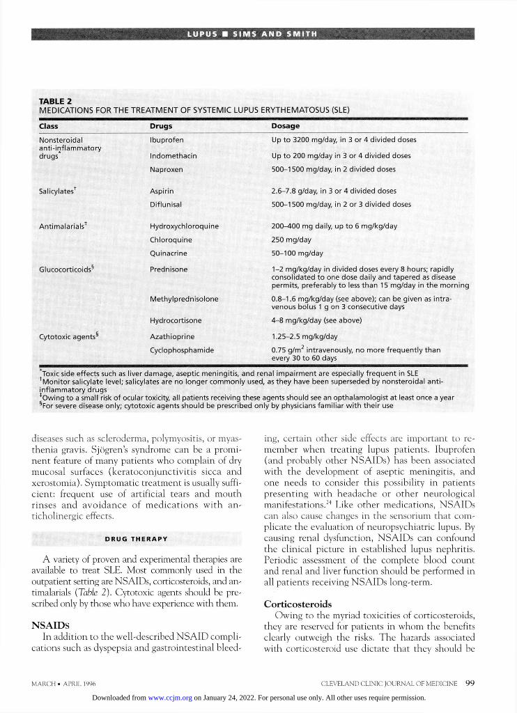
TABLE 2 MEDICATIONS FOR THE TREATMENT OF SYSTEMIC LUPUS ERYTHEMATOSUS (SLE) Class Drugs Dosage
Nonsteroidal anti-inflammatory drugs
Ibuprofen
Indomethacin
Up to 3200 mg/day, in 3 or 4 divided doses
Up to 200 mg/day In 3 or 4 divided doses
Naproxen 500-1500 mg/day, in 2 divided doses
Salicylatest Aspirin 2.6-7.8 g/day, in 3 or 4 divided doses
Diflunisal 500-1500 mg/day, in 2 or 3 divided doses
Antimalarials* Hydroxychloroquine 200-400 mg daily, up to 6 mg/kg/day
Chloroquine 250 mg/day
Quinacrine 50-100 mg/day
Glucocorticoids5 Prednisone 1-2 mg/kg/day in divided doses every 8 hours; rapidly consolidated to one dose daily and tapered as disease permits, preferably to less than 15 mg/day in the morning
Methylprednlsolone 0.8-1.6 mg/kg/day (see above); can be given as intra-venous bolus 1 g on 3 consecutive days
Hydrocortisone 4-8 mg/kg/day (see above)
Cytotoxic agents5 Azathioprine 1.25-2.5 mg/kg/day
Cyclophosphamide 0.75 g/m2 intravenously, no more frequently than every 30 to 60 days
* Toxic side effects such as liver damage, aseptic meningitis, and renal impairment are especially frequent in SLE tMonltor salicylate level; salicylates are no longer commonly used, as they have been superseded by nonsteroidal anti-inflammatory drugs * Owing to a small risk of ocular toxicity, all patients receiving these agents should see an opthalamologlst at least once a year §For severe disease only; cytotoxic agents should be prescribed only by physicians familiar with their use
diseases such as scleroderma, polymyositis, or myas-thenia gravis. Sjogren's syndrome can be a promi-nent feature of many patients who complain of dry mucosal surfaces (keratoconjunctivitis sicca and xerostomia). Symptomatic treatment is usually suffi-cient: frequent use of artificial tears and mouth rinses and avoidance of medications with an-ticholinergic effects.
DRUG T H E R A P Y
A variety of proven and experimental therapies are available to treat SLE. Most commonly used in the outpatient setting are NSAIDs, corticosteroids, and an-timalarials (Table 2). Cytotoxic agents should be pre-scribed only by those who have experience with them.
N S A I D s In addition to the well-described N S A I D compli-
cations such as dyspepsia and gastrointestinal bleed-
ing, certain other side effects are important to re-member when treating lupus patients. Ibuprofen (and probably other NSAIDs) has been associated with the development of aseptic meningitis, and one needs to consider this possibility in patients presenting with headache or other neurological manifestations.24 Like other medications, NSAIDs can also cause changes in the sensorium that com-plicate the evaluation of neuropsychiatrie lupus. By causing renal dysfunction, NSAIDs can confound the clinical picture in established lupus nephritis. Periodic assessment of the complete blood count and renal and liver function should be performed in all patients receiving N S A I D s long-term.
Corticosteroids Owing to the myriad toxicities of corticosteroids,
they are reserved for patients in whom the benefits clearly outweigh the risks. T h e hazards associated with corticosteroid use dictate that they should be
MARCH • APRIL 1996 CLEVELAND CLINIC JOURNAL OF MEDICINE 9 9
on January 24, 2022. For personal use only. All other uses require permission.www.ccjm.orgDownloaded from
L U P U S • S I M S A N D S M I T H
used judiciously. Complications of long-term use in-clude exacerbation of underlying diseases such as diabetes mellitus and hypertension, premature atherosclerosis, hypercortisolism, increased inci-dence of infection, cataract formation, osteonecrosis, and corticosteroid myopathy. Osteoporosis can be a major problem in patients treated long-term, and preventive strategies should be employed such as replacement estrogens, supplemental calcium, and modification of other risk factors, if possible.
If corticosteroid therapy is necessary, it is pru-dent to taper it as quickly as possible to the lowest dose necessary to control disease. Low doses, alter-nate-day doses, and short-term use are preferable when the clinical situation permits, as these may limit toxicity.
Antimalarial agents Antimalarial agents are generally safe and cause
few side effects. Hydroxychloroquine is used most commonly, followed by chloroquine. Atabrine is rarely used in this country because it is not widely available. Because patients usually require 2 to 4 months of treatment with these agents before they show improvement, concurrent treatment with other medications, such as N S A I D S , is often needed. A few patients treated with antimalarial agents experience dyspepsia, abdominal cramping, and diarrhea. These symptoms usually respond to dosage reduction and do not preclude long-term use. Adverse cutaneous reactions include pigment changes in the nails and skin, rashes, and dryness. Retinopathy is the most feared complication, though it is very rare in patients receiving less than 6.5 mg/kg/day of hydroxychloroquine. Fewer than 5 % of patients receiving 400 mg per day of hy-droxychloroquine develop corneal deposits, which are almost always asymptomatic.25 Corneal deposits and early retinal changes confined to the macula disappear with drug discontinuance. More ad-vanced retinal disease can result in irreversible vis-ual loss. To avoid this, a baseline ophthalmologic examination is recommended, with follow-up every 6 to 12 months.25
A C K N O W L E D G M E N T
The authors would like to thank Dr. Jeffrey Wisnieski for his helpful review of the manuscript.
R E F E R E N C E S
1. Hochberg MC. Systemic lupus erythematosus. Rheum His Clin North Am 1990; 1 6 ( 3 ) : 6 1 7 - 6 3 9 .
2. Tan EM, Cohen AS, Fries JF, et al. The 1982 revised criteria for the classification of systemic lupus erythematosus. Arthritis Rheum 1982; 2 5 : 1 2 7 1 - 1 2 7 7 .
3. Reichlin M. A N A s and antibodies to DNA: their use in clinical diagnosis. Bull Rheum Dis 1993; 4 2 ( 7 ) : 3 - 5 .
4. Reichlin M, Harley JB . Antinuclear antibodies: an overview. In: Wallace DJ, Hahn BH, editors. Dubois' lupus erythematosus. 4th ed. Philadelphia: Lea and Febiger, 1993 :188-194 .
5. Solinger AM. Drug-related lupus. Clinical and etiologic consid-erations. Rheum Dis Clin North Am 1988; 14( 1) : 187-202 .
6. Krupp LB, LaRocca N G , Muir J, Steinberg A D . A study of fatigue in systemic lupus erythematosus. J Rheumatol 1990; 1 7 : 1 4 5 0 - 1 4 5 2 .
7. Blumenfield M. Psychological aspects of systemic lupus erythe-matosus. Prim Care 1978 5 ( 1 ) : 1 5 9 - 1 7 1 .
8. Hellman DB, Petri M, Whiting-O'Keefe Q. Fatal infections in systemic lupus erythematosus: the role of opportunistic organisms. Medicine 1987; 6 6 : 3 4 1 - 3 4 8 .
9. Zysset MK, Montgomery MT, Redding SW, Dell'Italia LJ. Sys-temic lupus erythematosus: a consideration for antimicrobial pro-phylaxis. Oral Surg Oral Med Oral Pathol 1987; 6 4 : 3 0 - 3 4 .
10. Rothfield N . Cutaneous manifestations of systemic lupus erythe-matosus. Resid Staff Physician 1991; 3 9 ( 9 ) : 1 5 -23 .
11. Kreig AM. Environmental and infectious factors, pp.553—555. In: Steinberg AD, moderator. NIH conference. Systemic lupus erythematosus. A n n Intern Med 1991; 1 1 5 : 5 4 8 - 5 5 9 .
12. Wolfe F, Smythe H A , Yunus MB, et al. The American College of Rheumatology 1990 criteria for classification of fibromyalgia. Arthritis Rheum 1990; 3 3 : 1 6 0 - 1 7 2 .
13. Carrete S, Bell MJ, Reynolds W J , et al. Comparison of amitrip-tyline, cyclobenzaprine, and placebo in the treatment of fibromy-algia. Arthritis Rheum 1994; 3 7 : 3 7 - 4 0 .
14. Quismorio F. Pulmonary manifestations. In: Wallace DJ, Hahn BH, editors. Dubois' lupus erythematosus. 4th ed. Philadelphia: Lea and Febiger, 1993:343.
15. Stevens M, Ziminski C . Heart disease in systemic lupus erythe-matosus. Journal of Musculoskeletal Medicine 1992; 9 ( 6 ) : 4 1 - 4 6 .
16. Loeb DS, Ahlquist DA, Tulles N J . Management of gastroduo-denopathy associated with use of nonsteroidal anti-inflammatory drugs. Mayo Clin Proc 1992; 6 7 : 3 5 4 - 3 6 4 .
17. Schousboe J, Koch A, Chang R. Chronic lupus peritonitis with ascites; review of the literature with a case report. Semin Arthritis Rheum 1988; 1 8 ( 2 ) : 121 - 1 2 6 .
18. Pollak VE, Pirani C L . Lupus nephritis. In: Wallace DJ, Hahn BH, editors. Dubois' lupus erythematosus. 4th ed. Philadelphia: Lea and Febiger, 1993:525.
19. Love PE , Santero SA. Antiphospholipid antibodies: anticardi-olipin and the lupus anticoagulant in systemic lupus erythemato-sus (SLE) and in non-SLE disorders. Ann Intern Med 1990; 1 1 2 : 6 8 2 - 6 9 8 .
20. Lockshin MD. Antiphospholipid antibody syndrome. J A M A 1992; 2 6 8 : 1 4 5 1 - 1 4 5 3 .
21. Petrie M. Systemic lupus erythematosus and pregnancy. Rheum Dis Clin North Am 1994; 2 0 ( 1 ) : 8 7 - 1 1 9 .
22. Futrell N , Schultz L R , Millikan C. Central nervous system disease in patients with systemic lupus erythematosus. Neurology 1 9 9 2 ; 4 2 : 1 6 4 9 - 1 6 5 7 .
23. M c C u n e W J , Golbus J. Neuropsychiatry lupus. Rheum Dis Clin North Am 1988; 1 4 : 1 4 9 - 1 6 7 .
24. Kovacs JAJ , Urowitz MB, Gladman DD. Dilemmas in neurop-sychiatry lupus. Rheum Dis Clin North Am 1993; 1 9 : 7 9 5 - 8 1 3 .
25. Easterbrook M. T h e ocular safety of hydroxychloroquine. Semin Arthritis Rheum 1993; 2 3 ( 2 ) : 6 2 - 6 7 .
1 0 0 CLEVELAND CLINIC JOURNAL OF MEDICINE VOLUME 63 • NUMBER 2
on January 24, 2022. For personal use only. All other uses require permission.www.ccjm.orgDownloaded from